G1/S Cyclin-dependent Kinase Regulates Small GTPase Rho1p through Phosphorylation of RhoGEF Tus1p in Saccharomyces cerevisiae
←
→
Page content transcription
If your browser does not render page correctly, please read the page content below
Molecular Biology of the Cell
Vol. 19, 1763–1771, April 2008
G1/S Cyclin-dependent Kinase Regulates Small GTPase
Rho1p through Phosphorylation of RhoGEF Tus1p in
Saccharomyces cerevisiae
Keiko Kono,*† Satoru Nogami,* Mitsuhiro Abe,* Masafumi Nishizawa,‡
Shinichi Morishita,§ David Pellman,† and Yoshikazu Ohya*
Departments of *Integrated Biosciences and §Computational Biology, Graduate School of Frontier Sciences,
University of Tokyo, Chiba 277-8562, Japan; †Department of Pediatric Oncology, Dana-Farber Cancer Institute
and Division of Hematology/Oncology, Children’s Hospital Boston and Harvard Medical School, Boston, MA
02115; and ‡Department of Microbiology and Immunology, Keio University School of Medicine, Tokyo
160-8582, Japan
Submitted September 21, 2007; Revised January 16, 2008; Accepted January 30, 2008
Monitoring Editor: Mark Solomon
Rho1p is an essential small GTPase that plays a key role in the morphogenesis of Saccharomyces cerevisiae. We show here
that the activation of Rho1p is regulated by a cyclin-dependent kinase (CDK). Rho1p is activated at the G1/S transition
at the incipient-bud sites by the Cln2p (G1 cyclin) and Cdc28p (CDK) complex, in a process mediated by Tus1p, a guanine
nucleotide exchange factor for Rho1p. Tus1p interacts physically with Cln2p/Cdc28p and is phosphorylated in a Cln2p/
Cdc28p-dependent manner. CDK phosphorylation consensus sites in Tus1p are required for both Cln2p-dependent
activation of Rho1p and polarized organization of the actin cytoskeleton. We propose that Cln2p/Cdc28p-dependent
phosphorylation of Tus1p is required for appropriate temporal and spatial activation of Rho1p at the G1/S transition.
INTRODUCTION Rom2p, and Tus1p (Ozaki et al., 1996; Schmidt et al., 1997;
Bickle et al., 1998; Schmelzle et al., 2002). To date, the fol-
Asymmetric cell division is a fundamental process in devel- lowing effectors of Rho1p have been reported: Fks1/2p,
opment, maintaining stem cell pools and producing differ- Pkc1p, Bni1p, Sec3p, and Skn7p. Fks1p and its homologue
entiated cells. Budding yeast is a well-studied model for Fks2p are components of the -1,3-glucan (a major com-
asymmetric cell division. The assembly of the daughter cell ponent of the cell wall) synthase, which are essential for
or bud requires the polarization of the actin cytoskeleton cell wall biosynthesis (Drgonova et al., 1996; Mazur and
and the secretory apparatus. Cell polarization is under the Baginsky, 1996; Qadota et al., 1996). Pkc1p is a protein
control of the cell cycle machinery, absolutely requiring the kinase C homolog involved in the mitogen-activated pro-
activity of G1 cyclin/cyclin-dependent kinase (CDK) activ- tein (MAP) kinase cascade during various stress re-
ity. Despite intensive study, few cyclin/CDK substrates im- sponses and polarized cell growth. Pkc1p regulates actin
portant for polarized growth have been defined. patch distribution as well as the transcription of genes
In eukaryotic cells, Rho-type small GTPases play pivotal involved in G1/S transition and cell wall synthesis
roles in the process that underlies asymmetric cell division (Nonaka et al., 1995; Madden and Snyder, 1998, Levin,
(Hall, 1998; Kaibuchi et al., 1999). These GTPases cycle be- 2005). Bni1p, a formin family protein, assembles actin
tween the GTP-bound active states and the GDP-bound cables, which serve as tracks for the polarized delivery of
inactive states, and act as molecular switches. Rho-type materials necessary for polarized growth (Evangelista et
small GTPases are regulated by the following three classes al., 2002; Pruyne et al., 2002; Sagot et al., 2002a,b; Pring et
of proteins: guanine nucleotide exchange factors (GEFs), al., 2003) and for cytokinesis (Tolliday et al., 2002). Sec3p
GTPase-activating proteins (GAPs), and guanine nucleotide serves as a landmark protein for this polarized secretion
dissociation inhibitors (GDIs). GEFs are positive regulators (Finger et al., 1998; Guo et al., 2001). Skn7p regulates G1/S
and GAPs and GDIs are negative regulators (Matozaki et al., transition–specific and stress-induced transcription (Al-
2000; Takai et al., 2001). berts et al., 1998; Raitt et al., 2000). Because some functions
Rho1p is an essential Rho-type small GTPase in the bud- of these effectors have phase specificity during the cell
ding yeast S. cerevisiae that regulates many cellular processes cycle, it is possible that Rho1p is regulated in a cell
essential for cell morphogenesis. Rho1p is activated by spe- cycle– dependent manner.
cific GEFs (Matozaki et al., 2000; Takai et al., 2001), Rom1p, The localization of Rho1p changes during the cell cycle,
similar to many other polarity regulators. Rho1p localizes to
the prebud cell cortex in the G1-phase, to the plasma mem-
This article was published online ahead of print in MBC in Press branes of daughter cells in the S- to G2- phase, and around the
(http://www.molbiolcell.org/cgi/doi/10.1091/mbc.E07– 09 – 0950) contractile ring during cytokinesis (Yamochi et al., 1994; Qadota
on February 6, 2008. et al., 1996; Ayscough et al., 1999). Immunostaining with an
Address correspondence to: Yoshikazu Ohya, (ohya@k.u-tokyo. antibody specific for activated Rho1p (act-Rho1p) has revealed
ac.jp). that act-Rho1p localizes exclusively to the bud tip in small
© 2008 by The American Society for Cell Biology 1763K. Kono et al.
budded cells (Abe et al., 2003) and at the bud neck during g/ml pepstatin, 25 g/ml antipain, 25 g/ml aprotinin, 25 g/ml chymo-
cytokinesis (Yoshida et al., 2006), implying that the GDP/GTP statin, and 0.6% CHAPS), incubated with bead-bound glutathione-S-trans-
ferase (GST)-Pkc1RBD, washed, and subjected to 12.5% SDS-PAGE gels.
cycle of Rho1p is also cell cycle regulated. Several rho1ts mu- Bound Rho1p was detected by Western blot analysis using a polyclonal
tants arrest as nonbudded or small-budded cells (Yamochi et antibody against Rho1p (Qadota et al., 1996). Usually, the yeast cells were
al., 1994; Drgonova et al., 1999; Saka et al., 2001) and are defec- cultured to early log phase at 25°C. The cdc mutants were cultured to early log
tive in actin ring assembly during cytokinesis (Tolliday et al., phase at 25°C and then shifted to 37°C for 2 h. For cyclin overexpression,
YPH499 cells carrying pYO2344, pYO2345, and pYO2346, as well as YCp50-
2002). These observations further suggest that Rho1p functions GAL-CLN3 and YOC3008 cells were grown at 25°C in synthetic growth media
at specific stages of the cell cycle. with appropriate supplements and were then transferred to galactose media
In budding yeast, cell cycle– dependent cell morphogene- and incubated for 3 h at 25°C.
sis is regulated by the essential CDK Cdc28p (Lew and Reed,
1993, 1995; Nasmyth, 1993). Cdc28p-dependent phosphory- Microscopy
lation occurs preferentially on serine and threonine residues Visualization of Rho1p and act-Rho1p was performed as described previously
(Abe et al., 2003). To visualize myc-tagged proteins, the cells were processed
that are located within the minimum consensus (S/T P) or as described previously (Pringle et al., 1989). The anti-c-myc antibody (9E10,
full consensus (K/R S/T P X K/R) motifs of the target Calbiochem, La Jolla, CA) and fluorescein isothiocyanate (FITC)-conjugated
proteins (Songyang et al., 1994). Cdc28p is activated by the goat anti-mouse antibody (Jackson ImmunoResearch Laboratories, West
binding of cyclins (Lew and Reed, 1995), which are divided Grove, PA) were used as the primary antibody and secondary antibody,
respectively. Visualization of green fluorescent protein (GFP)-fused proteins
into three groups: three G1 cyclins (Cln1-3p), two S-phase was performed without antibody after formaldehyde fixation. Actin staining
B-type cyclins (Clb5p and Clb6p), and four M-phase B-type with rhodamine-labeled phalloidin (Molecular Probes, Eugene, OR) was car-
cyclins (Clb1-4p). Periodic activation of Cdc28p by each cyclin, ried out as described preciously (Pringle et al., 1989). Cells were observed by
which is expressed at the specific stage of the cell cycle is Zeiss Axioplan 2 imaging (Thornwood, NY). The images were captured by a
CCD camera (Cool SNAP HQ; Roper Scientific, Trenton, NJ) and the Meta-
required for orchestrating phase-specific events. morph Imaging software (Universal Imaging, West Chester, PA).
In this study, we focus on the cell cycle– dependent acti-
vation of the small GTPase Rho1p. We present evidence Coimmunoprecipitation of Cln2:HA/Cdc28p and Tus1:GFP
showing that the Rho1p-GEF Tus1p is a substrate of Cln2p/ The tus1 cln1 cln2::GALp-CLN2-HA (YOC3390) cells, which carry TUS1-GFP
Cdc28p, and that the phosphorylation of Tus1p is required on a centromeric plasmid, were grown to early log phase in glucose media.
for the efficient activation of Rho1p at the G1/S transition. Half of the culture was transferred to galactose media and cultured for an
additional 4 h. Cells were harvested and lysed in the lysis buffer (100 mM
Thus, Tus1p is an important integrator of cell cycle signals
and morphogenesis.
MATERIALS AND METHODS
Media, Strains, and Genetic Manipulations
Standard procedures were used for the DNA manipulations and Escherichia coli
transformations (Sambrook et al., 1989). The E. coli strain SCS1 (Stratagene, San
Diego, CA) was used for propagation of plasmids. The S. cerevisiae strains and
plasmids are listed in Tables S1 and S2, respectively. Yeast transformation was
carried out by the lithium acetate method (Ito et al., 1983). Genetic manipulations
for yeast were carried out as described previously (Kaiser et al., 1994; Sakumoto
et al., 1999). TUS1 mutagenesis was performed with the QuikChange Multi
Site-directed Mutagenesis Kit (Stratagene), using pBSA3 as the template. The
resulting plasmids were digested by BglII and then used for yeast transforma-
tion. The tagging of Tus1p with 13myc was performed as described previously
(Longtine et al., 1998). Yeast cells were grown either in rich medium (YPD; 1%
Bacto-yeast extract [Difco, Detroit, MI], 2% Bacto-peptone [Difco], 2% glucose
[Wako Chemicals, Osaka, Japan)] or in synthetic growth medium (0.67% yeast
nitrogen base [Difco], 2% glucose), with appropriate supplements.
Cell Synchronization and Culture Condition
Cells were cultured at 25°C unless otherwise indicated. Cell synchronization
was achieved using the yeast mating pheromone (␣-factor) or the microtu-
bule-depolymerizing reagent (nocodazole), as described previously (Marini et
al., 1996, Padmashree and Surana, 2001). After synchronization with nocoda-
zole, the cells were released into media that contained ␣-factor, to inhibit
subsequent budding. Analog sensitive cdc28-as1 allele was treated as de-
scribed previously (Bishop et al., 2000), but cells were arrested at G1/S-phase
by incubation at 37°C for 3 h (with cdc4-1 mutation). The concentration of
1-NM-PP1 used in our experiments was 20 M.
Purification of Glutathione-S-transferase Fusion Proteins
The regions that encode the Rho1p-binding domain of Pkc1p (377-640 amino
acids) was cloned into pGEX-3X and transformed into E. coli SCS1. The fusion Figure 1. GST:Pkc1RBD specifically pulls down act-Rho1p. (A)
protein was purified with glutathione-Sepharose 4B beads (Amersham Bio-
The fusion protein of the Pkc1p Rho1p-binding domain and
sciences, Uppsala, Sweden) as described previously (Frangioni and Neel,
1993). In addition, the purified proteins were clarified with Microcon Centrif- glutathione-S-transferase (GST:Pkc1RBD) was expressed in and
ugal Filter Devices (Microcon YM-50; Millipore, Bedford, MA). purified from E. coli and subjected to SDS-PAGE. The arrowhead
indicates GST:Pkc1RBD. (B) The strains with integrated 2xHA-
The Pulldown Assay for act-Rho1p RHO1 (YOC1678) harboring a centromeric plasmid of untagged
RHO1, RHO1(G19V), or RHO1(T24N) under the control of the
The pulldown assay for act-Rho1p was performed as described previously for
GAL1 promoter were cultured to log phase in glucose medium
mammalian RhoA (Kimura et al., 2000), with some modifications. Yeast cells
were lysed in lysis buffer (50 mM Tris-HCl, pH 7.5, 150 mM NaCl, 1 mM and transferred to galactose medium for 3.5 h. The collected cells
EDTA, 12 mM MgCl2, 1 mM DTT, 1 mM phenylmethylsulfonyl fluoride were subjected to the pulldown assay. Asterisks indicate the
[PMSF], 25 g/ml N-tosyl-l-phenylalanine chloromethyl ketone [TPCK], 25 signal for 2xHA-Rho1p, which is tagged to be distinguished from
g/ml N-tosyl-l-lysine chloromethyl ketone [TLCK], 25 g/ml leupeptin, 25 untagged Rho1 mutant proteins expressed from the plasmid.
1764 Molecular Biology of the CellG1/S CDK Regulates Rho1p through Tus1p
NaCl, 1 mM Na4P2O7, 5 mM NaF, 1 mM EDTA, 1% Triton X-100, 0.5% DOC, phosphate, 5 mM EDTA, 1 mM PMSF, 25 g/ml TPCK, 25 g/ml TLCK, 25
50 mM Tris-Cl, pH 7.5, 1 mM PMSF, 1 mM sodium orthovanadate, 2 g/ml g/ml leupeptin, 25 g/ml pepstatin, 25 g/ml antipain, 25 g/ml aproti-
aprotinin, 2 g/ml leupeptin, and 2 g/ml pepstatin). The cell lysate was nin, ad 25 g/ml chymostatin), boiled, and then separated by SDS-PAGE (8%
incubated for 2 h at 4°C with protein G-Sepharose (Amersham Biosciences) gel), followed by Western blot analysis using anti-c-myc antibody (9E10;
coupled with the anti-hemagglutinin (HA) antibody (16B12, BAbCO, Rich- Calbiochem). Equal amounts of protein (50 g) were loaded in each lane. For
mond, CA). The beads were washed and subjected to SDS-PAGE (10% gel) observing the cell cycle– dependent mobility shift of Tus1p(1-300):13myc (Fig-
and Western blot analysis with antibodies against HA (BAbCO), PSTARE ure 7D), cells were synchronized with ␣-factor, washed, and then released to
(Santa Cruz Biotechnology, Santa Cruz, CA), and GFP (Boehringer Ingelheim the fresh media. Samples are collected and suspended in 2⫻ sample buffer,
GmbH, Ingelheim, Germany), respectively. boiled for 5 min, treated with liquid N2, and then boiled for 5 min again.
Supernatants were subjected to SDS-PAGE (7% gel), followed by Western blot
Immunoprecipitation of Tus1p(1-300):13myc or analysis as described above.
Tus1p:13myc and Phosphatase Treatment Image Processing
For Figure 6, cells expressing Tus1p(1-300):myc or Tus1p:13myc were treated Image processing with the CalMorph (ver. 1.0) software was performed as
with 110 mM NaOH, boiled in 1⫻ sample buffer, and the supernatant was described previously (Ohtani et al., 2004). Briefly, log phase yeast cells were fixed
diluted with RIPA buffer without SDS (final SDS concentration was 0.1%). The with formaldehyde and then stained with rhodamine-phalloidin (for actin),
cell extract was incubated for 2 h at 4°C with protein G-Sepharose (Amersham FITC-concanavalin A (ConA; for the cell wall), and DAPI (for the DNA). Images
Biosciences) coupled with the anti-c-myc antibody (9E10, Calbiochem). The were captured using the Zeiss Axioplan 2 CCD camera (Cool SNAP HQ; Roper
beads were washed and treated with the Mn2⫹-dependent protein serine/thre- Scientific) and the Metamorph Imaging software (Universal Imaging). Captured
onine/tyrosine phosphatase (-PPase; New England BioLabs, Beverly, MA) in images were processed with the CalMorph software. A tiny bud was defined as
the presence or absence of its inhibitors, according to the manufacturer’s instruc- a bud that appeared as ⬍70 pixels after image processing.
tions. The beads were boiled and subjected to the SDS-PAGE [7% gel for
Tus1(300 a.a.):13myc, 5% gel for Tus1p:13myc], followed by Western blot anal-
ysis using the anti-c-myc antibody (9E10, Roche, Indianapolis, IN). RESULTS
The Levels of Act-Rho1p Peak at the G1/S Transition and
Western Blot Analysis of Tus1:13myc and Tus1(1-300):13myc
at the Late M-phase
For Figure 7B, cells were lysed in the lysis buffer (50 mM Tris-HCl, pH 7.5,
10% glycerol, 1% Triton X-100, 0.1% SDS, 150 mM NaCl, 50 mM NaF, 1 mM To measure active Rho1p (act-Rho1p) levels, we established
sodium orthovanadate, 50 mM -glycerol phosphate, 5 mM sodium pyro- a pulldown assay using a GST fusion to the Rho1p-binding
Figure 2. Rho1p is activated at the G1/S
boundary and during cytokinesis. (A) Wild-
type cells (YPH499) in early log phase were
arrested at the G1-phase by the addition of
mating pheromone and then released into the
fresh medium to resume the cell cycle. Sam-
ples were collected at the indicated time
points, and subjected to the pulldown assay.
(B) The bud index (E) and the relative act-
Rho1p signal intensities normalized by those
of total Rho1p (f) are shown. (C) Wild-type
cells in early log phase were arrested before
mitosis by the addition of nocodazole for 3 h.
Samples were collected and subjected to the
pulldown assay. (D) The relative signal inten-
sities of C are shown. Error bars, SD of three
independent experiments. (E) Wild-type cells
in early log phase were arrested before mitosis
with nocodazole and then released into fresh
medium to resume the cell cycle. Samples
were collected at the indicated time points and
subjected to the pulldown assay. (F) The bud
index (E) and the relative act-Rho1p signal
intensities normalized to those of total Rho1p
(f) are shown. (G) The cdc mutants (YOC931,
YOC934, YOC937, YOC938, YOC940, and
YOC946) were cultured to early log phase at
25°C and then shifted to 37°C for 2 h. Collected
cells were subjected to the pulldown assay. (H)
The relative signal intensities of act-Rho1p in
G normalized to those of total Rho1p are
shown.
Vol. 19, April 2008 1765K. Kono et al.
domain of Pkc1p (GST:Pkc1RBD), which specifically bound tively active form of Rho1p [Rho1(G19V)p] in yeast cell
to act-Rho1p (Nonaka et al., 1995). The bacterially expressed extract (Figure 1B). However, it displayed little affinity for
GST:Pkc1RBD (Figure 1A) efficiently bound to the constitu- Rho1(T24N)p (Figure 1B), which is a presumable nucleotide-
free form of Rho1p (Nonaka et al., 1995). Our pulldown
assay therefore specifically detects act-Rho1p. Next, wild-
A CLN3 CLN2 CLB2 CLB3 CLB5 type cells were arrested in the G1-phase by mating phero-
D G D G D G D G D G mone, released into fresh medium, and subjected to the
pulldown assay at intervals after release. During the cell
Act-Rho1p
cycle arrest, the level of act-Rho1p was relatively high,
Rho1p which is consistent with previous reports that Rho1p is
essential for mating (Drgonova et al., 1999). The act-Rho1p
B level declined sharply after release and then more signifi-
CLN + - cantly peaked 30 min after release, when the cells started
Act-Rho1p
budding (i.e., at the G1/S border; Figure 2, A and B). These
changes in act-Rho1p levels are not due to the changes of the
Rho1p steady-state protein level of Rho1p, which were unaltered
during the course of the experiment (Figure 2A, bottom
panel).
C
2
3
2
3
5
WT
To characterize the act-Rho1p levels at the later stages of
cln
cln
clb
clb
clb
the cell cycle, we performed the pulldown assay using cells
Act-Rho1p released form the nocodazole-arrest at the G2/M boundary.
The cells were collected at 10-min intervals and subjected to
Rho1p the pulldown assay. Act-Rho1p levels were relatively low in
nocodazole-treated cells (Figure 2, C and D), consistent with
D our recent finding that Rho1p is activated before mitotic exit
a 31.4%
(Yoshida et al., 2006; Figure 2, E and F). We also noticed that
the peak of act-Rho1p levels appears after most of the cells
b 9.8%
Figure 3. Cln2p/Cdc28p activates Rho1p. (A) Cells with cyclin
genes under the control of the GAL1 promoters were grown expo-
nentially in glucose media (D) and then transferred to galactose
media for 3 h (G). Collected cells were subjected to the pulldown
assay. (B) cln1 GAL1p-CLN2 cln3 (YOC4248) cells were cultured in
galactose media, and glucose was added and incubated for 4 h.
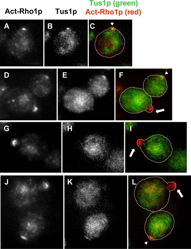
Collected cells were subjected to the pulldown assay. (C) Wild-type Figure 4. Tus1 colocalizes with act-Rho1p at the prebud sites. tus1
(WT) and cyclin mutants (YOC3383, YOC3384, YOC3385, YOC3386, cells with TUS1:GFP were fixed with formaldehyde and subjected to
and YOC3387) were cultured to log phase and subjected to the the immunofluorescence analysis with anti-act-Rho1p antibody. (A,
pulldown assay. (D) cdc4-1 cdc28-as1 (PY5543) cells were shifted to D, G, and J) Act-Rho1p; (B, E, H, and K) Tus1p:GFP; (C, F, I, and L)
37°C for 3 h and treated with DMSO (a) or 1-NM-PP1 (b), and overlay. Arrowheads indicate colocalized act-Rho1p signals and
act-Rho1p localization was observed by the immunofluorescence Tus1p:GFP signals. Arrows indicate only act-Rho1p signals without
analysis with anti-act-Rho1p antibody. Tus1p:GFP signals.
1766 Molecular Biology of the CellG1/S CDK Regulates Rho1p through Tus1p
PC Rh-ph
A C
α-fact.
Time after
release
(min) 10 20 30 40 50 60
Act-Rho1p Wild-
type
Rho1p
B 100 3
Relative signal intensity
Bud index (%) ( )
80
2
(Unit) ( )
Figure 5. Tus1p is required for the activation 60
of Rho1p at the G1/S boundary. (A) tus1
40
(YOC3388) cells were cultured to early log 1
phase, arrested at the G1-phase with mating 20 tus1
pheromone, and then released into fresh me-
dium to resume the cell cycle. Samples were 0 0
collected at the indicated time points and sub- 0 10 20 30 40 50 60
jected to the pulldown assay. (B) The bud in- Time after release (min)
dex (E) and the relative act-Rho1p signal inten-
sities normalized to those of total Rho1p (f) are D Wild-type tus1 tus1 E
shown. (C) Wild-type and tus1 (YOC3388) cells (Vector) (Vector) (PKC1[R398P])
at 30 min after release from the alpha-factor
CLN2 off CLN2 on
treatment were fixed with formaldehyde, and
stained with rhodamine-phalloidin. (D) Wild-
Control
Control
type with empty vector, tus1 (YOC3388) cells
Rh-ph
tus1
tus1
with empty vector, and tus1 (YOC3388) cells
with PKC1(R398P) (pYO1714) were cultured to
log phase, fixed with formaldehyde, and stained Act-Rho1p
with rhodamine-phalloidin. (E) tus1 cln1 pGAL1-
CLN2:3xHA (YOC3390) cells and the isogenic Rho1p
control (YOC3389) cells were grown exponen-
DIC
tially in glucose media (CLN2 off) and then trans- Cln2p:HA
ferred to galactose media for 4 h (CLN2 on) to
induce the expression of Cln2p:3HA. Collected
cells were subjected to the pulldown assay.
complete cytokinesis, suggesting that Rho1p may be involved (cdc) mutants were examined. Act-Rho1p levels were low in
not only in actin ring formation but also in cell separation. cdc25 cells (early G1-phase arrest), cdc7 cells (S-phase arrest),
To verify the results obtained with synchronized wild- and cdc17 cells (S-phase arrest), whereas the act-Rho1p level
type cells, various temperature-sensitive cell division cycle was high in cdc4 cells (G1/S-phase arrest), cdc14 cells (an-
Figure 6. Tus1p is a substrate of Cln2p/
Cdc28p. (A) cdc28-as1 cdc4-1 TUS(1-300):13myc
(PY5544) cells and cdc28-as1 cdc4-1 TUS1:13myc
(PY5543) cells were shifted to 37°C for 3 h to
arrest cells at the G1/S boundary, followed by
the treatment with 1-NM-PP1 or DMSO (control)
for 15 or 30 min as indicated. Cells were collected
and subjected to SDS-PAGE, followed by West-
ern blot analysis with anti-myc antibody. For
phosphatase treatment, cells were cultured to
early log phase at 25°C and shifted to 37°C for
3 h. Cell extracts were subjected to immunopre-
cipitation with anti-myc antibody, treated with
-PPase in the presence or absence of its inhibi-
tors. Bead-bound proteins were subjected to the
SDS-PAGE, followed by Western blot analysis
with anti-myc antibody. (B) tus1 cln1 pGAL1-
CLN2:3xHA (YOC3390) cells with TUS1:GFP
plasmid were cultured to early log phase in glu-
cose medium (Cln2:HA repressed condition: ⫺)
and transferred to galactose medium for 4 h
(Cln2:HA overexpressed condition: ⫹). Collected
cells were subjected to immunoprecipitation anal-
ysis with the anti-HA (12CA5) antibody, followed
by SDS-PAGE and Western blot analysis with the
anti-GFP antibody, 12CA5, and the anti-PSTARE
antibody. The asterisk indicates Pho85p, which is
also recognized by the anti-PSTARE antibody.
Vol. 19, April 2008 1767K. Kono et al.
aphase arrest), and cdc15 cells (anaphase arrest; Figure 2, G (Bishop et al., 2000). We first arrested cdc28-as1 cdc4-1 cells at
and H). These results provide independent evidence that the G1/S boundary by the cdc4-1 mutation, and then the
act-Rho1p peaks both at the G1/S boundary and later at act-Rho1p signal was detected by immunofluorescence mi-
M-phase. These results are consistent with our previous croscopy with an antibody specifically recognizing act-
work that active Rho1p signal is observed at the bud site Rho1p (Abe et al., 2003), in the presence or absence of 1-NM-
during budding (Abe et al., 2003) and at the bud neck in PP1 (Figure 3D). Act-Rho1p signals were observed at the
anaphase (Yoshida et al., 2006). bud tip in cells arrested at the G1/S boundary (36.8% n ⫽
106) and DMSO-treated control cells (31.4%, n ⫽ 175). On
Involvement of Cln2p and Clb2p in the Regulation of the other hand, only 9.8% of cells treated with 1-NM-PP1 for
act-Rho1p Levels 15min showed the polarized localization of act-Rho1p signal
Overexpression of G1 cyclins induces hyperpolarized (n ⫽ 205). These data indicate that the activity of G1/S
growth (Lew and Reed, 1993) because G1 cyclins are key transition Cdc28p is required for the spatially proper acti-
regulators during bud emergence and the following polar- vation of Rho1p.
ized cell morphogenesis. We tested whether the G1 cyclin
Cln2p activates Rho1p at the G1/S transition. GAL1 promo- Tus1p Is Required for the Activation of Rho1p at the G1/S
ter–mediated overexpression of Cln2p, but not of the other Transition
cyclins, increased the amount of act-Rho1p (Figure 3A). We tested the hypothesis that any of the three known GEFs
Furthermore, depletion of all the G1 cyclins, i.e., Cln1p, for Rho1p, Tus1p, Rom1p, and Rom2p (Ozaki et al., 1996;
Cln2p, and Cln3p, resulted in diminished levels of act- Schmelzle et al., 2002), were CDK substrate. The best candi-
Rho1p (Figure 3B). These findings suggest that the level of date was Tus1p, which contains two full-consensus se-
act-Rho1p depends on the expression of the G1 cyclin Cln2p. quences for CDK phosphorylation (12RTPEK16, 134RSPNK138),
Similar to Cln2p-overexpressing cells, hyperpolarized and was identified as a Clb/Cdc28p substrate (Ubersax et al.,
growth is also induced in cells that lack the major mitotic 2003).
cyclin Clb2p (Lew and Reed, 1993). Consistent with this Consistent with this idea, we found that 80.4% of Tus1:
observation, the act-Rho1p levels were increased in clb2 cells 3GFP signal colocalized with act-Rho1p signal in unbudded
(Figure 3C). cells (n ⫽ 163; Figure 4, arrowheads), and the colocalization
Next, we tested whether the activation of Rho1p at the was lost after bud emergence (Figure 4, arrows). We found
G1/S boundary depends on Cdc28p. We used an analog- that the deletion of TUS1 abolished the peak of act-Rho1p at
sensitive version of CDC28 allele (cdc28-as1) that can be the G1/S transition (Figure 5, A and B). tus1 cells at 30 min
specifically inhibited by the adenine-analog 1-NM-PP1 after the cell cycle release, which was a peak of the act-Rho1
A 40
D Time (min) 0 15 30 45
75 kDa
polarized signal (%)
Unbudded cells with
30 Tus1p(1-300)
:13myc
20
tus1-2Ap(1-300)
75 kDa
:13myc
10
0 tus1-9Ap(1-300) 75 kDa
TUS1 tus1-2A tus1-9A :13myc Figure 7. Phosphorylation of Tus1p is re-
quired for Cln2p-dependent activation of
B Rho1p. (A) Tus1p:3GFP, tus1-2Ap:3GFP and
100 Tus1p(1-300): tus1-10Ap:GFP were expressed in asynchro-
13myc
CLN2 off CLN2 on tus1-2Ap(1-300):
nous tus1 cells (YOC3388) and observed under
: :
75 the fluorescent microscope after fixation. Un-
Bud index (%)
13myc
tus1-9Ap(1-300):
13myc budded cells with polarized Tus1p signal
A
A
A
A
were counted (n ⬎ 200). Error bars show SE.
1-2
1-9
1-2
1-9
50
S1
S1
(B) GAL1p-CLN2 cells with myc-tagged Tus1p,
TU
tus
tus
TU
tus
tus
:
25 Tus1-2Ap or Tus1-9Ap (YOC4245, YOC4246
and YOC 4247, respectively) were cultured,
0 : : and CLN2-expression was induced by the ad-
0 15 30 45 dition of galactose for 4 h. Equal amount of
Time (min) proteins are loaded in each lane. (C) Cells
(YOC4096, YOC3868, and YOC3869) were cul-
C TUS1 tus1-2A tus1-9A
tured to log phase in glucose medium (D), and
transferred to galactose medium (G) for 4 h.
D G D G D G Collected cells were subjected to the pulldown
assay. (D) Cells with Tus1(1-300):myc (YKK79),
tus1-2A(1-300):myc (YKK98), and tus1-9A (1-
Act-Rho1p
300):myc (YKK89) in early log phase were ar-
rested at the G1-phase by the addition of mat-
Rho1p ing pheromone, and then released into fresh
medium to resume the cell cycle. Samples
Cln2p-HA were collected at the indicated time points and
subjected to SDS-PAGE, followed by Western
blot analysis with anti-myc antibody. The bud
index of each strain is shown (n ⬎ 200).
1768 Molecular Biology of the CellG1/S CDK Regulates Rho1p through Tus1p
level in wild-type cells, were larger than wild-type cells, and A
actin patches were dispersed to the mother cells (Figure 5C). ROM2 rom2
This is not a secondary consequence of mating pheromone
treatment because the actin patches were dispersed in asyn-
chronous culture as well (Figure 5D). This phenotype was TUS1
suppressed by the active form of Pkc1 (Pkc1[R398P]). In
addition, the deletion of TUS1 prevented ectopic accumula-
tion of act-Rho1p induced by CLN2 overexpression (Figure
5E). Therefore, Tus1p is required for the Cln2p/Cdc28p-
dependent activation of Rho1p at the G1/S boundary, pre-
sumably regulating actin polarity through Pkc1.
tus1-2A
Tus1p Is a Cln2p/Cdc28p Substrate
Next we tested the possibility that Tus1p is a direct substrate
of Cln2p/Cdc28p in vivo. We found that the N-terminus of
Tus1p is phosphorylated in a Cln2p/Cdc28p-dependent
manner. First, cells were arrested at the G1/S boundary by
the mutation of cdc4-1 at 37°C, in which Tus1p N-terminal tus1-9A
fragment (amino acids 1-300) showed slower migration (Fig-
ure 6A). This slower migration was sensitive to phosphatase
treatment, indicating that Tus1p is phosphorylated at the
G1/S boundary. Importantly, this slower migrating band is, B
at least in part, sensitive to Cdc28p inhibition with 1-NM-
Depolarized actin patches
in tiny budded cells (%)
PP1, indicating that Tus1 (1-300 a.a.) is phosphorylated by
Cdc28p in vivo. Moreover, the Cdc28p-dependent phos-
phorylation of full-length Tus1p:13 myc was also detected
(Figure 6A, bottom panels). Consistent with Tus1p being a
direct CDK substrate in vivo, Tus1p:GFP and Cln2p:HA
could be coimmunoprecipitated (Figure 6B).
Phosphorylation of Tus1p Is Required for the Activation
of Rho1p
tus1-2A rom2
tus1-9A rom2
tus1-9A
TUS1 rom2
tus1-2A
TUS1
To test whether the phosphorylation of Tus1p by CDK is
required for the activation of Rho1p, we constructed the
following tus1 phosphorylation site mutants, integrated at
the TUS1 locus. tus1-2A harbors alanine replacement muta-
tions (T13A and S135A) in both of the full-consensus se- Figure 8. Phosphorylation of Tus1p is required for the polarized
quences for CDK phosphorylation (12RTPEK16 and 134RSPNK138, organization of actin cytoskeleton. (A) TUS1, tus1-2A, and tus1-9A
(YOC4088, YOC3862, and YOC4089, respectively) cells with or with-
respectively). tus1-9A harbors nine alanine replacement mu- out rom2 were cultured to early log phase, fixed with formaldehyde,
tations (S8A, T13A, T93A, S122A, S126A, S163A, S170A, and stained with rhodamine-phalloidin. (B) Percentiles of depolar-
S173A, and S176A) in the minimal consensus sequence of ized actin in tiny budded cells are scored. Error bars, SE.
nine S/T P motifs for CDK phosphorylation. These alanine
substitution mutant proteins have slight defects in subcellu-
lar targeting (Figure 7A), but were expressed at comparable
levels to Tus1p (Figure 7B), indicating that the alanine re- tin patches (n ⬎ 200; Figure 8, A and B). This effect was
placement mutations do not affect steady-state expression. strikingly enhanced by deletion of ROM2 (rom2 23.4%,
When Cln2p was overexpressed under the control of the tus1-2A rom2 40.8%, tus1-9A rom2 92.9%; n ⬎ 200), suggest-
GAL1 promoter, in comparison with TUS1 strains (100%), ing that the Cdc28p-dependent activation of Rho1p through
the levels of act-Rho1p decreased in the tus1-2A (15%) and Tus1p serves as a parallel pathway to Rom2p-dependent
tus1-9A (3%; Figure 7C). The phosphorylation of Tus1p N- Rho1p activation. Consistent with this phenotype, tus1-2A
terminus (1-300 amino acids) at the G1/S boundary was rom2, tus1-9A, and tus1-9A rom2 exhibited slow growth (Fig-
decreased in these mutants (Figure 7D), consistent with the ure 9). The growth defect was suppressed by addition of 1 M
idea that the phosphorylation of Tus1p by CDK is required sorbitol. These results suggest that these mutants have a
for the activation of Rho1p at the G1/S boundary. defect in cell integrity pathway. Taken together, phosphor-
ylation of Tus1p is required for the normal polarized orga-
Phosphorylation of Tus1p Is Necessary for Normal Actin nization of actin patches, and phosphorylation of Tus1p
Organization regulates Rho1p activation parallel to Rom2p.
We examined cell morphology in tus1-2A and tus1-9A cells.
Exponentially growing cells were fixed and triple-stained DISCUSSION
with FITC-ConA, DAPI, and rhodamine-phalloidin, to visu-
alize the cell wall, nuclei, and actin cytoskeleton, respec- How the cell cycle and cell morphogenesis is orchestrated is
tively. Images were processed by our automated morpho- a longstanding question. To investigate the cell cycle– de-
metric software, CalMorph (Ohtani et al., 2004). pendent regulation of Rho1p, the key regulator of cell mor-
In cultures of TUS1 (control) cells with tiny buds, only phogenesis, we established a pulldown assay for act-Rho1p.
11.6% of the cells showed delocalized actin patches (n ⬎ Using this assay and the antibody specific for act-Rho1p, we
200). On the other hand, 21.4 and 55.6% of the tus1-2A and uncovered one important linkage between CDK and Rho1p.
tus1-9A mother cells, respectively, exhibited delocalized ac- We found that a G1 cyclin/CDK complex Cln2p/Cdc28p-
Vol. 19, April 2008 1769K. Kono et al.
25oC 25oC 37oC 37oC
YPD YPD+1M Sor. YPD YPD+1M Sor.
TUS1
tus1-2A
tus1-9A Figure 9. tus1 point mutants show synthetic growth
defect with rom2. TUS1, tus1-2A, tus1-9A, TUS1 rom2,
TUS1 rom2 tus1-2A rom2, and tus1-9A rom2 (YOC4088, YOC3862,
YOC4089, YOC4093, YOC4094, and YOC4095, respec-
tus1-2A rom2 tively) cells were spotted by 10-fold dilution on YPD or
tus1-9A rom2 YPD ⫹ 1 M sorbitol plates and incubated at 25°C or
37°C for 2 d.
dependent phosphorylation of RhoGEF Tus1p is important ACKNOWLEDGMENTS
for the activation of Rho1p at the G1/S boundary. Moreover,
We thank K. M. Shokat (University of California, San Francisco) for the kind
this activation is required for the polarized organization of gift of 1-NM-PP1, S. Buttery for reviewing manuscript, and S. Yoshida and S.
the actin cytoskeleton. These findings indicate that Rho1p is Bartolini for constructing plasmids and primers and helpful comments
a key target of CDK for cell morphogenesis in budding throughout this study. This work was supported by a grant (16026205) for
yeast. Our finding is consistent with the previous observa- Scientific Research from the Ministry of Education, Science, Sports, and
Culture of Japan and by the Institute for Bioinformatics and Research and
tion that Mpk1, which is in one downstream pathway of Development of the Japan Science and Technology Corporation.
Rho1p, is activated at the G1/S boundary (Zarzov et al.,
1996).
Previously Gulli et al. (2000) have shown that the activated REFERENCES
form of Cdc42p, which is another essential Rho-type small Abe, M., Qadota, H., Hirata, A., and Ohya, Y. (2003). Lack of GTP-bound
GTPase, is sufficient for the establishment of cell polarity in Rho1p in secretory vesicles of Saccharomyces cerevisiae. J. Cell Biol. 162, 85–97.
cells without Cln1/2/3p. Importantly, they have also shown
Alberts, A. S., Bouquin, N., Johnston, L. H., and Treisman, R. (1998). Analysis
that although cells with the activated form of Cdc42p are of RhoA-binding proteins reveals an interaction domain conserved in hetero-
able to form buds, they lyse shortly after polarization. This trimeric G protein beta subunits and the yeast response regulator protein
observation indicates that Cdc42p-dependent cell polariza- Skn7. J. Biol. Chem. 273, 8616 – 8622.
tion is not sufficient for maintaining cell wall integrity at the Audhya, A., and Emr, S. D. (2002). Stt4 PI 4-kinase localizes to the plasma
polarized sites. Taken together with our results, an attractive membrane and functions in the Pkc1-mediated MAP kinase cascade. Dev.
explanation (Sopko et al., 2007; Zheng et al., 2007) is that Cell 2, 593– 605.
Cdc42p and Rho1p are both required for G1-cyclin– depen- Ayscough, K. R., Eby, J. J., Lila, T., Dewar, H., Kozminski, K. G., and Drubin,
dent polarized morphogenesis; Cdc42p establishes cell po- D. G. (1999). Sla1p is a functionally modular component of the yeast cortical
actin cytoskeleton required for correct localization of both Rho1p-GTPase and
larity and Rho1p may maintain polarized growth and cell Sla2p, a protein with talin homology. Mol. Biol. Cell 10, 1061–1075.
wall integrity during subsequent bud growth.
Interestingly, although the peak of act-Rho1p is severely Bickle, M., Delley, P. A., Schmidt, A., and Hall, M. N. (1998). Cell wall
integrity modulates RHO1 activity via the exchange factor ROM2. EMBO J.
decreased in tus1 cells (Figure 5, A and B), tus1 deletion is 17, 2235–2245.
not lethal. Considering that rom2 tus1 is nearly lethal in
Bishop, A. C. et al. (2000). A chemical switch for inhibitor-sensitive alleles of
normal growth conditions (Schmelzle et al., 2002), Rom2p any protein kinase. Nature 407, 395– 401.
must play an overlapping function with Tus1p. Tus1p local-
izes to the prebud sites at the G1/S boundary (Figure 4) and Dephoure, N., Howson, R. W., Blethrow, J. D., Shokat, K. M., and O’Shea,
E. K. (2005). Combining chemical genetics and proteomics to identify protein
disperses to the cytosol after the bud emergence. In contrast, kinase substrates. Proc. Natl. Acad. Sci. USA 102, 17940 –17945.
Rom2p localizes globally to the bud cortex even after bud
Drgonova, J., Drgon, T., Tanaka, K., Kollar, R., Chen, G. C., Ford, R. A., Chan,
emergence (Manning et al., 1997, Audhya and Emr, 2002, C. S., Takai, Y., and Cabib, E. (1996). Rho1p, a yeast protein at the interface
Abe et al., 2003). We suggest that Tus1p provides a boost of between cell polarization and morphogenesis. Science 272, 277–279.
Rho1p activation in the early stage of bud emergence, Drgonova, J., Drgon, T., Roh, D. H., and Cabib, E. (1999). The GTP-binding
whereas Rom2p stimulates a basal level of act-Rho1p protein Rho1p is required for cell cycle progression and polarization of the
throughout the bud cortex. Considering that Pho85p is yeast cell. J. Cell Biol. 146, 373–387.
working as a backup in the absence of CLN1/2 (Espinoza et Espinoza, F. H., Ogas, J., Herskowitz, I., and Morgan, D. O. (1994). Cell cycle
al., 1994; Measday et al., 1994) and that Rom2p is a Pho85p control by a complex of the cyclin HCS26 (PCL1) and the kinase PHO85.
substrate (Dephoure et al., 2005), one possibility is that the Science 266, 1388 –1391.
Pho85p-Rom2p pathway is working parallel to the Cdc28p- Evangelista, M., Pruyne, D., Amberg, D. C., Boone, C., and Bretscher, A.
Tus1p pathway. Alternatively, Wsc1p-Rom2p pathway may (2002). Formins direct Arp2/3-independent actin filament assembly to polar-
be activated by membrane flux during bud emergence as ize cell growth in yeast. Nat. Cell Biol. 4, 260 –269.
proposed by Gray et al. (1997). Finger, F. P., Hughes, T. E., and Novick, P. (1998). Sec3p is a spatial landmark
In this work we found that the G1 cyclin/CDK complex for polarized secretion in budding yeast. Cell 92, 559 –571.
Cln2p/Cdc28p phosphorylates the Rho1p-GEF Tus1p, re- Frangioni, J. V., and Neel, B. G. (1993). Solubilization and purification of
sulting in the activation of Rho1p at the G1/S boundary. enzymatically active glutathione S-transferase (pGEX) fusion proteins. Anal.
Two lines of evidence suggest that this regulation could be Biochem. 210, 179 –187.
conserved in higher eukaryotes. First, botulinum C3 exoen- Gray, J. V., Ogas, J. P., Kamada, Y., Stone, M., Levin, D. E., and Herskowitz,
zyme (an inhibitor of Rho) treatment of interphase mamma- I. (1997). A role for the Pkc1 MAP kinase pathway of Saccharomyces cerevisiae
in bud emergence and identification of a putative upstream regulator. EMBO J.
lian cells leads to the cell cycle arrest in G1-phase 16, 4924 – 4937.
(Yamamoto et al., 1993). Second, expression of constitutively
Gulli, M. P., Jaquenoud, M., Shimada, Y., Niederhauser, G., Wiget, P., and
active Rho is sufficient for the G1/S transition (Olson et al., Peter, M. (2000). Phosphorylation of the Cdc42 exchange factor Cdc24 by the
1995). The activation of Rho at the G1/S boundary by CDK PAK-like kinase Cla4 may regulate polarized growth in yeast. Mol. Cell 6,
could be a conserved mechanism among eukaryotes. 1155–1167.
1770 Molecular Biology of the CellG1/S CDK Regulates Rho1p through Tus1p
Guo, W., Tamanoi, F., and Novick, P. (2001). Spatial regulation of the exocyst Pringle, J. R., Preston, R. A., Adams, A. E., Stearns, T., Drubin, D. G., Haarer,
complex by Rho1 GTPase. Nat. Cell Biol. 3, 353–360. B. K., and Jones, E. W. (1989). Fluorescence microscopy methods for yeast.
Methods Cell Biol. 31, 357– 435.
Hall, A. Rho GTPases and the actin cytoskeleton. (1998). Science 279, 509 –514.
Pruyne, D., Evangelista, M., Yang, C., Bi, E., Zigmond, S., Bretscher, A., and
Ito, H., Fukuda, Y., Murata, K., and Kimura, A. (1983). Transformation of
Boone, C. (2002). Role of formins in actin assembly: nucleation and barbed-
intact yeast cells treated with alkali cations. J. Bacteriol. 153, 163–168.
end association. Science 297, 612– 615.
Kaibuchi, K., Kuroda, S., and Amano, M. (1999). Regulation of the cytoskel-
eton and cell adhesion by the Rho family GTPases in mammalian cells. Annu. Qadota, H., Python, C. P., Inoue, S. B., Arisawa, M., Anraku, Y., Zheng, Y.,
Rev. Biochem. 68, 459 – 486. Watanabe, T., Levin, D. E., and Ohya, Y. (1996). Identification of yeast Rho1p
GTPase as a regulatory subunit of 1,3-beta-glucan synthase. Science 272,
Kaiser, C., Michaelis, S., and Mitchel, A. (1994). Methods in Yeast Genetics: A 279 –281.
Cold Spring Harbor Laboratory Course Manual, Cold Spring Harbor, NY:
Cold Spring Harbor Laboratory Press. Raitt, D. C., Johnson, A. L., Erkine, A. M., Makino, K., Morgan, B., Gross, D. S.,
and Johnston, L. H. (2000). The Skn7 response regulator of Saccharomyces
Kimura, K., Tsuji, T., Takada, Y., Miki, T., and Narumiya, S. (2000). Accumu- cerevisiae interacts with Hsf1 in vivo and is required for the induction of heat
lation of GTP-bound RhoA during cytokinesis and a critical role of ECT2 in shock genes by oxidative stress. Mol. Biol. Cell 11, 2335–2347.
this accumulation. J. Biol. Chem. 275, 17233–17236.
Sagot, I., Rodal, A. A., Moseley, J., Goode, B. L., and Pellman, D. (2002a). An
Levin, D. E. (2005). Cell wall integrity signaling in Saccharomyces cerevisiae. actin nucleation mechanism mediated by Bni1 and profilin. Nat. Cell Biol. 4,
Microbiol. Mol. Biol. Rev. 69, 262–291. 626 – 631.
Lew, D. J., and Reed, S. I. (1993). Morphogenesis in the yeast cell cycle: Sagot, I., Klee, S. K., and Pellman, D. (2002b). Yeast formins regulate cell
regulation by Cdc28 and cyclins. J. Cell Biol. 120, 1305–1320. polarity by controlling the assembly of actin cables. Nat. Cell Biol. 4, 42–50.
Lew, D. J., and Reed, S. I. (1995). Cell cycle control of morphogenesis in Saka, A., Abe, M., Okano, H., Minemura, M., Qadota, H., Utsugi, T., Mino, A.,
budding yeast. Curr. Opin. Genet. Dev. 5, 17–23. Tanaka, K., Takai, Y., and Ohya, Y. (2001). Complementing yeast rho1 muta-
Longtine, M. S., McKenzie, A., 3rd, Demarini, D. J., Shah, N. G., Wach, A., tion groups with distinct functional defects. J. Biol. Chem. 276, 46165– 46171.
Brachat, A., Philippsen, P., and Pringle, J. R. (1998). Additional modules for Sakumoto, N. et al. (1999). A series of protein phosphatase gene disruptants in
versatile and economical PCR-based gene deletion and modification in Sac- Saccharomyces cerevisiae. Yeast 15, 1669 –1679.
charomyces cerevisiae. Yeast 14, 953–961.
Sambrook, J., Fritsch, E. F., and Maniatis, T. (1989). Molecular Cloning: A
Madden, K., and Snyder, M. (1998). Cell polarity and morphogenesis in Laboratory Manual, 2nd Ed., Cold Spring Harbor, NY: Cold Spring Harbor
budding yeast. Annu. Rev. Microbiol. 52, 687–744. Laboratory Press.
Manning, B. D., Padmanabha, R., and Snyder, M. (1997). The Rho-GEF Rom2p
Schmelzle, T., Helliwell, S. B., and Hall, M. N. (2002). Yeast protein kinases
localizes to sites of polarized cell growth and participates in cytoskeletal
and the RHO1 exchange factor TUS1 are novel components of the cell integ-
functions in Saccharomyces cerevisiae. Mol. Biol. Cell 8(10), 1829 –1844.
rity pathway in yeast. Mol. Cell Biol. 22, 1329 –1339.
Marini, N. J., Meldrum, E., Buehrer, B., Hubberstey, A. V., Stone, D. E.,
Schmidt, A., Bickle, M., Beck, T., and Hall, M. N. (1997). The yeast phospha-
Traynor-Kaplan, A., and Reed, S. I. (1996). A pathway in the yeast cell division
tidylinositol kinase homolog TOR2 activates RHO1 and RHO2 via the ex-
cycle linking protein kinase C (Pkc1) to activation of Cdc28 at START. EMBO J.
change factor ROM2. Cell 88, 531–542.
15, 3040 –3052.
Songyang, Z., Blechner, S., Hoagland, N., Hoekstra, M. F., Piwnica-Worms,
Matozaki, T., Nakanishi, H., and Takai, Y. (2000). Small G-protein networks:
H., and Cantly, L. C. (1994). Use of an oriented peptide library to determine
their crosstalk and signal cascades. Cell Signal 12, 515–524.
the optimal substrates of protein kinases. Curr. Biol. 4, 973–982.
Mazur, P., and Baginsky, W. (1996). In vitro activity of 1,3-beta-D-glucan
synthase requires the GTP-binding protein Rho1. J. Biol. Chem. 271, 14604 – Sopko, R., Huang, D., Smith, J. C., Figeys, D., and Andrews, B. J. (2007).
14609. Activation of the Cdc42p GTPase by cyclin-dependent protein kinases in
budding yeast. EMBO J. 26, 4487– 4500.
Measday, V., Moore, L., Ogas, J., Tyers, M., and Andrews, B. (1994). The PCL2
(ORFD)-PHO85 cyclin-dependent kinase complex: a cell cycle regulator in Takai, Y., Sasaki, T., and Matozaki, T. (2001). Small GTP-binding proteins.
yeast. Science 266, 1391–1395. Physiol. Rev. 81, 153–208.
Nasmyth, K. (1993). Control of the yeast cell cycle by the Cdc28 protein Tolliday, N., VerPlank, L., and Li, R. (2002). Rho1 directs formin-mediated
kinase. Curr Opin Cell Biol. 5, 166 –179. actin ring assembly during budding yeast cytokinesis. Curr. Biol. 12, 1864 –
1870.
Nonaka, H., Tanaka, K., Hirano, H., Fujiwara, T., Kohno, H., Umikawa, M.,
Mino, A., and Takai, Y. (1995). A downstream target of RHO1 small GTP- Ubersax, J. A., Woodbury, E. L., Quang, P. N., Paraz, M., Blethrow, J. D., Shah,
binding protein is PKC1, a homolog of protein kinase C, which leads to K., Shokat, K. M., and Morgan, D. O. (2003). Targets of the cyclin-dependent
activation of the MAP kinase cascade in Saccharomyces cerevisiae. EMBO J. 14, kinase Cdk1. Nature 425, 859 – 864.
5931–5938. Yamamoto, M., Marui, N., Sakai, T., Morii, N., Kozaki, S., Ikai, K., Imamura,
Ohtani, M., Saka, A., Sano, F., Ohya, Y., and Morishita, S. (2004). Development S., and Narumiya, S. (1993). ADP-ribosylation of the rhoA gene product by
of image processing program for yeast cell morphology. J. Bioinform. Com- botulinum C3 exoenzyme causes Swiss 3T3 cells to accumulate in the G1
put. Biol. 1, 695–709. phase of the cell cycle. Oncogene 8, 1449 –1455.
Olson, M. F., Ashworth, A., and Hall, A. (1995). An essential role for Rho, Rac, Yamochi, W., Tanaka, K., Nonaka, H., Maeda, A., Musha, T., and Takai, Y.
and Cdc42 GTPases in cell cycle progression through G1. Science 269, 1270 – (1994). Growth site localization of Rho1 small GTP-binding protein and its
1272. involvement in bud formation in Saccharomyces cerevisiae. J. Cell Biol. 125,
1077–1093.
Ozaki, K., Tanaka, K., Imamura, H., Hihara, T., Kameyama, T., Nonaka, H.,
Hirano, H., Matsuura, Y., and Takai, Y. (1996). Rom1p and Rom2p are Yoshida, S., Kono, K., Lowery, D. M., Bartolini, S., Yaffe, M. B., Ohya, Y., and
GDP/GTP exchange proteins (GEPs) for the Rho1p small GTP binding pro- Pellman, D. (2006). Polo-like kinase Cdc5 controls the local activation of Rho1
tein in Saccharomyces cerevisiae. EMBO J. 15, 2196 –2207. to promote cytokinesis. Science 313, 108 –111.
Padmashree, C. G., and Surana, U. (2001). Cdc28-Clb mitotic kinase nega- Zarzov, P., Mazzoni, C., and Mann, C. (1996). The SLT2(MPK1) MAP kinase
tively regulates bud site assembly in the budding yeast. J. Cell Sci. 114, is activated during periods of polarized cell growth in yeast. EMBO J. 15,
207–218. 83–91.
Pring, M., Evangelista, M., Boone, C., Yang, C., and Zigmond, S. H. (2003). Zheng, X.-D. Lee, R. T., Wang, Y.-M., Lin, Q.-S., and Wang, Y. (2007). Phos-
Mechanism of formin-induced nucleation of actin filaments. Biochemistry 42, phorylation of Rga2, a Cdc42 GAP, by CDK/Hgc1 is crucial for Candida
486 – 496. albicans hyphal growth. EMBO J. 26, 3760 –3769.
Vol. 19, April 2008 1771You can also read