Bip inhibition in glioma stem cells promotes radiation-induced immunogenic cell death - Nature
←
→
Page content transcription
If your browser does not render page correctly, please read the page content below
Yang et al. Cell Death and Disease (2020)11:786
                                    https://doi.org/10.1038/s41419-020-03000-z                                                                                                             Cell Death & Disease

                                     ARTICLE                                                                                                                                                    Open Access

                                    Bip inhibition in glioma stem cells promotes
                                    radiation-induced immunogenic cell death
                                    Wei Yang1, Zenghe Xiu1, Yuping He1, Wenpeng Huang1, Yanyan Li2 and Ting Sun2

                                      Abstract
                                      Tumor regression in sites distant to the irradiated field are thought to be associated with emission of damage-
                                      associated molecular patterns (DAMPs) molecules and generation of immunogenic cell death (ICD). Glioma stem cells
                                      (GSCs) are resistant to high doses of radiation, and ultimately select the outgrowth of a more aggressive tumor. This
                                      study showed high-dose IR triggered fewer DAMPs molecules exposure and release in GSCs comparing to matched
                                      non-GSCs. Downregulation of binding immunoglobulin protein (Bip) promoted IR-mediated endoplasmic reticulum
                                      stress to generate DAMPs molecules by PERK and IRE1-α phosphorylation, and increased dendritic cells mature and
                                      effector T lymphocytes activation. GSCs treated with Bip knockdown and IR efficiently prevented tumor generation,
                                      and reduced post-radiotherapy tumor recurrence. These data suggest that Bip plays a critical role in inhibition of IR-
                                      induced ICD in GSCs, and Bip inhibition may be a promising strategy on adjuvant therapy by ameliorating tumor
                                      immune microenvironment.
                  1234567890():,;
1234567890():,;
                  1234567890():,;
1234567890():,;

                                    Introduction                                                                                        non-GSCs, however, GSCs can resist even high doses of
                                      Glioblastoma (GBM) is the most aggressive primary                                                 radiation to ultimately select the outgrowth of a more
                                    brain tumor with a high mortality rate. Despite advanced                                            aggressive tumor5. Many, although not all, clinical trials
                                    multimodality treatment consisting of resection, radio-                                             have failed to show a benefit to radiation dose escalation,
                                    therapy (RT), chemotherapy, and other adjuvant therapy,                                             radiosurgery boost, or brachy therapy boost.
                                    median survival remains dismal at 12–15 months1,2. GBM                                                RT is generally used as a primary therapy of localized
                                    patients typically respond initially to therapy, but tumor                                          tumors by inducing DNA damage and blocking the cell
                                    ultimately relapses within the high-dose irradiation field,                                          division. Increasing evidences reported tumor regression
                                    suggesting the presence of a subpopulation of resistant                                             observed following RT alone6 or combination with
                                    cells. The small and rare cell subpopulation, termed glioma                                         immunotherapy7,8 in sites distant to the irradiated field
                                    stem cells (GSCs), with stem-like properties including self-                                        recently. RT provokes the emission of immunogenic sig-
                                    renewal, multi-lineage differentiation potential and resis-                                         nals conveyed by damage-associated molecular patterns
                                    tance to conventional treatments, has the ability to reca-                                          (DAMPs) molecules such as plasma membrane-exposed
                                    pitulate the entire cell repertoire of the whole tumor3,4. RT                                       calreticulin (CRT), ATP and high mobility group box1
                                    may initially reduce the bulk of the tumor by targeting                                             (HMGB1) during the radiation-induced immunogenic cell
                                                                                                                                        death (ICD)9. DAMP molecules play a key role in the
                                                                                                                                        immunogenic potential to attract and activate dendritic
                                    Correspondence: Wei Yang (detachedy@aliyun.com) or                                                  cells (DCs) to phagocytose dying tumor cells, to process
                                    Ting Sun (sunting1979st@aliyun.com)
                                    1                                                                                                   and present released tumor antigens to T cells9,10.
                                     State Key Laboratory of Radiation Medicine and Protection, School of
                                    Radiation Medicine and Protection and Collaborative InnovationCenter of                               At present, there are no effective therapeutic strategies
                                    Radiation Medicine of Jiangsu Higher Education Institutions, Soochow                                for the elimination of GSCs. Due to an enhanced repair
                                    University, Suzhou, Jiangsu, China
                                    2                                                                                                   capacity, GSCs recover rapidly from conventional ther-
                                     Neurosurgery and Brain and Nerve Research Laboratory, The First Affiliated
                                    Hospital of Soochow University, 215006 Suzhou, Jiangsu, China                                       apeutic stress, which leads to resistance and eventual
                                    Edited by A. Stephanou

                                    © The Author(s) 2020
                                                        Open Access This article is licensed under a Creative Commons Attribution 4.0 International License, which permits use, sharing, adaptation, distribution and reproduction
                                                        in any medium or format, as long as you give appropriate credit to the original author(s) and the source, provide a link to the Creative Commons license, and indicate if
                                    changes were made. The images or other third party material in this article are included in the article’s Creative Commons license, unless indicated otherwise in a credit line to the material. If
                                    material is not included in the article’s Creative Commons license and your intended use is not permitted by statutory regulation or exceeds the permitted use, you will need to obtain
                                    permission directly from the copyright holder. To view a copy of this license, visit http://creativecommons.org/licenses/by/4.0/.

                                    Official journal of the Cell Death Differentiation Association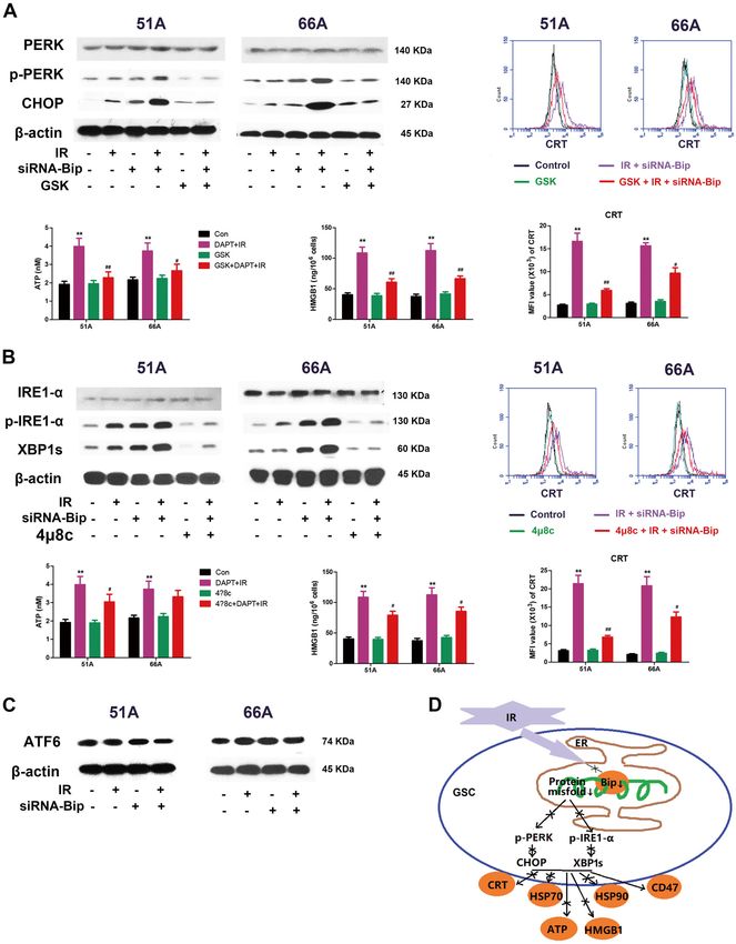
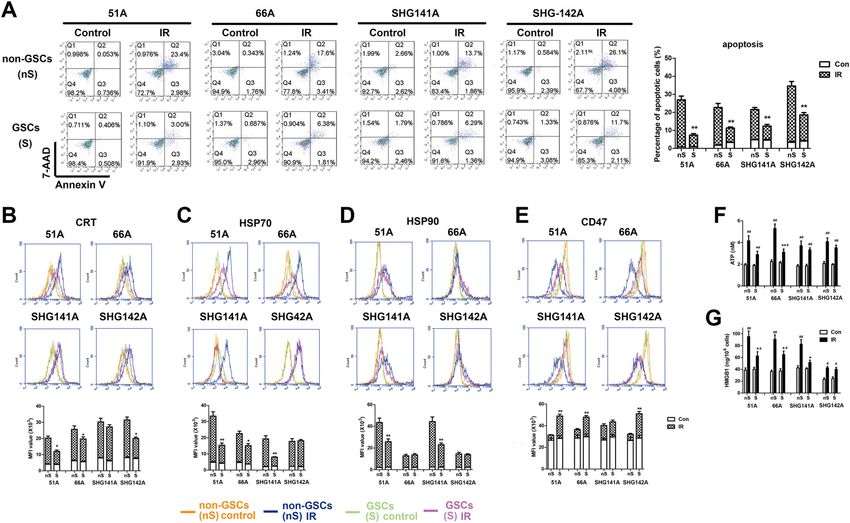
Yang et al. Cell Death and Disease (2020)11:786                                                                                      Page 2 of 13

disease relapse in glioma patients. Augment of RT-                         Results
induced endoplasmic reticulum (ER) stress might block                      IR induces less DAMP molecules exposure and release in
self-recovery of GSCs and make cells to die. As a broad                    GSCs comparing to non-GSCs
specificity molecular chaperone within ER, binding                            The results of Annexin V and 7-AAD stain showed that
immunoglobulin protein (Bip), also known as 78-kDa                         less cell apoptosis was induced in GSCs comparing to non-
glucose regulated protein (GRP78), correctly folds nascent                 GSCs after 10 Gy IR (Fig. 1a). It has been shown that IR
polypeptides and regulates the unfolded protein response                   triggers ICD in cancer cells14–16. Emission of ICD hall-
(UPR) ensuring protection of the cell from denatured                       mark molecules from non-GSCs was significantly
protein and reinforcing its anti-apoptotic role, when the                  increased following 10 Gy IR (Supplementary Fig. 1). Next,
cell is under stress11. In addition, Bip is responsible for                we analyzed whether ICD can be induced by the same
maintaining “stemness” in cancer cells12,13. To demon-                     dosage of IR in cancer stem cells. The data showed less cell
strate the mechanism of GSCs resistance to IR-induced                      surface exposure of CRT, heat shock protein (HSP) 70 and
ICD, the role of Bip was evaluated in ER stress-activated                  HSP90 in all detected GSCs compared with non-GSCs
ICD. In this study, we found high-dose ionizing radiation                  except for CRT in SHG141A, HSP70 in SHG142A and
(IR) triggered fewer DAMPs molecules exposure and                          HSP90 in 66A and SHG142A (Fig. 1b–d). CD47 expres-
release comparing to non-GSCs, which made the immune                       sion on cell surface was significantly decreased after IR in
response elicited by RT insufficient to eliminate GSCs. Bip                 both GSCs and non-GSCs, and less decrease on CD47
inhibition efficiently enhanced ER stress and promoted                      expression in GSCs comparing to matched non-GSCs
IR-mediated DAMP molecules exposure and release in                         were observed except for SHG141A (Fig. 1e).
GSCs. These data suggested that promoting GSCs ICD                           We further detected the release of ATP by chemolu-
should be a promising strategy to prevent or delay post-                   minescent assay and HMGB1 using Elisa assay. We
radiotherapy recurrence of GBM.                                            observed the release of ATP (Fig. 1f) and HMGB1 (Fig. 1g)

  Fig. 1 IR induces less cell apoptosis, immunogenic molecule exposure and release in GSCs compared with non-GSCs. GSCs and non-GSCs
  were incubated for 24 h after 10 Gy IR for apoptosis assay and detection of CRT, HSP90, HSP70, CD47, ATP, and HMGB1. a Cell apoptosis was
  determined by Annexin V and 7-AAD staining. The expressions of CRT (b), HSP70 (c), HSP90 (d), and CD47 (e) on cell surface were measured by flow
  cytometry. The level of extracellular ATP (f) using a chemiluminescence assay and HMGB1 release (g) in supernatant using an elisa assay were
  detected. *P < 0.05, **P < 0.01 vs matched non-GSCs, #P < 0.05, ##P < 0.01 vs control.

Official journal of the Cell Death Differentiation Association
Yang et al. Cell Death and Disease (2020)11:786                                                                                          Page 3 of 13

produced by IR from all detected GSCs and non-GSCs.                           24 h following 10 Gy IR were detected when GSCs were
IR-induced less ATP release in supernatant of 51A and                         pretreated with Bip inhibitor or siRNA-Bip transfection.
66A GSCs and less HMGB1 release in 51A, 66A and                                 We first found significant upregulation of Bip expres-
SHG141A GSCs compared with matched non-GSCs.                                  sion in all detected GSCs comparing to non-GSCs (Sup-
                                                                              plementary Fig. 2), and downregulation of Bip expression
Bip inhibition increased IR-induced ER stress and ICD in                      was showed postirradiation in non-GSCs but not GSCs
GSCs                                                                          (Fig. 2a). As shown in Fig. 2b, we next found the
  Emerging evidences demonstrate the exposure and                             expression levels of ER stress associated protein were
release of ICD hallmark molecules through ER stress9,                         upregulated in GSCs 51A and 66A post-IR. Pretreatment
which causes an overload and misfold of proteins in the                       with tauroursodeoxycholic acid (TUDCA), a chemical
ER11. To demonstrate whether stemness maintenance                             chaperone for blocking ER stress, of 2.5 mM for 6 h
resulting from Bip upregulation gave rise to less DAMPs                       effectively inhibited expression of ER stress associated
exposure and release in GSCs, specific ICD molecules                           protein. To measure whether Bip downregulation can

  Fig. 2 Bip inhibition promoted IR-mediated ER stress and ICD generation. a Bip expression was detected using western blot in GSCs and non-
  GSCs after cells were irradiated with 10 Gy. b EGCG or siRNA-Bip enhanced the increase of IR-induced ER stress associated protein in GSCs. GSCs were
  transfected with siRNA-Bip or pcDNA-Bip. After pretreatment with or without 2.5 mM TUDCA for 6 h, cells were irradiated with 10 Gy, then indicated
  protein expressions were detected by western blot. c The level of CRT exposure on cell surface was measured using flow cytometry, and MFI was
  analyzed. Cells were transfected or untransfected with siRNA-Bip following 10 Gy radiation. d The level of extracellular ATP was measured using a
  chemiluminescence assay. e HMGB1 in supernatant was detected using an elisa assay. *P < 0.05, **P < 0.01 vs control, #P < 0.05, ##P < 0.01 vs IR.

Official journal of the Cell Death Differentiation Association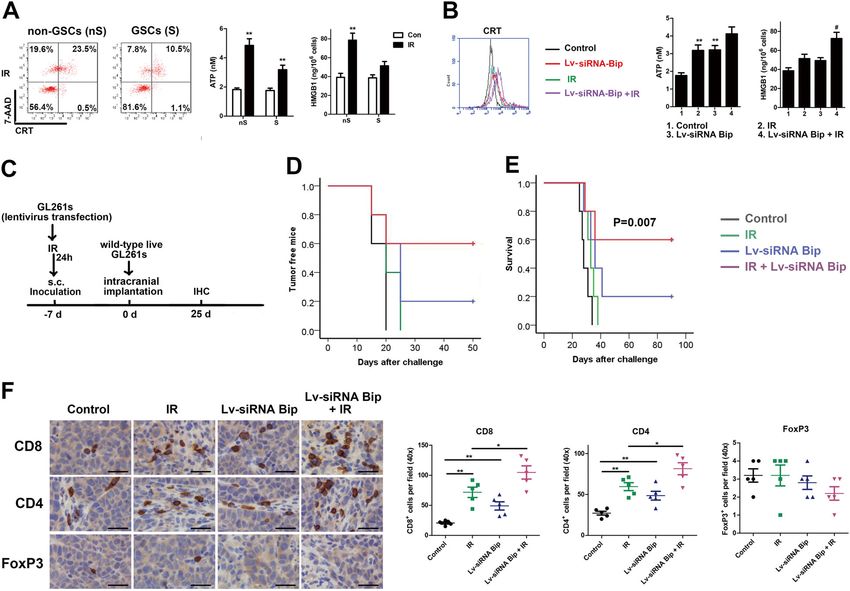
Yang et al. Cell Death and Disease (2020)11:786 Page 4 of 13
promote ER stress in GSCs after IR, a Bip inhibitor, epi- phosphorylated IRE1-α and XBP1s protein coupled with
gallocatechin gallate (EGCG, Sigma Aldrich)17, was used reduction in CRT exposure, extracellular ATP level and
to inhibit protein synthesis. The results showed that HMGB1 release in IR and siRNA-Bip treating cells. These
expressions of ER stress associated protein were sig- finding suggested that IRE1-α pathway was also involved
nificantly upregulated by 50 μM EGCG pretreatment for in ICD induced by IR and Bip inhibition (Fig. 3b).
24 h and more obvious following IR (Fig. 2b and Sup- For ATF6 pathway analysis, we found no difference in
plementary Fig. 3). EGCG showed similar function on cleavage of ATF6 in GSCs with or without Bip knock-
increasing expressions of ER stress associated protein as down after irradiation. We could exclude the participation
Bip knockdown (siRNA-Bip transfection in GSCs) fol- of ATF6 pathway activation in anti-ICD efficacy of Bip
lowing IR. The results suggested Bip inhibition or upregulated GSCs. (Fig. 3c).
knockdown efficiently triggered IR-mediated ER stress in Together, these findings advocate that IR can promote
GSCs. partial UPR protein expression in GSCs, and the Bip
Transfection with siRNA-Bip increased the exposure of inhibition enhances radiation-induced ICD, which is
CRT on cell surface (Fig. 2c), increased extracellular ATP mediated by ER stress and is specifically dependent on
level (Fig. 2d) and HMGB1 release (Fig. 2e) induced by IR PERK and IRE1-α pathways (Fig. 3d).
in 51A and 66A GSCs, suggesting the increase of IR-
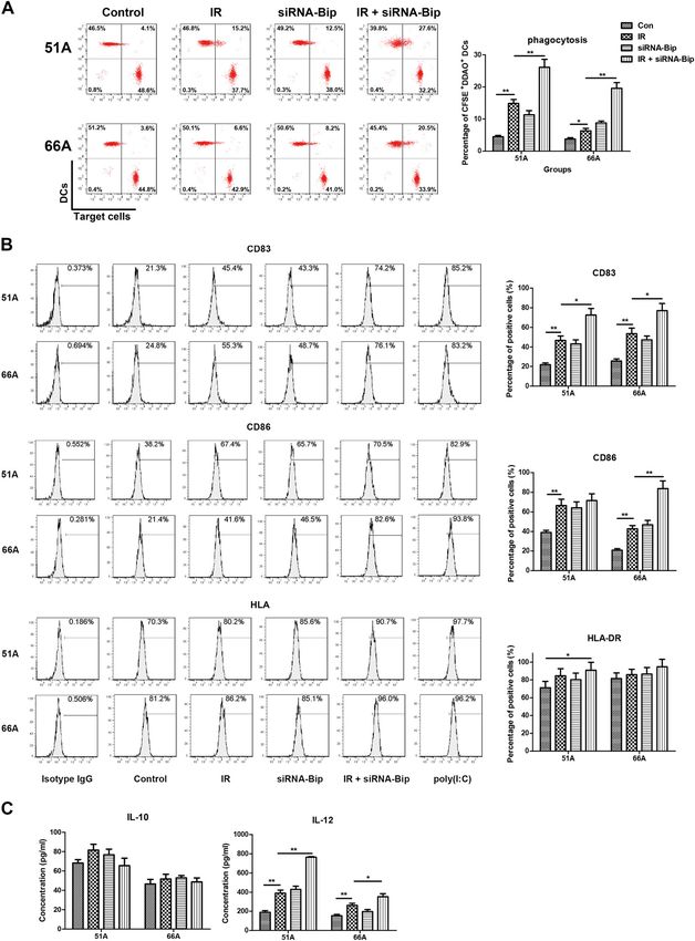
induced ICD hallmark molecules by Bip inhibition in Bip inhibition elevates phagocytosis of DCs on IR-treated
GSCs. Meanwhile, Bip overexpression decreased the GSCs
change of IR-induced ICD hallmark molecules in non- Dying tumor cells with CRT translocation on cell sur-
GSCs (Supplementary Fig. 4). face were engulfed by dendritic cells (DCs)19. We ana-
lyzed whether Bip knockdown and IR-induced ICD on
Bip inhibition enhances IR-mediated PERK and IRE1-α GSCs would impact on phagocytosis of DCs. As shown in
activation Fig. 4a, the percentage of DCs phagocytosis is (26.1 ±
The mechanism of ICD induction is associated with ER 2.5)% in 51A and (19.6 ± 1.8)% in 66A GSCs in combi-
stress, a conserved cellular program that tries to cope with nation of siRNA-Bip transfection and IR treatment.
UPR resulting from dysfunctions18. In normal cells, the The capability of IR and/or siRNA-Bip transfection
UPR is regulated by three major pathways involving treated cells to induce the maturation of monocyte-
activating protein kinase RNA-like ER kinase (PERK), derived DCs was examined by flow cytometry. Phagocy-
inositol-requiring protein 1-α (IRE1-α) and activating tosis of IR-treated cells yielded a mature-DC phenotype of
transcription factor 6 (ATF6)18. CD83 and CD86 as compared to control, and combina-
For PERK pathway analysis, we observed no increased tion of IR and siRNA-Bip transfection increased DCs
phosphorylated PERK expression following irradiation in maturation, which was evidenced by increased expression
51A and 66A GSCs. Transfection with siRNA-Bip before of specific markers including CD83, CD86 and HLA,
IR significantly upregulated the expression of phos- except for CD86 expression in 51A GSCs and HLA
phorylated PERK and CHOP, relative to IR treatment expression (Fig. 4b). Isotype IgG or poly(I:C) stimulation
alone, using western blot assay (Fig. 3a). To examine the was used as negative or positive control, respectively.
role of the PERK pathway in Bip inhibition-enhanced UPR Transfection with siRNA-Bip increased the production of
in GSCs, GSK2606414, a specific pharmacological inhi- an important cytokines related to DCs function, IL-12,
bitor of PERK phosphorylation, was used. When GSCs from DCs after IR in detected cells, however, the level of
were treated with GSK2606414+IR+siRNA-Bip, reduc- IL-10 showed no obvious change (Fig. 4c). These data
tion in the levels of phosphorylated PERK was accom- indicate that ICD-induced DCs phagocytosis, specific
panied by a significant decrease in CRT exposure on cell phenotypic maturation and cytokine secretion of IR-
surface, extracellular ATP level and HMGB1 release, treated GSCs are increased by Bip knockdown.
indicating the involvement of PERK signaling in Bip-
inhibited immunogenic death (Fig. 3a). Bip downregulation of GSCs is critical for activation of
For IRE1-α pathway analysis, the expression of phos- effector T lymphocyte
phorylated IRE1-α in 51A and 66A GSCs and splicing T lymphocyte stimulatory capacity of DCs pulsed with
XBP1 (XBP1s) in 51A GSCs was upregulated following siRNA-Bip and/or IR-treated cells was evaluated after
irradiation. SiRNA-Bip transfection further enhanced the T cells were co-incubated with the target GSCs. The
expression of phosphorylated IRE1-α and downstream results were presented in Fig. 5. Combination of siRNA-
XBP1s following irradiation (Fig. 3b). To investigate the Bip transfection and IR treatment resulted in more Ki67+
role of IRE1-α pathway in GSCs with Bip knockdown and and CD137+ T cells in 51A GSCs as target cells (Fig. 5a), a
IR treatment, we used 4μ8c, a specific inhibitor of IRE1-α. significantly higher production of IFN-γ in 51A GSCs
Pretreatment with 4μ8c showed a significant decrease in (Fig. 5b), and an elevated cell lysis in 51A and 66A GSCs
Official journal of the Cell Death Differentiation Association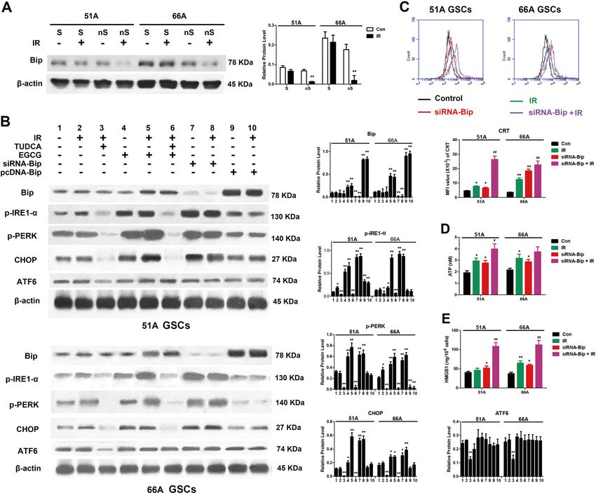
Yang et al. Cell Death and Disease (2020)11:786                 Page 5 of 13

  Fig. 3 (See legend on next page.)

Official journal of the Cell Death Differentiation Association
Yang et al. Cell Death and Disease (2020)11:786 Page 6 of 13
(see figure on previous page)
Fig. 3 Bip knockdown enhances the activation of IR-induced specific UPR branch pathways. GSCs transfected or untransfected with siRNA-Bip
were treated with IR 10 Gy in the presence or absence of UPR pathway inhibitors, then UPR proteins and DAMPs were detected at 24 h after
irradiation. a The changes of PERK pathway proteins and DAMPs were analyzed with or without pretreatment with 2 μM GSK2606414, a specific PERK
pathway inhibitor, for 1 h before irradiation. b The expressions of IRE1-α pathway proteins and DAMPs were measured in presence or absence of
10 μM 4u8c, a specific IRE1-α pathway inhibitor, for 1 h before irradiation. c Protein level of ATF6 was determined by immunoblot. d The mechanism
of IR-mediated decreased ICD in GSCs. *P < 0.05, **P < 0.01 vs control, #P < 0.05, ##P < 0.01 vs IR.
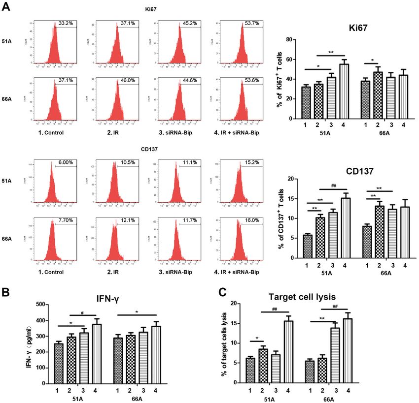
(Fig. 5c) compared with IR alone. These data again indi- immunity induced by high-dose irradiation and Bip inhi-
cating that Bip inhibition triggered a more potent anti- bition promoted antitumor efficacy of radiochemotherapy.
glioma immune response compared with IR alone.
The relative analysis of Bip expression and immune cells
GSCs transfected with Bip siRNA following IR as a vaccine infiltration in GBM patients
prevent efficiently from tumor generation We analyzed the relationship of Bip gene (HSPA5) and
Less ICD hall markers were showed after mouse GSCs immune cells infiltration in glioma patients using the web-
GL261s comparing to matched non-GSCs GL261 were site of Tumor Immune Estimation Resource (https://
irradiated at a dose of 10 Gy (Fig. 6a). Stable transfection cistrome.shinyapps.io/timer/). The analyzed result showed
with siRNA-Bip lentivirus significantly enhanced the that Bip expression was negative correlation with CD8+ T
production of DAMPs molecules in GL261s (Fig. 6b). To cell infiltration (Fig. 7d) and GBM patients’ survival (Fig. 7e),
estimate whether high-dose-IR and Bip inhibition-treated consistent with our results in vitro and animal models.
cells have an effect of preventing the generation of glioma
from GSCs as a vaccine in vivo, the syngenic C57BL/6J Discussion
mice were administrated with lentivirus transfection- and/ GBM is well-known to contain few immune cells in its
or IR-treated GL261s GSCs subcutaneously before microenvironment19. Immunotherapy for glioma malig-
untreated GL261s GSCs were implanted intracranially for nancies has met significant challenges. Research results
tumor model establishment. As shown in Fig. 6d, e, pre- are inconsistent about whether neoadjuvant PD-1 regi-
treatment with siRNA-Bip- and IR-treated GL261s GSCs men significantly improved both overall and progression-
was highly efficient in preventing the growth of live free survival of GBM patients in clinical trials20,21. How-
GL261s injected 7 days later. The efficacy of vaccination ever, more immune cell infiltration was observed in long-
from IR-treated GL261s was attenuated without Bip- term responders, supporting a local immuno-modulatory
siRNA transfection. These results confirmed that GSCs effect of treatment21.
with Bip inhibition following IR as a vaccine could prevent The biologic features of stem cells are relevant to
tumor generation efficiently. immune therapies, since stem cells are essential for the
Next, immune cell subset were evaluated by IHC stain on repair of tissue damage22. In this study, we analyzed the
formalin fixed paraffin embedded (FFPE) tissues (Fig. 6f). mechanism of GSCs resistance to RT-induced immune
Increase of CD4+ and CD8+ T cells were observed in the response to protect themselves from immune damage,
group of Lv-siRNA-Bip transfection and IR combination and found the notable correlation of GSCs and IR-
comparing to other treatment groups, indicating the car- induced ICD. To promote DAMP molecules exposure
dinal role of Bip in facilitating high-dose irradiation and release following IR in GSCs, the knockdown of Bip,
induced immunogenicity. an ER stress sensor, was designed to enhance IR-induced
ICD. Our results demonstrated that Bip downregulation
Bip inhibition- and IR-induced immunity has a significantly elicited apoptosis, ER stress and DAMPs molecules
adjuvant antitumor efficacy to radiochemotherapy in vivo exposure on cell surface and extracellular release. The
To determine whether the immunity induced by high- phagocytosis and maturation of DCs and the proliferation
dose-IR and Bip inhibition refuses tumor rechallenge and of T lymphocytes following IR were augmented when
promotes antitumor efficacy of radiochemotherapy, the s.c. GSCs were transfected with siRNA-Bip. Combination of
allografts of C57BL/6J mice were treated with Bip inhibitor Bip inhibition and IR increased CD4+ and CD8+ immune
and 10 Gy irradiation, then primary tumors were resected, cells and decreased Treg cells infiltration in vivo. Notable
and mice were rechallenged intracranially with live GL261s prevention function was showed using Bip knockdown-
(Fig. 7a). Radiochemotherapy plus Bip inhibitor and IR- and IR-treated GSCs as a vaccine, and tumor delayed
induced immunity efficiently extended survival of glioma growth and longer survival were showed following Bip
bearing mice (Fig. 7b), and reduced tumor sizes after inhibition treatment and local IR far from tumor
GL261s rechallenge (Fig. 7c). These results suggested the implantation region.
Official journal of the Cell Death Differentiation Association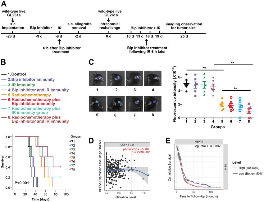
Yang et al. Cell Death and Disease (2020)11:786                 Page 7 of 13

  Fig. 4 (See legend on next page.)

Official journal of the Cell Death Differentiation Association
Yang et al. Cell Death and Disease (2020)11:786                                                                                            Page 8 of 13

  (see figure on previous page)
  Fig. 4 ICD induced phagocytosis of IR-treated GSCs, DCs phenotypic maturation, cytokine production. DCs were generated from PBMCs of
  three healthy doors after culture of adherent monocytes with rhGM-CSF and rhIL-4 for 7 days. GSCs transfected with or without siRNA-Bip were
  irradiated with 10 Gy and incubated for 24 h, then were added to human DCs at ratio 1:1 and incubated for additional 24 h, then the characteristics of
  DCs were determined. a The percentage of DCs positive for CFSE dye after co-culture with GSCs for far red dye was assessed by flow cytometry, and
  the percentage of double positive cells (DCs/GSCs) was calculated. b Specific markers expression of DCs maturation was evaluated by flow cytometry
  using monoclonal antibodies. c The levels of IL-10 and IL-12 was examined from cell culture supernatants by elisa. #P < 0.05, ##P < 0.01.

  Fig. 5 Effector T lymphocyte activation were notably occurred in GSCs of combinative treatment with IR and Bip knockdown. DCs were co-
  cultured with autologous isolated T lymphocytes after pulsed with siRNA-Bip- and/or IR-treated GSCs. Activated T lymphocytes as effector cells were
  re-stimulated with target GSCs, then Ki67+ and CD137+ T lymphocytes using flow cytometry and IFN-γ secretion in supernatant using elisa were
  detected. Target cells were labeled with CFSE, and co-cultured with unlabeled T lymphocytes pulsed by DCs. Lysis of target cells was measured using
  7-AAD stain. The percentage of Ki67+ and CD137+T lymphocyte (a), IFN-γ secretion in supernatant (b) and percentage of target cell lysis (c) were
  analyzed after target cells were added to DCs-activated T lymphocyte.

Official journal of the Cell Death Differentiation Association
Yang et al. Cell Death and Disease (2020)11:786                                                                                                 Page 9 of 13

  Fig. 6 GSCs treated with Bip inhibition following high-dose IR as a vaccine prevent efficiently tumor generation. GL261s GSCs with lentivirus
  transfection following 10 Gy IR were inoculated subcutaneously into the left flank of C57BL/6J mice, then untreated live cells were intracranially
  implanted into the mouse cerebrum 7 days later. a IR induces ICD hallmark molecules exposure in mouse GSCs and non-GSCs in vitro. Mouse glioma
  cell line GL261 and GSCs GL261s were irradiated with 10 Gy, CRT on cell surface, ATP and HMGB1 release in supernatant were measured 24 h after IR
  treatment. b Bip knockdown promoted IR-mediated ICD in mouse GSCs GL261s. GL261s transfected or untransfected with siRNA-Bip lentivirus were
  irradiated with 0 or 10 Gy, ICD hallmark molecules were detected 24 h later. *P < 0.05, **P < 0.01 vs control, #P < 0.05, ##P < 0.01 vs IR. c The schedules
  of preventing treatment were indicated. d Tumor-free mice were calculated after live GL261s implantation. e Survival curves of C57BL/6 mice was
  analyzed after live GL261simplantation. f Immune subsets were showed using anti-CD4, CD8 and FoxP3 antibodies in intracranial allografts 25 days
  after cell implantation. Average number of stained cells were showed. Scale bar = 50 μm, *P < 0.05, **P < 0.01.

  A new strategy suggests a combination of immune                                theory, increased Bip reduces protein misfold in GSCs to
intervention and local irradiation for solid tumor, due to                       maintain GSCs growth and stemness characteristics.
irradiated tumors triggering a better antitumor immune                           Our results in western blot showed higher Bip expres-
response (6–8). Previous studies showed that cancer cells                        sion levels in GSCs comparing to non-GSCs, which was
exposed to a high dose of RT14 or chemotherapy reagent                           consistent with previous studies12,13. We hypothesized
in vitro, such as mitoxantrone23, oxaliplatin24, and dox-                        that knockdown of Bip augmented misfold of nascent
orubicin25, were capable of vaccinating syngeneic mice                           polypeptides, which would induce ER stress and UPR,
against a subsequent challenge with living cells of the                          and trigger GSCs ICD. In order to verify whether fewer
same type. In this study, we demonstrated fewer DAMPs                            DAMPs release was associated with high Bip expression,
were exposed and released in IR-treated GSCs, resistant                          siRNA-Bip was used to investigate the change of IR-
to RT and chemotherapy, compared with non-GSCs. Less                             induced GSCs ICD. Our results showed Bip knockdown
ICD generation becomes one mechanism of GBM                                      increased IR-induced apoptosis, the translocation of
recurrence. New regimens for promoting GSCs ICD may                              CRT, HSP70 and HSP90, the release of ATP and
be efficient to inhibiting GBM recurrence.                                        HMGB1, and decreased CD47 expression on cell sur-
  ER stress initially serves as an adaptive measure to                           face, suggesting IR-induced ICD enhancement by Bip
protect the cell from irreversible damage in cancer11. In                        inhibition.

Official journal of the Cell Death Differentiation Association
Yang et al. Cell Death and Disease (2020)11:786                                                                                          Page 10 of 13

  Fig. 7 Bip inhibition and IR immunity following radiochemotherapy have a significant antitumor efficacy, and Bip expression is relative to
  immune infiltration. C57BL/6J mice were subcutaneously implanted with untreated GL261s, then the allografts were irradiated and treated with Bip
  inhibitor. After removal of primary tumors, the mice were intracranially implanted with untreated live GL261s cells. a The schedules of therapeutic
  regimen were indicated. b Survival curves of mice was analyzed under indicated therapeutic regimen. c Tumor size were measured after live GL261s
  rechallenge. d Bip gene expression has a negative correlation with CD8+ T cell infiltration. e Bip gene expression and GBM patients’ survival showed a
  negative correlation. *P < 0.05, **P < 0.01.

  The accumulation of misfolded polypeptides in the ER                        dissociation of BiP from PERK and IRE1-α, thus
lumen results in UPR, which is involved in the molecular                      removing its inhibitory effects26.
mechanisms underlying ICD hallmark18. Our results
showed Bip inhibition activated two of three UPR signal                       Materials and methods
pathways induced by IR, including PERK and IRE1-α                             Primary cell culture of human GSCs and cell lines
pathways, in radioresistant GSCs. PERK activated by                             GSCs 51A and 66A are from GBM patients, as gifts
combined treatment of Bip inhibition and IR was phos-                         from Professor Yihong Zhou at the UC Irvine Brain
phorylated and selectively enhanced CHOP expression.                          Tumor Research Laboratory. GSCs SHG141A and
Similarly, combination of Bip inhibition and IR treat-                        SHG142A were isolated from GBM surgical specimen in
ment dissociated IRE1-α from BiP, and IRE1-α under-                           the First Affiliated Hospital of Soochow University. GSCs
went dimerization and autophosphorylation, then                               were cultured in DMEM/F12 medium that was supple-
activated and spliced the target, XBP1. The results were                      mented with 10% fetal bovine serum (FBS) to induce
consistent with the mechanism of BiP binding to the ER                        differentiation for one month, considering as non-GSCs.
luminal domains, preventing homodimerizatio26. In the                         Mouse glioma cell line GL261 were cultured in high
presence of cellular stress, accumulation of misfolded                        glucose DMEM with 10% FBS. Then these cells were
proteins within the ER bound BiP competitively, causing                       cultured in stem cell medium for 12 passages to produce

Official journal of the Cell Death Differentiation Association
Yang et al. Cell Death and Disease (2020)11:786                                                               Page 11 of 13

GSCs GL261s, and cells were used as GSCs after identi-          conjugated anti-rabbit and anti-mouse secondary anti-
fication. All cells are isolated, cultured and identified as      bodies were used and the chemiluminescent signal was
previously described27.                                         detected by using electrochemiluminescence (ECL)
                                                                reagents (Invitrogen).
Ionizing radiation
  The cells and s.c. allografts of mice were exposed to         ATP and HMGB1 assays
10 Gy X-ray (160 kV) at room temperature using a linear           5 × 10⁵ cells following IR were incubated for 24 h, then
accelerator (RadSource, Suwanee, GA, USA) at a dose rate        extracellular ATP and HMGB1 release were measured.
of 0.50 Gy/min. Radiotherapy on intracranial allografts of      Extracellular ATP after IR was measured by an ATP Assay
mice was subjected to 8 Gy (2 Gy/d once every 3 days for        Kit     (Promega,      Madison,     USA)     based      on
4 times) X-ray irradiation (6 MV, the dose rate was             luciferin–luciferase conversion following the manu-
100 cGy/min) by a PRIMUS accelerator (SIEMENS                   facturer’s instructions. The chemoluminescent signal was
Medical Solutions, Erlangen, Germany) at room tem-              read by a Synergy Neo2 Hybrid Multi-Mode Reader
perature. Irradiation was locally confined to the tumors by      (BioTek, USA). HMGB1 in supernatant was determined
shielding the rest of the body with lead.                       by a HMGB1 ELISA kit (Biorbyt, UK) according to
                                                                manufacturer’s instructions. The microplates were read
Flow cytometry                                                  using a multiskan spectrophotometer model 1510
  Neurospheres were dissociated into single cells, and          (Thermo Fisher Scientific, Finland) for protein con-
adherent cells were trypsinized and collected. Cells were       centration assessment.
stained using FITC-Annexin V/7-AAD apoptotic Kit
(Biolegend) according to the manufacturer’s instructions.       SiRNA and plasmid transfection
Cells were incubated with antibodies for 30 min at room           For upregulation or downregulation of Bip, siRNA-Bip
temperature. The primary antibodies include APC-                or pcDNA-Bip, purchased from Shanghai Genepharma
conjugated nestin mouse antibody (1:10; Invitrogen),            Co., Ltd., was transfected into 51A and 66A GSCs using
PE-conjugated CD133 antibody (1:11; Miltenyi Biotech            Lipofectamine 2000 (invitrogen) according to the manu-
GmbH), PE-conjugated anti-CRT rabbit antibody (1:50,            facturer’s instructions. The cell were transfected with
Cell Signaling), PE-conjugated anti-HSP90 rabbit mAb            nontargeting control (NTC) siRNA as a control.
(1:50, Cell Signaling), Alexa Fluor® 488-conjugated anti-
HSP70 rabbit antibody (1:500, Abcam), FITC-conjugated           DCs generation and maturation
anti-CD47 mouse antibody (1:20, eBioscience™), PE-                Human peripheral blood mononuclear cells (PBMCs)
conjugated anti-HLA-DR mouse antibody (1:20, Cell               from healthy donors were purified by Ficoll-Hypaque
Signaling), PE-conjugated anti-CD83 mouse antibody              gradient centrifugation of heparinized blood. PBMCs
(1:20, eBioscience™), PE-conjugated anti-CD86(B7-2)             were divided into two parts, one half was used for DCs
mouse antibody (1:20, eBioscience™), PE-conjugated anti-        culture, and the other half was frozen until they were used
Ki67 rabbit antibody (1:50, Cell Signaling), PE-conjugated      as effector cell production in later experiments.
anti-CD137 rabbit antibody (1:50, Cell Signaling). Labeled      Monocyte-derived DCs were obtained from CD14+ per-
cells were analyzed by a flow cytometic Beckton Dick-            ipheral blood monocytes isolated using magnetic beads
inson FACScan (BD Biosciences). Data were analyzed              (MiltenyiBiotec). Immature DCs (iDCs) were obtained by
using FlowJo Software version 7.0 and presented by mean         culturing for seven days in RPMI-1640 medium in pre-
fluorescence intensities (MFI) or positive cell number.          sence of 10% FBS, 800 U/ml rhGM-CSF and 50 ng/ml
                                                                rhIL-4. Immature DCs were stained with CellTraceTM
Western blot analysis                                           CFSE cell proliferation Kit (Invitrogen) for flow cyto-
  Cell lysates were prepared in RIPA lysis buffer con-          metry. GSCs were stained with CellTraceTM Far Red cell
taining phenylmethylsulfonyl fluoride (PMSF). The pro-           proliferation Kit (Invitrogen). 1 × 10⁶ NTC, irradiated
tein samples were separated by 10% sodium dodecyl               and/or siRNA-Bip transfected GSCs were added to iDCs
sulfate-polyacrylamide gel (SDS-PAGE), then were                in 1:1 ratio for 24 h culture for analysis of phagocytosis.
transferred onto the polyvinylidene fluoride (PVDF)              DCs that phagocytosed GSCs were double positive cells of
membrane. The membrane was then blocked with 5%                 CFSE and far red using flow cytometry. Phenotypical
non-fat dry milk for 1 h. Primary rabbit antibodies include     assessment of DCs was determined by flow cytometry
anti-Bip (1:1000, Cell Signaling), anti-CHOP (1:1000, Cell      using fluorescein labeled monoclonal antibodies to HLA-
Signaling), anti-PERK (1:1000, Cell Signaling), phospho-        DR, CD83 and CD86. iDCs were incubated with 25 μg/mL
PERK (1:1000, Invitrogen), IRE1-α (1:1000, Cell Signal-         poly(I:C) (Polyinosinic–polycytidylic acid sodium salt,
ing), p-IRE1-α (1:1000; Invitrogen), splicing XBP1 (1:1000,     Sigma-aldrich) for 24 h as a positive control of DCs
Cell Signaling), ATF6 (1:1000, Cell Signaling). HRP-            maturation. IL-10 (Human IL-10 Pre-Coated ELISA Kit,

Official journal of the Cell Death Differentiation Association
Yang et al. Cell Death and Disease (2020)11:786 Page 12 of 13
BioGems) and IL-12 (Human IL-12 (p70) Pre-Coated with live GL261s GSCs. Mice were monitored daily until
ELISA Kit, BioGems) concentrations in cell culture severe neurological deficits appeared. No neurological
supernatants were determined according to the manu- deficit syndrome appearance was considered tumor-free
facturer’s instructions. mice at 50 day. The indicated treatment schedules were
given in Fig. 6c. Survival analysis was used to compare the
T cell isolation, target cell proliferation, and lysis assays differences of each group according to survival time.
T lymphocytes were purified by pan T cell isolation kit
(MiltenyiBiotec GmbH) from PBMCs, and labeled by Antitumor therapy
CFSE.DCs were pulsed with NTC, siRNA-Bip and/or IR- Flank tumors were established by subcutaneous injec-
treated GSCs, then co-cultured with autologous isolated T tion of 5 × 10⁶ live GL261s GSCs suspension in a volume
lymphocytes at ratio 10:1 for 7 days. 50 U/mL IL-2 was of 100 μL into the left flank. C57BL/6J mice with sub-
added on days 3 and 5. T lymphocytes as effector cells for cutaneous tumor were randomly divided into 8 groups:
antitumor immune response study were re-stimulated (1) Control group, no treatment; (2) Bip inhibitor
with target GSCs (1 × 10⁴ cells in 100 μl) treated with immunity group, Bip inhibitor was administrated when
original samples at an effector-to-target ratio (E:T) ratio subcutaneous tumor formation; (3) IR immunity group,
of 10:1 for 12 h. Flow cytometry detected Ki67+ and subcutaneous tumor was irradiated; (4) Bip inhibitor and
CD137+ T lymphocytes, and Elisa assay was used for IFN- IR immunity group, the mice was administrated with
γ secretion in supernatant. Lysis of target cells was mea- combination of Bip inhibitor and irradiation treatment
sured using flow cytometry. Target cells were labeled with when subcutaneous tumor formation; (5) radio-
far red, and co-cultured in a 96 U-bottom well plate with chemotherapy group, the mice was treated with fractio-
T lymphocytes pulsed by DCs. Dead cells were detected nated radiation and Bip inhibitor after rechallenge with
by flow cytometry using 7-AAD stain. GL261s GSCs; (6) radiochemotherapy plus Bip inhibitor
immunity group, the mice was treated with Bip inhibitor
Generation of stable cell lines after subcutaneous tumor formation, then fractionated
GL261s GSCs with stable integration of Bip cDNA and radiation and Bip inhibitor after intracranial GSCs
Bip-siRNA sequences were generated through lentiviral- rechallenge; (7) radiochemotherapy plus IR immunity
mediated gene transfection (Shanghai GenePharma Co., group, allograft was irradiated after subcutaneous tumor
Ltd., Shanghai, China) according to the manufacturer’s formation, then the mice was treated with fractionated
instructions. More than 90% of GSCs with the stably radiation and Bip inhibitor after rechallenge; 8. Radio-
integrated gene were considered GSCs of Bip over- chemotherapy plus Bip inhibitor and IR immunity group,
expression or Bip knockdown. the mice was administrated with combination of Bip
inhibitor and irradiation treatment when subcutaneous
Antitumor vaccination tumor formation, then fractionated radiation and Bip
The male syngenicC57BL/6J mice about 18–20 g were inhibitor after rechallenge.
bred and housed in a specific pathogen free condition. All Bip inhibitor EGCG was dosed at 25 mg/kg in a volume
animal experimental protocols were approved by the of 200 μL by intraperitoneal (i.p.) injection 15 days after
Institutional Animal Care and Use Committee of Soo- GL261 GSCs inoculation. Focal radiation was delivered in
chow University and complied with the code of ethics for one fraction of total 10 Gy at 6 h after Bip inhibitor
animal experimentation. treatment. Seven days postirradiation, subcutaneous
Twenty mice were randomly divided into four groups tumors were removed, and two days later mice were
including control (transfection with scramble lentivirus), rechallenged intracranially with 1 × 10⁵ untreated live
IR (transfection with scramble lentivirus following IR), Lv- GL261s GSCs. The mice were administrated i.p. injection
siRNA-Bip (transfection with siRNA-Bip lentivirus) and of Bip inhibitor at 25 mg/kg on day 10, 13, 16 and 19 and
IR plus Lv-siRNA-Bip (transfection with siRNA-Bip len- local IR 6 h later with the dose of 2 Gy after intracranial
tivirus following IR). GL261s GSCs stably transfected with rechallenge. Tumor size was visualized by IVIS image on
lentivirus were irradiated with 10 Gy, then cultured for intracranial postimplantation day 25, and survival time
24 h. 1 × 10⁷treated GL261s cells were inoculated sub- was recorded for survival analysis. The indicated treat-
cutaneously into the left flank of male syngenic C57BL/6J ment schedules were given in Fig. 7a.
mice as a vaccine. Whereas 1 × 10⁵ untreated live cells
were intracranially implanted into the frontal lobe of the Immunohistochemical (IHC) staining
mouse cerebrum by stereotactic implantation to establish Tumor tissues were fixed in paraffin, imbedded and cut
intracranial transplantation 7 days later. In vivo imaging for 4 mm sections. Tumor sections were incubated with
system (IVIS) 50 system was used to monitor the allo- primary antibodies, including anti-mouse CD4 (1:100,
grafts at day 15 and day 20 after intracranial implantation Cell Signaling), CD8 (1:400, Cell Signaling) and FoxP3
Official journal of the Cell Death Differentiation AssociationYang et al. Cell Death and Disease (2020)11:786 Page 13 of 13
(1:100, Cell Signaling) antibodies, at 4 °C overnight, and 2. Louis, D. N. et al. The 2016 World Health Organization Classification of Tumors
biotin-labeled secondary antibody for 30 min at 37 °C. The of the Central Nervous System: a summary. Acta Neuropathol. 131, 803–820
(2016).
final signal was developed using the 3,3′-diaminobenzi- 3. Lathia, J. D., Mack, S. C., Mulkearns-Hubert, E. E., Valentim, C. L. & Rich, J. N.
dine (DAB) substrate and the sections were observed Cancer stem cells in glioblastoma. Genes Dev. 29, 1203–1217 (2015).
under optical microscope. 4. Morgan, M. A. & Canman, C. E. Replication stress: an Achilles’ heel of glioma
cancer stem-like cells. Cancer Res. 78, 6713–6716 (2018).
5. Rivera, M., Sukhdeo, K. & Yu, J. Ionizing radiation in glioblastoma initiating cells.
Statistical analysis Front Oncol. 3, 74 (2013).
All samples in vitro were carried out in triplicate inde- 6. Rodríguez-Ruiz, M. E., Vanpouille-Box, C., Melero, I., Formenti, S. C. & Demaria, S.
Immunological mechanisms responsible for radiation-induced abscopal effect.
pendent experiments and represented as mean ± SD using Trends Immunol. 39, 644–655 (2018).
GraphPad Prism 5 software. Data were statistically 7. Golden, E. B. et al. Local radiotherapy and granulocyte-macrophage colony-
determined by one-way ANOVA, and the significance stimulating factor to generate abscopal responses in patients with metastatic
solid tumours: a proof-of-principle trial. Lancet Oncol. 16, 795–803 (2015).
level was considered at a value of P < 0.05. Overall mouse 8. Postow, M. A. et al. Immunologic correlates of the abscopal effect in a patient
survivals were estimated via Kaplan–Meier survival curves with melanoma. N. Engl. J. Med. 366, 925–931 (2012).
using SPSS software version 19.0. 9. Krysko, D. V. et al. Immunogenic cell death and DAMPs in cancer therapy. Nat.
Rev. Cancer 12, 860–875 (2012).
10. Demaria, O. et al. Harnessing innate immunity in cancer therapy. Nature 574,
Conclusions 45–56 (2019).
Together, these findings supported the notion that 11. Wang, W. A., Groenendyk, J. & Michalak, M. Endoplasmic reticulum stress
associated responses in cancer. Biochim. Biophys. Acta 1843, 2143–2149 (2014).
inhibition of Bip fostered the switch from IR-induced ICD- 12. Dauer, P. et al. ER stress sensor, glucose regulatory protein 78 (GRP78) reg-
resistant GSCs to ICD-sensitive GSCs by activating UPR ulates redox status in pancreatic cancer thereby maintaining “stemness”. Cell
pathway. Improved immunogenicity of GSCs could facil- Death Dis. 10, 132 (2019).
13. Chen, H. Y. et al. The endogenous GRP78 interactome in human head and
itate T lymphocytes recognition and overcome therapy neck cancers: a deterministic role of cell surface GRP78 in cancer stemness. Sci.
resistance. Enhancing immune response in GBM micro- Rep. 8, 536 (2018).
environment by facilitating DAMPs exposure and release 14. Obeid, M. et al. Calreticulin exposure is required for the immunogenicity of
gamma-irradiation and UVC light-induced apoptosis. Cell Death Differ. 14,
in adjuvant therapy makes it a promising strategy to pre- 1848–1850 (2007).
vent tumor generation and inhibit tumor recurrence. 15. Golden, E. B. et al. Radiation fosters dose-dependent and chemotherapy-
induced immunogenic cell death. Oncoimmunology 3, e28518 (2014).
Acknowledgements 16. Golden, E. B. & Apetoh, L. Radiotherapy and immunogenic cell death. Semin.
This work was supported by the National Natural Science Foundation of China Radiat. Oncol. 25, 11–17 (2015).
(81874080, 31870844, 31570851) and A Project Funded by the Priority 17. Gurusinghe, K. R. D. S. N. S., Mishra, A. & Mishra, S. Glucose-regulated protein 78
Academic Program Development of Jiangsu Higher Education Institutions substrate-binding domain alters its conformation upon EGCG inhibitor bind-
(PAPD). Thanks for Prof. Yihong Zhou supplying 51A and 66A GSCs. ing to nucleotide-binding domain: Molecular dynamics studies. Sci. Rep. 8,
5487 (2018).
Conflict of interest 18. Kepp, O. et al. Consensus guidelines for the detection of immunogenic cell
The authors declare that they have no conflict of interest. death. Oncoimmunology 3, e955691 (2014).
19. Keskin, D. B. et al. Neoantigen vaccine generates intratumoral T cell responses
in phase Ib glioblastoma trial. Nature 565, 234–239 (2019).
20. Cloughesy, T. F. et al. Neoadjuvant anti-PD-1 immunotherapy promotes a
Publisher’s note survival benefit with intratumoral and systemic immune responses in recur-
Springer Nature remains neutral with regard to jurisdictional claims in rent glioblastoma. Nat. Med. 25, 477–486 (2019).
published maps and institutional affiliations. 21. Schalper, K. A. et al. Neoadjuvant nivolumab modifies the tumor immune
microenvironment in resectable glioblastoma. Nat. Med. 25, 470–476 (2019).
Supplementary Information accompanies this paper at (https://doi.org/ 22. Clarke, M. F. Clinical and therapeutic implications of cancer stem cells. N. Engl.
10.1038/s41419-020-03000-z). J. Med. 380, 2237–2245 (2019).
23. Obeid, M. et al. Calreticulin exposure dictates the immunogenicity of cancer
Received: 3 May 2020 Accepted: 7 August 2020 cell death. Nat. Med. 13, 54–61 (2007).
24. Tesniere, A. et al. Immunogenic death of colon cancer cells treated with
oxaliplatin. Oncogene 29, 482–491 (2010).
25. Casares, N. et al. Caspase-dependent immunogenicity of doxorubicin-induced
tumor cell death. J. Exp. Med. 202, 1691–1701 (2005).
References 26. Hetz, C. The unfolded protein response: controlling cell fate decisions under
1. Stupp, R. et al. Effects of radiotherapy with concomitant and adjuvant ER stress and beyond. Nat. Rev. Mol. Cell Biol. 13, 89–102 (2012).
temozolomide versus radiotherapy alone on survival in glioblastoma in a 27. Yang, W., Li, Y., Gao, R., Xiu, Z. & Sun, T. MHC class I dysfunction of glioma stem
randomised phase III study: 5-year analysis of the EORTC-NCIC trial. Lancet cells escapes from CTL-mediated immune response via activation of Wnt/
Oncol. 10, 459–466 (2009). β-catenin signaling pathway. Oncogene 39, 1098–1111 (2020).
Official journal of the Cell Death Differentiation AssociationYou can also read