11βHSD2 Efficacy in Preventing Transcriptional Activation of the Mineralocorticoid Receptor by Corticosterone
←
→
Page content transcription
If your browser does not render page correctly, please read the page content below

Journal of the Endocrine Society, 2021, Vol. 5, No. 11, 1–11
                                                                               https://doi.org/10.1210/jendso/bvab146
                                                                                                     Research Article

Research Article

11βHSD2 Efficacy in Preventing Transcriptional
Activation of the Mineralocorticoid Receptor by

                                                                                                                                                              Downloaded from https://academic.oup.com/jes/article/5/11/bvab146/6363522 by guest on 12 December 2021
Corticosterone
Yusuf             Ali,1,2           Maniselvan            Kuppusamy,3
Carolina Velarde-Miranda,3 Clara M. Gomez-Sanchez,4 Maria Plonczynski,2
Celso E. Gomez-Sanchez,1,2,3 and Elise P. Gomez-Sanchez2
1
 G.V. (Sonny) Montgomery VA Medical Center, University of Mississippi Medical Center, Jackson,
Mississippi 39216, USA; 2Department of Pharmacology and Toxicology, University of Mississippi Medical
Center, Jackson, Mississippi 39216, USA; 3Department of Medicine, University of Mississippi Medical
Center, Jackson, Mississippi 39216, USA; and 4Department of Surgery, University of California San
Francisco, San Francisco, California 94143 USA
ORCiD numbers: 0000-0003-4579-584X (M. Kuppusamy); 0000-0003-2936-3246 (C. M. Gomez-Sanchez); 0000-0002-9882-2082
(C. E. Gomez-Sanchez); 0000-0002-4432-4950 (E. P. Gomez-Sanchez).

Abbreviations: 11βHSD2, 11β-hydroxysteroid dehydrogenase type 2; CHO, Chinese hamster ovary cells; cDNA,
complementary DNA; doxy, doxycycline; EC50, 50% maximal mineralocorticoid receptor activation; FCS, fetal calf serum; GC,
glucocorticoid; G-luc, Gaussia luciferase; GR, glucocorticoid receptor; HRE, hormone response element; HRP, horseradish
peroxidase; HSD, hydroxysteroid dehydrogenase; MMTV, mouse mammary tumor virus; MR, mineralocorticoid receptor;
tet, tetracycline.
Received: 25 June 2021; First Published Online: 3 September 2021; Corrected and Typeset: 23 September 2021.

Abstract
Affinity of the mineralocorticoid receptor (MR) is similar for aldosterone and the gluco-
corticoids (GC) cortisol and corticosterone, which circulate at concentrations far exceeding
those of aldosterone. 11β-hydroxysteroid dehydrogenase type 2 (11βHSD2) inactivation
of GC within the immediate vicinity of the MR is credited with prereceptor specificity
for aldosterone in cells coexpressing MR and 11βHSD2. 11βHSD2 efficacy is also critical
to other recently described 11βHSD2 substrates. The aim of this work was to address
doubts that low levels of expression of 11βHSD2 in aldosterone target tissues suffice to
prevent the initiation of gene transcription by the MR activated by physiological con-
centrations of corticosterone. Cell models stably expressing an MR/Gaussia luciferase
reporter and various levels of constitutive or induced 11βHSD2 at concentrations lower
than those in rat kidney homogenates and microsomes were produced. Aldosterone and
corticosterone were equipotent transactivators of the MR reporter gene in cells without
11βHSD2. Rate of conversion of tritiated corticosterone to 11-dehydrocorticosterone in-
creased and corticosterone-induced nuclear translocation of MR decreased, as 11βHSD2
expression increased. The 50% maximal MR activation for the reporter gene stimula-
tion by corticosterone rose with increasing 11βHSD2 expression, shifting the steroid

ISSN 2472-1972

Published by Oxford University Press on behalf of the Endocrine Society 2021. This work is written by (a) US
Government employee(s) and is in the public domain in the US.
                                                                                                                           https://academic.oup.com/jes   1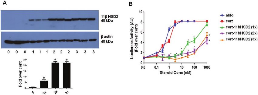
2                                                                            Journal of the Endocrine Society, 2021, Vol. 5, No. 11

dose-response curve for corticosterone-induced MR transactivation to the right. Several
stable cell lines expressing an easily and reproducibly measured MR reporter system
and consistent incremental amounts of 11βHSD2 protein were produced and used to
document that 11βHSD2 within low physiological levels inactivates relevant concentra-
tions of GC and decreases MR transactivation by GC in a dose-dependent fashion, laying
to rest doubts of the efficacy of this enzyme.
Key Words: 11βHSD2, mineralocorticoid receptor, glucocorticoids, corticosterone, Gaussia luciferase

The mineralocorticoid receptor (MR) is a member of the              were cloned and characterized by several laboratories,
steroid-thyroid hormone receptor superfamily of ligand-             as reviewed in [2]. 11βHSD2 is a high-affinity, NAD+-

                                                                                                                                       Downloaded from https://academic.oup.com/jes/article/5/11/bvab146/6363522 by guest on 12 December 2021
dependent transcription factors and has diverse functions.          dependent, obligate hydroxysteroid dehydrogenase (HSD)
It is unique among steroid hormone receptors in that it             with a Michaelis constant (4-14 nM) for corticosterone and
has 3 primary physiological agonists: aldosterone, cor-             cortisol that is low enough to be relevant to their circu-
tisol, and corticosterone [1]. Corticosterone is the main           lating levels [9]. The products, 11-dehydrocorticosterone
glucocorticoid (GC) in animals that do not express the              and cortisone, are inactive as ligands for the MR and GR.
17-hydroxylase in the zona fasciculata, including labora-           11βHSD2 is expressed with the MR in aldosterone target
tory rats and mice. Before the mineralocorticoid and the            cells, where it confers aldosterone specificity to the MR
glucocorticoid receptors (GRs) were cloned, it was thought          over the much more abundant GCs.
that the MR was expressed primarily in epithelial cells in-             Despite the large amount of evidence accrued from clin-
volved in electrolyte and water transport such as those of          ical and experimental studies that is generally accepted [2],
the renal collecting tubule and colon, and that there were          others have reported studies casting doubt that the low
2 corticosteroid receptors, one with 10-fold higher affinity        levels of endogenous 11βHSD2 in the kidney in vivo are
than the other, reviewed in [2]. On cloning of the MR and           sufficient to confer prereceptor specificity for aldosterone
GR, it was demonstrated that the MR was the high-affinity           to the MR given the large amounts of free circulating GC
corticosteroid receptor and that it had a similar affinity for      [10, 11], reviewed in [12]. To address reservations about
corticosterone, cortisol, and aldosterone of about 0.5 to           whether expression and catalytic activity of 11βHSD2 is
1 nM [3]. At the time it was also known that total circu-           sufficient to prevent corticosterone binding and transcrip-
lating GC levels are 1000-fold higher and free, nonprotein-         tional activation of the MR, we produced several stable cell
bound GC concentrations are 100-fold higher than those              lines that constitutively express different levels of 11βHSD2
of aldosterone, providing a clear stoichiometric advantage          below those found in the kidney and others that expressed
to the GC, yet clearly aldosterone, not corticosterone or           it on induction to provide consistent graded concentrations
cortisol, acted through the MR to ensure water and elec-            of the enzyme and characterized its catalytic efficacy for the
trolyte homeostasis [3-5]. At the time the interconversion          conversion of active GC to their inactive 11-HSD metabol-
of cortisol and cortisone, and 11-dehydrocorticosterone by          ites. These cells were also engineered with an MR reporter
11β-hydroxysteroid dehydrogenase (11βHSD), enzymatic                gene to demonstrate the efficacy of different expression
activity was being studied in the context of patients with          levels of the 11βHSD2 enzyme on MR transcriptional ac-
a hereditary form of hypertension, apparent mineralocor-            tivation by physiologically relevant concentrations of cor-
ticoid excess [6]. In addition to the hypertension, hypo-           ticosterone and aldosterone.
kalemia, and alkalosis expected of excessive aldosterone
production, these patients had low aldosterone and low
urinary ratios of cortisol plus cortisol metabolites to cor-        Materials and Methods
tisone plus cortisone metabolites, indicating that they had         [1,2-3H]-Corticosterone was purchased from American
deficient 11βHSD activity. Within months of the demonstra-          Radiolabeled and unlabeled steroids from Steraloids.
tion that the MR had an equal affinity for aldosterone and          Channeled thin-layer chromatography plates (silica gel
the GC, 2 different laboratories demonstrated that 11βHSD           GF254, 60 Å) were obtained from Analtech and reagent-
was coexpressed with the MR in aldosterone target tissues           grade solvents from Fisher Scientific. A bicinchoninic
and provided selectivity to the MR by converting the more           acid kit from Pierce Biotechnology was used to measure
abundant cortisol and corticosterone into inactive corti-           protein concentrations. Solvents and other reagents were
sone and 11-dehydrocorticosterone and their metabolites,            purchased from Millipore Sigma. The antibodies for
respectively [7, 8]. Within a few years, 2 11βHSD enzymes           11βHSD2 (C.E.G.S., University of Mississippi Medical
Journal of the Endocrine Society, 2021, Vol. 5, No. 11 3
Center, catalog No. 2147, RRID:AB_2892988; https:// CV1 monkey kidney cells (CLS catalog No. 605471/
antibodyregistry.org/search.php?q=AB_2892988) p715_CV-1, RRID:CVCL_0229; https://scicrunch.org/
and MR (DSHB catalog No. rMR1-18 1D5, scicrunch/resolver/CVCL_0229?i=5fbef78f0143b73be5
RRID:AB_1157909; https://antibodyregistry.org/search. 149fff) stably transduced with a lentivirus containing a
php?q=AB_1157909) were developed in house against the hormone-response element from the mouse mammary tumor
recombinant rat 11βHSD2 protein in sheep [13] and the virus (MMTV) and a G-luc reporter gene (pBM14-MMTV-
rat MR protein in mice, respectively [14, 15]. Peroxidase- Gluc) were kindly provided by Dr William E. Rainey [17].
conjugated rabbit antibodies against β-actin and These cells were transduced with a lentivirus carrying the
glyceraldehyde-3-phosphate dehydrogenase (GAPDH) rat MR (pWPT-rMR) cDNA and the resulting CV1-rMR-
were obtained from Proteintech, catalog Nos. HRP- MMTV-Gluc cells were transduced with a tet-inducible
60008, RRID:AB_2819183; https://www.ptglab.com/ plasmid (pCW57.1-r11βHSD2) carrying the rat 11βHSD2
products/ACTB-Antibody-HRP-60008.htm and HRP- cDNA and a puromycin selection gene. Antibiotic selection
Downloaded from https://academic.oup.com/jes/article/5/11/bvab146/6363522 by guest on 12 December 2021
60004, RRID:AB_2737588; https://scicrunch.org/re- with puromycin and G418 produced the stably transduced
solver/RRID:AB_2737588, respectively. The antilamin CV1-rMR MMTV-Gluc.tet-inducible-r11βHSD2. Cells
A/C antibody (DSHB catalog No. MANLAC1[4A7], were maintained in Dulbecco’s modified Eagle’s medium
RRID:AB_2618203; https://scicrunch.org/scicrunch/re- containing 10% fetal calf serum (FCS) under a humidified
solver/RRID:AB_2618203) was from Developmental atmosphere of 5% CO2, at 37 ºC. CV1-rMR MMTV-Gluc.
Studies and Hybridoma Bank (DSHB, University of Iowa), tet-inducible-r11βHSD2 and CHO-rMR-pBM14-pTAT3-
MANLAC1 (4A7). The horseradish peroxidase (HRP)- Gluc.tet-inducible-r11βHSD2 cells were incubated in media
conjugated donkey antisheep secondary antibody was with several concentrations of doxycycline (doxy) 0.1 to
purchased from Jackson ImmunoResearch Labs (catalog 1.0 µg/mL for 48 hours to induce 11βHSD2 expression
No. 713-001-003, RRID:AB_2340702; https://www. before experiments. Before each experiment the cells were
jacksonimmuno.com/catalog/products/713-001-003). transferred to medium in which the FCS had been treated
with 1% charcoal to remove steroids.
Production, Culture, and Characterization of
Stable Cell-Line Models Western Blot Analyses
Chinese hamster ovary cells (CHO) (CLS catalog No. The levels of 11βHSD2 protein expression in each stably
603479/p746_CHO, RRID:CVCL_0213; https://scicrunch. transfected cell line was measured by Western blot. Cells
org/scicrunch/resolver/CVCL_0213?i=5dc224499898c9 were cultured in 6-well plates until subconfluent. The
58a92d4333) were grown in Dulbecco’s modified Eagle’s CV1-rMR-MMTV-Gluc.tet-inducible-r11βHSD2 and
medium supplemented with 5% newborn calf serum until CHO-rMR-pBM14-pTAT3-Gluc.tet-inducible-r11βHSD2
50% confluent, were transduced with a lentivirus carrying cells were treated with doxy at several doses, 0 to
the rat MR complementary DNA (cDNA) (pWPT-rMR), 1.0 µg/mL, for 48 hours to provide a stable induction of
cloned, then transduced with a lentivirus with a reporter 11βHSD2. Cells were solubilized in a mixture of ice-cold
gene Gaussia luciferase (G-luc) and 3 hormone response radioimmunoprecipitation assay buffer and 1× protease in-
elements (HREs; pBM14-TAT3-Gluc), and selected with hibitor (Thermo Fisher). The cell lysates were centrifuged,
0.5 mg/mL of G418 (geneticin) as previously described [16]. the supernatants mixed with 2× Laemmli buffer, and then
The resulting CHO-rMR-pBM14-TAT3-Gluc cells were heated at 65 °C for 20 minutes. The proteins were separ-
then transduced by a lentivirus pCDH-CMV-.r11βHSD2 ated using 12.5% sodium dodecyl sulfate–polyacrylamide
(SBI, System Biosciences). Additional cells were also trans- gel electrophoresis gels and transferred electrophoretically
duced with an all-in-one tetracycline (tet)-inducible plasmid to polyvinylidene difluoride membrane (EMD Millipore).
(pCW57.1, Addgene.org plasmid 41393, from the labora- The membranes were blocked using 1% bovine serum al-
tory of Dr David Root) carrying the rat 11βHSD2 cDNA. bumin for 1 hour and then incubated in 1% bovine serum
Transduced cells were selected with 5-µg/mL puromycin. albumin containing the rat 11βHSD2 antibody (sheep
The CHO-rMR-pBM14-TAT3-Gluc cells were transduced antirat, 1:3000 dilution) overnight at 4 °C. The membranes
with pCDH-puro-rβ11HSD2 for 1, 2, or 3 times to pro- were further incubated with HRP-conjugated secondary
duce 3 stably transduced cells with progressively greater antibody (donkey antisheep, 1:5000 dilution) for 1 hour
11βHSD2 expression. The resulting stably transfected at room temperature and then washed in Tris-buffered sa-
cell lines are designated CHO-rMR-pBM14-TAT3-Gluc line. Chemiluminescence was performed for visualization
r11βHSD2 (×1-3) and CHO-rMR-pBM14-pTAT3-Gluc. using a luminal reagent prepared as described by Haan
tet-inducible-r11βHSD2. and Behrmann [18]. Protein bands were imaged with a4 Journal of the Endocrine Society, 2021, Vol. 5, No. 11
ChemiDoc imager (Bio-Rad). The membranes were stripped to the steroids were located under UV light, scraped, and
and reincubated with a HRP-conjugated anti–β-actin anti- eluted with 0.5 mL of isopropanol and counted using a li-
body (rabbit, 1:10 000 dilution) for protein normalization. quid scintillation counter. All experiments were performed
The quantification of signal densities from triplicate wells in quadruplicate. The 4 types of CHO-rMR-TAT3-Gluc
was performed by Image J software (National Institutes of cell lines created by titrated infection (0, 1, 2, and 3 times)
Health). with lentivirus carrying the 11βHSD2 were grown then
seeded on a 12-well plate (1 mL/well) and cultured until
subconfluent. The media were replaced with media con-
Gaussia Luciferase Assay for the Reporter Gene taining 500 000 cpi of [3H]-corticosterone and the steroid
Cell lines were grown in 96-well plates (0.2 mL/well) using extraction and measurements performed as described
phenol-red free growth media until confluent, then changed earlier.
Downloaded from https://academic.oup.com/jes/article/5/11/bvab146/6363522 by guest on 12 December 2021
to 1% charcoal-treated FCS media plus steroid ligand over-
night. CHO-rMR-pBM14-TAT3-Gluc-r11βHSD2 (×1-3) Nuclear Translocation of the Mineralocorticoid
were incubated overnight in steroid stripped media with Receptor Induced by Aldosterone and
0.1- to 1000-nM aldosterone or corticosterone and 10-µM Corticosterone
(100-fold excess) mifepristone to prevent GR activation and
transactivation of the reporter HRE. Cells stably transduced The CHO-rMR-pBM14-pTAT3-Gluc.tet-inducible-
with the tet-inducible plasmid pCW57.1-r11βHSD2were r11βHSD2 cells grown in 10-cm dishes were incubated
incubated for 48 hours without or with 0- to 1.0-µg/mL without and with doxy at the indicated doses for 48 hours,
doxy, then incubated with 1% charcoal-treated FCS media serum-starved overnight with the same concentration of
containing the same amounts of doxy plus aldosterone or doxy, and then treated with 10-nM aldosterone or cor-
corticosterone overnight with suppression of GR transacti- ticosterone for 1 hour. After trypsinization and phosphate-
vation with mifepristone as described earlier. All experiments buffered saline wash, a portion of the whole cells was taken
were performed in quadruplicate. After overnight incubation and lysed to measure the11βHSD2 and MR in total pro-
with or without an MR agonist, 25 µL of media was used for tein. The remaining cells were homogenized in ice-cold su-
G-luc analysis with 50 µL of the substrate coelenterazine (di- crose 0.25 M, HEPES 20 mM, Molybdate 20-mM buffer
luted 1:100 in 50-mM Tris and 150-mM NaCl buffer) [16]. (pH 7.4) supplemented with a protease inhibitor cocktail
Luminescence was measured with a BMG microplate reader (Goldbio.com), and centrifuged at 800g for 10 minutes.
according to the manufacturer’s instructions. The ligand The pellet was resuspended in an isolation buffer con-
concentration that produced 50% maximal MR activation taining 1.8-M sucrose and 0.5% Igepal with HEPES and
(EC50) was calculated from the dose-response curves. molybdate, centrifuged at 60 000g for 40 minutes to pellet
the nuclei. The nuclear pellet was washed once with the
same buffer and lysed. The supernatant of the 800g spin
was further centrifuged at 100 000g for 1 hour to separate
Conversion of Corticosterone to the cytosolic fraction in the supernatant. All centrifugations
11-Dehydrocorticosterone were performed at 4 °C. Protein concentration of all frac-
To assess the enzymatic activity, CV1-rMR-MMTV-Gluc tions was determined by bicinchoninic acid kit. To assess
and CHO-rMR-pTAT3-Gluc cells stably transduced with the purity of the nuclear and cytosolic fractions, we per-
tet-inducible plasmid pCW57.1-r11βHSD2 were seeded formed immunoblotting for Lamin A/C and GAPDH, nu-
on 24-well plates (0.5 mL/well) and treated for 48 hours clear and cytosolic markers, respectively.
with different doses of doxy. Cells were serum-starved for
1 hour, then the media were replaced with fresh phenol
red-free media containing 1% charcoal-stripped FCS and Statistical Analysis
500 000 cpm of [1,23H]-corticosterone per well. After a Results are expressed as mean ± SEM. Differences between
2-hour incubation, the supernatants were collected in glass a single data set and a grouped data set were analyzed by
tubes, mixed well with 2 mL of methylene chloride, the 1-way and 2-way analysis of variance, respectively, fol-
aqueous phase aspirated and discarded, 20 µg of unlabeled lowed by Bonferroni multiple comparisons. The differences
corticosterone and 11-dehydrocorticosterone added, and were considered significant at P less than .05. Statistical
the samples dried by evaporation under vacuum. Steroids analyses were performed using GraphPad/Prism (v6 for
were then dissolved in 50-µL isopropanol and separated on Windows software; GraphPad Software).
channeled silica-gel thin-layer chromatography plates using
acetone-methylene chloride (18:82). Areas correspondingJournal of the Endocrine Society, 2021, Vol. 5, No. 11 5
Results region of the rat 11βHSD2 was enough to decrease the
EC50 for corticosterone by 2 orders of magnitude.
Consistent with a previous report [19], endogenous ex-
To demonstrate that expression of 11βHSD2 protein in
pression of 11βHSD2 mRNA and protein were not de-
the several model cells was within low physiological ranges
tected in the CHO and CV1 cells by reverse-transcriptase
reported in other aldosterone target tissues, it was com-
polymerase chain reaction and Western blot analysis (data
pared to its relatively high endogenous expression in rat
not shown). Expression of the MR/G-luc was stable as
kidney homogenate and microsomes. Fig. 2A and 2B are
reported for our original model cell [15]. 11βHSD2 pro-
representative Western blots of 11βHSD2 protein in CV1-
tein levels analyzed by Western blot were reproducibly
rMR MMTV-Gluc.tet-inducible-r11βHSD2 and CHO-
commensurate with the times the CHO-rMR-pBM14-
rMR-pBM14-pTAT3-Gluc.tet-inducible-r11βHSD2 cells
TAT3-Gluc cells were stably transduced with the lenti-
treated with graded doses of doxy. Doxy at 0.3 and 1.0 µg/
virus carrying the rat Hsd11b2 cDNA (Fig. 1A), and
Downloaded from https://academic.oup.com/jes/article/5/11/bvab146/6363522 by guest on 12 December 2021
mL dose-dependently increased the 11βHSD2 protein in
concentration of doxy used to induce the expression of
these stably transduced cells. The same amount of total
the gene (Fig. 2). To elucidate the effect of different levels
protein, 20 µg, was loaded in each lane. The proportion of
of 11βHSD2 expression on agonist-induced MR trans-
11βHSD2 protein in the model cells was stable (not shown)
activation, luciferase activity was measured in the media
and lower than in rat kidney homogenates and rat kidney
of the cell models after stimulation with aldosterone or
microsomes.
corticosterone at concentrations from 0 to 1000 nM.
Fig. 3 shows the results of representative MR trans-
Mifepristone was added to block binding of corticosterone
activation assays using a Gluc reporter construct
to endogenous GR to determine the effect of different
in CV1-rMR MMTV-Gluc.tet-inducible-r11βHSD2
levels of 11βHSD2 protein on the ability of the steroids to
(Fig. 3A) and CHO-rMR-pBM14-pTAT3-Gluc.tet-
stimulate MR transcriptional activity (Fig. 1B). MR trans-
inducible-r11βHSD2 (Fig. 3B) cells. The general pat-
activation was enhanced similarly by aldosterone or cor-
terns of transactivation were similar in the 2 cell lines.
ticosterone in cells that did not express 11βHSD2 (red and
Corticosterone-stimulated MR transactivation of the
blue lines). Corticosterone-induced MR transcriptional
luciferase reporter gene significantly decreased with
activity was significantly attenuated in an 11βHSD2
increasing doxy concentration in the incubation media,
concentration-dependent manner, with the level of inacti-
commensurate with an increase in 11βHSD2 protein
vation correlating with the concentration of enzyme (see
measured in Fig. 2, shifting the curves to the right in
Fig. 1A and 1B). A single transduction with the pCDH-
a dose-responsive manner. The EC50 for transactivation
puro-r11βHSD2 lentivirus containing the entire coding
Figure 1. A, Western blot analysis of rat 11β-hydroxysteroid dehydrogenase type 2 (11βHSD2) protein in cell lysates of control: CHO-rMR-pBM14-TAT3-
Gluc 1 (0); CHO-rMR-pBM14-TAT3-Gluc r11HSD2 (1×), CHO-rMR-pBM14-TAT3-Gluc r11HSD2 (2×) cells, and CHO-rMR-pBM14-TAT3-Gluc r11HSD2 (3×)
cells [3]. B, Corticosterone-induced mineralocorticoid receptor transactivation assessed by luciferase secretion into the media by stably transfected
CHO-rMR-pBM14-TAT3-Gluc cells infected 0 (red and blue lines), 1, 2, or 3 times with a virus carrying 11βHSD2. For A and B, mean values are based
on data from quadruplicate wells for each concentration of steroids from 3 separate experiments and expressed as fold over control (0), without
doxycycline. Results are shown as mean ± SEM. *P less than .05 vs control. Results are shown as mean ± SEM. *P less than .05 vs the same concen-
tration of corticosterone in control cells without doxycycline.6 Journal of the Endocrine Society, 2021, Vol. 5, No. 11
by aldosterone in the CV1-rMR MMTV-Gluc.tet- Dose-dependent activity of the 11βHSD2 in the model
inducible-r11βHSD2 cells was about 0.9 nM, and by cells was assessed by performing a radioactive sub-
corticosterone was 3.0 nM without doxy (see Fig. 3A), strate conversion assay. Fig. 4A and 4B are representa-
and after 11βHSD2 induction with 0.01, 0.03, 0.1, 0.3, tive results of the conversion of tritiated corticosterone
and 1 µg/mL of doxy, the EC50 for corticosterone was to 11-dehydrocorticosterone in CV1-rMR MMTV-Gluc.
significantly increased to 29.29, 30.0, 29.87, 78.51, and tet-inducible-r11βHSD2 (see Fig. 4A), and CHO-rMR-
164.5 nM, respectively. The EC50 in CHO-rMR-pBM14- pBM14-pTAT3-Gluc.tet-inducible-r11βHSD2 (see Fig. 4B)
pTAT3-Gluc.tet-inducible-r11βHSD2 for aldosterone cells as a function of the concentration of doxy. Conversion
was 2.28 nM, similar to the EC50 for corticosterone increased commensurate with the dose of doxy in both cell
without doxy, 2.84 nM. Induction of 11βHSD2 with lines. The efficacy of induction was greater in the CHO-
0.01, 0.03, 0.1, 0.3, and 1 µg/mL of doxy resulted in a rMR-pBM14-pTAT3-Gluc.tet-inducible-r11βHSD2 cells.
significant increase in EC50 values for corticosterone to
Downloaded from https://academic.oup.com/jes/article/5/11/bvab146/6363522 by guest on 12 December 2021
Similarly, conversion increased significantly with each suc-
2.98, 9.8, 10.2, 51.06, and 56.43 nM, respectively (see cessive infection with the lentivirus carrying the r11βHSD2
Fig. 3B). cDNA in CHO-rMR-pBM14-TAT3-Gluc cells (Fig. 4C).
Figure 2. 11β-hydroxysteroid dehydrogenase type 2 (11βHSD2) protein measured by Western blot analysis in lysates of A, CV1-rMR MMTV-Gluc.
tet-inducible-r11βHSD2, and B, CHO-rMR-pBM14-pTAT3-Gluc.tet-inducible-r11βHSD2cells incubated with graded concentrations of doxycycline for
48 hours. Kidney homogenates and kidney microsomes were used as controls. A total of 20 µg of protein was used in all lanes. Results are shown as
mean ± SEM. *P less than .05 vs control (0), without doxycycline.
Figure 3. Corticosterone-induced mineralocorticoid receptor transactivation in A, CV1-rMR MMTV-Gluc.tet-inducible-r11βHSD2, and B, CHO-rMR-
pBM14-pTAT3-Gluc.tet-inducible-r11βHSD2cells assessed by a Gaussia luciferase reporter gene system. Results are shown as mean ± SEM. *P less
than .05 vs the same concentration of corticosterone in control cells without doxycycline (doxy). Mean values based on data from quadruplicate wells
for each concentration of steroids from 3 separate experiments were plotted and expressed as fold over control.Journal of the Endocrine Society, 2021, Vol. 5, No. 11 7
Downloaded from https://academic.oup.com/jes/article/5/11/bvab146/6363522 by guest on 12 December 2021
Figure 4. Determination of dose-dependent enzymatic activity by measuring the conversion of corticosterone to 11-dehydrocorticosterone by cells
with variable amounts of enzyme. 11β-hydroxysteroid dehydrogenase type 2 (11β-HSD2) is induced by graded doses of doxycycline (doxy) indicated
by the x-axis in A, CV1-rMR MMTV-Gluc.tet-inducible-r11βHSD2, and B, CHO-rMR MMTV-Gluc.tet-inducible-r11βHSD2 cells. C, CHO-rMR-pBM14-
pTAT3-Gluc.tet-inducible-r11βHSD2 cells stably infected 1 to 3 times with the virus 11βHSD2. Results are presented as the mean of percentage of
conversion ± SEM. *P less than .05 vs control (0), without doxycycline induction for A and B and without infection of the complementary DNA for
r11βHSD2. Mean values based on data from quadruplicate wells were plotted.
Fig. 5 represents the results of experiments enzyme 11βHSD2, yet significantly inhibited the nuclear
demonstrating the effect of increasing levels of transport and consequent activation of the MR by cor-
11βHSD2 expression on corticosterone-induced MR ticosterone. Lamin A/C and GAPDH are nuclear and
nuclear translocation in CHO-rMR-pBM14-pTAT3- cytosolic markers, respectively, and were measured to
Gluc.tet-inducible-r11βHSD2 cells by isolating nuclei determine the purity of the isolated cell fractions.
and cytosolic fractions and measuring MR by Western
blot. 11βHSD2 was undetected in whole-cell lysates
(upper left panel) in the absence of doxy, then increased Discussion
after 48 hours of incubation with increasing concen- Circulating levels of the major physiological agonists of the
trations of the inducing agent. As in the original model MR, aldosterone and the GCs cortisol and corticosterone,
cell [15], MR/G-luc expression was stable and not sig- are regulated by distinct physiological signals including the
nificantly affected by different 11βHSD2 levels. MR renin-angiotensin-aldosterone system and hypothalamic-
expression levels in the nuclear fraction was signifi- pituitary-adrenal axis, respectively. As the affinity of the MR
cantly and similarly increased by corticosterone and for the GCs is 10-fold that of the GR, in cells with both re-
aldosterone (upper right panel) in the absence of doxy, ceptors but no 11βHSD2, only MRs are occupied at lower
when 11βHSD2 was undetected in whole-cell lysates. concentrations of GCs [1]. 11βHSD1 and 11βHSD2 fur-
Treatment with 0.1, 0.3, and 1 µg/mL of doxy markedly ther modulate intracellular concentrations of the GCs, thus
attenuated the corticosterone-induced movement of the their occupation of both MR and GR. The effect of GC
MR from cytosol into the nucleus. The lowest dose of binding to the MR is gene, cell-type, and physiological con-
doxy produced minimally detectable expression of the text dependent. It may activate gene transcription as does8 Journal of the Endocrine Society, 2021, Vol. 5, No. 11
Downloaded from https://academic.oup.com/jes/article/5/11/bvab146/6363522 by guest on 12 December 2021
Figure 5. Nuclear translocation of the mineralocorticoid receptor (MR) in CHO-rMR-pBM14-pTAT3-Gluc.tet-inducible-r11βHSD2 cells induced by
10-nM aldosterone or corticosterone for 1 hour. Cells were lysed and the nuclear and cytosol fractions separated by centrifugation. Laminin A/C and
glyceraldehyde-3-phosphate dehydrogenase (GAPDH) are markers for nuclei and cytosol, respectively. *P less than .05 vs no treatment control, and
#P less than .05 vs corticosterone, without doxycycline.
aldosterone or maintain the MR in a quiescent state unless and corticosterone were equally potent in producing trans-
there are secondary factors, in particular inflammation and activation of the MR reporter gene in our model cells
oxidative stress, that render the GC a full agonist for the that did not express the enzyme 11βHSD2. While gener-
MR [20]. ally accepted, the concept that 11βHSD2 in aldosterone
The premise that 11βHSD2 protects the MR from in- target cells confers aldosterone specificity to the MR by
appropriate activation by GCs has been challenged pri- inactivating GCs has been disputed on the grounds of the
marily from data from in vivo studies and those using small amount of enzyme relative to steroid substrate. Study
whole-organ homogenates [12]. In vitro systems have limi- of the enzymatic characteristics of 11βHSD2 in vivo has
tations; however, they allow the determination of specific been difficult because of its restricted distribution and ex-
enzyme and receptor functions and interactions within the pression, as well as coexpression with 11βHSD1 in several
milieu in which they function. For example, in the kidney, tissues, including the kidney. The results presented herein
MR expression is limited to only a few cell types; expression dispel doubts about the efficacy of low levels of 11βHSD2
of 11βHSD2 is even more limited and occurs in cells also in inactivating corticosterone and preventing its activation
expressing MR [15, 21, 22]. In vitro models such as ours of MR transcriptional activities.
for this report reflect the nature of aldosterone target cells Corticosterone- and aldosterone-stimulated MR trans-
in which MR and 11βHSD2 are coexpressed and produce activation in model cells that did not express 11βHSD2
their physiological effects, not the whole organ or organism. were the same. The HRE used for the transcription reporter
Our cells used for these studies model the aldosterone for these studies is shared by the MR and GR, as are most
target cell. Like the aldosterone-target MR of the renal in vivo. The affinity of cortisol and corticosterone for the
tubular epithelium of patients who are genetically defi- GR is about one-tenth that for the MR, thus in cells lacking
cient in 11βHSD2 who suffer from apparent mineralocor- 11βHSD2 the GR is activated at higher steroid concentra-
ticoid excess described earlier, the transcriptional effect of tions after most or all MRs are occupied. Addition of the
GC and aldosterone in the aldosterone target cells is the GR antagonist mifepristone to the media limited transcrip-
same. Therefore, studies in which model cells are incu- tion of the reporter gene by the GR activated by the highest
bated with both ligands were not conducted. Aldosterone concentrations of corticosterone in the culture media.Journal of the Endocrine Society, 2021, Vol. 5, No. 11 9
Total basal serum concentrations of corticosterone in of MR by GCs. In addition to limiting GC access to the
the rat are about 5 to 20 µg/dL, or 0.14 to 0.58 µM; most MR, 11βHSD2 limits access of GCs to the GR. GRs are
corticosterone is protein-bound and cannot enter the cell. expressed in many, though not all, cells that express MR,
Free GC ranges typically from 14 to 60 nM, concentra- including some kidney tubular epithelial cells, and many
tions that still exceeded that of aldosterone by 100-fold. GR-mediated transcriptional and functional activities
The lower concentrations of corticosterone used for the have profound, often oppositional effects on those of
MR transactivation experiments are well within basal the MR [27]. MR and GR share most, but not all, HRE
physiological concentrations; the highest concentration on the DNA, as well as cell- and context-specific chap-
used is within stress concentrations reported to saturate erone proteins and transcription coregulators. MR most
and cause product inhibition of the 11βHSD2 [2]. Thus, frequently activates gene transcription; GR may activate
both the expression levels of 11βHSD2 and the concen- or suppress gene transcription depending on the context
trations of corticosterone were within physiological limits. and cell type [1]. Thus at higher concentrations of GCs,
Downloaded from https://academic.oup.com/jes/article/5/11/bvab146/6363522 by guest on 12 December 2021
Sequential increments in 11βHSD2 protein in our cell activation of GR represses transcription at some HREs
models shifted the corticosterone-induced activation curves that MR activates [28]. The same cotranscription factor
toward the right and increased the EC50 for corticosterone may have different effects depending on the steroid re-
for MR transcriptional activity, indicating that the levels of ceptor. ELL (eleven-nineteen lysine-rich leukemia) is a
11βHSD2 transcription, translation, and activity are within coactivator for the ligand-bound MR, but a corepressor
the functional dynamic range relevant for prereceptor re- for the ligand-bound GR [29]. 11β-HSD2 reduces the
duction of GC binding to the MR. amount of GC available to bind to the GR, thus pro-
The plots for the proportion of corticosterone converted tecting the transcriptional activity of the MR at the level
to 11-dehydrocorticosterone also indicated that the enzyme of HRE binding. In the brain, where 11βHSD2 is limited
expression and concentrations of corticosterone studied to very few neurons, basal levels of GCs activate the MR,
were in a dynamic range. It is not necessary that a large pro- which mediates essential trophic processes and neuron
portion of the total GCs within the cell be inactivated. The activation. High-stress levels of GCs activate the GR,
11βHSD2 protein spans the membrane of the endoplasmic which dampens many MR-mediated effects and modu-
reticulum and nucleus so that the C-terminus comprising lates the stress response [30].
the catalytic domain and structural features that promote In conclusion, the results of these enzyme function
a close association with the MR are in close proximity in and transactivation studies demonstrate that the low
the cytoplasm [23]. Inactivation of the GC need only occur physiological levels of 11βHSD2 convert physiological
within the microenvironment of the receptor (reviewed in amounts of corticosterone to 11-dehydrocorticosterone
[2]). It has been suggested that in tissues in which 11βHSD1 and prevent transactivation of the MR by corticosterone,
and 11βHSD2 are both expressed, for example, the aortic dispelling doubts about its efficacy. The importance of
endothelium where 11βHSD2 expression is lower than this work is not limited to the MR and GR. The search
that of 11βHSD1, the net activity within the cell is that of for the mechanism for extrinsic ligand specificity for the
a reductase [24, 25]. However, in addition to the affinity of MR diverted attention from other crucial functions of
11βHSD2 for the active steroids being an order of magnitude the 11βHSD2. The affinity of 11βHSD2 and 11βHSD1
greater than that of 11βHSD1 for 11-dehydrocorticosterone for other endogenous and exogenous sterols is similar
and cortisone, the catalytic site of 11βHSD2 is in the cytosol, or greater than for the GC, as reviewed in [2]. Among
that of the 11βHSD1 is in the endoplasmic reticulum lumen. these are bile and cholesterol metabolites and adrenal
Thus, the net tissue or cell 11βHSD activity does not neces- androgens. 11βHSD2 also regulates concentrations of
sarily reflect that of the microenvironment of the receptor. 7β, 27-dihydroxycholesterol, an agonist of the retinoid-
Our present studies confirm that low physiological levels of related orphan receptor-γ [31], and catalyzes the forma-
the rat 11βHSD2 inactivate physiologically relevant concen- tion of the active adrenal androgens 11-ketotestosterone
trations of corticosterone and thereby prevent corticosterone and 11-ketodihydrotestosterone from their inactive 11β-
from activating the MR and initiating the recruitment of pro- hydroxy forms [32]. Estrogens formed from 11-keto an-
teins required for its translocation into the nucleus, where it drogens by aromatization are potent activators of the
can initiate transcription. Translocation of the MR from the estrogen receptor [33]. While their circulating levels may
cytosol to the nucleus is a separate event from its nuclear be negligible [33], like aldosterone, they may be physio-
transcription activities. Both require ligand binding and acti- logically relevant within the cells where they are formed.
vation of the receptor [26]. Our findings suggest that the low levels of 11βHSD2
The effect of 11βHSD2 on MR-mediated effects are will prove to be physiologically relevant in the context
more complex than preventing inappropriate activation of these less well-studied substrates as well.10 Journal of the Endocrine Society, 2021, Vol. 5, No. 11
Acknowledgments 12. Morris DJ, Latif SA, Brem AS. Interactions of mineralocortic-
oids and glucocorticoids in epithelial target tissues revisited.
Financial Support: This work was supported by the National Heart,
Steroids. 2009;74(1):1-6.
Lung and Blood Institute (grant No. R01 HL144847), the National
13. Gomez-Sanchez EP, Ganjam V, Chen YJ, Liu Y, Clark SA,
Institute of General Medical Sciences (grant No. 1U54GM115428),
Gomez-Sanchez CE. The 11beta hydroxysteroid dehydrogenase
and the Department of Veteran Affairs (grant No. BX004681).
2 exists as an inactive dimer. Steroids. 2001;66(11):845-848.
14. Zhou MY, Gomez-Sanchez EP, Cox DL, Cosby D, Gomez-Sanchez CE.
Cloning, expression, and tissue distribution of the rat nicotinamide
Additional Information
adenine dinucleotide-dependent 11 beta-hydroxysteroid dehydro-
Correspondence: Elise P. Gomez-Sanchez, DVM, PhD, Department genase. Endocrinology. 1995;136(9):3729-3734.
of Pharmacology and Toxicology, University of Mississippi Medical 15. Gomez-Sanchez EP, Ganjam V, Chen YJ, et al. Regulation of 11
Center, 2500 N State St, Jackson, MS 39216 USA. Email: Egomez-
beta-hydroxysteroid dehydrogenase enzymes in the rat kidney
sanchez@umc.edu.
by estradiol. Am J Physiol Endocrinol Metab. 2003;285(2):E27
Downloaded from https://academic.oup.com/jes/article/5/11/bvab146/6363522 by guest on 12 December 2021
Disclosures: The authors have nothing to disclose.
2-E279.
Data Availability: Some or all data generated or analyzed during
16. Kuppusamy M, Gomez-Sanchez EP, Beloate LN, et al. Interaction
this study are included in this published article. Data sharing is not
of the mineralocorticoid receptor with RACK1 and its role in al-
applicable to this article because no data sets were generated or ana-
dosterone signaling. Endocrinology. 2017;158(7):2367-2375.
lyzed during the present study.
17. Campana C, Rege J, Turcu AF, et al. Development of a novel cell
based androgen screening model. J Steroid Biochem Mol Biol.
2016;156:17-22.
References 18. Haan C, Behrmann I. A cost effective non-commercial ECL-
1. Gomez-Sanchez EP. Brain mineralocorticoid receptors solution for Western blot detections yielding strong signals and
in cognition and cardiovascular homeostasis. Steroids. low background. J Immunol Methods. 2007;318(1-2):11-19.
2014;91:20-31. 19. Morita H, Zhou M, Foecking MF, Gomez-Sanchez EP,
2. Gomez-Sanchez EP, Gomez-Sanchez CE. 11β-hydroxysteroid Cozza EN, Gomez-Sanchez CE. 11 beta-Hydroxysteroid de-
dehydrogenases: a growing multi-tasking family. Mol Cell hydrogenase type 2 complementary deoxyribonucleic acid
Endocrinol. 2021;526:111210. stably transfected into Chinese hamster ovary cells: specific
3. Arriza JL, Weinberger C, Cerelli G, et al. Cloning of human min- inhibition by 11 alpha-hydroxyprogesterone. Endocrinology.
eralocorticoid receptor complementary DNA: structural and 1996;137(6):2308-2314.
functional kinship with the glucocorticoid receptor. Science. 20. Mihailidou AS, Loan Le TY, Mardini M, Funder JW.
1987;237(4812):268-275. Glucocorticoids activate cardiac mineralocorticoid receptors
4. Sheppard K, Funder JW. Mineralocorticoid specificity of during experimental myocardial infarction. Hypertension.
renal type I receptors: in vivo binding studies. Am J Physiol. 2009;54(6):1306-1312.
1987;252(2 Pt 1):E224-E229. 21. Shimojo M, Ricketts ML, Petrelli MD, et al. Immunodetection
5. Sheppard K, Funder JW. Type I receptors in parotid, colon, of 11 beta-hydroxysteroid dehydrogenase type 2 in human min-
and pituitary are aldosterone selective in vivo. Am J Physiol. eralocorticoid target tissues: evidence for nuclear localization.
1987;253(4 Pt 1):E467-E471. Endocrinology. 1997;138(3):1305-1311.
6. Ulick S, Levine LS, Gunczler P, et al. A syndrome of apparent 22. Hirasawa G, Sasano H, Suzuki T, et al. 11Beta-hydroxysteroid de-
mineralocorticoid excess associated with defects in the per- hydrogenase type 2 and mineralocorticoid receptor in human fetal
ipheral metabolism of cortisol. J Clin Endocrinol Metab. development. J Clin Endocrinol Metab. 1999;84(4):1453-1458.
1979;49(5):757-764. 23. Odermatt A, Arnold P, Frey FJ. The intracellular local-
7. Funder JW, Pearce PT, Smith R, Smith AI. Mineralocorticoid ization of the mineralocorticoid receptor is regulated by
action: target tissue specificity is enzyme, not receptor, medi- 11beta-hydroxysteroid dehydrogenase type 2. J Biol Chem.
ated. Science. 1988;242(4878):583-585. 2001;276(30):28484-28492.
8. Edwards CR, Stewart PM, Burt D, et al. Localisation of 11 beta- 24. Christy C, Hadoke PW, Paterson JM, Mullins JJ, Seckl JR,
hydroxysteroid dehydrogenase–tissue specific protector of the Walker BR. 11beta-hydroxysteroid dehydrogenase type 2 in
mineralocorticoid receptor. Lancet. 1988;2(8618):986-989. mouse aorta: localization and influence on response to gluco-
9. Krozowski ZS, Rundle SE, Wallace C, et al. Immunolocalization corticoids. Hypertension. 2003;42(4):580-587.
of renal mineralocorticoid receptors with an antiserum against 25. Gong R, Morris DJ, Brem AS. Variable expression of 11beta
a peptide deduced from the complementary deoxyribonucleic hydroxysteroid dehydrogenase (11beta-HSD) isoforms in vas-
acid sequence. Endocrinology. 1989;125(1):192-198. cular endothelial cells. Steroids. 2008;73(11):1187-1196.
10. Funder J, Myles K. Exclusion of corticosterone from epithe- 26. Galigniana MD, Erlejman AG, Monte M, Gomez-Sanchez C,
lial mineralocorticoid receptors is insufficient for selectivity Piwien-Pilipuk G. The hsp90-FKBP52 complex links the
of aldosterone action: in vivo binding studies. Endocrinology. mineralocorticoid receptor to motor proteins and persists
1996;137(12):5264-5268. bound to the receptor in early nuclear events. Mol Cell Biol.
11. Brem AS, Bina RB, King TC, Morris DJ. Localization of 2 2010;30(5):1285-1298.
11beta-OH steroid dehydrogenase isoforms in aortic endothe- 27. Gomez-Sanchez E, Gomez-Sanchez CE. The multifaceted min-
lial cells. Hypertension. 1998;31(1 Pt 2):459-462. eralocorticoid receptor. Compr Physiol. 2014;4(3):965-994.Journal of the Endocrine Society, 2021, Vol. 5, No. 11 11
28. Pascual-Le Tallec L, Lombès M. The mineralocorticoid receptor: 31. Beck KR, Inderbinen SG, Kanagaratnam S, et al.
a journey exploring its diversity and specificity of action. Mol 11β-Hydroxysteroid dehydrogenases control access of 7β,27-
Endocrinol. 2005;19(9):2211-2221. dihydroxycholesterol to retinoid-related orphan receptor γ. J
29. Pascual-Le Tallec L, Simone F, Viengchareun S, Meduri G, Lipid Res. 2019;60(9):1535-1546.
Thirman MJ, Lombès M. The elongation factor ELL 32. Storbeck KH, Bloem LM, Africander D, Schloms L,
(eleven-nineteen lysine-rich leukemia) is a selective Swart P, Swart AC. 11β-Hydroxydihydrotestosterone and
coregulator for steroid receptor functions. Mol Endocrinol. 11-ketodihydrotestosterone, novel C19 steroids with andro-
2005;19(5):1158-1169. genic activity: a putative role in castration resistant pros-
30. de Kloet ER, Meijer OC, de Nicola AF, de Rijk RH, Joëls M. tate cancer? Mol Cell Endocrinol. 2013;377(1-2):135-146.
Importance of the brain corticosteroid receptor balance in 33. Barnard L, Schiffer L, Louw du-Toit R, et al. 11-oxygenated estrogens
metaplasticity, cognitive performance and neuro-inflammation. are a novel class of human estrogens but do not contribute to the
Front Neuroendocrinol. 2018;49:124-145. circulating estrogen pool. Endocrinology. 2021;162(3):bqaa231.
Downloaded from https://academic.oup.com/jes/article/5/11/bvab146/6363522 by guest on 12 December 2021You can also read


