Electrospun Combination of Peppermint Oil and Copper Sulphate with Conducive Physico-Chemical properties for Wound Dressing Applications - MDPI
←
→
Page content transcription
If your browser does not render page correctly, please read the page content below
polymers
Article
Electrospun Combination of Peppermint Oil and
Copper Sulphate with Conducive Physico-Chemical
properties for Wound Dressing Applications
Saravana Kumar Jaganathan 1,2,3, *, Mohan Prasath Mani 4 and Ahmad Zahran Md Khudzari 3
 1   Department for Management of Science and Technology Development, Ton Duc Thang University,
     Ho Chi Minh City, Vietnam
 2   Faculty of Applied Sciences, Ton Duc Thang University, Ho Chi Minh City, Vietnam
 3   IJNUTM Cardiovascular Engineering center, School of Biomedical Engineering and Health Sciences,
     Faculty of Engineering, Universiti Teknologi Malaysia, Skudai 81310, Malaysia; zahran.kl@utm.my
 4   School of Biomedical Engineering and Health Sciences, Faculty of Engineering, Universiti Teknologi
     Malaysia, Skudai 81310, Malaysia; mohanprasathutm@gmail.com
 *   Correspondence: saravana@tdtu.edu.vn; Tel.: +84-8-37-755-037

 Received: 27 February 2019; Accepted: 20 March 2019; Published: 1 April 2019

 Abstract: The ultimate goal in tissue engineering is to fabricate a scaffold which could mimic
 the native tissue structure. In this work, the physicochemical and biocompatibility properties of
 electrospun composites based on polyurethane (PU) with added pepper mint (PM) oil and copper
 sulphate (CuSO4 ) were investigated. Field Emission Electron microscope (FESEM) study depicted
 the increase in mean fiber diameter for PU/PM and decrease in fiber diameter for PU/PM/CuSO4
 compared to the pristine PU. Fourier transform infrared spectroscopy (FTIR) analysis revealed the
 formation of a hydrogen bond for the fabricated composites as identified by an alteration in PU
 peak intensity. Contact angle analysis presented the hydrophobic nature of pristine PU and PU/PM
 while the PU/PM/CuSO4 showed hydrophilic behavior. Atomic force microscopy (AFM) analysis
 revealed the increase in the surface roughness for the PU/PM while PU/PM/CuSO4 showed a
 decrease in surface roughness compared to the pristine PU. Blood compatibility studies showed
 improved blood clotting time and less toxic behavior for the developed composites than the pristine
 PU. Finally, the cell viability of the fabricated composite was higher than the pristine PU as indicated
 in the MTS assay. Hence, the fabricated wound dressing composite based on PU with added PM and
 CuSO4 rendered a better physicochemical and biocompatible nature, making it suitable for wound
 healing applications.

 Keywords: polyurethane; PM/CuSO4 ; physico-chemical properties; biocompatibility; wound
 healing applications

1. Introduction
     The largest organ in the human body is skin which plays a critical role in homeostasis and
inhibits microorganism invasion. A wound is an injury to the skin when the skin tissue gets damaged.
An injury is caused due to trauma, burns, or other by other factors like foot ulcers due to diabetes.
Once the skin gets affected, it should be treated immediately, otherwise it can cause acute pain, serious
infections, and also wound burden [1]. The treatment for the damaged skin involves the use of wound
dressing which acts as a skin barrier and aids wound healing [2]. An Rideal wound dressing should
be biocompatible, non-allergic, providing a moist environment, facilitates gas exchange, adsorbs
exudates, and reduces pain and risk of infection [3]. Further, it should display good biocompatibility

Polymers 2019, 11, 586; doi:10.3390/polym11040586                               www.mdpi.com/journal/polymers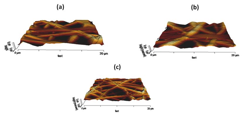
Polymers 2019, 11, 586                                                                               2 of 15

and also resemble the native ECM structure for supporting new tissue formation [1,4]. Now-a-days,
the nanofibrous scaffold was reported to be widespread in the tissue engineering applications.
      Nanofibers were fabricated using various processing techniques such as emulsion freeze drying,
self-assembly, and phase separation [1]. The nanofibers fabricated using the above techniques were
used in several applications such as energy applications [5], treatment of water and environment [6],
healthcare, and biomedical engineering [7,8]. However, the nanofibers produced using these techniques
does not meet the requirements of the ideal wound dressing membrane because of their large diameters
and low porosity [9]. In addition, the nanofibers based on the electrospinning technique were widely
used in biomedical applications because of their large surface area to volume ratios and porous
structure [10]. The small interconnected pores of the electrospun membranes do not help suitable
cellular response and tissue ingrowth. However, the pore size can be tailor made by optimizing or
tuning the electrospinning parameters such as voltage, flow rate and collector distance [11]. Tang et al.
reported that the increase in flow rate resulted in an increase in fiber diameter [12]. Similarly, Zhang et
al observed that the low voltage favors the smaller fiber diameter while the high voltage results in
broader fiber diameter [13]. Further, Yuan et al. reported that the thin fiber was formed for a larger
collector distance and vice versa [14]. Tarus et al reported that the solvent with lower surface tension
causes smoother fibers and fibers will be higher for other solvents with higher surface tension [15].
Further, electrospinning is a cost effective, facile, and well established technique used to fabricate
nanofibers from both natural and synthetic polymers [16]. Some of the natural polymers used in the
wound dressing were chitosan, collagen, alginate, gelatin, chitin, and silk fibroin. While the synthetic
polymers used were poly(vinyl alcohol), poly(lactic acid), polycaprolactone (PCL), and poly(ethylene
oxide) [1]. In this study, the Tecoflex EG 80A was used to fabricate the wound dressing scaffold.
It belongs to the family of aliphatic polyether polyurethanes. It possesses good mechanical and
ultraviolet (UV) stability [17]. For the past few decades, it was widely used in biomedical applications
because of its biocompatible and biodegradable behaviour, oxidative and thermal stability [18,19].
      An ideal scaffold for wound dressing must possess improved biocompatibility for influencing
the fibroblast adhesion and also have antimicrobial activity to inhibit the microbes penetration.
Unnithan et al. studied electrospun polyurethane (Mw = 110,000, Medical grade) scaffold with added
emu oil fibers. It was shown that the emu oil incorporated PU scaffolds showed enhanced adhesion
of fibroblast cells compared to the pristine polyurethane [20]. In another study, Jaganathan et al.
fabricated polyurethane (Tecoflex EG 80A) scaffold added with grape seed oil, honey, and propolis.
It was reported that the addition of grape seed improved the fibroblast adhesion compared to the
pristine PU [21]. Hence these studies have proved that the addition of essential oil influenced the
fibroblast adhesion and proliferation. Hence, these types of studies motivated us to use pepper mint
(PM) oil for wound dressing applications. Mentha piperita L., known as peppermint oil, is a medicinal
plant belongs to the Lamiaceae family [22]. It is widely found in temperate areas of the world, mainly
in North America, Europe, and North Africa. Peppermint is composed of 29–48% menthol, about
20–31% menthone, 6.8% menthofuran, and 3–10% menthyl acetate. Other bioactive compounds include
flavonoids, caffeic acid, polyphenols, tocopherols, carotenes, tannins, betaine, and choline [23–25].
Owing to their various bioactive constituents, it was used in food, cosmetic, and clinical applications.
It has been utilized for treating colds, cancers, cramps, indigestion, sore throat, nausea, and toothaches.
The polyphenolic constituents of PM oil might stimulate the immune system and help in preventing
colds and similar viral infections [26]. These constituents also help in protecting the cells from free
radicals and exert anticancer activity [27]. Further, it has been reported to possess some biomedical
activities of an antibacterial, antiviral, and antioxidant nature [22]. Furthermore for wound dressing
applications, it must impart or enhance the antimicrobial activity. Hence, it is necessary to add some
efficient antibacterial agents into the fabricated scaffolds. In this research the copper sulphate (CuSO4 )
is selected. Literature reported that silver, copper, and zinc are less toxic with excellent antimicrobial
activity compared to the other metallic particles. In addition to promising antimicrobial activity, copper
also plays a vital role in aiding the various physiological and metabolic processes like promoting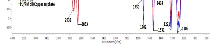
Polymers 2019, 11, 586                                                                             3 of 15

endothelial cells growth, accelerating angiogenesis, and equilibrium of extracellular skin proteins [28].
The aim of this research is to develop and characterize a novel wound dressing scaffold based on
polyurethane added with PM oil and CuSO4 .

2. Experimental

2.1. Materials
    PU (Medical grade Tecoflex EG 80A) was supplied by Lubrizol, Wickliffe, OH, USA. PM oil
was purchased from AEON, Johor, Malaysia. Copper sulphate (CuSO4.5 H2 O) was supplied by
Sigma-Aldrich, Gillingham, UK. The solvent for PU is Dimethylformamide (DMF) which was obtained
from Merck, Burlington, NJ, USA. The reagents utilized in clotting study was obtained from Diagnostic
Enterprise, Thiruvananthapuram, India.

2.2. Preparation of PU, Composite Solution and Electrospinning process
      The homogeneous solution of PU, PM oil, and CuSO4 was prepared at 9 wt %, 4 v/v % and 4 wt %
respectively. To make 9 wt % of PU solution, 0.315 g of PU pellets was added to 3.5 mL of DMF and
stirred for 12 h at room temperature. Similarly, 80 µL of PM oil was added to 2 mL of DMF and stirred
for 1 hr maximum to make 4 v/v % PM solution. Further, 4 wt % of CuSO4 solution was done by
adding 0.080 g of CuSO4 in 2 mL of DMF and stirred for 2 h respectively. The prepared PM and CuSO4
homogeneous solution were added to the PU solution at an 8:1 v/v % for PU/PM and 8:0.5:0.5 v/v %
for PU/PM/CuSO4 . In this research, the electrospinning technique was used to develop a fibrous
scaffold from the prepared homogenous solution. They were electrospun at a voltage of 11 kV, a flow
rate of 0.3 ml/h, and a distance of 20 cm respectively [21]. The parameters were kept constant for all
solutions for even comparison. The attained fibers were collected using aluminum foil and vacuum
dried to remove any residues.

2.3. Morphological and Structural Characterization
     Field emission scanning electron microscopy (FESEM) unit (Hitachi SU8020, Tokyo, Japan)
was employed to analyze the morphology of as-spun membranes. The samples were coated with
gold and imaged at different magnifications. The fiber diameter was measured using Image J
software by choosing 30 locations randomly from the captured image. The wettability of the fibrous
membranes was determined through static video contact angle equipment (AST products, Inc.,
Billerica, MA, USA). Sample with a size of 1 × 1 cm2 was placed on the measuring surface and
deposited with a water droplet (0.5 µL). The image of the water droplet was captured within a
few seconds via video camera. The computer integrated software measure the manual contact
angle and the experiment was repeated for three trials. For IR analysis, the electrospun samples
were measured over wavelengths between 600 and 4000 cm−1 at 4 cm−1 resolution in Nicolet iS 5,
Thermo Fischer Scientific, Waltham, MA, USA. The ATR crystal employed is Zinc Selenium (ZnSe).
The thermal stability was measured in thermogravimetric analysis (TGA) unit (PerkinElmer, Waltham,
MA, USA) by heating a small piece of electrospun membrane (3 mg) between 30–1000 degree celcius
under the nitrogen atmosphere. The remaining weight residue was measured at each temperature
point and the obtained values were exported in an excel sheet. Finally, the surface morphology
was determined using atomic force microscopy (AFM) unit (NanoWizard®, JPK Instruments, Berlin,
Germany) under normal atmosphere by scanning the electrospun membrane in size of 20 um × 20 µm
area and images in 256 resolution were obtained. Ra was calculated at three different locations using
the integrated JPKSPM software.

2.4. Mechanical Characterization
    Mechanical properties of the electrospun PU, PU/PM, and PU/PM/CuSO4 composite was
measured by universal testing equipment (Gotech Testing Machines, AI-3000). The samples with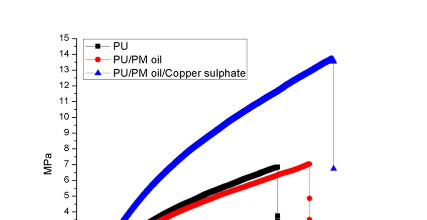
Polymers 2019, 11, 586                                                                             4 of 15

a size of 40 mm × 15 mm was clamped in the testing machine with a gauge length of 20 mm.
The test was performed at a velocity of 10 mm/min with a load cell of 500 N at room temperature.
The computer integrated software displays the stress–strain curve from which the average tensile
strength at maximum stress was measured.

2.5. Coagulation Assays

2.5.1. Activated Partial Thromboplastin Time (APTT) and Prothrombin Time (PT) Assay
      PU, PU/PM, and PU/PM/CuSO4 scaffolds were cut into a size of 1 × 1 cm2 and incubated with
50 µl of platelet poor plasma (PPP) for 1 min at 37 ◦ C. Afterwards, 50 µl of rabbit brain cephaloplastin
reagent was further added and incubated for 2 min at 37◦ C. Finally, the blood clot was initiated
by adding 50 µL of CaCl2 and the formation of the clot was investigated with a sterile steel needle.
The time taken for the blood clot was noted using a chronometer. Similarly for PT assay, the samples
size of 1 × 1 cm2 was incubated with 50 µl of obtained PPP for 1 min at 37 ◦ C. After, it was added with
50 µL of NaCl–thromboplastin reagent (Factor III) which initiate the blood clot. The time taken for the
clot formation was measured by chronometer.

2.5.2. Hemolysis Assay
      Hemolysis assay was done to investigate the toxicity of electrospun membranes with red blood
cells. Initially, the samples (1 × 1 cm2 ) were soaked in 0.9% w/v of physiological saline at 37 ◦ C for
30 min. Next, they were added with a mixture of aliquots of citrated blood and diluted saline (4:5) for
1 h at 37 ◦ C. After, the samples were retrieved and the mixture was centrifuged at 3000 rpm for 15 min.
Finally, the supernatant was aspirated and the optical density was recorded at 542 nm which depicts
the release of haemoglobin. The percentage of hemolysis or hemolytic index was calculated using the
formula [21]:
                             Hemolysis ratio (HR)=(TS−NC)/(PC−NC)×100                                (1)

where TS, NC, and PC are measured absorbance values of the test sample, negative control, and
positive control at 542 nm, respectively.

2.6. Characterization of In Vitro Biocompatibility
     HDF (Human Skin Fibroblast Cells 1184, ECACC, UK, primary cells obtained from the dermis
of the skin) were cultured in DMEM medium supplied with 10% fetal bovine serum and incubated
at 37 ◦ C with 5% CO2 . The medium was replaced every 3 days. The electrospun membranes with
dimensions of 0.5 cm × 0.5 cm were cut and placed in the 96 well plates. Once the cells reached
80% confluence, they were seeded onto the scaffolds at a density of 10 × 103 cells/ cm2 . MTS assay
was used to determine the cell viability of the cells after 5 days culturing. After 5 days of culture,
the medium in which cell is grown on the electrospun membranes was added with 20% of MTS
solution and incubated for 4 h. After 4 hr, the absorbance was measured at a wavelength of 490 nm
using a spectrophotometric plate reader.

2.7. Statistical Analysis
     One way ANOVA followed by Dunnett post hoc test was performed for experiments results with
three trails to determine statistical significance. The analyzed data were expressed as mean ± SD for
three trials. A representative of three images was presented for qualitative experiments.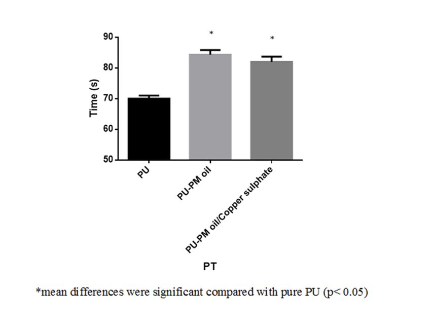
Polymers 2019, 11, 586 5 of 15
3. Result and discussion
3.1. FESEM Investigation
FESEM images of the prepared electrospun fibrous mats are shown in Figure 1. All the
as-spun nanofibrous scaffolds showed a non-woven structure with smooth and beadless morphology.
The average fiber size for pure PU was found to be 915 ± 137 nm, while the PU incorporated with
PM and
Polymers PM/
2018, 10, xCuSO4
FOR PEER REVIEW fiber sizes of 997 ± 134 nm and 359 ± 166 nm, respectively
exhibited 5 (mean
of 16
differences were significant compared with pure PU (p < 0.05)). Hence, the addition of PM increased
were significant
the fiber diameter compared with pure
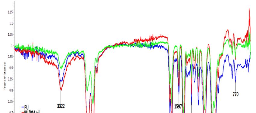
of the pristine PU,PU (p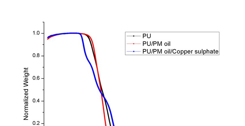
1531 and 1597 cm−1                  Vibrations of N–H
                          1414 cm−1                      Vibrations of C–H
                   1221, 1105, and 770 cm−1    C–O corresponding to the alcohol group

       The band at 3322 cm−1 indicates the stretching of NH group, peaks at 2932 and 2853 cm−1
 represent   the asymmetric and symmetric stretching of CH, and twin peaks at 1730 and 1702 cm−1
Polymers 2019, 11, 586                                                                                         6 of 15
 were attributed to the CO group. The vibration of the NH group was observed at 1531 and 1597 cm−1
 and for CH vibrations it was shown at 1414 cm−1. The other sharp peaks at 1220, 1078, and 770 cm−1
 indicate
3.2. FTIRthe    CO group with respect to alcohol [21]. For composite membranes, it was found that no
            Analysis
 new peaks were observed but the intensity of PU was altered (increasing for PU/PM and decreasing
      To evaluate 4the
 for PU/PM/CuSO             chemical
                       ) indicating    composition
                                    a formation       of the developed
                                                of a stronger             fibrous
                                                              hydrogen bond   [20]. membrane,
                                                                                    The hydrogenFTIR
                                                                                                   bondspectra
                                                                                                        was    were
inspected   in wavelength
 formed between      moleculesrange
                                 of PMofand
                                         600–4000    cm−PU.
                                            CuSO4 with    1 as indicated in Figure 2. The typical peaks present in
                                                              The hydrogen bond formation was due to the
 combination
the  pristine PUof OH
                    are and   CH molecules
                          shown   in Table 2.present in PM oil with the molecules of the pristine PU. Hence,
 the FTIR study confirms the existence of PM and CuSO4 in the polyurethane matrix.

         Figure 2. Fourier transform infrared spectroscopy (FTIR) of PU, PU/PM, and PU/PM/CuSO4.
          Figure 2. Fourier transform infrared spectroscopy (FTIR) of PU, PU/PM, and PU/PM/CuSO4.
                          Table 2. Functional groups in the electrospun PU membranes.
 3.3. Wettability Measurements
                            Peaks                                         Band name
      The contact angle is a measure of the wettability of the developed membranes. The static
                          3322 cm−1 of the pure PU and their composites N–H
 contact angle (CA) measurements                                              were presented. The pure PU
 fibrous membrane2932showed
                          and a2853  cm−angle
                                contact  1    of 106° ± 3° indicating its hydrophobic
                                                                             C–H           behavior. Upon the
 incorporation of PM    into  the polyurethane
                    1730 and 1702 cm−1
                                                 matrix,  the contact  angle   was
                                                                             C=O      increased  to 111° ± 2°,
 indicating higher hydrophobicity than the pristine PU. On another hand, the addition of CuSO4 to
 the PU/PM resulted 1531       1597 cm−1 nature showing a contact angle
                          andhydrophilic
                      in the                                         Vibrations
                                                                            of 82°of± N–H
                                                                                       1°. Hence, the addition
 of CuSO4 improved the1414     cm−1 of the polymer matrix. Jaganathan
                           wettability                                      et al. electrospun
                                                                     Vibrations    of C–H       polyurethane
                  1221, 1105, and 770 cm−1                C–O corresponding to the alcohol group

     The band at 3322 cm−1 indicates the stretching of NH group, peaks at 2932 and 2853 cm−1
represent the asymmetric and symmetric stretching of CH, and twin peaks at 1730 and 1702 cm−1
were attributed to the CO group. The vibration of the NH group was observed at 1531 and 1597 cm−1
and for CH vibrations it was shown at 1414 cm−1 . The other sharp peaks at 1220, 1078, and 770 cm−1
indicate the CO group with respect to alcohol [21]. For composite membranes, it was found that no
new peaks were observed but the intensity of PU was altered (increasing for PU/PM and decreasing
for PU/PM/CuSO4 ) indicating a formation of a stronger hydrogen bond [20]. The hydrogen bond was
formed between molecules of PM and CuSO4 with PU. The hydrogen bond formation was due to the
combination of OH and CH molecules present in PM oil with the molecules of the pristine PU. Hence,
the FTIR study confirms the existence of PM and CuSO4 in the polyurethane matrix.
Polymers 2019, 11, 586                                                                                                          7 of 15

3.3. Wettability Measurements
     The contact angle is a measure of the wettability of the developed membranes. The static contact
angle (CA) measurements of the pure PU and their composites were presented. The pure PU fibrous
membrane showed a contact angle of 106◦ ± 3◦ indicating its hydrophobic behavior. Upon the
incorporation of PM into the polyurethane matrix, the contact angle was increased to 111◦ ± 2◦ ,
indicating higher hydrophobicity than the pristine PU. On another hand, the addition of CuSO4 to the
PU/PM resulted in the hydrophilic nature showing a contact angle of 82◦ ± 1◦ . Hence, the addition of
CuSO4 improved the wettability of the polymer matrix. Jaganathan et al. electrospun polyurethane
(Tecoflex EG 80A) scaffold incorporated with zinc particles for wound dressing applications. It was
reported that    the
          Polymers    incorporation
                   2018,                    of zinc particles improved the wettability of the pristine
                         10, x FOR PEER REVIEW                                                                        7 of 16 PU which
correlates with our observation. The contact of polyurethane was reported to be 100◦ ± 0.5774◦ , while
          (Tecoflex EG 80A) scaffold incorporated with zinc particles for wound dressing applications. It was
polyurethane added with zinc nitrate displayed contact angle of 84◦ ± 4.041◦ . Further, the scaffold
          reported that the incorporation of zinc particles improved the wettability of the pristine PU which
with improved
          correlateswettability       exhibited
                      with our observation.     Theenhanced       fibroblastwas
                                                    contact of polyurethane      cells  adhesion
                                                                                     reported         than
                                                                                               to be 100°     the pristine
                                                                                                          ± 0.5774°,  while PU [30].
The addition    of CuSO
          polyurethane      added
                               4 improved
                                    with  zinc the  wettability
                                               nitrate displayed    of the
                                                                   contact   PU/PM
                                                                            angle  of 84° which
                                                                                          ± 4.041°.might
                                                                                                     Further,be  suitable
                                                                                                               the  scaffold  for better
          with  improved      wettability exhibited
fibroblast cell adhesion and proliferation.         enhanced   fibroblast  cells adhesion  than  the pristine PU   [30]. The
            addition of CuSO4 improved the wettability of the PU/PM which might be suitable for better
            fibroblast cell adhesion and proliferation.
3.4. TGA Analysis
          3.4. TGA Analysis
     The thermal      properties of electrospun PU, PU/PM, and PU/PM/CuSO4 are shown in Figure 3.
                The  thermal
In the case of pristine PU,    properties  of electrospun
                                      the initial           PU, PU/PM,
                                                    degradation            and PU/PM/CuSO
                                                                       temperature         was4 are  shown into
                                                                                                observed       Figure  3. In ◦ C, while
                                                                                                                  be 266
          the case of pristine PU, the initial degradation temperature was observed       ◦       to be 266 °C, while for
for electrospun PU/PM the temperature was increased to 286 C and for PU/PM/CuSO4 , it was
          electrospun◦ C.PU/PM    the temperature was increased to 286 °C and for PU/PM/CuSO4, it was decreased
decreasedtoto227
               227         Hence,
                  °C. Hence,          the thermal
                               the thermal            properties
                                             properties              werewith
                                                         were increased      increased      withofthe
                                                                                  the addition     PM.addition
                                                                                                        However, of    PM. However,
                                                                                                                   adding
adding CuSO        intothe
          CuSO44 into     the  polymer
                             polymer        matrix
                                        matrix        decreased
                                               decreased   its thermalitsstability.
                                                                           thermal  Thestability.
                                                                                         decrease inThe   decrease
                                                                                                      the thermal     in the thermal
                                                                                                                  stability
          was due
stability was   duetoto thethe
                            moisture
                               moistureevaporation   present inpresent
                                             evaporation         the copperinsulphate
                                                                               the copperpentahydrate.
                                                                                              sulphate  Further, DTG was
                                                                                                            pentahydrate.       Further,
          performed for the electrospun membranes as indicated in Figure 4 and their weight loss peaks were
DTG was performed for the electrospun membranes as indicated in Figure 4 and their weight loss
          listed in Table 3. From the results obtained, the electrospun PU/PM showed only three weight loss
peaks were    listed
          peaks        in Table
                   compared     to 3.
                                    theFrom    thePU
                                         pristine   results
                                                        showingobtained,     the electrospun
                                                                    four weight                      PU/PMelectrospun
                                                                                     loss peaks. However,       showed only three
weight loss  peaks     compared       to the  pristine   PU    showing      four    weight   loss  peaks.
          PU/PM/CuSO4 showed weight loss peaks as that of pristine PU, but their weight loss intensity was   However,      electrospun
          decreased.    Hence,  the  reduced   weight  loss peaks  and   decrease   in weight loss
PU/PM/CuSO4 showed weight loss peaks as that of pristine PU, but their weight loss intensity was    intensity confirms  the
          reduced weigh loss of the fabricated nanocomposites. Moreover, this attribute also confirms the
decreased.    Hence, the reduced weight loss peaks and decrease in weight loss intensity confirms
          presence of PM and CuSO4 in the polyurethane matrix.
the reduced weigh loss of the fabricated nanocomposites. Moreover, this attribute also confirms the
presence of PM and CuSO4 in the polyurethane matrix.

                                    Figure 3. TGA of PU, PU/PM, and PU/PM/CuSO4.
Polymers 2018, 10, x FOR PEER REVIEW 8 of 16
Figure 3. TGA of PU, PU/PM, and PU/PM/CuSO4.
Polymers 2019, 11, 586 8 of 15
Figure 4. Weight residue of PU, PU/PM, and PU/PM/CuSO4.
Figure 4. Weight residue of PU, PU/PM, and PU/PM/CuSO4.
Table 3. Weight loss peaks of electrospun PU, PU/PM, and PU/PM/CuSO4.
Table 3. Weight loss peaks of electrospun PU, PU/PM, and PU/PM/CuSO4.
S.NO PU PU/PM PU/PM/CuSO4
S.NO
First weight loss ◦
210 C to 302 PU◦ C PU/PM
◦
228 C to 327 C ◦ PU/PM/CuSO
206 ◦ C to
4
270 ◦ C
First weight loss 210 °C to 302 °C 228 °C◦ to 327 °C◦ 206 °C to 270 °C
Second weight loss 302 ◦ C to 353 ◦ C 327 C to 434 C 270 ◦ C to 353 ◦ C
Second weight loss 302 °C to 353 °C 327 °C to 434 °C 270 °C to 353 °C
Third weight ◦ C to 494 ◦ C ◦ C to 476 ◦ C
Thirdloss
weight loss353 353 °C to 494 °C - - 353476
353 °C to °C
Fourthloss
Fourth weight 494 ◦494
weight loss C to°C760 ◦ C °C
to 760 - - 476 °C to
476 ◦ C°C
664 to 664 ◦ C
3.5. AFM Analysis
3.5. AFM Analysis
To analyse the effect of PM and CuSO4 incorporation on the surface roughness of the pristine
To analyse the effect of PM and CuSO4 incorporation
PU, AFM was performed. The representative
on the surface roughness of the pristine PU,
3D images of the fibrous membranes are shown in
AFMFigure
was performed.
5. The average The representative
roughness (Ra) 3D images
of the of the
pristine PUfibrous
rangedmembranes
776 ± 468 nm, are while
shownthe in Figure
PU 5.
The average roughness
incorporated with PM and (R ) of the pristine PU ranged 776 ± 468 nm, while the PU
a PM/CuSO4 have an average roughness of 1039 ± 198 nm and 515 ± 123 nm, incorporated with
PM and PM/CuSO
respectively. have an
The4 surface averagemeasurements
roughness 1039 ± that
roughness ofdepicted 198 the and 515 ±
nm fabricated 123 nm,
PU/PM respectively.
composites
have rougher
The surface surfaces,
roughness while PU/PM/CuSO
measurements depicted4 had
thatsmoother surfacesPU/PM
the fabricated compared to the pristine
composites havePU.
rougher
Hyung
surfaces, Hwan
while Kim et al studied
PU/PM/CuSO 4 hadthe effect
smoother of fiber diameter
surfaces on
compared surface
to roughness
the pristine in electrospun
PU. Hyung Hwan
polycaprolactone membrane. It was reported that the average surface roughness
Kim et al studied the effect of fiber diameter on surface roughness in electrospun polycaprolactone was increased with
an increase in fiber diameter. Our electrospun PU/PM oil showed a larger fiber diameter which might
membrane. It was reported that the average surface roughness was increased with an increase in
favor the improvement in the surface roughness. In contrast, the electrospun PU/PM/CuSO4
fiber diameter. Our electrospun PU/PM oil showed a larger fiber diameter which might favor the
displayed smaller fiber morphology which results in smooth surfaces [31]. Huag et al reported that
improvement in the
the fibroblast surface
cells prefer roughness. In contrast,
smoother surface the and
to adhere electrospun PU/PM/CuSO
proliferate [32]. The smooth4 displayed smaller
surface will
fiberhave
morphology
a smaller fiber diameter which would be suitable for the fibroblast cells to adhere and grow cells
which results in smooth surfaces [31]. Huag et al reported that the fibroblast
prefer smoother
[18,31]. surface
On other to adhere
hand, Sharifi and
et al.proliferate
reported that[32].surface
The smooth
modifiedsurface will have a smaller
polycaprolactone fibrous fiber
diameter whichwith
membrane would be suitable
increased surfaceforroughness
the fibroblast cells tothe
also favors adhere and grow
improved [18,31].
adhesion and On other
growth ofhand,
Sharifi et al. reported that surface modified polycaprolactone fibrous membrane with increased surface
roughness also favors the improved adhesion and growth of fibroblast cells [33]. The influence of
surface roughness on the cellular response is still unclear and the various physico-chemical properties
of the manufactured composites might have a role in the cell adhesion behavior. To state, the smaller
fiber diameter favors the enhanced adhesion of protein which results in large cell attachment and
Polymers 2018, 10, x FOR PEER REVIEW                                                                   9 of 16

fibroblast
Polymers    cells
         2019,      [33].
               11, 586The influence of surface roughness on the cellular response is still unclear9 of and
                                                                                                         15
the various physico-chemical properties of the manufactured composites might have a role in the cell
adhesion behavior. To state, the smaller fiber diameter favors the enhanced adhesion of protein which
proliferation [29]. Further, the adequate porosity also helps in efficient transport of nutrients and waste
results in large cell attachment and proliferation [29]. Further, the adequate porosity also helps in
removal for better cell adhesion and growth [34].
efficient transport of nutrients and waste removal for better cell adhesion and growth [34].

                       Figure 5. AFM images of (a) PU, (b) PU/PM, and (c) PU/PM/CuSO4.

3.6. Tensile Testing
                            Figure 5. AFM images of (a) PU, (b) PU/PM, and (c) PU/PM/CuSO4.
     Mechanical
 Polymers 2018, 10, xtesting of the
                      FOR PEER      electrospun membranes was indicated in Figure 6 and Table 4.
                                REVIEW                                                        The tensile
                                                                                                   10 of 16
testing clearly indicated
3.6. Tensile Testing       that the tensile strength of the pristine PU was enhanced on incorporating PM
and CuSO4 . PU showed a tensile strength of 6.83 MPa and, on adding PM and CuSO4 , it was increased
      Mechanical
to 7.04            testing
        MPa and 13.60       of the
                         MPa,      electrospun membranes was indicated in Figure 6 and Table 4. The
                                respectively.
tensile testing clearly indicated that the tensile strength of the pristine PU was enhanced on
incorporating PM and CuSO4. PU showed a tensile strength of 6.83 MPa and, on adding PM and
CuSO4, it was increased to 7.04 MPa and 13.60 MPa, respectively.

                              Figure 6. Tensile strength of PU, PU/PM, and PU/PM/CuSO4.

                               Figure 6. Tensile strength of PU, PU/PM, and PU/PM/CuSO4.

                               Table 4. Mechanical testing values of electrospun membranes.

                                           Tensile                Elastic              Elongation at
                Membrane
                                     strength(miao)MPa       Modulus(miao)MPa         break(miao)(%)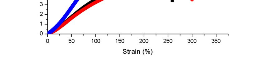
Polymers 2019, 11, 586                                                                             10 of 15

                         Table 4. Mechanical testing values of electrospun membranes.

                                 Tensile Strength      Elastic Modulus       Elongation at Break
               Membrane
                                       MPa                   MPa                     (%)
                    PU                 6.83                  3.51                   259.16
                 PU/PM                 7.04                  2.95                   300.10
            PU/PM/CuSO4                13.60                 5.37                   331.15

     The increase in tensile strength was perhaps due to the homogeneous dispersion of PM and CuSO4
in the polymer matrix. Unnithan et al. electrospun wound dressing scaffold utilizing polyurethane
(Mw = 110,000), Medical grade) and emu oil. It was observed that the polyurethane/emu oil having
smaller fiber diameter favored the enhancement of the tensile strength. They concluded that the
addition of emu oil might favor the attachment of fibers owing to the hydrogen bond formation
between polyurethane and emu oil molecules [20]. As reported earlier, FTIR analysis revealed the
hydrogen bond formation in the fabricated composites which might have favored the enhancement of
the tensile strength of PU/PM/CuSO4 . Jaganathan et al. fabricated a wound dressing polyurethane
(Tecoflex EG 80A) scaffold added with zinc nitrate. It was found that the zinc nitrate incorporation
into the polyurethane exhibited improvement in the mechanical strength which correlates with our
findings. They concluded this behavior was because of the smaller fiber diameter (568 ± 136.69 nm)
of the electrospun composites compared to the polyurethane (1159 ± 147.48 nm) [30]. Our fiber
diameter reduction was found to be similar to that reported above, which might have resulted in the
improvement of the mechanical strength.

3.7. Blood Compatibility Measurements
      The dressing materials used for wound treatment will frequently come in contact with body
fluids such as leakage of plasma and other blood components. If the dressing material does not
have hemo-compatible properties, there might be risk of activating an undesired immune response
which ultimately result in formation of thrombosis, inflammation, and foreign body reaction. This
in turn delays the wound healing process. Hence, an ideal wound dressing should possess better
blood compatible properties [35]. Blood clotting time of the electrospun PU, PM, and PM/CuSO4
was measured using APTT and PT assay. The coagulation assays showed the enhanced anticoagulant
nature of the electrospun PU/PM and PU/PM/CuSO4 compared to the PU membrane as presented
in Figures 7 and 8. The developed PU/PM and PU/PM/CuSO4 mat exhibited blood clotting time
of 175 ± 4 s and 172 ± 4 s (mean differences were significant compared with pure PU (p < 0.05)),
while the PU showed blood clotting time of 155 ± 2 s as calculated in the APTT assay. Similarly,
the developed PU/PM and PU/PM/CuSO4 mat exhibited blood clotting time of 84 ± 2 s and 82 ± 2 s
(mean differences were significant compared with pure PU (p < 0.05)), while the PU showed blood
clotting time of 70 ± 1 s as calculated in the PT assay. The existence of PM and CuSO4 in the PU
matrix delayed the blood clotting time. Further, the hemolytic assay was measured to analyse the
electrospun membranes toxicity with red blood cells. The results of hemolytic assay represent that
the index value of the electrospun PU/PM and PU/PM/CuSO4 mat was found to be lower than
pristine PU. The pristine PU showed an index value of 2.7% and for the electrospun PU/PM and
PU/PM/CuSO4 mat, it was 1.66% and 1.85% (mean differences were significant compared with pure
PU (p < 0.05)) as denoted in Figure 9. The fabricated composite was considered as non-hemolytic
because its index value was less than 2% according to ASTMF756-00(2000) [21]. In blood compatibility
assessments, the electrospun PU/PM oil showed prolonging blood clotting time compared to the
pure polyurethane. However, there is a slight reduction in the blood clotting time while blending
CuSO4 to the PU/PM but the observed blood clotting values were still higher than the pristine PU.
According to Huang et al, the blood compatibility is influenced by multiple surface parameters such as
wettability, fiber diameter, and surface roughness [36]. Jaganathan et al. electrospun a novel scaffold
Polymers 2019, 11, 586 11 of 15
based on polyurethane and mustard oil. They found the improvement in the blood compatibility of
the pristine PU with the addition of mustard oil and reported this might be because of an increase in
surface roughness [37]. In another study, Ayyar et al. electrospun polyurethane scaffold incorporated
with indhulekha oil and determined its anticoagulant nature. It was found that the electrospun
PU/indhulekha oil composite with hydrophobic behavior showed prolonged blood clotting time than
the pristine PU 2018,
Polymers [38].10,Further,
x FOR PEERJaganathan
REVIEW et al. 2017 developed polyurethane scaffold loaded
12 of 16 with
castor oil using the electrospinning technique. The developed PU/castor oil showed prolonged blood
clotting time compared to the pristine PU and this may be due to their smaller fiber diameter [39].
Hence, the addition of PM and CuSO4 into the PU matrix caused reduced fiber diameter morphology
(PU/PM/CuSO 4 ) and
Polymers 2018, 10,hydrophobic behavior (PU/PM) which might have resulted in the improvement
x FOR PEER REVIEW 12 of 16
of the anticoagulant nature.
Figure 7. APTT assay of PU, PU/PM, and PU/PM/CuSO4.
Figure 7. APTT assay of PU, PU/PM, and PU/PM/CuSO4.
Figure 7. APTT assay of PU, PU/PM, and PU/PM/CuSO4.
Figure 8. PT assay PU, PU/PM, and PU/PM/CuSO4.Polymers 2018, 10, x FOR PEER REVIEW 13 of 16
Figure 8. PT assay PU, PU/PM, and PU/PM/CuSO4.
Polymers 2019, 11, 586 12 of 15
Figure 9. Hemolytic assay PU, PU/PM, and PU/PM/CuSO4.
3.8. Cytocompatibility Measurements
Figure 9. Hemolytic assay PU, PU/PM, and PU/PM/CuSO4.
Figure 103.8. presents the Measurements
Cytocompatibility HDF cell viability in the electrospun membranes evaluated through
MTS assay. After five days of
Figure 10 presents the cell
HDF culture, it was
cell viability in the observed that the evaluated
electrospun membranes HDF cells were
through MTS well adhered
and proliferated
assay. in allfive
After electrospun membranes
days of cell culture, compared
it was observed that thetoHDF
thecells
control plates.
were well adhered The
andcell viability
percentage forproliferated
the PU in all electrospun
membrane wasmembranes
reportedcompared
to be 130 to ±the4%,
control plates.
while theThe cell viabilityPU/PM and
electrospun
percentage for the PU membrane was reported to be 130 ± 4%, while the electrospun PU/PM and
PU/PM/CuSO 4 composites
PU/PM/CuSO showed cell viability of 133 ± 11% and 144 ± 3%, respectively. Further,
4 composites showed cell viability of 133 ± 11% and 144 ± 3%, respectively. Further,
HDF cells viability
HDF cells inviability
the electrospun PU/PM/CuSO
in the electrospun composites
PU/PM/CuSO4 4composites was observed
was observed to be higher tothan
be higher
the than the
PU/PM composites. As reported in the introduction section, PM oil contains polyphenols as one of
PU/PM composites. As reported in the introduction section, PM oil contains polyphenols as one of
their bioactive constituents. It has been reported that the phenolic components help in protecting the
their bioactive constituents.
fibroblast cells againstIt oxidative
has been reported
stress caused by that the phenolic
hydrogen components
peroxide resulting help in protecting the
in cell proliferation
fibroblast cells against oxidative stress caused by hydrogen peroxide resulting in cell
and migration. The phenolic compounds present in the PM oil might have influenced cell proliferation and
viability
[40]. The reason for the improved HDF cell viability was perhaps due to their smaller fiber diameter
migration. The phenolic compounds present in the PM oil might have influenced cell viability [40].
(PU/PM) and hydrophilic behavior (PU/PM/ CuSO4) [41,42]. Hence, the improved cellular response
The reason forof thethe improved
developed HDF
composites cellbeviability
might suitable forwas
wound perhaps due to their smaller fiber diameter
dressing applications.
Polymers 2018, 10, x FOR PEER REVIEW 14 of 16
(PU/PM) and hydrophilic behavior (PU/PM/ CuSO4 ) [41,42]. Hence, the improved cellular response
of the developed composites might be suitable for wound dressing applications.
Figure 10. MTS assay PU, PU/PM, and PU/PM/CuSO4.
Figure 10. MTS assay PU, PU/PM, and PU/PM/CuSO4.
4. Conclusion
This work successfully evaluated the physicochemical and biocompatibility properties of
electrospun composites based on polyurethane (PU) added with pepper mint (PM) oil and copperPolymers 2019, 11, 586 13 of 15
4. Conclusion
This work successfully evaluated the physicochemical and biocompatibility properties of
electrospun composites based on polyurethane (PU) added with pepper mint (PM) oil and copper
sulphate (CuSO4 ). The fabricated composites showed improved physicochemcial properties compared
to the pristine PU. Blood compatibility studies showed improved anti-coagulation time and less toxic
behavior for the developed composites than the pristine PU. Finally, the cell viability of the fabricated
composite was higher than the pristine PU as indicated in MTS assay. Hence, the fabricated wound
dressing composite based on PU/PM and PU/PM/CuSO4 rendering better physicochemical and
biocompatible nature, mark its suitability for wound healing applications. In the future, it would be
interesting to explore the cell proliferation and morphology characteristics of HDF cells using SEM
analysis at different time points, which will further validate the claims of cytotoxicity.
Author Contributions: Conceptualization: S.K.J. and A.Z.M.K.; Methodology: S.K.J., M.P.M., A.Z.M.K.; Software:
S.K.J. and M.P.M.; Validation: S.K.J., M.P.M., A.Z.M.K.; Formal Analysis: S.K.J. and A.Z.M.K.; Investigation: S.K.J.,
M.P.M., A.Z.M.K.; Resources: S.K.J. and A.Z.M.K.; Data Curation: S.K.J., M.P.M., A.Z.M.K.; Writing-Original Draft
Preparation: M.P.M.; Writing-Review & Editing: S.K.J., M.P.M. and A.Z.M.K.; Visualization: S.K.J. and A.Z.M.K.;
Supervision: S.K.J. and A.Z.M.K.; Project Administration: S.K.J. and A.Z.M.K.; Funding Acquisition: A.Z.M.K.
and S.K.J.
Funding: This project was majorly supported by MRUN funding with the Grant reference no. PY/2015/05186.
This work was also partly supported by the Ministry of Higher Education Malaysia with the Grant no.
Q.J130000.2545.17H00 and Q.J130000.2545.20H00.
Acknowledgments: Authors acknowledge the support of UPMU technical service.
Conflicts of Interest: The authors declare no conflict of interest.
References
1. Li, X.; Wang, C.; Yang, S.; Liu, P.; Zhang, B. Electrospun Pcl/mupirocin and chitosan/lidocaine hydrochloride
multifunctional double layer nanofibrous scaffolds for wound dressing applications. Int. J. Nanomed. 2018,
13, 5287. [CrossRef] [PubMed]
2. Dhivya, S.; Padma, V.V.; Santhini, E. Wound dressings—A review. Bio Med. 2015, 5. [CrossRef] [PubMed]
3. Chen, X.; Zhao, R.; Wang, X.; Li, X.; Peng, F.; Jin, Z.; Gao, X.; Yu, J.; Wang, C. Electrospun mupirocin loaded
polyurethane fiber mats for anti-infection burn wound dressing application. J. Biomater. Sci. Polym. 2017, 28, 162–176.
[CrossRef] [PubMed]
4. Yildirimer, L.; Thanh, N.T.; Seifalian, A.M. Skin regeneration scaffolds: A multimodal bottom-up approach.
Trends Biotechnol. 2012, 30, 638–648. [CrossRef]
5. Fu, K.; Lu, Y.; Dirican, M.; Chen, C.; Yanilmaz, M.; Shi, Q.; Bradford, P.D.; Zhang, X. Chamber-confined silicon–carbon
nanofiber composites for prolonged cycling life of Li-ion batteries. Nanoscale 2014, 6, 7489–7495. [CrossRef]
6. Wang, Y.; Zhang, X.; He, X.; Zhang, W.; Zhang, X.; Lu, C. In situ synthesis of MnO2 coated cellulose nanofibers
hybrid for effective removal of methylene blue. Carbohydr. Polym. 2014, 110, 302–308. [CrossRef]
7. Lim, C.T. Nanofiber technology: Current status and emerging developments. Prog. Polym. Sci. 2017, 1, 70:1–70:7.
8. Rasouli, R.; Barhoum, A.; Bechelany, M.; Dufresne, A. Nanofibers for Biomedical and Healthcare Applications.
Macromol. Biosci. 2018, 28, 1800256. [CrossRef]
9. Ali, I.H.; Khalil, I.A.; El-Sherbiny, I.M. Single-dose electrospun nanoparticles-in-nanofibers wound dressings
with enhanced epithelialization; collagen deposition, and granulation Properties. ACS Appl. Mater. Interface
2016, 8, 14453–14469. [CrossRef] [PubMed]
10. Agarwal, S.; Wendorff, J.H.; Greiner, A. Use of electrospinning technique for biomedical applications. Polymer
2008, 49, 5603–5621. [CrossRef]
11. Rnjak-Kovacina, J.; Weiss, A.S. Increasing the pore size of electrospun scaffolds. Tissue Eng. Part B Rev. 2011,
17, 365–372. [CrossRef] [PubMed]
12. Tang, X.; Si, N.; Xu, L.; Liu, H. Effect of flow rate on diameter of electrospun nanoporous fibers. Therm. Sci.
2014, 18, 1447–1449. [CrossRef]
13. Zhang, C.; Yuan, X.; Wu, L.; Han, Y.; Sheng, J. Study on morphology of electrospun poly (vinyl alcohol) mats.
Eur. Polym. J. 2005, 41, 423–432. [CrossRef]Polymers 2019, 11, 586 14 of 15
14. Yuan, X.; Zhang, Y.; Dong, C.; Sheng, J. Morphology of ultrafine polysulfone fibers prepared by
electrospinning. Polym. Int. 2004, 53, 1704–1710. [CrossRef]
15. Tarus, B.; Fadel, N.; Al-Oufy, A.; El-Messiry, M. Effect of polymer concentration on the morphology and mechanical
characteristics of electrospun cellulose acetate and poly (vinyl chloride) nanofiber mats. Alex. Eng. J. 2016,
55, 2975–2984. [CrossRef]
16. Abrigo, M.; McArthur, S.L.; Kingshott, P. Electrospun nanofibers as dressings for chronic wound care:
Advances, challenges, and future prospects. Macromol. Biosci. 2014, 14, 772–792. [CrossRef] [PubMed]
17. Tecoflex™ TPU - Lubrizol. Available online: https://www.lubrizol.com/Life-Sciences/products/Tecoflex-TPU
(accessed on 13 March 2019).
18. Jaganathan, S.K.; Mani, M.P.; Palaniappan, S.K.; Rathanasamy, R. Fabrication and characterisation of
nanofibrous polyurethane scaffold incorporated with corn and neem oil using single stage electrospinning
technique for bone tissue engineering applications. J. Polym. Res. 2018, 25, 146. [CrossRef]
19. Detta, N.; Errico, C.; Dinucci, D.; Puppi, D.; Clarke, D.A.; Reilly, G.C.; Chiellini, F. Novel electrospun
polyurethane/gelatin composite meshes for vascular grafts. J. Mater. Sci. Mater. Med. 2010, 21, 1761–1769.
[CrossRef]
20. Unnithan, A.R.; Pichiah, P.T.; Gnanasekaran, G.; Seenivasan, K.; Barakat, N.A.; Cha, Y.S.; Jung, C.H.;
Shanmugam, A.; Kim, H.Y. Emu oil-based electrospun nanofibrous scaffolds for wound skin tissue
engineering. Colloids. Surf. A Phys. Eng. Asp. 2012, 415, 454–460. [CrossRef]
21. Chao, C.Y.; Mani, M.P.; Jaganathan, S.K. Engineering electrospun multicomponent polyurethane scaffolding
platform comprising grapeseed oil and honey/propolis for bone tissue regeneration. PLoS ONE 2018, 13,
e0205699. [CrossRef]
22. Singh, R.; Shushni, M.A.; Belkheir, A. Antibacterial and antioxidant activities of Mentha piperita L. Arab. J. Chem.
2015, 8, 322–328. [CrossRef]
23. Karuza, L.; Blazevic, N.; Soljic, Z. Isolation and structure of flavonoids from peppermint (Mentha piperita)
leaves. Acta Pharm. 1996, 46, 315–320.
24. Shimada, K.; Fujikawa, K.; Yahara, K.; Nakamura, T. Antioxidative properties of xanthan on the autooxidation
of soybean oil in cyclodextrin. J. Agric. Food Chem. 1992, 40, 945–948. [CrossRef]
25. Sokovic, M.D.; Vukojevic, J.; Marin, P.D.; Brkic, D.D.; Vajs, V.; van Griensven, L.J.L.D. Chemical composition
of essential oils of Thymus and Mentha species and their antifungal activities. Molecules 2009, 14, 238–249.
[CrossRef]
26. Grigore, A. Plant phenolic compounds as immunomodulatory agents. In Phenolic Compounds-Biological
Activity; InTech: London, UK, 2017.
27. Zhang, Y.J.; Gan, R.Y.; Li, S.; Zhou, Y.; Li, A.N.; Xu, D.P.; Li, H.B. Antioxidant phytochemicals for the
prevention and treatment of chronic diseases. Molecules 2015, 20, 21138–21156. [CrossRef] [PubMed]
28. Amna, T.; Hassan, M.S.; Yang, J.; Khil, M.S.; Song, K.D.; Oh, J.D.; Hwang, I. Virgin olive oil blended
polyurethane micro/nanofibers ornamented with copper oxide nanocrystals for biomedical applications. Int.
J. Nanomed. 2014, 9, 891. [CrossRef] [PubMed]
29. Jaganathan, S.K.; Mani, M.P. Electrospun polyurethane nanofibrous composite impregnated with metallic
copper for wound-healing application. 3 Biotech 2018, 8, 327. [CrossRef] [PubMed]
30. Jaganathan, S.K.; Mani, M.P. Single-stage synthesis of electrospun polyurethane scaffold impregnated with
zinc nitrate nanofibers for wound healing applications. J. Appl. Polym. Sci. 2019, 136, 46942. [CrossRef]
31. Kim, H.H.; Kim, M.J.; Ryu, S.J.; Ki, C.S.; Park, Y.H. Effect of fiber diameter on surface morphology, mechanical
property, and cell behavior of electrospun poly (ε-caprolactone) mat. Fibers Polym. 2016, 17, 1033–1042.
[CrossRef]
32. Chou, S.H.; Don, T.M.; Lai, W.C.; Cheng, L.P. Formation of microporous poly (hydroxybutyric acid)
membranes for culture of osteoblast and fibroblast. Polym. Adv. Technol. 2009, 20, 1082–1090.
33. Sharifi, F.; Irani, S.; Zandi, M.; Soleimani, M.; Atyabi, S.M. Comparative of fibroblast and osteoblast cells adhesion on
surface modified nanofibrous substrates based on polycaprolactone. Prog. Biomater. 2016, 5, 213–222. [CrossRef]
34. Vernon, B. (Ed.) Injectable Biomaterials: Science and Applications; Elsevier: Amsterdam, the Netherlands,
24 January 2011.
35. Balaji, A.; Jaganathan, S.K.; Ismail, A.F.; Rajasekar, R. Fabrication and hemocompatibility assessment of
novel polyurethane-based bio-nanofibrous dressing loaded with honey and carica papaya extract for the
management of burn injuries. Int. J. Nanomed. 2016, 11, 4339.Polymers 2019, 11, 586 15 of 15
36. Huang, N.; Yang, P.; Leng, Y.X.; Chen, J.Y.; Sun, H.; Wang, J.; Wang, G.J.; Ding, P.D.; Xi, T.F.; Leng, Y.
Hemocompatibility of titanium oxide films. Biomaterials 2003, 24, 2177–2187. [CrossRef]
37. Jaganathan, S.; Mani, M.; Ismail, A.; Ayyar, M. Manufacturing and characterization of novel electrospun
composite comprising polyurethane and mustard oil scaffold with enhanced blood compatibility. Polymers
2017, 9, 163. [CrossRef]
38. Ayyar, M.; Mani, M.P.; Jaganathan, S.K.; Rathanasamy, R. Preparation, characterization and blood compatibility
assessment of a novel electrospun nanocomposite comprising polyurethane and ayurvedic-indhulekha oil for tissue
engineering applications. Biomed. Eng./Biomed. Tech. 2018, 63, 245–253. [CrossRef] [PubMed]
39. Jaganathan, S.K.; Mani, M.P.; Ayyar, M.; Supriyanto, E. Engineered electrospun polyurethane and castor oil
nanocomposite scaffolds for cardiovascular applications. J. Mater. Sci. 2017, 52, 10673–10685. [CrossRef]
40. Pitz, H.D.; Pereira, A.; Blasius, M.B.; Voytena, A.P.; Affonso, R.C.; Fanan, S.; Trevisan, A.C.; Ribeiro-do-Valle, R.M.;
Maraschin, M. In vitro evaluation of the antioxidant activity and wound healing properties of Jaboticaba
(Plinia peruviana) fruit peel Hydroalcoholic extract. Oxidative Med. Cell. Longev. 2016, 2016. [CrossRef] [PubMed]
41. Tian, F.; Hosseinkhani, H.; Hosseinkhani, M.; Khademhosseini, A.; Yokoyama, Y.; Estrada, G.G.; Kobayashi, H.
Quantitative analysis of cell adhesion on aligned micro-and nanofibers. J. Biomed. Mater. Res. Part A 2008, 84, 291–299.
[CrossRef]
42. Nandakumar, V.; Suresh, G.; Chittaranjan, S.; Doble, M. Synthesis and Characterization of Hydrophilic
High Glycolic Acid–Poly (DL-Lactic-co-Glycolic Acid)/Polycaprolactam/Polyvinyl Alcohol Blends and Their
Biomedical Application as a Ureteral Material. Ind. Eng. Chem. Res. 2012, 52, 751–760. [CrossRef]
© 2019 by the authors. Licensee MDPI, Basel, Switzerland. This article is an open access
article distributed under the terms and conditions of the Creative Commons Attribution
(CC BY) license (http://creativecommons.org/licenses/by/4.0/).You can also read