Development of Versatile Vectors for Heterologous Expression in Bacillus - MDPI
←
→
Page content transcription
If your browser does not render page correctly, please read the page content below
microorganisms
Article
Development of Versatile Vectors for Heterologous
Expression in Bacillus
Øivind Larsen and Gro Elin Kjæreng Bjerga *              ID

 Centre for Applied Biotechnology, Uni Research AS, Thormøhlens gt. 55, N-5006 Bergen, Norway;
 oivind.larsen@uni.no
 * Correspondence: gro.bjerga@uni.no; Tel.: +47-55-58-45-54

 Received: 28 March 2018; Accepted: 5 June 2018; Published: 7 June 2018

 Abstract: The discovery of new enzymes for industrial application relies on a robust discovery
 pipeline. Such a pipeline should facilitate efficient molecular cloning, recombinant expression and
 functional screening procedures. Previously, we have developed a vector set for heterologous
 expression in Escherichia coli. Here, we supplement the catalogue with vectors for expression in
 Bacillus. The vectors are made compatible with a versatile cloning procedure based on type IIS
 restriction enzymes and T4 DNA ligase, and encompass an effective counter-selection procedure and
 complement the set of vectors with options for secreted expression. We validate the system with
 expression of recombinant subtilisins, which are generally challenging to express in a heterologous
 system. The complementarity of the E. coli and Bacillus systems allows rapid switching between the
 two commonly used hosts without comprehensive intermediate cloning steps. The vectors described
 are not limited to the expression of certain enzymes, but could also be applied for the expression of
 other enzymes for more generalized enzyme discovery or development.

 Keywords: cloning; recombinant DNA technology; ccdB; subtilisin; Bacillus

1. Introduction
     Due to their wide application range, the discovery and development of proteases have a great
economic potential. An enzyme discovery pipeline should facilitate efficient molecular cloning,
recombinant expression and functional screening procedures [1,2], but often requires adaptation to the
enzyme of interest. Serine proteases constitute about a third of known proteolytic enzymes, including
the subtilisin family [3]. The subtilisins derived their name from Bacillus subtilis, from which the
enzyme was first isolated [4], but they are widespread, being found in bacteria, archaea, viruses and
eukaryotes [5]. Certain members of the subtilisin family, such as the extracellular subtilisin proteases
(ESPs), have been extensively researched and used in the detergent, leather and food industries [6].
Endogenous ESPs are produced as inactive precursor proteins consisting of a leader sequence [7]
that directs their export, a pro-sequence required for folding [8,9] and the catalytic domain. The latter
classifies as a Peptidase S8 (PF00082) domain in the Pfam classification [10]. The leader sequence in
ESPs is a typical secretory (Sec) sequence [11] directing export of the enzyme using the Sec-dependent
pathway, which is the most common pathway for secretion [11]. The pro-sequence has a dual role and
acts as both an inhibitor and as a molecular chaperone that guide correct folding of the enzyme [12–14],
and is removed by autoproteolysis. The autoproteolytic maturation poses a challenge for heterologous
production. It has, however, been shown that ESPs eliminated for the leader sequence but retaining
the pro-domain can be produced in the commonly used host Escherichia coli [12,15,16]. Furthermore,
a comprehensive pipeline has been generated that facilitates parallel, directional cloning of genes to
a vector set compatible with recombinant expression in E. coli [16]. The established system has proven
successful for expression of more than 100 enzymes, albeit not limited to proteases [17]. However,

Microorganisms 2018, 6, 51; doi:10.3390/microorganisms6020051             www.mdpi.com/journal/microorganisms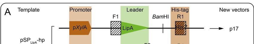
Microorganisms 2018, 6, 51                                                                           2 of 12

metagenomic discovery efforts, where E. coli is frequently used as a host, often suffer from a low
number of positive clones [18] despite the relatively high number of subtilisins per genome and their
wide representation in organisms [19]. The difficulties in expressing subtilisins and obtaining them in
an active form are likely due to the complex maturation and intrinsic proteolytic activity if unregulated.
However, yields for downstream characterization and application are often insufficient in E. coli.
Secretion of enzymes from the host is an attractive and cost-efficient production system due to limited
needs for cell processing and elaborate purifications. Bacillus subtilis is such an attractive host because
of its large capacity to export enzymes. Besides, it is non-pathogenic and is generally regarded as safe
(GRAS) by the US Food and Drug Administration, which make it a suitable host for enzymes that will
be used in food applications. However, regular B. subtilis species produce a number of extracellular
proteases that can potentially be detrimental for heterologous expression. Moreover, native proteases
pose a challenge for the assessment of recombinant proteases as they provide a background activity in
biochemical assays. For this purpose, protease-deficient strains, such as the WB800-derivatives [20],
are preferred.
      To facilitate efficient molecular cloning of a large number of genes in parallel, a range of assembly
cloning techniques have recently been developed based on type IIS restriction enzymes and T4 DNA
ligase [21–23]. With several of these methods a counter-selection approach using the coupled cell
division B gene (ccdB) gene is used [23]. The negative selection is based on the presence of the
ccdB-gene in the cloning region of the vector which, upon sub-cloning, is replaced with the gene of
interest. This promotes the emergence of positive clones, as negative clones will express a cytotoxic
protein encoded by the ccdB-gene that cause gyrase-mediated chromosomal damage and ultimately
cell death [24]. For high-throughput cloning, counter selection is identified as particularly useful
to limited elaborate screening for positive clones. This has previously shown great advantages for
recombinational cloning [25,26].
      In this study, we have developed three vectors for heterologous expression in Bacillus,
here‘explored in B. subtilis WB800N, and validated these by production of both intracellular
(green fluorescent protein) and extracellular (subtilisin) proteins. The vectors are compatible
with a versatile cloning regime based on fragment exchange (FX), encompassing an effective
counter-selection procedure and implementing secreted expression. The Bacillus system complement
the previously developed system for E. coli [16], and allows rapid switching between two commonly
used heterologous host systems without comprehensive intermediate cloning steps.

2. Materials and Methods

2.1. Construction of Fragment Exchange (FX)-Compatible Vectors for Bacillus Expression
     Three vectors were designed for FX-compatible cloning [23] and recombinant expression in
Bacillus (Table S1). The p17-vector allows intracellular or secreted expression depending on the
absence or presence of native leader sequences in the sub-cloned gene, respectively. The p18-
and p19-vectors contain leader sequences reported to efficiently direct export of the recombinant
proteins [27]. All vectors contain C-terminal hexahistidine tags. As starting templates, two vectors
that had successfully been used in Bacillus, pSPLipA -hp and pSPYocH -hp (MoBiTec, Table S1),
were used [27]. To enable FX compatibility, a counter selection cassette containing the ccdB gene
and a chloramphenicol-resistance gene (camR) flanked by SapI restriction sites were added to the
cloning region of the templates using a megapriming polymerase chain reaction (PCR) cloning
method [28]. The cassette was amplified from the p1 vector [16,23] using cloning primers listed
in Table S2 and Phusion polymerase (New England Biolabs, Hitchin, UK) protocol and purified
with the QIAquick PCR purification kit (QIAGEN, Valencia, CA, US). The cassette was inserted to
BamHI linearized pSPLipA -hp and pSPYocH -hp vectors, and used as templates in the linear plasmid
amplification reaction as described elsewhere [16,28–30]. To identify whether product formation
occurred, the reaction mixture was screened by PCR using primers flanking the insertion site (Table S2).
Microorganisms 2018, 6, 51                                                                             3 of 12

To remove template DNA, the PCR products were digested with 10 U DpnI (New England Biolabs,
Hitchin, UK) and transformed to E. coli MC1061 cells (Table S1). Plasmids were isolated using the
NucleoSpin plasmid purification kit (MACHEREY-NAGEL GmbH & Co, Düren, Germany). Sanger
sequencing was used to confirm correct cloning of all vectors. Plasmids and strains used in this study
are detailed in Table S1.

2.2. Sub-Cloning of gfp and apr Genes to the FX-Compatible Bacillus Vectors
     The gfp gene (coding for residues 2–247 of green fluorescent protein, GFP) was sub-cloned from
the B. megaterium optimized pSSBm85 plasmid [27] into the pINITIAL cloning vector [23] and verified
by sequencing. Codon-optimized apr genes (Genscript, Piscataway, NJ, USA) encoding subtilisins
from B. licheniformis DSM13, B. paralicheniformis ATCC 9945A, B. subtilis subsp. subtilis str. 168 and
B. amyloliquefaciens (GenBank IDs: AAU40017, AGN35600, CAB12870 and AAB05345, respectively,
herein termed: B13, B9945, BSU and BAM) [16] served as templates for PCRs using Phusion polymerase
(NEB). Genes were integrated to the pINITIAL cloning vector [16,23] and sequenced to confirm correct
cloning. Sub-cloning of genes from pINITIAL to the FX-compatible Bacillus expression vectors were
carried out as described previously [16,23]. Empty vectors were generated by replacing the ccdB-camR
cassette with a GSGSGS (GS) linker to allow their propagation in E. coli MC1061 cells, as described
previously [16], and use as background controls in experiments.

2.3. Transformation of Bacillus subtilis by Natural Competence
      The Bacillus subtilis WB800N strain [20], utilized for heterologous expression, was transformation-
based using a protocol developed by Anagnostopoulus and Spizizen [31]. A single colony of B. subtilis
WB800N from an lysogeny broth (LB) agar plate (1% (w/v) tryptone, 0.5% (w/v) yeast extract,
1% (w/v) NaCl, 1.5% (w/v) agar-agar) was inoculated in freshly prepared minimal medium (60 mM
K2 HPO4 , 40 mM KH2 PO4 , 3 mM trisodium citrate, 20 mM potassium-L-glutamate, 3 mM MgSO4 ,
1% glucose, 20 µg/mL L-tryptophane, 0.1% casamino acids) and grown for 16–20 h at 37 ◦ C and
250 rpm. The culture was diluted to an optical density at 600 nm (OD600 ) of 0.2 with minimal medium,
and grown for 4 h at the conditions given above. Cells were harvested, diluted 10-fold and distributed
in 1 mL aliquots for individual transformations. Typically 0.5–1.0 µg plasmid DNA was added to
cells, and incubated for 6 h at the conditions given above. Cells were harvested and spread on LB agar
plates supplemented with 10 µg/mL tetracycline. Colonies were checked for the presence of correct
plasmid by PCR.

2.4. Heterologous Expression of Green Fluorescent Protein (GFP) and Subtilisins in Bacillus subtilis
     1 mL LB media (1% (w/v) tryptone, 0.5% (w/v) yeast extract, 1% (w/v) NaCl) containing 10 µg/mL
tetracycline was added to deep 24-well plates, and inoculated with single colonies from agar plates and
grown at 37 ◦ C and 750 rpm. 100 µL pre-culture was used to inoculate 4 mL 2YT media (1.6% (w/v)
tryptone, 1% (w/v) yeast extract and 0.5% (w/v) NaCl) with 10 µg/mL tetracycline. Cells were
incubated for 3–4 h to reach log phase, prior to the induction of expression with 0.1% (v/v) D-xylose
(Sigma-Aldrich, St. Louis, MO, USA) for 16–20 h at 20 ◦ C (GFP) or 37 ◦ C (proteases) and 750 rpm.
Cells were harvested using an Allegra X-12R benchtop centrifuge (Beckman Coulter, Brea, CA, USA)
at 4750 rpm for 15 min. Proteins in 1 mL supernatants were precipitated with trichloroacetic acid
(TCA; 10% final concentration) for 1 h at 37 ◦ C, washed twice in 500 µL acetone, and resuspended
in 40 µL 1× Laemmli sample buffer (Bio-Rad Laboratories, Hercules, CA, USA) for sodium dodecyl
sulfate polyacrylamide gel electrophoresis (SDS-PAGE) analysis [32]. Cells were resuspended in 1 mL
8.5 N lysis buffer (50 mM Tris HCl pH 8.5, 50 mM NaCl, 0.25 mg/mL lysozyme, 10% (v/v) glycerol),
and lysed by ultrasound as previously described [16]. Cleared lysates (soluble fraction) and pellets
(insoluble fraction) were analyzed by SDS-PAGE [32].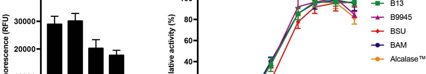
Microorganisms 2018, 6, 51                                                                        4 of 12

2.5. GFP Fluorescence Measurement
    Fluorescence from 100 µL cell cultures containing GFP was measured with excitation at 485 nm
and emission at 520 nm using a Sense microplate reader (Hidex, Turku, Finland).

2.6. Detection of Recombinant Subtilisins by Immunoblotting
     Proteins from supernatants and cleared lysates were analyzed by SDS-PAGE, and transferred
onto a nitrocellulose membrane [33] using the Trans-Blot Turbo (Bio-Rad Laboratories) transfer system.
A mouse monoclonal anti-polyhistidine antibody (Cat. No. H1029, Sigma-Aldrich) was used to
identify expression of recombinant subtilisins with C-terminal histidine affinity tags. The primary
antibodies were detected with a secondary rabbit horseradish peroxidase linked mouse IgG (NA931V,
GE Healthcare, Little Chalfont, UK). The HRP-reaction was developed with the Clarity Western ECL
Substrate (Bio-Rad Laboratories), and imaged in the Chemi-Doc gel imager (Bio-Rad Laboratories).

2.7. Subtilisin Activity Assays
     Proteolytic activity was assessed using EnzChek™ Protease Assay Kit (Thermo Fisher Scientific,
Waltham, MA, USA). 10 µg/mL BODIPY FL casein was prepared by resuspending the substrate in
50 mM Tris HCl pH 8.5 (at RT) and 50 mM NaCl. 12.5 µL of BODIPY-FL casein was used per reaction,
with 10 µL supernatant from expression in assay buffer I (50 mM NaCl, 50 mM TrisHCl pH 8.5 at room
temperature) in a final volume of 100 µL in MicroFluor 1 plates (Thermo Fisher Scientific). Samples
were incubated at 37 ◦ C for 1 h unless otherwise stated, and fluorescence was read. Fluorescence
was measured at excitation 485 nm and emission 520 nm using the Hidex Sense microplate reader.
Routinely, Alcalase™ 2.4L (Sigma-Aldrich) was used at a dilution 1:10,000 in 8.5 N lysis buffer.
     Temperature activity profiles for recombinant subtilisins were determined from 15 µL
supernatants in assay buffer II (80 mM NaCl, 80 mM TrisHCl buffer pH 8.2 at the experimental
temperatures) and 3.3 µM N-succinyl-L-Ala-L-Ala-L-Pro-L-Ala (AAPA) p-nitroanilide (Bachem, Weil am
Rhein, Germany; resuspended in dimethyl sulfoxide) in a total volume of 150 µL. The assay was
carried out for 10 min at the relevant temperatures, and stopped by addition of acetic acid at a final
concentration of 80 mM. 150 µL of the reaction volume was then transferred to a microplate for
absorbance reading at 405 nm in the Hidex Sense microplate reader. pH activity profiles were
determined for 15 µL supernatant as above, but using either 80 mM citrate buffer pH 3.0–6.0, 80 mM
Tris-HCl buffer pH 7.3, 8.1, 9.0 and 80 mM glycine buffer pH 10.0 Reaction was run at 37 ◦ C and
increase in absorbance at 405 nm was monitored. In both experiments, the obtained absorbance data
were background subtracted (backgrounds are supernatants from cells expressing the GS-linker from
vectors), and presented in relative activity (% of maximum activity). The measurements were carried
out with two technical replicates in three biological replicates. GraphPad Prism 7 (GraphPad Software,
La Jolla, CA, USA) was used to prepare plots and to perform statistical analyses.

3. Results

3.1. Preparation of FX-Compatible Bacillus Vectors
      To facilitate screening of industrially relevant enzymes, such as subtilisins, efficient systems
for high-throughput cloning and expression is essential. Here, we have developed three vectors
for heterologous expression in Bacillus [20], named p17, p18, and p19, by adapting existing
Bacillus-compatible plasmids to the FX-cloning principle (Figure 1). The ccdB-camR cassette was inserted
to the pSPLipA -hp and pSPYocH -hp plasmids by linear amplification using a PCR product containing
the cassette sequence as a megaprimer [28]. To confirm product formation before continuing with
transformation, the PCR reaction itself was screened using primers flanking the insertion site. In these
cases, the screening process identified a mix of both templates and products. During DpnI-digestions,
the templates were removed, and positive clones selected. The three vectors allow intracellular as well
as secreted expression. The p17 vector does not harbor a leader sequence, thus allowing exploitation of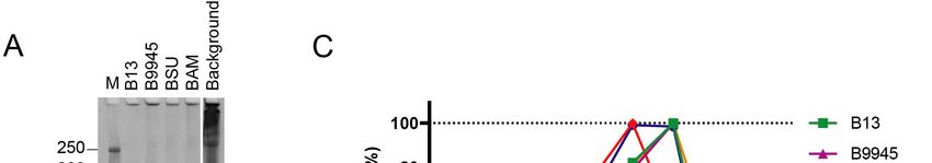
Microorganisms 2018, 6, 51                                                                                                        5 of 12

the putative native leader sequence from the enzyme of interest. The p18 and p19 vectors harbor known
LipA   and YocH
Microorganisms 2018,leader  sequences,
                     6, x FOR           respectively, which are known to facilitate export in Bacillus [34].
                              PEER REVIEW                                                               5 of 12

      Figure 1. Design of fragment exchange (FX) compatible Bacillus expression vectors. (A) The vector
      Figure 1. Design of fragment exchange (FX) compatible Bacillus expression vectors. (A) The vector
      templates, pSPLipA-hp and pSPYocH-hp, have a common backbone consisting of two origins of
      templates, pSPLipA -hp and pSPYocH -hp, have a common backbone consisting of two origins of
      replication (one for E. coli and one for Bacillus), two antibiotic resistance genes (for ampicillin
      replication (one for E. coli and one for Bacillus), two antibiotic resistance genes (for ampicillin selection
      selection in E. coli and tetracycline selection in Bacillus) and the Bacillus gene encoding the regulatory
      in E. coli and tetracycline selection in Bacillus) and the Bacillus gene encoding the regulatory protein,
      protein, XylR. The highlighted regions (upper panel) consist of the Bacillus pXylA promoter, two
      XylR. The highlighted regions (upper panel) consist of the Bacillus pXylA promoter, two different
      different leader sequences (LipA and YocH leader sequences, respectively), and C-terminal histidine
      leader sequences (LipA and YocH leader sequences, respectively), and C-terminal histidine affinity tags
      affinity tags
      (His-tag)       (His-tag) for purification
                  for downstream       downstreamofpurification
                                                        the recombinantof theproteins.
                                                                               recombinant        proteins.
                                                                                           The hatched        The (F-
                                                                                                           boxes    hatched   boxes
                                                                                                                       and R-sites)
      (F- and the
      indicate   R-sites)   indicate the
                     vector-specific        vector-specific
                                        regions                regions of
                                                   used for insertion    used
                                                                            the for    insertion
                                                                                 ccdB-camR          of the BamHI
                                                                                                 cassette.   ccdB-camR     cassette.
                                                                                                                      indicates the
      BamHI     indicates  the  restriction  site  used  for linearization,   and    arrows    indicate   position
      restriction site used for linearization, and arrows indicate position and direction of screening primers;      and  direction
      of screening
      (B) The region  primers;
                        inserted(B)   The region
                                   between          inserted
                                              the F-sites  andbetween    theofF-sites
                                                                the R-site      the pSP  and the   R-site of the pSPLipA-hp and
                                                                                           LipA -hp and pSPYocH -hp templates
      pSP  YocH-hp templates to generate the three vectors p17, p18 and p19, as shown in A, by whole
      to generate the three vectors p17, p18 and p19, as shown in A, by whole plasmid amplification.
      plasmid
      The  regionamplification.
                    contains theThe ccdBregion   contains
                                          and camR    genes,theand
                                                                ccdB  and camR
                                                                    flanking   SapI genes,
                                                                                       sites. and  flanking
                                                                                              Hatched    boxesSapI  sites. Hatched
                                                                                                                  marked   F and R
      boxes    marked     F  and   R  are  specific   to  each   vector   design     and    consists   of  two
      are specific to each vector design and consists of two parts; it has an overlapping region for gene-specificparts; it has an
      overlapping (filled
      amplification    regionboxes),
                                  for gene-specific
                                         and extensionsamplification         (filled boxes),
                                                            that are complementary           to theand     extensions
                                                                                                     insertion            that
                                                                                                                 site (open     are
                                                                                                                             boxes)
      complementary
      in the templates to  (F the  insertion
                               = either  F1, F2site
                                                  or(open
                                                     F3; R boxes)
                                                            = R1, asinindicated
                                                                       the templatesin A).(FThe= either
                                                                                                  insertF1,  F2 ordesignates
                                                                                                          region     F3; R = R1,the
                                                                                                                                 as
      indicated
      region       in A).
               in the finalThe   insert
                             vectors     region
                                       that        designates
                                            is replaced   uponthe    region in of
                                                                  sub-cloning      the   final vectors
                                                                                      recombinant         that and
                                                                                                       genes,   is replaced   upon
                                                                                                                     arrows show
      sub-cloning
      position   andof   recombinant
                      direction          genes, and
                                  of sequencing        arrows show position and direction of sequencing primers.
                                                    primers.

3.2. Expression of GFP in Bacillus subtilis
     To validate the three vectors, the gfp gene encoding green fluorescent protein was used for
simple fluorescence-based monitoring of recombinant protein production. The gene encoding GFP
was sub-cloned from the B. megaterium optimized pSSBm85 plasmid [27] into the pINITIAL cloning
vector, and sub-cloned into the Bacillus vectors. Expression was achieved by xylose induction.
Fluorescence measurements were taken from the cultures directly. GFP, which does not contain a leader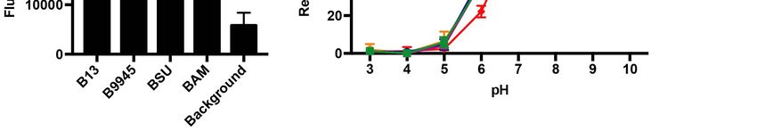
Microorganisms 2018, 6, 51                                                                                                       6 of 12

3.2. Expression of GFP in Bacillus subtilis
     To validate the three vectors, the gfp gene encoding green fluorescent protein was used for simple
fluorescence-based monitoring of recombinant protein production. The gene encoding GFP was
sub-cloned from the B. megaterium optimized pSSBm85 plasmid [27] into the pINITIAL cloning
vector, and sub-cloned into the Bacillus vectors. Expression was achieved by xylose induction.
Fluorescence measurements were taken from the cultures directly. GFP, which does not contain
a leaderMicroorganisms
          sequence,2018,
                       was 6, xfound
                                FOR PEERtoREVIEW
                                           fluoresce in the p17-based cultures (Figure 2A), indicating          6 of 12 soluble
expression of an intracellular GFP. SDS-PAGE analysis of fractions from expression revealed that
        sequence,
recombinant     GFP waswas
                        found    to fluoresce
                             indeed           in the p17-based
                                        expressed     from p17,cultures (Figure 2A), in
                                                                  and primarily      indicating solublefraction
                                                                                        the cellular    expression   of
                                                                                                                 (Figure    2B).
        an intracellular GFP. SDS-PAGE analysis of fractions from expression revealed that recombinant
Export was low or not obtained by adding the LipA or YocH leader sequences in front of the gfp
        GFP was indeed expressed from p17, and primarily in the cellular fraction (Figure 2B). Export was
gene (Figure
        low or 2).
                not obtained by adding the LipA or YocH leader sequences in front of the gfp gene (Figure 2).

               Figure 2. Recombinant expression of green fluorescent protein (GFP) in B. subtilis WB800N. (A) GFP
                2. Recombinant
      Figure expression                 expression of green fluorescent protein (GFP) in B. subtilis WB800N.
                            from the p17 to p19 vectors was assessed by fluorescence measurements taken from the
      (A) GFPexpression
                 expression    from the
                            cultures.     p17 to p19
                                      Fluorescence  wasvectors
                                                         comparedwastoassessed   by controls
                                                                        background   fluorescence     measurements
                                                                                               (in which                taken from
                                                                                                          a GSGSGS-linker
      the expression     cultures.GFP
               (GS) is replacing    Fluorescence    wasData
                                        in the vectors).  compared
                                                               is showntofrom
                                                                          background      controls experiment,
                                                                               one representative    (in which aandGSGSGS-linker
                                                                                                                       error
               bars represent
      (GS) is replacing     GFP deviation  in two technical
                                  in the vectors).    Data replicates;
                                                             is shown(B)    Sodium
                                                                          from  one dodecyl     sulfate polyacrylamide
                                                                                      representative      experiment,gel  and error
               electrophoresis
      bars represent             (SDS-PAGE)
                         deviation              analysis ofreplicates;
                                      in two technical       fractions from   expression
                                                                         (B) Sodium        of GFPsulfate
                                                                                         dodecyl     from the   p17 to p19
                                                                                                             polyacrylamide     gel
               vectors. S1, supernatant fraction; T, total protein in the cellular fraction; S2, soluble fraction of protein
      electrophoresis (SDS-PAGE) analysis of fractions from expression of GFP from the p17 to p19 vectors.
               in cells. M, Precision Plus protein standard (Bio-Rad Laboratories).
      S1, supernatant fraction; T, total protein in the cellular fraction; S2, soluble fraction of protein in cells.
      M, 3.3.
          Precision   Plusofprotein
              Validation               standard
                              Vectors by          (Bio-Rad
                                          Expression          Laboratories).
                                                      of Active  Recombinant Subtilisin Proteins in Bacillus subtilis
               To assess the capacity for secreted expression, the apr gene encoding extracellular subtilisin
3.3. Validation of from
        proteases  Vectors   by Expression
                        B. licheniformis    of Active
                                         DSM13        Recombinant
                                                was used (Figure 3A).Subtilisin
                                                                      The entireProteins
                                                                                 apr gene,in Bacillussubtilisin
                                                                                           encoding   subtilis
     To with  the native leader sequence (residues 1–379), was sub-cloned from pINITIAL to p17. The
         assess  the capacity for secreted expression, the apr gene encoding extracellular subtilisin
        truncated gene encoding pro-subtilisin (residues 30–379) without the leader sequence, was
proteases from B. licheniformis DSM13 was used (Figure 3A). The entire apr gene, encoding subtilisin
        sub-cloned to p18 and p19. In the latter two constructs, the native leader sequences of subtilisin were
with thereplaced
         native leader
                  with thesequence
                           LipA and (residues
                                    YocH leader 1–379),   wasencoded
                                                  sequences    sub-cloned
                                                                        by thefrom   pINITIAL
                                                                                vectors,          to p17.
                                                                                         respectively.     The truncated
                                                                                                       SDS-PAGE
gene encoding
        analysis from heterologous expression showed that the recombinant subtilisin was secreted to the to p18
                  pro-subtilisin  (residues    30–379)    without   the  leader   sequence,    was   sub-cloned
and p19.media  afterlatter
           In the     induction
                           twoand  expressionthe
                                constructs,    from  all three
                                                   native      vectors
                                                            leader     (Figure 3B).ofRecombinant
                                                                    sequences                       subtilisin
                                                                                      subtilisin were          was with
                                                                                                           replaced
         not identified in the cellular fractions. Immunoblots using anti-his antibodies against the C-terminal
         his-tags of the recombinant subtilisins supported these findings (Figure 3C). To measure the activity
         of the recombinant subtilisin, we used an in vitro casein-based protease assay. The activity detected
Microorganisms 2018, 6, 51 7 of 12
the LipA and YocH leader sequences encoded by the vectors, respectively. SDS-PAGE analysis from
heterologous expression showed that the recombinant subtilisin was secreted to the media after
induction and expression from all three vectors (Figure 3B). Recombinant subtilisin was not identified
in the cellular fractions. Immunoblots using anti-his antibodies against the C-terminal his-tags of
the recombinant
Microorganisms 2018, 6,subtilisins
x FOR PEER supported
REVIEW these findings (Figure 3C). To measure the activity of the
7 of 12
recombinant subtilisin, we used an in vitro casein-based protease assay. The activity detected in
the supernatant
in the supernatant fractions was
fractions was3–11
3–11times
timeshigher
higherthan
thaninincontrol
controlsamples
samplesdepending
depending on on conditions
(Figure 3D),
3D), and
and all
all versions
versions of
of the
the recombinant
recombinant subtilisin
subtilisin showed
showed comparable
comparableactivity.
activity.
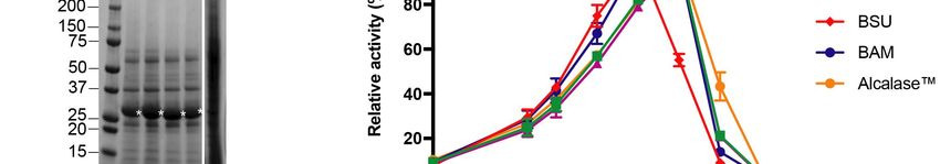
Figure 3.
Figure 3. Recombinant
Recombinantexpression
expressionand and activity of of
activity subtilisins from
subtilisins fromFX-compatible
FX-compatible Bacillus vectors.
Bacillus (A)
vectors.
Cartoon of recombinant subtilisin versions expressed from p17, p18 and p19
(A) Cartoon of recombinant subtilisin versions expressed from p17, p18 and p19 vectors, showing the vectors, showing the
leader sequence (triangles), pro-domain (grey boxes), catalytic domain (white
leader sequence (triangles), pro-domain (grey boxes), catalytic domain (white boxes) and C-terminal boxes) and C-terminal
histidine affinity
histidine affinity tag
tag (brown
(brown rectangles).
rectangles). Green
Green pins
pins point
point toto residues
residues involved
involved in in catalysis
catalysis (catalytic
(catalytic
triad). Dotted
triad). Dottedlinelineshows
showsthethe region
region of the
of the protein
protein encoded
encoded by theby the sub-cloned
sub-cloned BacillusBacillus licheniformis
licheniformis DSM13
DSM13 apr gene. In the p18 and p19 vectors, the vector-encoded LipA or YocH
apr gene. In the p18 and p19 vectors, the vector-encoded LipA or YocH leader sequences (green triangles) leader sequences
(green the
replace triangles) replacesequence
native leader the native(whiteleader sequence
triangle) (white respectively.
in subtilisin, triangle) in Arrows
subtilisin,
showrespectively.
processing
Arrows show processing sites for leader sequence removal and pro-domain cleavage.
sites for leader sequence removal and pro-domain cleavage. Illustration is drawn to scale; (B) SDS-PAGE Illustration is
drawn to
analysis ofscale; (B) SDS-PAGE
recombinant subtilisinsanalysis
expressed of in
recombinant subtilisins
B. subtilis WB800N (leftexpressed
panel) andin B. subtilis
empty vectorWB800N
controls
(left panel)
(right panel).and empty
Lanes 1, 4vector
and 7 controls (right panel).
show supernatant Lanesfrom
fractions 1, 4 and 7 show
cultures supernatant
expressing fractions
subtilisin fromfrom
p17,
cultures expressing subtilisin from p17, p18 and p19, respectively. Lanes 2, 5 and
p18 and p19, respectively. Lanes 2, 5 and 8 contains soluble fractions from cleared cell lysates. Lanes 3, 68 contains soluble
fractions
and from insoluble
9 contains cleared cell lysates.
fractions fromLanes 3, 6 andLanes
cell lysates. 9 contains insoluble
10–18 are organizedfractions from cell
accordingly, lysates.
but contain
Lanes 10–18
fractions fromare organized
cultures accordingly,
with empty but contain
vectors (subtilisin fractionswith
is replaced from thecultures with
GS-linker). M,empty vectors
Precision Plus
(subtilisin is replaced with the GS-linker). M, Precision Plus protein standard are shown to the left of
both panels; (C) Immunoblot of recombinant subtilisin using antibodies against the C-terminal
his-tag. Supernatants and soluble fractions of subtilisin expression from p17 (lanes 1–2), p18 (lanes
3–4) and p19 vectors (lanes 5–6) in B. subtilis WB800N, respectively. As control, fractions from
expression of empty p17 are shown in lanes 7–8. Asterisks indicate bands that correspond to the
expected mass of matured subtilisins (28 kDa). Two bands from the Precision Plus protein standard
are shown to the left; (D) The supernatants of B. subtilis WB800N expression cultures containing the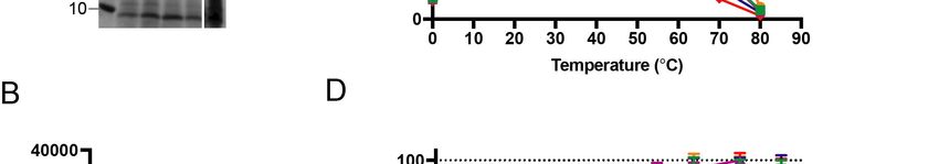
Microorganisms 2018, 6, 51                                                                                                           8 of 12

      protein standard are shown to the left of both panels; (C) Immunoblot of recombinant subtilisin using
      antibodies against the C-terminal his-tag. Supernatants and soluble fractions of subtilisin expression
      from p17 (lanes 1–2), p18 (lanes 3–4) and p19 vectors (lanes 5–6) in B. subtilis WB800N, respectively.
      As control, fractions from expression of empty p17 are shown in lanes 7–8. Asterisks indicate bands
      that correspond to the expected mass of matured subtilisins (28 kDa). Two bands from the Precision
      Plus protein standard are shown to the left; (D) The supernatants of B. subtilis WB800N expression
      cultures containing the subtilisin versions (expressed from p17, p18 and p19 vectors, respectively)
      were screened for proteolytic activity against BODIPY-conjugated casein for 1.5 and 20 h. Fluorescence
      values were made relative to empty vector controls (GS-linker), and errors show deviation in three
      biological replicates.

      WeMicroorganisms
            performed   2018,a6,comparative
                                x FOR PEER REVIEW study on four subtilisins, including the one encoded            8 of 12 by the

B. licheniformis DSM13 apr gene and three other apr genes from B. paralicheniformis ATCC 9945A,
               We performed a comparative study on four subtilisins, including the one encoded by the B.
B. subtilis  subsp. subtilis
         licheniformis  DSM13str.  apr 168    andB.three
                                        geneand      amyloliquefaciens
                                                         other apr genespreviously     used in a similar
                                                                           from B. paralicheniformis  ATCC mini
                                                                                                             9945A,screen
                                                                                                                       B.   [16].
The three    additional
         subtilis subsp. subtilis   str. 168apr
                            full-length      andgenes    with native previously
                                                  B. amyloliquefaciens  leader sequences
                                                                                  used in a were
                                                                                             similarsub-cloned
                                                                                                      mini screen into
                                                                                                                   [16]. the p17
vector. The
         Each    of the
              three       four subtilisins
                     additional                 was
                                   full-length apr    expressed,
                                                    genes           and
                                                          with native     thesequences
                                                                       leader supernatants     were tested
                                                                                        were sub-cloned    intofor
                                                                                                                the proteolytic
                                                                                                                    p17
         vector.  Each   of the  four  subtilisins was  expressed,  and  the supernatants  were  tested
activity in the in vitro casein assay (Figure 4). All four subtilisins were exported from the host      for proteolytic
cell andactivity   in the in vitro casein assay (Figure 4). All four subtilisins were exported from the host cell
            identified     as soluble enzymes in the supernatant (Figure 4A), and found to be active
         and identified as soluble enzymes in the supernatant (Figure 4A), and found to be active (p-value
(p-value
Microorganisms 2018, 6, 51 9 of 12
3.4. Initial Characterisation of Four Different Subtilisin Proteins
To demonstrate the functionality of the recombinant enzymes, we characterized their activity
profiles. The temperature activity profiles of the recombinant subtilisins were assessed with
a synthetic peptide, succinyl-L-Ala-L-Ala-L-Pro-L-Ala (AAPA), commonly used to address substrate
specificity [35,36]. The two recombinant subtilisins from B. licheniformis and B. paralicheniformis and
the commercial enzyme formula Alcalase™ 2.4L, that originates from B. licheniformis, peaked at
60 ◦ C (Figure 4C). The B. subtilis homolog had an optimal temperature at 50 ◦ C, and only 55% of
its activity remained at 60 ◦ C. The B. amyloliquefaciens homolog had a broader temperature profile,
with optimal temperature at 50 ◦ C, which remained largely unchanged at 60 ◦ C. All of the enzymes
had lost most of the activity at 80 ◦ C. The recombinant subtilisins were all active at pH 5.0 and above,
where a peak of activity was reached at pH 8.0–10.0 for all enzymes (Figure 4D). Subtilisins from
B. subtilis, B. licheniformis and B. amyloliquefaciens are, however, more active at pH 10 than the other
enzymes (p-value < 0.00001).
The two recombinant subtilisin homologs from B. licheniformis and B. paralicheniformis appeared to
share similar activity profiles, both in terms of temperature and pH preferences. They also aligned
well with the activity profile of the commercial Alcalase™ 2.4L (Figure 4C,D).
4. Discussion
The Bacillus fragment exchange (FX) vector system was designed to complement the intracellular
E. coli system [16] by facilitating secreted expression. These vectors would be useful for naturally
secreted proteins, but also for exploring the export of proteins that otherwise would be expressed in
the intracellular compartment.
The pSPLipA -hp and pSPYocH -hp plasmids, initially designed for mediating high-yield production
in B. megaterium [27], were used as templates to design FX-cloning compatible vectors for heterologous
expression. To develop the vectors, we used an approach exploring linear amplification that uses the
insert DNA fragment as a megaprimer for polymerase-mediated elongation [28]. The method has
previously been used to design new vectors [30,37,38]. Commonly, such long-range amplifications
are carried out on circular templates (plasmids), but we found that BamHI-linearized templates gave
higher product formation than circular templates as assessed by agarose gel electrophoresis [39].
Evaluation of this as a general optimization procedure for large and difficult plasmid amplifications is
outside the scope of this study.
The new vectors facilitate expression in the presence of xylose due to a strong inducible pXylA
promoter [38], which is repressed in the absence of inducer by the XylR repressor (Figure 1).
The promoter has previously been shown to drive heterologous expression in B. subtilis [40].
In a previous screen of leader sequences applicable for high-yield production, the YocH and LipA
were shown to mediate efficient secretion of a thermophilic ester-hydrolase into the growth medium in
B. megaterium [27]. The LipA and YocH leader sequences originate from an esterase and a cell wall
binding protein in B. megaterium [27,41], respectively, but are conserved in B. subtilis [11]. As addressed
in previous reports, the YocH and LipA leader sequence show high similarity with the consensus
sequence for type I leader sequences directing Sec-dependent export. We were, therefore, confident that
the vector systems would be suitable for heterologous expression not only in B. megaterium, but also in
B. subtilis WB800N.
The system was initially validated with expression of the GFP from the p17 vector (Figure 2).
Expression trials with p18 and p19 vectors were not giving traceable amounts of GFP (Figure 2),
which is likely explained by the fact that GFP is not directed for export using the Sec pathway [42].
To validate Sec-dependent export of proteins, subtilisin from B. licheniformis DSM13 was used and
shown to be successfully expressed in all vectors (Figure 3B). This study confirms that these vectors
can be used for secreted expression in B. subtilis. As such, the vectors could be explored for the
replacement of native leader sequences with known sequences, particularly useful when the native
enzyme sequence is divergent and may escape recognition by the Bacillus secretion systems.Microorganisms 2018, 6, 51 10 of 12
Comparison to protein yields in B. megaterium, for which the vector backbones were optimized,
is outside the scope of this study. In the aforementioned leader sequence screen, however, it was
found that the YocH leader sequence promoted higher yields of exported target enzyme than the LipA
sequence [27]. In our study, based on expression and activity levels of recombinant subtilisin from
B. licheniformis DSM13, it was not possible to discriminate between results obtained with native and
artificial leader sequences (Figure 3D). Induction at shorter intervals (1.5 h) did not change the results.
To identify the optimal leader sequence, empirical testing on a higher number of recombinant proteins
may be required, but the fact that results vary between reports demonstrates the merit of including
several construct designs in a screen at this time point.
The Bacillus system has been assessed by expressing four homologous subtilisins that are divergent
in sequence and expected to have different temperature activity profiles [16]. These were previously
expressed in an active form in E. coli. Using our p17 vector and exploring the native leader sequences
for export, all subtilisins were identified as soluble and active in the growth medium (Figure 4B).
These subtilisins used in the mini screen are all alkaline, as confirmed by their activity at high pH
(Figure 4D). They differ somewhat in the activity profiles, with the B. subtilis homolog having a lower
optimal temperature than the B. licheniformis and B. paralicheniformis homologs. Apparently, the latter
two homologs share similar activity profiles. This can be explained by a 98% sequence identity
according to a pairwise sequence alignment [16]. Moreover, they share a profile with the commercial
Alcalase™, which also originates from B. licheniformis. The B. amyloliquefaciens homolog has an optimal
activity at 50 ◦ C, which aligns well with other reports [43], but appears to have a broader temperature
optimum range than the other homologs. This trait could be useful in industrial applications to avoid
the enzyme activity from dropping due to temperature fluctuation. Apparently, most activity is lost at
80 ◦ C which aligns with industrial conditions for enzyme inactivation that commonly occur at 90 ◦ C.
5. Conclusions
To conclude, the Bacillus system herein reported complements the previously developed E. coli
system [16], and allows rapid switching between two commonly used heterologous host systems for
comparative expression. Moreover, we show that the vectors described are not limited to the expression
of certain enzymes, here exemplified by the expression of both subtilisins and green fluorescent protein,
but could also be applied to other enzymes for more generalized enzyme discovery or development.
Supplementary Materials: The following are available online at http://www.mdpi.com/2076-2607/6/2/51/s1,
Table S1: Plasmids and strains used in this study; Table S2: Primers used in this study.
Author Contributions: G.E.K.B. conceived and designed the experiments and prepared the manuscript; Ø.L. and
G.E.K.B. performed the experiments and analyzed the data. Both authors have discussed the results and read,
edited and approved the final manuscript.
Acknowledgments: We would like to thank Stephan Thies and Andreas Knapp for providing excellent advice and
protocols. We would also like to thank Rebekka Biedendieck for kindly sharing the pSSBm85 plasmid. This study
was financed by the Norwegian Research Council (grant ref.: 221568; NorZymeD) and Regional Research Funds
in Norway (grant ref.: 259224, iEnzyme).
Conflicts of Interest: The authors declare no conflict of interest.
References
1. Kwon, K.; Hasseman, J.; Latham, S.; Grose, C.; Do, Y.; Fleischmann, R.D.; Pieper, R.; Peterson, S.N.
Recombinant expression and functional analysis of proteases from Streptococcus pneumoniae, Bacillus anthracis,
and Yersinia pestis. BMC Biochem. 2011, 12, 17. [CrossRef] [PubMed]
2. Sroga, G.E.; Dordick, J.S. A strategy for in vivo screening of subtilisin E reaction specificity in E. coli periplasm.
Biotechnol. Bioeng. 2002, 78, 761–769. [CrossRef] [PubMed]
3. Siezen, R.J.; Leunissen, J.A. Subtilases: The superfamily of subtilisin-like serine proteases. Protein Sci. 1997, 6,
501–523. [CrossRef] [PubMed]Microorganisms 2018, 6, 51 11 of 12
4. Güntelberg, A.V.; Ottesen, M. Preparation of Crystals containing the Plakalbumin-forming Enzyme from
Bacillus subtilis. Nature 1952, 170, 802. [CrossRef] [PubMed]
5. Rawlings, N.D.; Barrett, A.J. Families of serine peptidases. Methods Enzymol. 1994, 244, 19–61. [PubMed]
6. Gupta, R.; Beg, Q.K.; Lorenz, P. Bacterial alkaline proteases: Molecular approaches and industrial applications.
Appl. Microbiol. Biotechnol. 2002, 59, 15–32. [CrossRef] [PubMed]
7. Wong, S.L.; Doi, R.H. Determination of the signal peptidase cleavage site in the preprosubtilisin of Bacillus
subtilis. J. Biol. Chem. 1986, 261, 10176–10181. [PubMed]
8. Wells, J.A.; Ferrari, E.; Henner, D.J.; Estell, D.A.; Chen, E.Y. Cloning, sequencing, and secretion of Bacillus
amyloliquefaciens subtilisin in Bacillus subtilis. Nucleic Acids Res. 1983, 11, 7911–7925. [CrossRef] [PubMed]
9. Vasantha, N.; Thompson, L.D.; Rhodes, C.; Banner, C.; Nagle, J.; Filpula, D. Genes for alkaline protease
and neutral protease from Bacillus amyloliquefaciens contain a large open reading frame between the regions
coding for signal sequence and mature protein. J. Bacteriol. 1984, 159, 811–819. [PubMed]
10. Finn, R.D.; Mistry, J.; Tate, J.; Coggill, P.; Heger, A.; Pollington, J.E.; Gavin, O.L.; Gunasekaran, P.; Ceric, G.;
Forslund, K.; et al. The Pfam protein families database. Nucleic Acids Res. 2009, 38, D211–D222. [CrossRef]
[PubMed]
11. Tjalsma, H.; Antelmann, H.; Jongbloed, J.D.H.; Braun, P.G.; Darmon, E.; Dorenbos, R.; Dubois, J.-Y.F.;
Westers, H.; Zanen, G.; Quax, W.J.; et al. Proteomics of protein secretion by Bacillus subtilis: Separating the
‘secrets’ of the secretome. Microbiol. Mol. Biol. Rev. 2004, 68, 207–233. [CrossRef] [PubMed]
12. Ikemura, H.; Takagi, H.; Inouye, M. Requirement of pro-sequence for the production of active subtilisin E in
Escherichia coli. J. Biol. Chem. 1987, 262, 7859–7864. [PubMed]
13. Ohta, Y.; Hojo, H.; Aimoto, S.; Kobayashi, T.; Zhu, X.; Jordan, F.; Inouye, M. Pro-peptide as an intramolecular
chaperone: Renaturation of denatured subtilisin E with a synthetic pro-peptide. Mol. Microbiol. 1991, 5,
1507–1510. [CrossRef] [PubMed]
14. Zhu, X.L.; Ohta, Y.; Jordan, F.; Inouye, M. Pro-sequence of subtilisin can guide the refolding of denatured
subtilisin in an intermolecular process. Nature 1989, 339, 483–484. [CrossRef] [PubMed]
15. Ikemura, H.; Inouye, M. In vitro processing of pro-subtilisin produced in Escherichia coli. J. Biol. Chem. 1988,
263, 12959–12963. [PubMed]
16. Bjerga, G.E.K.; Arsın, H.; Larsen, Ø.; Puntervoll, P.; Kleivdal, H.T. A rapid solubility-optimized screening
procedure for recombinant subtilisins in E. coli. J. Biotechnol. 2016, 222, 38–46. [CrossRef] [PubMed]
17. Martínez-Martínez, M.; Coscolín, C.; Santiago, G.; Chow, J.; Stogios, P.J.; Bargiela, R.; Gertler, C.;
Navarro-Fernández, J.; Bollinger, A.; Thies, S.; et al. The INMARE Consortium, Determinants and Prediction
of Esterase Substrate Promiscuity Patterns. ACS Chem. Biol. 2018, 13, 225–234. [CrossRef] [PubMed]
18. Ferrer, M.; Martínez-Martínez, M.; Bargiela, R.; Streit, W.R.; Golyshina, O.V.; Golyshin, P.N. Estimating
the success of enzyme bioprospecting through metagenomics: Current status and future trends.
Microb. Biotechnol. 2016, 9, 22–34. [CrossRef] [PubMed]
19. Tripathi, L.P.; Sowdhamini, R. Genome-wide survey of prokaryotic serine proteases: Analysis of distribution
and domain architectures of five serine protease families in prokaryotes. BMC Genom. 2008, 9, 549. [CrossRef]
[PubMed]
20. Nguyen, H.D.; Phan, T.T.P.; Schumann, W. Analysis and application of Bacillus subtilis sortases to anchor
recombinant proteins on the cell wall. AMB Express. 2011, 1, 22. [CrossRef] [PubMed]
21. Engler, C.; Kandzia, R.; Marillonnet, S. A one pot, one step, precision cloning method with high throughput
capability. PLoS ONE 2008, 3, e3647. [CrossRef] [PubMed]
22. Engler, C.; Gruetzner, R.; Kandzia, R.; Marillonnet, S. Golden gate shuffling: A one-pot DNA shuffling
method based on type IIs restriction enzymes. PLoS ONE 2009, 4, e5553. [CrossRef] [PubMed]
23. Geertsma, E.R.; Dutzler, R. A versatile and efficient high-throughput cloning tool for structural biology.
Biochemistry 2011, 50, 3272–3278. [CrossRef] [PubMed]
24. Bernard, P.; Couturier, M. Cell killing by the F plasmid CcdB protein involves poisoning of
DNA-topoisomerase II complexes. J. Mol. Biol. 1992, 226, 735–745. [CrossRef]
25. Hartley, J.L.; Temple, G.F.; Brasch, M.A. DNA cloning using in vitro site-specific recombination. Genome Res.
2000, 10, 1788–1795. [CrossRef] [PubMed]
26. Busso, D.; Delagoutte-Busso, B.; Moras, D. Construction of a set Gateway-based destination vectors for
high-throughput cloning and expression screening in Escherichia coli. Anal. Biochem. 2005, 343, 313–321.
[CrossRef] [PubMed]Microorganisms 2018, 6, 51 12 of 12
27. Stammen, S.; Müller, B.K.; Korneli, C.; Biedendieck, R.; Gamer, M.; Franco-Lara, E.; Jahn, D. High-yield intra-
and extracellular protein production using Bacillus megaterium. Appl. Environ. Microbiol. 2010, 76, 4037–4046.
[CrossRef] [PubMed]
28. Van den Ent, F.; Löwe, J. RF cloning: A restriction-free method for inserting target genes into plasmids.
J. Biochem. Biophys. Methods 2006, 67, 67–74. [CrossRef] [PubMed]
29. Lund, B.A.; Leiros, H.-K.S.; Bjerga, G.E.K. A high-throughput, restriction-free cloning and screening strategy
based on ccdB-gene replacement. Microb. Cell Fact. 2014, 13, 38. [CrossRef] [PubMed]
30. Bjerga, G.E.K.; Williamson, A.K. Cold shock induction of recombinant Arctic environmental genes.
BMC Biotechnol. 2015, 15, 78. [CrossRef] [PubMed]
31. Anagnostopoulos, C.; Spizizen, J. Requirements for transformation in Bacillus subtilis. J. Bacteriol. 1961, 81,
741–746. [PubMed]
32. Laemmli, U.K. Cleavage of structural proteins during the assembly of the head of bacteriophage T4. Nature
1970, 227, 680–685. [CrossRef] [PubMed]
33. Towbin, H.; Staehelin, T.; Gordon, J. Electrophoretic transfer of proteins from polyacrylamide gels to
nitrocellulose sheets: Procedure and some applications. Proc. Natl. Acad. Sci. USA 1979, 76, 4350–4354.
[CrossRef] [PubMed]
34. Tjalsma, H.; Bolhuis, A.; Jongbloed, J.D.; Bron, S.; van Dijl, J.M. Signal peptide-dependent protein transport
in Bacillus subtilis: A genome-based survey of the secretome. Microbiol. Mol. Biol. Rev. 2000, 64, 515–547.
[CrossRef] [PubMed]
35. Jones, D.D. Recombining low homology, functionally rich regions of bacterial subtilisins by combinatorial
fragment exchange. PLoS ONE 2011, 6, e24319. [CrossRef] [PubMed]
36. Tindbaek, N.; Svendsen, A.; Oestergaard, P.R.; Draborg, H. Engineering a substrate-specific cold-adapted
subtilisin. Protein Eng. Des. Sel. 2004, 17, 149–156. [CrossRef] [PubMed]
37. Unger, T.; Jacobovitch, Y.; Dantes, A.; Bernheim, R.; Peleg, Y. Applications of the Restriction Free (RF) cloning
procedure for molecular manipulations and protein expression. J. Struct. Biol. 2010, 172, 34–44. [CrossRef]
[PubMed]
38. Rygus, T.; Hillen, W. Inducible high-level expression of heterologous genes in Bacillus megaterium using the
regulatory elements of the xylose-utilization operon. Appl. Microbiol. Biotechnol. 1991, 35, 594–599. [CrossRef]
[PubMed]
39. Bjerga, G.E.K. Uni Research, Bergen, Norway. RF-cloning using linear and circular plasmids as templates, 2016.
40. Kim, L.; Mogk, A.; Schumann, W. A xylose-inducible Bacillus subtilis integration vector and its application.
Gene 1996, 181, 71–76. [CrossRef]
41. Ruiz, C.; Blanco, A.; Pastor, F.I.J.; Diaz, P. Analysis of Bacillus megaterium lipolytic system and cloning of
LipA, a novel subfamily I.4 bacterial lipase. FEMS Microbiol. Lett. 2002, 217, 263–267. [CrossRef] [PubMed]
42. Feilmeier, B.J.; Iseminger, G.; Schroeder, D.; Webber, H.; Phillips, G.J. Green fluorescent protein functions as
a reporter for protein localization in Escherichia coli. J. Bacteriol. 2000, 182, 4068–4076. [CrossRef] [PubMed]
43. Peng, Y.; Huang, Q.; Zhang, R.H.; Zhang, Y.Z. Purification and characterization of a fibrinolytic enzyme
produced by Bacillus amyloliquefaciens DC-4 screened from douchi, a traditional Chinese soybean food.
Comp. Biochem. Physiol. B Biochem. Mol. Biol. 2003, 134, 45–52. [CrossRef]
© 2018 by the authors. Licensee MDPI, Basel, Switzerland. This article is an open access
article distributed under the terms and conditions of the Creative Commons Attribution
(CC BY) license (http://creativecommons.org/licenses/by/4.0/).You can also read