VHL, BAP1, PBRM1, SETD2 expression in clear cell renal car-cinoma, association with PD-1, PD-L1, PD-L2 mRNA level
←
→
Page content transcription
If your browser does not render page correctly, please read the page content below
Preprints (www.preprints.org) | NOT PEER-REVIEWED | Posted: 21 July 2021                          doi:10.20944/preprints202107.0495.v1

             Article

             VHL, BAP1, PBRM1, SETD2 expression in clear cell renal car-
             cinoma, association with PD-1, PD-L1, PD-L2 mRNA level
             Liudmila Spirina 1,2,*, Zahar Yurnazov 1, Evgeny Usynin 1Firstname 1 , Irina Kondakova 1, Ekaterine Ladutko 2
             and Evgeny Choynzonov 1,2

                                          1 Cancer Research Institute, Tomsk National Research Medical Center; Russia, info@oncology.tomsk.ru
                                          2 Siberian State Medical University; Tomsk, Russia, rector@ssmu.ru
                                          * Correspondence: spirinalvl@mail.ru; Tel.: +79609758577

                                          Abstract: Novel mechanism of ccRCC progression is essential, including PBRM1, BAP1, and
                                          SETD2 in histone-modifying and chromatin remodeling genes. The study aimed to investigate
                                          VHL, PD-1, PD-L1, PD-L2. BAP1, PBRM1, SETD2 expression in ccRCC primary cancers and met-
                                          astatic tissues associated with the cancer dissemination. A total of 62 patients with RCC were
                                          enrolled in the study. Investigation of mRNA level of VHL, PD-1, PD-L1, PD-L2. PCR in real-
                                          time performed BAP1, PBRM1, SETD2 with the previous RNA isolation. Western Blotting anal-
                                          ysis was used for detecting the p-VHL protein content in tissues. The VHL expression and p-
                                          VHL content determined the aggressive cancer behavior and elevated in disseminated tumors.
                                          The cancer dissemination was accompanied by an increase in both mRNA and VHL content. The
                                          PD-L2 prevalence in metastatic cancers is crucial in tumor progression. ccRCC progression in
                                          VHL overexpression is associated with the decrease in BAP1 gene expression. It is revealed the
                                          heterogeneity in molecular markers in primary tumors and metastases. The low mRNA level of
                                          BAP1, PBRM1, SETD2, PD-1, PD-L1, PD-L2 in metastases compared with primary tumors were
                                          found. We show a novel mechanism for VHL tumor progression and present a new instrument
                                          and factor targeting tumor-related pathologies with p-VHL/HIF altered function.

                                          Keywords: kidney cancers; VHL; PD-1; PD-L1; PD-L2; BAP1; PBRM; SETD2; primary tumor;
                                          metastasis.

                                          1. Introduction
                                                   VHL is mutated in most patients with clear cell renal cell carcinoma (ccRCC),
                                              with conflicting clinical relevance. Recent studies have identified recurrent mutations
                                              in histone-modifying and chromatin remodeling genes, including BAP1, PBRM1, and
                                              SETD2. Current evidence suggests that BAP1 mutations are associated with
                                              aggressive disease [1]. The von Hippel-Lindau gene (VHL) product acts as the
                                              substrate-recognition component of an E3 ubiquitin ligase complex that ubiquitylates
                                              the catalytic alpha subunit of hypoxia-inducible factor (HIF) for oxygen-dependent
                                              destruction. Somatic mutations or loss of von Hippel-Lindau (pVHL) happen in most
                                              VHL disease tumors, which present a constitutively active Hypoxia Inducible Factor
                                              (HIF), essential for tumor growth [2].
                                                   A decrease in pVHL expression from the adjacent healthy tissues to the tumor's
                                              ones was found in recent studies [3]. But the content of pVHL in ccRCC primary
                                              tumors and metastatic sites remains unknown. There are current data repost the
                                              presence of VHL mutations is not concordant with mRNA or protein expression.
                                              Nonsense mutations of the VHL gene appear to be related to poorer prognosis and
                                              survival [2]. Multiple molecular targets have been developed based on blocking major

            © 2021 by the author(s). Distributed under a Creative Commons CC BY license.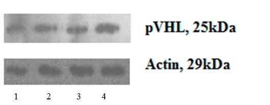
Preprints (www.preprints.org) | NOT PEER-REVIEWED | Posted: 21 July 2021              doi:10.20944/preprints202107.0495.v1

                                                                                                                       2 of 14

                                        signaling pathways directly or indirectly involved in ccRCC tumor progression,
                                        metastasis, angiogenesis, and survival [4].
                                              Inactivation of the VHL gene is an early event in ccRCC carcinogenesis [5]. The
                                        pVHL targets could be regulated independently of VHL mutation (and possibly by
                                        hypoxia alone); this data suggests that other pVHL marks may be more crucial in
                                        renal carcinogenesis [6]. VHL loss in clear-cell renal cell carcinoma (RCC) leads to
                                        increased levels of hypoxia-inducible factors (HIF) and overexpression of HIF target
                                        genes, such as VEGF and others. VEGF-targeted agents are standard in advanced
                                        clear-cell RCC, but biomarkers of activity are lacking. Several potential biomarkers
                                        along the VHL/HIF-1α/HIF-2α axis were not predictive for targeted therapy in
                                        patients with advanced clear-cell RCC. Additional efforts must continue to identify
                                        biomarkers associated with clinical outcomes to VEGF-targeted agents in metastatic
                                        RCC [7].
                                              Several novel recurrent mutations of histone-modifying and chromatin
                                        remodeling genes have been identified in renal cell carcinoma. These mutations cause
                                        loss of function of several genes located close to VHL and include PBRM1, BAP1, and
                                        SETD2. Chromosome 3p harbors BRCA1-associated protein-1 (BAP1) [8]. PBRM1
                                        encodes for BAF180, a component of the SWI/SNF chromatin remodeling complex,
                                        and is inactivated in, on average, 36% of clear cell renal cell carcinoma (ccRCC).
                                              Mutations of BAP1 encode for the histone deubiquitinase BRCA1 associated
                                        protein-1 and are present in 10% of RCCs. They are essentially mutually exclusive
                                        with PBRM1 mutations. Mutations to SETD2, a histone methyltransferase, occur in 10%
                                        of ccRCC. BAP1- or SETD2-mutated ccRCCs have been associated with poor overall
                                        survival, while PBRM1 mutations seem to identify a favorable group of ccRCC tumors
                                        [9]. It is found PRBM-1 loss is associated with more aggressive mccRCC and could be
                                        therapeutic targets for anti-cancer therapy [10].
                                              The relationship between gene mutation, loss of protein expression, and the
                                        correlations with clinicopathological parameters is essential for understanding renal
                                        cancer progression. The combined functional inactivation of PBRM1, BAP1, SETD2,
                                        and pVHL is critical for ccRCC advancement [11]
                                              Tumor immunogenicity is one of the urgent problems in modern oncology.
                                        Receptor PD-1 - (Programmed cell death 1; CD279) plays a role in the cellular
                                        differentiation of immune cells [12]. Under physiological conditions, when PD-1
                                        interacts with its ligands (PD-L1 PD-L2), an inhibitory signal is transmitted that
                                        prevents the development of an excessive immune response, triggering the processes
                                        of apoptosis of cytotoxic lymphocytes [13].
                                              The recent analysis highlights the VHL impact on the biological processes,
                                        including metabolism, immune regulation, apoptosis, and cell movement. Its
                                        overexpression promotes immune system activation and sensitivity to interferon
                                        therapy [14]. Recent studies showed that PD-L1 expression levels positively correlate
                                        with VHL mutation and HIF-2α expression. VHL mutations positively correlate with
                                        PD-L1 expression in ccRCC and may influence the response to ccRCC anti-PD-L1/PD-
                                        1 immunotherapy [15].
                                              In the presented works, the relationship of the positive expression of PD-L1 with
                                        the sporadic and hereditary ccRCC aggressiveness is currently noted [16]. It was
                                        revealed an association between the VHL biallelic inactivation and the PD-L1
                                        increased expression [15]. In addition, there is evidence if protein E3 of ubiquitin
                                        ligase is functional, an increase in the PD-L1 / PD-1 will be observed [17]. Unlike PD-
                                        L1, PD-L2 has a less pronounced association with disease prognosis [18].
                                              Currently, the prognostic role of the PD-L1 level with the cancer aggressiveness
                                        and the unfavorable outcome development is prominent [19]. Its function is currently
                                        not clearly defined. However, blocking antitumor immunity and inhibition of
                                        cytotoxic T cells and NK cells is possible, among other things, by increasing the
Preprints (www.preprints.org) | NOT PEER-REVIEWED | Posted: 21 July 2021 doi:10.20944/preprints202107.0495.v1
3 of 14
expression of PD-L2 [20]. However, there is evidence of a greater affinity of this ligand
for PD-1 receptors of immunocompetent cells [21]. It is known that the expression of
PD-1, PD-L1, and PD-L2 differs in primary tumor tissues and metastases [22].
The changes in AKT/mTOR and MAPK signaling cascades in the ccRCC can
influence tumorigenesis [23-24]. In addition, the expression of p-AKT is closely
associated with the expression of ligands PD-L1, PD-L2 in tumor cells [25]. It is known
that tumor withdrawal from immune surveillance occurs due to the production of
ligands PD-L1 PD-L2 by tumor cells, which is associated with the development of
hypoxia and the level of the HIF-1 protein [26-27]. In particular, for kidney cancer, an
association of PD-L1 expression with a high level of HIF-2α by the presence of
lymphocytic infiltration has been shown [28]. The study aimed to investigate VHL,
PD-L1, PD-L2. BAP1, PBRM1, SETD2 expression in ccRCC primary cancers and
metastatic tissues associated with the cancer dissemination.
2. Results
2.1. VHL, PD, PD-L1, PD-L2. BAP1, SEDT1, PRBM distribution in ccRCC tumor size
growth and dissemination.
ccRCC begins with an intragenic mutation in the von Hippel-Lindau (VHL) gene
and loss of 3p (where VHL is located). But several studies revealed the presence of
other abnormalities. We tried to found the association of VHL, PD, PD-L1, PD-L2.
BAP1, SEDT1, PRBM with clinical signs of the disease.
Table 1. Distribution of increased and decreased gene’s expression in ccRCC depended on the
tumor size
Expression, Tumor size χ2-
Relative Units T1N0M0 T2N0M0-1 T3N0M1 distribution
VHL, >1.0 7 (77.78) 9 (52.94) 6 (85.71) χ2- 29.25
(n, %) 1.0 9 (100.00) 7 (41.18) 3 (43.86) χ2- 95.07
(n, %)Preprints (www.preprints.org) | NOT PEER-REVIEWED | Posted: 21 July 2021 doi:10.20944/preprints202107.0495.v1
4 of 14
Table 2. Distribution of increased and decreased gene’s expression in ccRCC depended
on cancer dissemination
Cancer spreading
Expression, metastatic
Relative Units localized ccRCC ccRCC χ2-distribution
>1.0 6 (40.00) 16 (88.89) χ2- 50.47
VHL,
(n, %) 1.0 12 (80.00) 7 (38.89) χ2- 33.66
PD, (n,
%) 1.0 10 (66.67) 5 (27.78) χ2- 29.09
PD-L1,
(n, %) 1.0 13 (86.67) 11 (61.11) χ2- 15.78
PD- L2,
(n, %) 1.0 11 (68.75) 6 (33.33) χ2- 23.95
%) 1.0 8 (50.00) 8 (44.44) χ2- 0.42
(n, %) 1.0 11 (68.75) 8 (44.44) p=0.0008
2.2. VHL expression and p-VHL content increase in metastatic ccRCC.
All figures and tables should be cited in the main text as Figure 1, Table 1, etc. The
expression and content of VHL depended on the stage of the disease (criterion T) and
the extent of the tumor process in ccRCC. The VHL expression was increased by 12.5
times in patients with a T3-4N0-1M1 stage than T1-2N0M0. The found change was
accompanied by a 5-fold decrease in the p-VHL content (Figure 1). The most significant
changes were revealed when studying the prevalence of the tumor process. Substantial
changes in the expression and protein level under study were observed in the
metastatic tissue compared with the primary tumor and against the background of
targeted therapy. There was a decrease in the VHL expression in the metastases by 1.89
times and its content by 3.17 times compared with the primary tumor.
Figure 1. p-VHL content in ccRCC tissues and adjacent non-transformed ones.
The expression and content of VHL depended on the stage of the disease
(criterion T) and the extent of the tumor process in ccRCC (Table 3). The VHLPreprints (www.preprints.org) | NOT PEER-REVIEWED | Posted: 21 July 2021 doi:10.20944/preprints202107.0495.v1
5 of 14
expression was increased by 12.5 times in patients with a T3-4N0-1M1 stage than T1-2N0M0.
The change was accompanied by a 5-fold decrease in the p-VHL in the patients with
advanced cancers. The most significant changes were revealed when studying the
prevalence of the tumor process. The expression and content of VHL are associated
with the development of distant metastases. An increase in the VHL mRNA and its
protein was recorded in patients with disseminated form by 32.0 and 2.68 times,
respectively, of the disease, compared with patients with localized cancer.
Table 3. Expression and content of VHL in tumor tissue of patients depending on tumor size and disease
spreading, Me(Q1; Q3)
Indicator Tumor size Cancer dissemination
T1-2N0М0 T3-4N0-1М0-1 Localized Metastatic Metastases
cancer cancers (T1-4N0-
(T1-2N0М0) М1 )
1
VHL expression, Relative 7.00 (2.00; 87.50 (4.22; 2.00 (0.50;4.00) 64.00 (8.46; 1.09 (0.09;
Units 31.32) 128.00)* 128.00)* 5.44)**
VHL content,% to unal- 77.20 (42.25; 15.30 (11.30; 55.00 (20.10; 147.54 (100.54; 20.80
tered tissue 99.79) 46.07)* 93.61) 156.78)* (10.60;
58.80)**
Note: - * the significance of the differences with the localized form, pPreprints (www.preprints.org) | NOT PEER-REVIEWED | Posted: 21 July 2021 doi:10.20944/preprints202107.0495.v1
6 of 14
compared with T1N0M0. There was also a decrease in the level of PD-L2 mRNA during
tumor growth.
Table 5. PD, PD-L1, PD-L2 expression in ccRCC tissues depending on tumor size and disease spreading, Me(Q1;
Q3)
Indicator Tumor size Cancer dissemination
T1N0M0 T2N0M0-1 T3N0M1 Localized Disseminated Metastases
cancers cancers
PD-1 4.19 (3.45; 0.81 (0.11; 0.41 (0.20; 2.91 (1.44; 0.61 (0.12; 0.57 (0.135;
20.40) 2.00) 1.94) 7.63) 1.94)* 1.82)
Kruskal-Wallis Test: H =11.95785
p =0.0025
PD-L1 2.85 (0.40; 1.00 (0.19; 0.32 (0.28; 1.00 (0.25; 0.37 (0.24; 1.00) 0.75 (0.375;
6.77) 1.00) 0.50) 4.27) 1.50)
Kruskal-Wallis Test: H =3.792281
p =0.1501
PD-L2 11.8 (1.49; 1.14 (0.64; 1.77 (0.50; 1.28 (0.50; 6.81 (2.16; 0.605 (0.435;
21.10) 2.00) 10.54) 2.17) 17.8)* 0.755)**
Kruskal-Wallis Test: H =4.837951
p =0.0490
Note: - * - the significance of the differences with the localized cancers, pPreprints (www.preprints.org) | NOT PEER-REVIEWED | Posted: 21 July 2021 doi:10.20944/preprints202107.0495.v1
7 of 14
Figure 2. Scatter plots between VHL expression and p-VHL content.
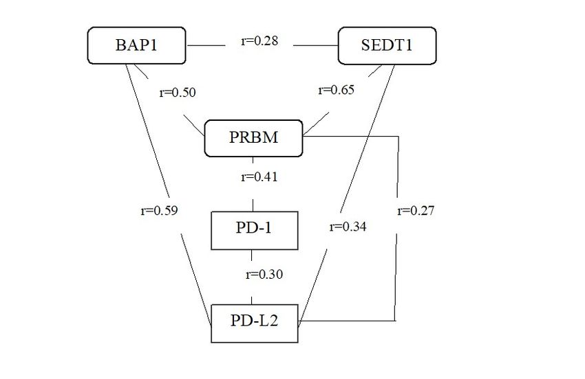
But there are no correlations between the VHL and PD-1, PD-L1, PD-L2, and
BAP1, PBRM1, SETD2 expression. The results revealed the direct connections between
the immune markers and genes participating in chromatin remodeling and epigenetic
regulation. Thus, BAP1 gene expression correlated with SEDT1 (r=0.28; pPreprints (www.preprints.org) | NOT PEER-REVIEWED | Posted: 21 July 2021 doi:10.20944/preprints202107.0495.v1
8 of 14
The data showed the presence of multiple regulatory triggers in PD-L2 activation and
immune system regulation.
3. Discussion
ccRCC accounts for 2-3% of all tumors, the most frequent solid lesion in the kidney.
VHL influences the progression of ccRCC tumors. The higher expression of VHL was
correlated with the better disease-free survival in ccRCC patients using The Cancer
Genome Atlas (TCGA) datasets [14].
We found that an increase in VHL mRNA level with a p-VHL decrease in ccRCC
was associated with increased tumor size. The prevalence of the disease was
accompanied by an increase in both mRNA and VHL content. p-VHL and gene
expression levels in the cancers are significantly reduced compared to the adjacent
untransformed tissue and correlate with the disease stage, which is especially
pronounced in metastatic ccRCC [3, 29-31].
Currently, about 32 significant mutations of the VHL gene and a large number of
insignificant gene changes have been found. Lots of modifications [2]. The intensity of
molecular processes in the oncogenesis of kidney cancer is associated with the role of
protein in the regulation of essential cellular processes [5, 32-33]. These findings reveal
a critical HIF-dependent molecular pathway connecting VHL, an established
"gatekeeper" of the renal epithelium [32].
Renal cell carcinoma (RCC) includes diverse tumor types characterized by various
genetic abnormalities. Genetic changes, like mutations, deletions, and epigenetic
alterations, play a crucial role in modifying signaling networks, tumor pathogenesis,
and prognosis. Loss of von Hippel-Lindau (VHL) gene and upregulation of hypoxia-
inducible factors (HIF), the signature of most sporadic ccRCC, promote multiple
growth factors [4].
The presence of significant relationships between the expression and the content
of the von Hippel-Lindau protein was confirmed by correlation analysis [34, 35]. An
increase in the VHL mRNA level is associated with decreased related protein content
and expression of the transcription factor NF-κB p65. However, an increase in p-VHL
content in ccRCC tissues was associated with a decrease in the NF-κB p65, NF-κB p50,
and VEGF expression. The presence of VHL mutations without taking into account the
biological behavior of the tumor cannot determine the course, the outcome of the
disease, and the effectiveness of targeted therapy [36]. The HIF overexpression
associated with the p-VHL deficiency leads to cancer progression [7, 30-31, 37].
Significant changes in molecular factors also determine the development of disease
metastases. In metastases, a decrease in the expression and content of p-VHL compared
with the primary tumor was revealed, which was accompanied by an increase in the
mRNA level of HIF-1, HIF-2, NF-κB p50, NF-κB p65, VEGF, and CAIX in the foci of
secondary growth as compared with the primary tumor.
The hypoxia-induced transcription factor activation by VHL mutation is
associated with p-VHL protein loss. The VHL protein, promoting degradation of HIFα,
is involved in the modification of the transcriptional activity of the nuclear factor NF-
κB [38]. The primary mechanism is direct hydroxylation of the NF-κB factor and its
repressor IκBα [39]. It is known that in the presence of the normal VHL protein, NF-κB
signaling is suppressed [38].
Although VHL mutations are triggers of ccRCC, the results demonstrated that
genetic so on epigenetic factors impact the ccRCC invasive and metastatic properties
due to VHL modifications. HIF-1 dependent protein CAIX is associated with increased
aggressiveness [40]. The lncRNA FGD5-AS1 was significantly associated with VHL and
can serve as a novel biomarker of ccRCC [41].
Recent genome-wide sequencing studies of ccRCC have revealed that mutations
of genes coding for epigenome modifiers and chromatin remodelers,Preprints (www.preprints.org) | NOT PEER-REVIEWED | Posted: 21 July 2021 doi:10.20944/preprints202107.0495.v1
9 of 14
including PBRM1, SETD2, and BAP1, are the most common somatic genetic
abnormalities after VHL mutations in these tumors [42, 43].
Biological characteristics in tumor grade and aggressiveness across cancer types
are well known poorly understood. BAP1- or SETD2-mutated ccRCCs have been
associated with poor overall survival, while PBRM1 mutations seem to identify a
favorable group of ccRCC tumors [9]. We detected the BAP expression in ccRCC is
decreased with tumor size growth and metastasis development. BAP1 is crucial for
kidney function and cooperates with VHL in renal oncogenesis [38]. Moreover, it is
considered the driver of tumor grade, promoted by activation of mTORC1 [44].
The critical oncogenic event in ccRCC is VHL overexpression affecting the
numerous processes. It results in inhibited epithelial-mesenchymal transition (EMT),
sterol regulatory element-binding protein 1 regulated triglyceride synthesis, and cell
proliferation [14]. VHL overexpression led to upregulation of proteins associated with
antigen processing and interferon-responsive proteins, rendering ccRCC cells with high
VHL expression more sensitive to interferon treatment. The data indicate that the
subset of ccRCC patients with high VHL expression benefit from immunotherapy [14].
Recent studies showed that PD-L1 expression levels positively correlate with VHL
mutation and HIF-2α expression. VHL mutations positively correlate with PD-L1
expression in ccRCC and may influence the response to ccRCC anti-PD-L1/PD-1
immunotherapy [15].
The significance of immune system regulation has been verified in correlation
analysis. The prevalence and distribution of PD-L2 are found to be correlated
significantly with PD-L1 [16]. The PD-L2 level is found to be dependent on the BAP1,
PBRM1, SETD2 expression. Previous studies have shown a relationship between the
PD-L1 expression with a ccRCC poor prognosis [16-18]. The study revealed high levels
of PD-L2 mRNA in patients with metastatic ccRCC. PD-L2 expression was observed in
all tumor types and present in the stroma, tumor, and endothelial cells. However, PD-
L2 was detected in the absence of PD-L1 in some tumor types [13]. The low rates in PD-
1, PD-1, and PD-L2 were found in the tissue of metastases. The revealed data
substantiate the contribution of heterogeneity and biological characteristics to the
progression of the disease [22].
An increase in tumor immunogenicity during tumor progression is the foremost
progression step associated with VHL inactivation [19, 20, 23-24, 28]. Previous studies
have revealed a relationship between VHL expression and components of the
AKT/mTOR signaling cascade and transcriptional and growth factors [35], which
possibly determines the response to targeted therapy. In addition, the activation of the
AKT mTOR signaling pathway and an increase in gene expression and the content of
transcriptional and growth factors have been shown [30-31] in ccRCC.
4. Materials and Methods
A total of 62 patients with RCC were enrolled in the study. The retrospective
study included patients with histopathologically verified RCC admitted to and
nephrectomized at the Cancer Research Institute, Tomsk National Research Center,
Russian Academy of Medical Sciences, Tomsk, Russian Federation. The patients
underwent a physical examination, chest radiography, and computer tomography (CT)
of the abdomen. When vena cava tumor thrombus invasion was suspected, cavography
or magnetic resonance imaging (MRI) was performed. Patients with skeletal-associated
pain or elevated serum alkaline phosphatase were assessed with bone scintigraphy. The
patients were followed up according to a program, including regular clinical and
radiological examinations. The median age of the patients was 57 years. The RCC
therapy depends on the tumor size and its spreading to other parts of the body.
Localized RCC (T1-3N0M0) was diagnosed in 18 patients, metastatic RCC (T2-
4N0-1M1) in 44 patients. All patients with localized RCC underwent surgery (partialPreprints (www.preprints.org) | NOT PEER-REVIEWED | Posted: 21 July 2021 doi:10.20944/preprints202107.0495.v1
10 of 14
nephrectomy or simple nephrectomy) and then followed up according to a program
including regular clinical and radiological examinations. Patients with metastatic RCC
received two cycles of preoperative targeted therapy with pazopanib at a dose of 800
mg daily for two months. Tumor response to targeted therapy was evaluated according
to RECIST criteria. All patients underwent radical nephrectomy. The diagnosis was ver-
ified based on biopsy results.
The Local Committee approved the study for Medical Ethics, and all patients
provided written informed consent (protocol code 4; 16.11.2018). Tumor tissue samples,
histologically normal tissue samples adjacent to tumors, and metastatic tissues were
used for investigation. Specimens were reviewed separately by two independent
pathologists.
RNA extraction. The postoperative tumor samples were incubated in RNAlater
solution (Ambion, USA) for 24-hours at + 4 °C and then stored at -80 oC. Total RNA
was extracted using the RNeasy Mini Kit (Qiagen).
RT-qPCR. PCR was conducted in 25 μl reaction volumes containing 12.5 μl Bio-
Master HS-qPCR SYBR Blue (2X) (“Biolabmix” Russia) and 300 nanoM of each primers.
.VHL: F 5´ - GGCAGGCGAATCTCTTGA-3´ , R 5´-CTATTTCCTTTACTCAGCAC-
CATT-3´; BAP1: F 5´ - GCCACGGACAGCAGAGAG-3´, R 5´-CTTCCTCTTCTCTAC-
CTCCTCCT-3´; PRBM1 : F 5´ - CCCAGTAGCCTTGTCGCA-3´, R 5´-TAGTCAATAA-
GAGCAGAGTTCAATCA-3´; SEDT1: F 5´ - CAGAGTTATGCCCAGCCAA-3´, R 5´-
GAGTTCCCAGGTCCATCTCA-3´; PD-L2: F 5-GTTCCACATACCTCAAGTCCAA-3,
R 5-ATAGCACTGTTCACTTCCCTCTT-3; PD-L1: F 5-AGGGA-
GAATGATGGATGTGAA-3, R 5-ATCATTCACAACCACACTCACAT-3; PD-1-1: F
5-CTGGGCGGTGCTACAACT-3, R 5-CTTCTGCCCTTCTCTCTGTCA-3; GAPDH: F
5´ - GGAAGTCAGGTGGAGCGA-3´ , R 5´-GCAACAATATCCACTTTACCAGA-3´ .
At 95°C for 10 min, a pre-incubation was to activate the Hot Start DNA polymerase and
denature DNA and was followed by 45 amplification cycles of 95°C denaturations at 95
0 for 10 sec, 60°C annealings at 60 0 for 20 sec (iCycler iQ™, BioRad).
The fold changes were calculated by ΔΔCt method (the total ΔΔCt = fold of can-
cerous/normal tissue gene level), using normal tissue. A ratio of specific mRNA/
GADPH (GADPH as a respective control) amplification was then calculated.
Determination of p-VHL level in tissues. Electrophoresis SDS-PAGE (Laemmli) was
used. The protein was transferred to a 0.2-/xm pore-sized PVDF membrane (GE
Healthcare, UK), either at 150 mA or 100 V for one h by using a Bio-Rad Mini Trans-
Blot electrophoresis cell. The membrane was incubated in a 1:2500 dilution of monoclo-
nal mouse anti-p-VHL (Ser68) (Affinity Biosciences, USA) at four ºС overnight.
PVDF samples were incubated in Amersham ECL western blotting detection
analysis system (Amersham, USA). The results were standardized using the beta-ac-
tin expression in a sample and were expressed in percentages to the protein content in
non-transformed tissues. The analysis of the results was carried out using the Chemi-
DocTMTouch Imaging System, and their density was assessed using the ImageLab
computer program (BioRad, USA). The level of protein in normal non-altered tissue
was indicated as 100%.
Statistical analysis. Statistical analysis was performed using SPSS 19.0 software.
Data were expressed as median and ranges. Mann-Whitney test was used for
comparing differences in mean values. Nonparametric one-way ANOVA on ranks was
carried out to test whether samples originate from the same distribution, which is used
to compare two or more independent samples of equal or different sample sizes.
Nonparametric correlation analysis was performed, and the Spearmen coefficient was
calculated. The significance of differences in the frequencies of qualitative traits was
evaluated using the χ2 criterion with the Yates correction.Preprints (www.preprints.org) | NOT PEER-REVIEWED | Posted: 21 July 2021 doi:10.20944/preprints202107.0495.v1
11 of 14
5. Conclusions
The von Hippel-Lindau gene product acts as the substrate-recognition component
of an E3 ubiquitin ligase complex that ubiquitylates the catalytic alpha subunit of the
hypoxia-inducible factor for oxygen-dependent destruction. VHL abnormalities and
alterations guide in ccRCC oncogenesis. We found the prevalence of VHL expression
in increased tumor growth and metastases development. The VHL expression and p-
VHL content determine the aggressive cancer behavior and are elevated in
disseminated tumors. ccRCC progression in VHL overexpression is associated with the
decrease in BAP1 gene expression. The prevalence of PD-L2, PD-1 ligand, is found in
metastatic cancers. It should be considered as an independent factor of cancer
aggressiveness.
It is revealed the heterogeneity in molecular markers in primary tumors and me-
tastases. We found the low mRNA level of BAP1, PBRM1, SETD2, PD-1, PD-L1, PD-L2
in ccRCC metastases compared with primary tumors. Direct and indirect relationships
in molecular factors determine the biological behavior and therapeutic effect. There are
opposite data on the VHL expression in ccRCC and its significance in cancer develop-
ment. This work shows a novel mechanism for VHL tumor progression and presents a
new instrument and factor for targeting tumor-related pathologies with p-VHL/HIF
altered function. The relationship between the expression and content of the VHL pro-
tein is of decisive importance in ccRCC oncogenesis. This new insight in ccRCC pro-
gression may offer prominent opportunities for therapeutic intervention
Supplementary Materials: No
Author Contributions: Conceptualization, Liudmila Spirina. and Zahar Yrmazov; methodol-
ogy, Evgeny Usynin; formal analysis, Ekaterine Ladutko; investigation, Liudmila Spirina; re-
sources, Evgeny Choynzonov.; data curation, Zahar Yurmazov.; writing—original draft prepa-
ration, Spirina L.V..; writing—review and editing, Spirina L.V. All authors have read and agreed
to the published version of the manuscript.”
Funding: This research received no external funding
Institutional Review Board Statement: The study was conducted according to the guidelines of
the Declaration of Helsinki, and approved by the Institutional Review Board (or Ethics Commit-
tee) (protocol code 4; 16.11.2018).
Informed Consent Statement: Written informed consent has been obtained from the patient(s)
to publish this paper.
Acknowledgments: No
Conflicts of Interest: The authors declare no conflict of interest.
Appendix A
Not acceptable
Appendix B
Figure 1. Note: 1, 3 –cancers, 2,4 – non-transformed tissues; p-VHL content is a
key oncogenic event in ccRCC development. It promotes and triggers the main
biological processes resulting in modification of metabolism, immune regulation,
apoptosis, and cell movement.
Figure 2. Note: VHL expression and p-VHL content are the decisive points on
oncogenesis governing kidney cancer spreading and disease progression.
VHL/HIF/VEGF pathway and VHL/PD-1/PD-L1/PD-L2 axis determine the ccRCC
aggressive behavior.Preprints (www.preprints.org) | NOT PEER-REVIEWED | Posted: 21 July 2021 doi:10.20944/preprints202107.0495.v1
12 of 14
Figure 3. Note: The roles of PBRM1, BAP1, and SETD2 in the development and
progression of ccRCC and their potential for future personalized approaches are con-
nected with immune system regulation. PD-L2 level is a potential target
References
1. Gossage, L.; Murtaza, M.; Slatter, A.F.; Lichtenstein, C.P.; Warren, A.; Haynes, B.; Marass, F.; Roberts, I.; Shanahan, S.J.;
Claas, A.; Dunham, A; May, A.P.; Rosenfeld, N.; Forshew, T.; Eisen, T. Clinical and pathological impact of VHL, PBRM1,
BAP1, SETD2, KDM6A, and JARID1c in clear cell renal cell carcinoma. Genes Chromosomes Cancer 2014, 53(1), 38-51. DOI:
10.1002/gcc.22116.
2. Alves, M.R.; Carneiro, F.C.; Lavorato-Rocha, A.M.; da Costa, W.H.; da Cunha, I.W.; de Cássio Zequi, S.; Guimaraes, G.C.;
Soares, F.A.; Carraro, D.M.; Rocha, R.M. Mutational status of VHL gene and its clinical importance in renal clear cell carci-
noma. Virchows Arch 2014, 465(3), 321-30. DOI: 10.1007/s00428-014-1629-z.
3. Ferchichi, I.; Kourda, N.; Sassi, S.; Romdhane, K.B.; Balatgi, S.; Cremet, J.Y.; Prigent, C.; Elgaaied, A.B.; Aurora-A overex-
pression and pVHL reduced expression are correlated with a bad kidney cancer prognosis. Dis Markers 2012, 33(6), 333-40.
DOI: 10.3233/DMA-2012-00942.
4. Tumkur Sitaram, R.; Landström, M.; Roos, G.; Ljungberg, B. Significance of PI3K signalling pathway in clear cell renal cell
carcinoma in relation to VHL and HIF status. J Clin Pathol 2021, 74(4), 216-222. DOI: 10.1136/jclinpath-2020-206693.
5. Arias-González, L.; Moreno-Gimeno, I.; del Campo, A.R.; Serrano-Oviedo, L.; Valero, M.L.; Esparís-Ogando, A.; de la Cruz-
Morcillo, M.Á.; Melgar-Rojas, P.; García-Cano, J.; Cimas, F.J.; Hidalgo, M.J.; Prado, A.; Callejas-Valera, J.L.; Nam-Cha, S.H.;
Giménez-Bachs, J.M.; Salinas-Sánchez, A.S.; Pandiella, A.; del Peso, L.; Sánchez-Prieto, R. ERK5/BMK1 is a novel target of
the tumor suppressor VHL: implication in clear cell renal carcinoma. Neoplasia 2013, 15(6), 649-59. DOI: 10.1593/neo.121896.
6. Nyhan, M.J.; El Mashad, S.M.; O'Donovan, T.R.; Ahmad, S.; Collins, C.; Sweeney, P; Rogers, E.; O'Sullivan, G.C.; McKenna,
S.L. VHL genetic alteration in CCRCC does not determine de-regulation of HIF, CAIX, hnRNP A2/B1 and osteopontin. Cell
Oncol (Dordr) 2011, 34(3), 225-34. DOI: 10.1007/s13402-011-0029-5.
7. Choueiri, T.K.; Fay, A.P.; Gagnon, R.; Lin, Y.; Bahamon, B.; Brown, V.; Rosenberg, J.E.; Hutson, T.E.; Baker-Neblett, K.L.;
Carpenter, C.; Liu, Y.; Pandit, L.; Signoretti, S. The role of aberrant VHL/HIF pathway elements in predicting clinical out-
come to pazopanib therapy in patients with metastatic clear-cell renal cell carcinoma. Clin Cancer Res 2013, 19(18), 5218-26.
DOI: 10.1158/1078-0432.CCR-13-0491.
8. Wang, S.S.; Gu, Y.F.; Wolff, N.; Stefanius, K.; Christie, A.; Dey, A.; Hammer, R.E.; Xie, X.J.; Rakheja, D.; Pedrosa, I.; Carroll,
T.; McKay, R.M.; Kapur, P.; Brugarolas, J. Bap1 is essential for kidney function and cooperates with Vhl in renal tumorigen-
esis. Proc Natl Acad Sci U S A 2014, 111(46), 16538-43. DOI: 10.1073/pnas.1414789111.
9. Piva, F.; Santoni, M.; Maturana, M.R.; Satti, S.; Giulietti, M.; Occhipinti, G.; Massari, F.; Cheng, L.; Lopez-Beltran, A.; Scar-
pelli, M.; Principato, G.; Cascina, S.; Montironi, R. BAP1, PBRM1 and SETD2 in clear-cell renal cell carcinoma: molecular
diagnostics and possible targets for personalized therapies. Expert Rev Mol Diagn 2015, 15(9), 1201-10. DOI:
10.1586/14737159.2015.1068122.
10. Moreira, M.; Pobel, C.; Epaillard, N.; Simonaggio, A.; Oudard, S.; Vano, Y.A. Resistance to cancer immunotherapy in meta-
static renal cell carcinoma. Cancer Drug Resist 2020, 3, 454-471. DOI: 10.20517/cdr.2020.16
11. Bihr, S.; Ohashi, R.; Moore, A.L.; Rüschoff, J.H.; Beisel, C.; Hermanns, T.; Mischo, A.; Corrò, C.; Beyer, J.; Beerenwinkel, N.;
Moch, H.; Schraml, P. Expression and Mutation Patterns of PBRM1, BAP1 and SETD2 Mirror Specific Evolutionary Subtypes
in Clear Cell Renal Cell Carcinoma. Neoplasia 2019, 21(2), 247-256. DOI: 10.1016/j.neo.2018.12.006.
12. Kushlinskii, N.E.; Fridman, M.V.; Morozov, A.A.; Gershtei, E.S.; Kadagidze, Z.G.; Matveev, V.B. Modern approaches to
kidney cancer immunotherapy. Cancer Urology 2018, 14(2), 54-67. DOI: 10.17650/1726-9776-2018-14-2-54-67.
13. Yearley, J.H.; Gibson, C.; Yu, N.; Moon, C.; Murphy, E.; Juco, J.; Lunceford, J.; Cheng, J.; Chow, L.Q.M.; Seiwert, T.Y.; Handa,
M.; Tomassini, J.E.; McClanahan, T. PD-L2 Expression in Human Tumors: Relevance to Anti-PD-1 Therapy in Cancer. Clin
Cancer Res 2017, 23(12), 3158-3167. DOI: 10.1158/1078-0432.CCR-16-1761.
14. Zhu, S.; Ding, W.; Chen, Y.; Wang, W.; Xu, R.; Liu, C.; Liu, X.; Deng, H.; High, V.H.L. Expression Reverses Warburg Pheno-
type and Enhances Immunogenicity in Kidney Tumor Cells. Genomics Proteomics Bioinformatics 2021, S1672-0229(21), 00039-
5. DOI: 10.1016/j.gpb.2019.12.002.
15. Messai, Y.; Gad, S.; Noman, M.Z.; Le Teuff, G.; Couve, S.; Janji, B.; Kammerer, S.F.; Rioux-Leclerc, N.; Hasmim, M.; Ferlicot,
S.; Baud, V.; Mejean, A.; Mole, D.R.; Richard, S.; Eggermont, A.M.; Albiges, L.; Mami-Chouaib, F.; Escudier, B.; Chouaib,
S. Renal Cell Carcinoma Programmed Death-ligand 1, a New Direct Target of Hypoxia-inducible Factor-2 Alpha, is Regu-
lated by von Hippel-Lindau Gene Mutation Status. Eur Urol 2016, 70(4), 623-632. DOI: 10.1016/j.eururo.2015.11.029.
16. Hong, B.; Cai, L.; Wang, J.; Liu, S.; Zhou, J.; Ma, K.; Zhang, J.; Zhou, B.; Peng, X.; Zhang, N.; Gong, K. Differential Expression
of PD-L1 Between Sporadic and VHL-Associated Hereditary Clear-Cell Renal Cell Carcinoma and Its Correlation With
Clinicopathological Features. Clin Genitourin Cancer. 2019, 17(2), 97-104.el. DOI: 10.1016/j.clgc.2018.11.001.
17. Kammerer-Jacquet, S.F.; Crouzet, L.; Brunot, A.; Dagher, J.; Pladys, A.; Edeline, J.; Laguerre, B.; Peyronnet, B.; Mathieu, R.;
Verhoest, G.; Patard, J.J.; Lespagnol, A.; Mosser, J.; Denis, M.; Messai, Y.; Gad-Lapiteau, S.; Chouaib, S.; Belaud-Rotureau,
M.A.; Bensalah, K.; Rioux-Leclercq, N. Independent association of PD-L1 expression with noninactivated VHL clear cell
renal cell carcinoma-A finding with therapeutic potential. Int J Cancer 2017, 140(1), 142-148. DOI: 10.1002/ijc.30429.Preprints (www.preprints.org) | NOT PEER-REVIEWED | Posted: 21 July 2021 doi:10.20944/preprints202107.0495.v1
13 of 14
18. Lu, Y.; Song, Y.; Xu, Y.; Ou, N.; Liang, Z.; Hu, R.; Zhang, W.; Kang, J.; Wang, X.; Liu, L.; Yang, Y.; Liu, X. The prevalence and
prognostic and clinicopathological value of PD-L1 and PD-L2 in renal cell carcinoma patients: a systematic review and meta-
analysis involving 3,389 patients. Transl Androl Urol 2020, 9(2), 367-381. DOI: 10.21037/tau.2020.01.21.
19. Ueda, K.; Suekane, S.; Kurose, H.; Chikui, K.; Nakiri, M.; Nishihara, K.; Matsuo, M.; Kawahara, A.; Yano, H.; Igawa, T.
Prognostic value of PD-1 and PD-L1 expression in patients with metastatic clear cell renal cell carcinoma. Urol Oncol 2018,
36(11), 499.e9-499.e16. DOI: 10.1016/j.urolonc.2018.07.003.
20. Tanegashima, T.; Togashi, Y.; Azuma, K.; Kawahara, A.; Ideguchi, K.; Sugiyama, D.; Kinoshita, F.; Akiba, J.; Kashiwagi, E.;
Takeuchi, A.; Irie, T.; Tatsumi, K.; Hoshino, T.; Eto, M.; Nishikawa, H. Immune Suppression by PD-L2 against Spontaneous
and Treatment-Related Antitumor Immunity. Clin Cancer Res 2019, 25(15), 4808-4819. DOI: 10.1158/1078-0432.CCR-18-3991.
21. Philips, E.A.; Garcia-España, A.; Tocheva, A.S.; Ahearn, I.M.; Adam, K.R.; Pan, R.; Mor, A.; Kong, X.P. The structural features
that distinguish PD-L2 from PD-L1 emerged in placental mammals. J Biol Chem 2020, 295(14), 4372-4380. DOI:
10.1074/jbc.AC119.011747.
22. Zhang, X.; Yin, X.; Zhang, H.; Sun, G.; Yang, Y.; Chen, J.; Zhu, X.; Zhao, P.; Zhao, J.; Liu, J.; Chen, N.; Wang, J.; Shen, P.; Zeng,
H. Differential expressions of PD-1, PD-L1 and PD-L2 between primary and metastatic sites in renal cell carcinoma. BMC
Cancer 2019, 19(1), 360. DOI: 10.1186/s12885-019-5578-4.
23. Lastwika, K.J.; Wilson, W. 3rd.; Li, Q.K.; Norris, J.; Xu, H.; Ghazarian, S.R.; Kitagawa, H.; Kawabata, S.; Taube, J.M.; Yao, S.;
Liu, L.N.; Gills, J.J.; Dennis, P.A. Control of PD-L1 Expression by Oncogenic Activation of the AKT-mTOR Pathway in Non-
Small Cell Lung Cancer. Cancer Res 2016, 76(2), 227-38. DOI: 10.1158/0008-5472.CAN-14-3362.
24. Tsai, T.F.; Lin, J.F.; Lin, Y.C.; Chou, K.Y.; Chen, H.E.; Ho, C.Y.; Chen, P.C.; Hwang, T.I. Cisplatin contributes to programmed
death-ligand 1 expression in bladder cancer through ERK1/2-AP-1 signaling pathway. Biosci Rep 2019, 39(9), BSR20190362.
DOI: 10.1042/BSR20190362.
25. Dong, L.; Lv, H.; Li, W.; Song, Z.; Li, L.; Zhou, S; Qiu, L.; Qian, Z.; Liu, X.; Feng, L.; Meng, B.; Fu, K.; Wang, X.; Pan-Ham-
marström, Q.; Wang, P.; Wang, X.; Zhang, H. Co-expression of PD-L1 and p-AKT is associated with poor prognosis in diffuse
large B-cell lymphoma via PD-1/PD-L1 axis activating intracellular AKT/mTOR pathway in tumor cells. Oncotarget 2016,
7(22), 33350-62. DOI: 10.18632/oncotarget.9061.
26. Noman, M.Z.; Desantis, G.; Janji, B.; Hasmim, M.; Karray, S.; Dessen, P.; Bronte, V.; Chouaib, S. PD-L1 is a novel direct target
of HIF-1α, and its blockade under hypoxia enhanced MDSC-mediated T cell activation. J Exp Med 2014, 211(5), 781-90. DOI:
10.1084/jem.20131916.
27. Ruf, M.; Moch, H.; Schraml, P. PD-L1 expression is regulated by hypoxia-inducible factor in clear cell renal cell carcinoma.
Int J Cancer 2016, 139(2), 396-403. DOI: 10.1002/ijc.30077.
28. Tatli Dogan, H.; Kiran, M.; Bilgin, B.; Kiliçarslan, A.; Sender, M.A.N.; Yalçin, B.; Ardiçoglu, A.; Atmaca, A.F.; Gumuskaya,
B. Prognostic significance of the programmed death-ligand 1 expression in clear cell renal cell carcinoma and correlation
with the tumor microenvironment and hypoxia-inducible factor expression. Diagn Pathol 2018, 13(1), 60. DOI:
10.1186/s13000-018-0742-8.
29. Xiao-Fen, W; Ting, C; Jie, L; Deng-Yang, M; Qing-Feng, Z, Xin, L. Correlation analysis of VHL and Jade-1 gene expression
in human renal cell carcinoma. Open Med (Wars) 2016, 11(1), 226-230. DOI: 10.1515/med-2016-0043.
30. Spirina, L.V.; Yurmazov, Z.A.; Gorbunov, A.K.; Usynin, E.A.; Lushnikova, N.A.; Kovaleva, I.V. Molecular Protein and Ex-
pression Profile in the Primary Tumors of Clear Cell Renal Carcinoma and Metastases. Cells 2020, 9(7), 1680. DOI:
10.3390/cells9071680.
31. Spirina, L.V.; Kondakova, I.V.; Yurmazov, Z.A.; Usynin, E.A.; Slonimskaya, E.M.; Lushnikova, N.A.; Podnebesnova, D.V.
VHL Expression in Kidney Cancer: Relation to Metastasis Development, Transcription and Growth Factors and Component
of Akt/m-TOR Signaling Pathway. Bull Exp Biol Med 2019, 167(5), 671-675. DOI: 10.1007/s10517-019-04596-9.
32. Evans, A.J.; Russell, R.C.; Roche, O.; Burry, T.N.; Fish, J.E.; Chow, V.W.; Kim, W.Y.; Saravanan, A.; Maynard, M.A.; Gervais,
M.L.; Sufan, R.I.; Roberts, A.M.; Wilson, L.A.; Betten, M.; Vandewalle, C.; Berx, G.; Marsden, P.A.; Irwin, M.S.; The, B.T.;
Jewett, M.A.; Ohh, M. VHL promotes E2 box-dependent E-cadherin transcription by HIF-mediated regulation of SIP1 and
snail. Mol Cell Biol 2007, 27(1), 157-69. DOI: 10.1128/MCB.00892-06.
33. Mikhaylova, O.; Stratton, Y.; Hall, D.; Kellner, E.; Ehmer, B.; Drew, A.F.; Gallo, C.A.; Plas, D.R.; Biesiada, J.; Meller, J.;
Czyzyk-Krzeska, M.F. VHL-regulated MiR-204 suppresses tumor growth through inhibition of LC3B-mediated autophagy
in renal clear cell carcinoma. Cancer Cell 2012, 21(4), 532-46. DOI: 10.1016/j.ccr.2012.02.019.
34. Spirina, L.V.; Usynin, E.A.; Yurmazov, Z.A.; Slonimskaya, E.M.; Kondakova, I.V. Effect of Targeted Therapy With Pazopanib
on Expression Levels of Transcription, Growth Factors and Components of AKT/m-TOR Signaling Pathway in Patients with
Renal Cell Carcinoma. Asian Pac J Cancer Prev. 2017, 18(11), 2977-2983. DOI: 10.22034/APJCP.2017.18.11.2977.
35. Spirina, LV, Kondakova, IV, Yurmazov, ZA, Usynin, EA, Slonimskaya EM, Lushnikova, NA, Podnebesnova, DV. VHL Ex-
pression in Kidney Cancer: Relation to Metastasis Development, Transcription and Growth Factors and Component of
Akt/m-TOR Signaling Pathway. Bull Exp Biol Med. 2019, 167(5), :671-675. doi: 10.1007/s10517-019-04596-9.
36. Kim, B.J; Kim, J.H; Kim, H.S; Zang, D.Y. Prognostic and predictive value of VHL gene alteration in renal cell carcinoma: a
meta-analysis and review. Oncotarget 2017, 8(8), 13979-13985. DOI: 10.18632/oncotarget.14704.
37. Less, F.; Mazzanti, C.M.; Tomei, S.; Di Cristofano, C.; Minervini, A.; Menicagli, M.; Apollo, A.; Masieri, L.; Collecchi, P.;
Minervini, R.; Carini, M.; Bevilacqua, G. VHL and HIF-1α: gene variations and prognosis in early-stage clear cell renal cell
carcinoma. Med Oncol 2014, 31(3), 840. DOI: 10.1007/s12032-014-0840-8.Preprints (www.preprints.org) | NOT PEER-REVIEWED | Posted: 21 July 2021 doi:10.20944/preprints202107.0495.v1
14 of 14
38. Wang, J.; Ma, Y.; Jiang, H.; Zhu, H.; Liu, L.; Sun, B.; Pan, S.; Kristiansen, G.W.; Sun, X. Overexpression of von Hippel-Lindau
protein synergizes with doxorubicin to suppress hepatocellular carcinoma in mice. J Hepatol 2011, 55(2), 359-68. DOI:
10.1016/j.jhep.2010.10.043.
39. Walmsley, S.R.; McGovern, N.N.; Whyte, M.K.; Chilvers, E.R. The HIF/VHL pathway: from oxygen sensing to innate im-
munity. Am J Respir Cell Mol Biol 2008, 38(3), 251-5. DOI: 10.1165/rcmb.2007-0331TR.
40. Diez-Calzadilla, N.A.; Noguera Salvá, R.; Soriano Sarrió, P.; Martínez-Jabaloyas, J.M. Genetic profile and immunohisto-
chemical study of clear cell renal carcinoma: Pathological-anatomical correlation and prognosis. Cancer Treat Res Commun
2021, 27, 100374. DOI: 10.1016/j.ctarc.2021.100374.
41. Yang, H.; Minamishima, Y.A.; Yan, Q.; Schlisio, S.; Ebert, B.L.; Zhang, X.; Zhang, L.; Kim, W.Y.; Olumi, A.F.; Kaelin, W.G.
Jr. pVHL acts as an adaptor to promote the inhibitory phosphorylation of the NF-kappaB agonist Card9 by CK2. Mol Cell
2007, 28(1), 15-27. DOI: 10.1016/j.molcel.2007.09.010.
42. Mehdi, A.; Riazalhosseini, Y. Epigenome Aberrations: Emerging Driving Factors of the Clear Cell Renal Cell Carcinoma. Int
J Mol Sci 2017, 18(8), 1774. DOI: 10.3390/ijms18081774.
43. Gao, W.; Li, W.; Xiao, T.; Liu, XS.; Kaelin, W.G. Jr. Inactivation of the PBRM1 tumor suppressor gene amplifies the HIF-
response in VHL-/- clear cell renal carcinoma. Proc Natl Acad Sci U S A 2017, 114(5), 1027-1032. doi: 10.1073/pnas.1619726114.
44. Gu, Y.F.; Cohn, S.; Christie, A.; McKenzie, T.; Wolff, N.; Do, Q.N.; Madhuranthakam, A.J.; Pedrosa, I.; Wang, T.; Dey, A.;
Busslinger, M.; Xie, X.J.; Hammer, R.E.; McKay, R.M.; Kapur, P.; Brugarolas, J. Modeling Renal Cell Carcinoma in
Mice: Bap1 and Pbrm1 Inactivation Drive Tumor Grade. Cancer Discov 2017, 7(8), 900-917. DOI: 10.1158/2159-8290.CD-17-
0292.You can also read