Evidence for diagnosis of early chronic pancreatitis after three episodes of acute pancreatitis: a cross sectional multicentre international study ...
←
→
Page content transcription
If your browser does not render page correctly, please read the page content below
www.nature.com/scientificreports

                OPEN              Evidence for diagnosis
                                  of early chronic pancreatitis
                                  after three episodes of acute
                                  pancreatitis: a cross‑sectional
                                  multicentre international study
                                  with experimental animal model
                                  Péter J. Hegyi1,2,3, Alexandra Soós4, Emese Tóth4, Attila Ébert5, Viktória Venglovecz5,
                                  Katalin Márta1, Péter Mátrai1, Alexandra Mikó1,2, Judit Bajor2, Patrícia Sarlós2, Áron Vincze2,
                                  Adrienn Halász6, Ferenc Izbéki6, Zoltán Szepes4, László Czakó4, György Kovács7,
                                  Mária Papp7, Zsolt Dubravcsik8, Márta Varga9, József Hamvas10, Balázs C. Németh4,
                                  Melania Macarie11, Ali Tüzün Ince12, Dmitry S. Bordin13,14,15, Elena A. Dubtsova13,
                                  Mariya A. Kiryukova13, Igor E. Khatkov13,15, Tanya Bideeva16, Artautas Mickevicius17,
                                  Elena Ramírez‑Maldonado18, Ville Sallinen19,20, Bálint Erőss1,2, Dániel Pécsi1,
                                  Andrea Szentesi1,4, Andrea Párniczky1,21, László Tiszlavicz22 & Péter Hegyi1,2,4*

                                  Chronic pancreatitis (CP) is an end-stage disease with no specific therapy; therefore, an early diagnosis
                                  is of crucial importance. In this study, data from 1315 and 318 patients were analysed from acute
                                  pancreatitis (AP) and CP registries, respectively. The population from the AP registry was divided into
                                  AP (n = 983), recurrent AP (RAP, n = 270) and CP (n = 62) groups. The prevalence of CP in combination
                                  with AP, RAP2, RAP3, RAP4 and RAP5 + was 0%, 1%, 16%, 50% and 47%, respectively, suggesting
                                  that three or more episodes of AP is a strong risk factor for CP. Laboratory, imaging and clinical
                                  biomarkers highlighted that patients with RAP3 + do not show a significant difference between RAPs
                                  and CP. Data from CP registries showed 98% of patients had at least one AP and the average number
                                  of episodes was four. We mimicked the human RAPs in a mouse model and found that three or more
                                  episodes of AP cause early chronic-like morphological changes in the pancreas. We concluded that
                                  three or more attacks of AP with no morphological changes to the pancreas could be considered as

                                  1
                                   Institute for Translational Medicine, Medical School, Szentágothai Research Centre, University of Pécs,
                                  Pécs, Hungary. 2Division of Gastroenterology, First Department of Medicine, Medical School, University of
                                  Pécs, Pécs, Hungary. 3Department of Gastroenterology, Slovak Medical University in Bratislava, Bratislava,
                                  Slovakia. 4Department of Medicine, University of Szeged, Szeged, Hungary. 5Department of Pharmacology
                                  and Pharmacotherapy, Szeged, Hungary. 6Szent György Teaching Hospital of County Fejér, Székesfehérvár,
                                  Hungary. 7Department of Internal Medicine, Division of Gastroenterology, University of Debrecen, Debrecen,
                                  Hungary. 8Bács-Kiskun County Hospital, Kecskemét, Hungary. 9Dr. Réthy Pál Hospital, Békéscsaba,
                                  Hungary. 10Peterfy Hospital and Trauma, Trauma Emergency Room, Esztergom, Hungary. 11County Emergency
                                  Clinical Hospital - Gastroenterology and, University of Medicine, Pharmacy, Sciences and Technology, Târgu
                                  Mureș, Romania. 12School of Medicine, Hospital of Bezmialem Vakif University, Istanbul, Turkey. 13A.S. Loginov
                                  Moscow Clinical Scientific Center, Moscow, Russia. 14Tver State Medical University, Tver, Russia. 15A.I. Yevdokimov
                                  Moscow State University of Medicine and Dentistry, Moscow, Russia. 16Semashko Central Clinical Hospital,
                                  Moscow, Russia. 17Clinic of Gastroenterology, Nephrourology and Abdominal Surgery, Faculty of Medicine, Vilnius
                                  University, Vilnius,, Lithuania. 18Consorci Sanitari del Garraf, Sant Pere de Ribes, Barcelona, Spain. 19Department
                                  of Abdominal Surgery, University of Helsinki and Helsinki University Hospital, Helsinki, Finland. 20Department of
                                  Transplantation and Liver Surgery, University of Helsinki and Helsinki University Hospital, Helsinki, Finland. 21Heim
                                  Pál National Institute of Pediatrics, Budapest, Hungary. 22Department of Pathology, University of Szeged, Szeged,
                                  Hungary. *email: hegyi.peter@pte.hu

Scientific Reports |   (2021) 11:1367                 | https://doi.org/10.1038/s41598-020-80532-6                                                   1

                                                                                                                                                Vol.:(0123456789)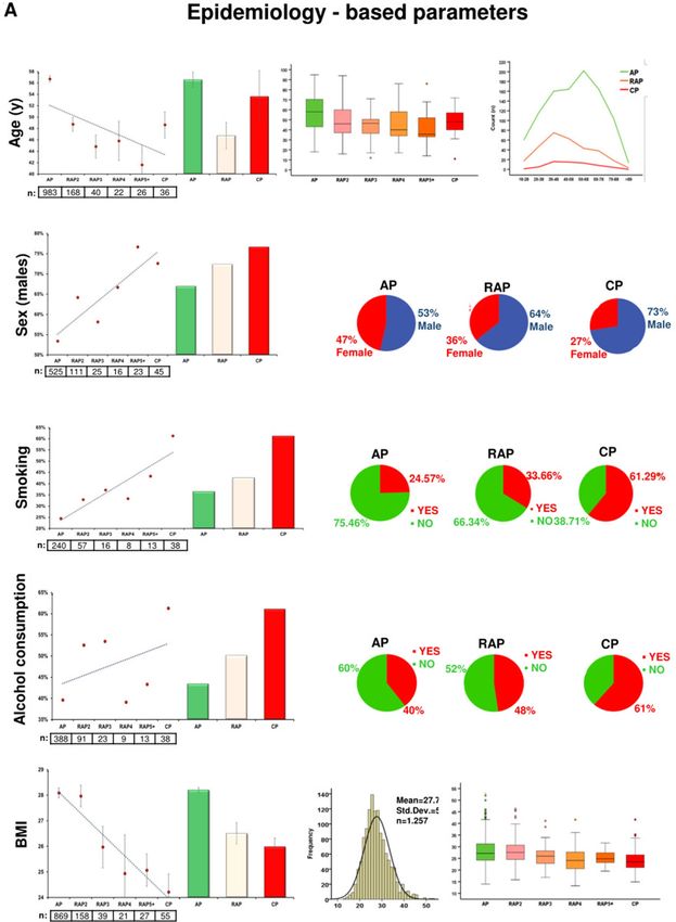
www.nature.com/scientificreports/
A Sex and age distribution across groups B Etiology of acute pancreatitis across study cohorts
Epidemiology Leading etiology
Groups n % Male Female Male% Female% Age (average/y) Age (SEM/y)
Groups AP RAP CP
AP 983 74.8% 525 458 53.4% 46.6% 56.7 0.55
number % number % number %
RAP-2 second AP 173 13.2% 111 62 64.2% 35.8% 52.6 1.21
RAP-3 third AP 43 3.3% 25 18 58.1% 41.9% 51.9 2.25
Biliary 493 50.2% 63 20.7% 7 11.3%*
RAP-4 fourth AP 24 1.8% 16 8 66.7% 33.3% 50.7 3.59 Alcohol 191 19.4% 119 39.1% 32 51.6%*
RAP-5+ fifth AP 30 2.3% 23 7 76.7% 23.3% 52.57 3.56 Lipid 48 4.9% 29 9.5% 1 1.6%
RAP 270 20.5% 175 95 64.8% 35.2% 52.7 0.93 Idiopathic 189 19.6% 72 23.8% 18 29.0%
CP 62 4.7% 45 17 72.6%* 27.4% 55.5 1.84
* p < 0.05 when comparing AP and CP
Total 1315 100.0% 745 570
* p < 0.05 when comparing AP and CP; p < 0.05
AP: acute pancreatitis; RAP: recurrent acute pancreatitis; CP: chronic pancreatitis; n: number;
SEM: standard error of the mean; y.: years;
C Etiology of recurrent acute pancreatitis
Leading aetiology
Groups RAP-2 (n=173) RAP-3 (n=43) RAP-4 (n=24) RAP-5+ (n=30)
number % number % number % number %
Biliary 43 24.9%a 4 9.3%b 2 8.3%c 6 20%d
Alcohol 65 37.6%a 21 48.8%b 9 37.5% 12 40.0%
Lipid 17 9.8% 1 2.3% 3 12.5% 3 10.0%
Idiopathic 41 23.7% 12 27.9% 6 25.0% 5 16.7%
a: p < 0.05 when comparing AP and RAP2
b: p < 0.05 when comparing AP and RAP3
c: p < 0.05 when comparing AP and RAP4
d: p < 0.05 when comparing AP and RAP5+
Figure 1. Epidemiology and etiology of pancreatitis in the study population. (A) Sex and age distribution
across groups. (B) Etiology of pancreatitis across study cohorts. (C) Etiology of recurrent acute pancreatitis
(RAP).
early CP (ECP).The new diagnostic criteria for ECP allow the majority of CP patients to be diagnosed
earlier. They can be used in hospitals with no additional costs in healthcare.
Chronic pancreatitis (CP) is a severe condition that greatly deteriorates quality of life and decreases life expec-
tancy. Importantly, there is no specific curative intervention available1–3. Patients with CP typically struggle with
pain, stigmatization, unemployment and depression4,5. The diagnostic criteria for CP only allow us to detect
the disease at the end stage, at which around 90% of pancreatic parenchyma is already irreversibly d amaged6.
Another issue is that 68% of cases are caused by alcohol, which negatively influences patients’ compliance with
any therapy7. Given these facts, it is not surprising that only 20 studies investigating CP are registered in the
leading trial registries (ISRCTN and clinicaltrials.gov), a number which is not comparable to the number of
studies on pancreatic cancer (114 trials).
One of the possibilities to increase research activity in the field is to diagnose CP earlier. The Japan Pancreatic
Society (JPS) has already provided a definition of early CP (ECP). They recommend that patients who fail to
qualify for a definitive or probable diagnosis of CP, continuously drink more than 80 g/day alcohol and show at
least two out of seven characteristic findings on endoscopic ultrasonography (EUS) should be diagnosed with
ECP. However, this diagnosis may be uncertain, since it includes patient- and observer-reported c omponents8.
The four leading pancreatic societies (IAP, APA, JPS and EPC) have attempted to reach an agreement on the
definition of ECP; however, even after long and exhaustive discussions, consensus has not been reached. The
board of investigators stated that ECP cannot be diagnosed by any single biomarker, since all early biomarkers
are nonspecific9.
Several pathomechanisms were proposed earlier to describe disease progression towards C P10,11. Sankaran
et al. based on their meta-analyzis investigated a frequency of progression from AP to CP. They has found that
more than 10% of patients with a first episode of AP and 36% of patients with recurrent AP develop CP12. Another
model approaching the transition of AP towards CP is the sentinel acute pancreatitis event (SAPE)13.
DeSouza et al. reported a high-quality MR study which was first to demonstrate ‚pancreas shrinkage ‘ after ≥ 3
attacks of AP14. However, large cohort analyses are still lacking. Theoretically, a high-quality acute pancreatitis
(AP) registry, in which the number of attacks is recorded and a large number of variables are collected, may help
us to identify biomarkers indicating the early stage of CP. If experimental data confirm significant differences in
the levels of these biomarkers in RAP and CP patients, we can propose an improved definition of ECP over the
currently available one from the JPS.
Results
One out of five patients with an acute episode suffers from RAP, as opposed to one out of
twenty who has CP. Out of the 1315 patients in the AP registry, a cohort of 983 (74.8%) patients had AP
without CP, as opposed to 270 (20.5%) suffering from RAP without CP; 62 (4.7%) of the acute episodes were
accompanied by already existing CP (shown in Fig. 1A, for the flowchart of patient selection from the AP regis-
try, see Sup Fig 1). Two-thirds of RAP cases had fewer than three attacks, as opposed to one-third experiencing
Scientific Reports | (2021) 11:1367 | https://doi.org/10.1038/s41598-020-80532-6 2
Vol:.(1234567890)
www.nature.com/scientificreports/
three or more attacks. As regards sex and age, there is a clear trend in the groups. The male/female ratio was
53.4/46.6% with AP, 64.8/35.2% with RAP, as opposed to 72.6/27.4% with CP. The average age at the first attack
in patients with AP was 56.7 ± 0.55, that of RAP was 52.7 ± 0.93, and that of CP was 55.5 ± 1.84 (Fig. 1A).
Bidirectional changes in alcoholic and biliary etiologies in AP, RAP and CP patients. As regards
etiology, 50.2% of AP cases were biliary; however, the rate of this etiological factor continuously decreased
towards CP (20.7% with RAP and 11.3% with CP). The distribution of alcoholic etiology moved in the opposite
direction: 19.4% with AP, 39.1% with RAP and 51.6% in the CP group (Fig. 1B). The increasing proportion of
cases with an unknown etiology deteriorating from AP (19.6%) to CP (29%) could either be due to a higher rate
of genetic predisposition15 or the attrition of alcohol-dependent patients (Fig. 1B). Leading etiologies in RAP
are shown in Fig. 1C.
Local complications are more frequent in CP than in AP or RAP, whereas systemic complica‑
tions and mortality are less so. The demographic, epidemiological and primary outcome parameters
suggest that after each acute episode of AP, patients move closer to CP. Therefore, investigating all biomarkers
collected from patients during episodes of AP, irrespective of its relationship with the pancreas, may help us to
recognise ECP. Thus, in the next part of the study, we systematically analysed all the 102 biomarkers (data quality
is shown in Sup Table 1). The primary outcomes of analysed data are shown in Fig. 2.
Fifteen out of 102 biomarkers showed significant alterations between the AP, RAP and CP
groups. Figure 3 shows the descriptive and comparative statistics of the fifteen biomarkers investigated.
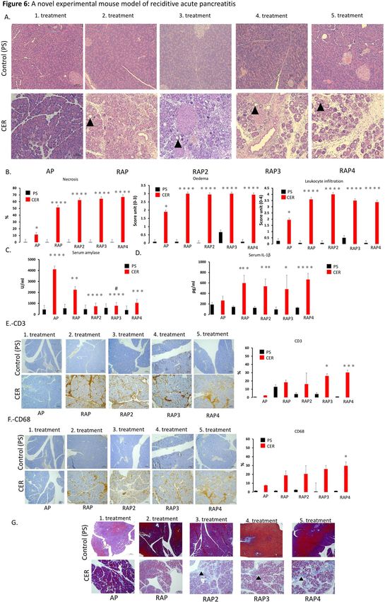
Five of them were epidemiology-based (age, sex, smoking, alcohol consumption and body mass index (BMI))
(Fig. 3A), six were etiology-based (biliary and alcoholic etiology, serum levels of bilirubin, gamma-glutamyl-
transferase (GGT), aspartate aminotransferase (AST) and alanine aminotransferase (ALT)) (Fig. 3B), one was
a laboratory parameter (red blood cell count (RBC)) (Fig. 3C), and three were pancreas-dependent parameters
(rate of pseudocyst formation, serum amylase and lipase) (Fig. 3D). In 13 biomarkers, there were significant dif-
ferences between the groups (Fig. 3A–D). In two parameters (serum lipase and amylase), due to the distorting
effect of the low n number in the CP group, no significant differences were detected between the AP and CP
groups. However, when we elevated the n numbers in the CP group by merging the CP and RAP5 + groups (as
the latter is biologically very close to the CP group), the statistical difference could be detected (Fig. 3E). All in
all, the dynamic changes of these parameters indicate that RAP represents a continuous transition from AP to
CP.
The significant differences between the biomarkers measured during acute episodes in AP
and CP disappear after 2–3 attacks. In these series of analyses, we aimed to determine the number of
episodes of AP (without pancreatic morphological changes) required for significant differences in biomarkers
to vanish compared to CP. The significant difference in eleven parameters disappears after the second episode
of AP (RAP2; age, sex, etiology, alcohol consumption, bilirubin, AST, GGT, RBC count, pseudocysts, amylase
and lipase). The significant difference in BMI and ALT disappears after the third episode of AP (RAP3). There is
no difference in smoking after the fourth episode of AP (RAP4) (Fig. 4). Calculating the rate of morphological
alterations after each episode revealed that 0.3% of AP, 1% of RAP2, 16% of RAP3, 33% of RAP4 and 32% of
RAP5 + cases already have either CT-, MRI-, US- or EUS-based morphological alterations.
These data indicate that there is a stage in CP development in which biomarkers show the disease progression
earlier than pancreatic morphological changes. Characteristics of patients with AP (non-ECP and non-CP), ECP
(non-CP) and CP are summarized in Sup Table 2.
In the RAP3 group, 16% of patients already have established CP, while the figure is nearly
50% in the RAP4 + group. We also investigated whether the incidence of recurrent episodes increases
the chance of CP development. Patients having had one or two episodes of acute inflammation had negligible
odds of developing CP (less than 1%); however, patients who had experienced three episodes had a 16% chance
of developing CP, and patients with four or more episodes had a 50% likelihood. These data demonstrate that
three or more episodes of AP are to be considered as a significant risk factor for the development of CP (Fig. 5).
RAP patients have an average of three attacks, whereas CP patients have an average of four
to five. RAP patients had an average of 3.07 ± 1.85 AP attacks at the time of diagnosis, whereas CP patients
had 3.76 ± 2.24 in the AP registry. We continued our analysis of data from the CP registry, which includes 366
patients. Out of this population, 324 cases had data on the number of attacks at the time of enrolment in the
CP registry (for the flowchart of patient selection from the CP registry, see Sup Fig. 2). In CP, 318 out of 324
patients had at least one acute episode: 69 CP patients had one acute episode (21.6%), 66 had two (20.8%), 66
had three (20.8%), 29 had four (9.1%), and 88 had five or more (27.7%). The average number of attacks was 4–5
(4.07 ± 3.82).
Experimental data confirm that three or more episodes of AP cause a significant decrease
in serum amylase levels and induce CP‑like morphological changes. In our clinical dataset, on-
admission pancreatic enzyme levels continuously decreased from AP to CP. However, we had two problems
with this dataset. Firstly, it only showed a significant difference when CP was merged with RAP5 + (due to the
low n number, see above). Secondly, our database did not provide clinical evidence on whether this continuous
Scientific Reports | (2021) 11:1367 | https://doi.org/10.1038/s41598-020-80532-6 3
Vol.:(0123456789)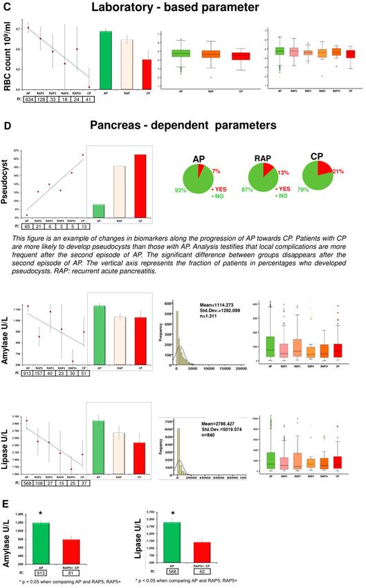
www.nature.com/scientificreports/

                                            Figure 2.  Representativeness of the study population. (A) Sex distribution. (B) Age distribution. (C) Lengths of
                                            hospitalization. (D) Mortality.

          Scientific Reports |   (2021) 11:1367 |               https://doi.org/10.1038/s41598-020-80532-6                                                  4

Vol:.(1234567890)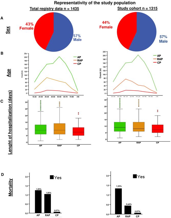
www.nature.com/scientificreports/

                                  Figure 3.  Descriptive and comparative statistics characterising fifteen biomarkers. (A) Epidemiology-based
                                  parameters: age, sex, smoking, alcoholic etiology and body mass index (BMI). (B) Etiology-based parameters:
                                  biliary or alcoholic etiology, bilirubin, gamma-glutamyl transferase (GGT), aspartate aminotransferase (AST)
                                  and alanine aminotransferase (ALT). (C) Laboratory-based parameter: red blood cell count (RBC). (D)
                                  Pancreas-dependent parameters: pseudocysts, amylase and lipase. (E) Differences in acute pancreatitis (AP) vs
                                  recurrent acute pancreatitis (RAP) 5 + and chronic pancreatitis (CP) comparisons in amylase and lipase.

Scientific Reports |   (2021) 11:1367 |               https://doi.org/10.1038/s41598-020-80532-6                                                  5

                                                                                                                                          Vol.:(0123456789)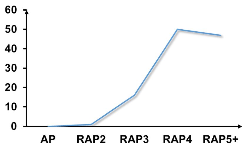
www.nature.com/scientificreports/

                                            Figure 3.  (continued)

          Scientific Reports |   (2021) 11:1367 |               https://doi.org/10.1038/s41598-020-80532-6   6

Vol:.(1234567890)
www.nature.com/scientificreports/
Figure 3. (continued)
Scientific Reports | (2021) 11:1367 | https://doi.org/10.1038/s41598-020-80532-6 7
Vol.:(0123456789)www.nature.com/scientificreports/
BMI Sex - Male
AP p < 0.001 CP AP p < 0.05 CP
AP 1 RAP2 p < 0.001 CP AP p > 0.05 RAP2 p > 0.05 CP
AP 0.186 RAP3 0.664 CP AP p > 0.05 RAP3 p > 0.05 CP
AP 0.109 RAP4 0.996 CP AP p > 0.05 RAP4 p > 0.05 CP
AP 0.063 RAP5+ 0.987 CP AP p > 0.05 RAP5+ p > 0.05 CP
Bilirubin Pseudocyst
AP p < 0.001 CP AP p < 0.05 CP
AP 0.002 RAP2 0.829 CP AP p > 0.05 RAP2 p > 0.05 CP
AP 0.018 RAP3 1 CP AP p > 0.05 RAP3 p > 0.05 CP
AP 0.114 RAP4 1 CP AP p > 0.05 RAP4 p > 0.05 CP
AP 0.846 RAP5+ 0.808 CP AP p > 0.05 RAP5+ p > 0.05 CP
Red blood cell count Biliary aetiology
AP p = 0.009 CP AP p < 0.05 CP
AP 0.946 RAP2 0.252 CP AP p < 0.05 RAP2 p > 0.05 CP
AP 0.893 RAP3 0.826 CP AP p < 0.05 RAP3 p > 0.05 CP
AP 0.835 RAP4 0.985 CP AP p < 0.05 RAP4 p > 0.05 CP
AP 0.963 RAP5+ 0.842 CP AP p < 0.05 RAP5+ p > 0.05 CP
GGT Alcohol aetiology
AP p < 0.001 CP AP p < 0.05 CP
AP 0.030 RAP2 0.624 CP AP p < 0.05 RAP2 p > 0.05 CP
AP 0.338 RAP3 0.935 CP AP p < 0.05 RAP3 p > 0.05 CP
AP 0.184 RAP4 1 CP AP p > 0.05 RAP4 p > 0.05 CP
AP 0.067 RAP5+ 1 CP AP p > 0.05 RAP5+ p > 0.05 CP
ALT Current alcohol consumption
AP p < 0.001 CP AP p < 0.05 CP
AP 0.189 RAP2 0.042 CP AP p < 0.05 RAP2 p > 0.05 CP
AP 0.004 RAP3 0.999 CP AP p > 0.05 RAP3 p > 0.05 CP
AP 0.054 RAP4 1 CP AP p > 0.05 RAP4 p > 0.05 CP
AP 0.117 RAP5+ 0.888 CP AP p > 0.05 RAP5+ p > 0.05 CP
AST Current smoking
AP p = 0.003 CP AP p < 0.05 CP
AP 0.108 RAP2 0.569 CP AP p > 0.05 RAP2 p < 0.05 CP
AP 0.023 RAP3 1 CP AP p > 0.05 RAP3 p > 0.05 CP
AP 0.068 RAP4 0.998 CP AP p > 0.05 RAP4 p > 0.05 CP
AP 0.386 RAP5+ 0.982 CP AP p > 0.05 RAP5+ p > 0.05 CP
Age at first attack of AP
AP p = 0.015 CP
AP p < 0.001 RAP2 1 CP
AP p < 0.001 RAP3 0.926 CP
AP 0.037 RAP4 0.990 CP
AP p < 0.001 RAP5+ 0.599 CP
Figure 4. Significant differences between thirteen biomarkers.
decrease is linked to the loss of pancreatic parenchyma. Therefore, we established a preclinical model of RAP
(and used a novel dosing schedule to induce pancreatitis) to investigate the effect of recurrent inflammation on
on-admission amylase activity, pro-inflammatory cytokine level (measuring IL-1ß), inflammatory cells (mac-
rophage and T cells) and pancreatic morphology. We found that repeated AP inductions (Sup Fig 3) can mimic
histological changes from RAP to CP. We found strong evidence that repeated AP induction leads to severe
fibrosis, necrosis, leukocyte infiltration, including T cells and macrophages, elevation of pro-inflammatory
cytokine level and edema of the pancreas (Fig. 6A–G). Serum amylase activity (Fig. 6C) was highly elevated in
the case of AP. After that, each episode reduced the rate of increase in serum amylase activity. For details, please
see Supplementary Document 1. Our data show that, after the second episode of AP, there is a significant drop
Scientific Reports | (2021) 11:1367 | https://doi.org/10.1038/s41598-020-80532-6 8
Vol:.(1234567890)www.nature.com/scientificreports/
Incidence of RAP towards to CP
50% 47%
Probability of CP (%)
16%
0% 1%
No CP (n): 983 173 43 24 30 1253
CP (n): 0 2 8 24 28 62
Total (n): 983 175 51 48 58 1315
Figure 5. Progression from the first acute pancreatitis (AP) episode through recurrent episodes (RAP) towards
chronic pancreatitis (CP).
in serum levels of amylase in each concomitant episode of AP, which is associated with pancreatic parenchymal
damage (i.e. episode-dependent acinar depletion).
Discussion
There is a large amount of data available on AP and CP; however, much less is known about RAP and ECP. There-
fore, it is not surprising that clinicians of the four major pancreatic associations failed to reach an agreement
on diagnostic criteria for ECP. The only association that has attempted to describe ECP is the Japan Pancreas
Society. However, their guidelines are complicated, with only a limited possibility for use in general practice8,16.
Two nationwide studies have already highlighted that repetitive inflammation of the pancreas can lead to CP17,18.
In a cross-sectional epidemiological study, Masamune et al. showed that 26.5% of ECP cases had previous acute
episodes17,18. Importantly, the incidence rate of RAP was much higher in a two-year prospective follow-up
study, in which 75% of patients had AP before ECP was diagnosed. Cho et al. in their nationwide cohort study
investigated the influence of cholecystectomy and RAP events on the risk of post-pancreatitis diabetes mellitus.
Patients who had 2 or ≥ 3 recurrent biliary events prior to cholecystectomy were at a significantly increased risk
of post-pancreatitis diabetes mellitus. The results of your study are in line with that s tudy19. These data support
the SAPE model describing the transition of AP towards CP. According to this model, the first (so-called sentinel)
episode of acute pancreatitis (SAPE) triggers a cellular activation cascade, which leads to chronic pancreatic
inflammation and fibrosis. The model proposes a tipping point in time when, in line with the multihit theory
model, the effects of risk factors, such as alcohol consumption and smoking, turn into etiological factors, trigger-
ing the cascade that ends up in CP20. Findings from a cohort study by Sheel et al. were in line with this model13.
In our international cohort, we investigated uniformly and prospectively collected 130,744 pieces of high-quality
data from 1315 patients. Our epidemiological analysis revealed that one out of five AP patients suffers from RAP,
whereas one out of twenty suffers from CP, which data were reported by the Japanese cohort studies. Of note,
almost all the CP cases (98%) had a previous episode of AP, which is surprisingly high compared to previous
data by Olesen et al. (47%)21.
We found 15 variables that were significantly different in the first AP and CP, and, importantly, the differences
start disappearing after recurrent episodes of AP. Epidemiological data showed that the male gender, younger age
and lower BMI are associated with RAP and CP, which data are in accordance with the findings of the Cleveland
cohort, where the average age of the first AP was 55.5 ± 16.6y, that of the second was 53.8 ± 18.5y, and that of the
third 45.2 ± 12.4y. Importantly, no further changes were observed after the third attacks of AP (45.7 ± 16.5y),
suggesting that three or more AP attacks may be a separate group of RAP22.
One of the key findings of this study is that the incidence of recurrent episodes increases the risk of CP devel-
opment. The first two attacks have small effects (0–1%) on the odds for developing CP, whereas the third and
fourth (16–50%) episodes have large ones. The striking difference between RAP2 and RAP3 could be explained
by at least three factors: (1) The biliary etiology decreased from 24.9 to 9.3%, whereas the alcoholic etiology
elevated from 37.6 to 48.8%. While the biliary etiology is usually a one-time hit on the pancreas, alcohol has
a continuous deteriorating effect. (2) RAP3 occurs in a more damaged pancreas, which is actually confirmed
by our experimental settings. (3) RAP3 seems to be more severe than RAP2 (mortality: 4.7% vs 2.3%; systemic
complications: 4.7% vs 2.3%).
In a longitudinal study by Lankisch et al.23 from Germany, CP was diagnosed after the second RAP in nine
patients, after the third RAP in seven and after the fourth in three. Heavy smoking (> 30 cigarettes per day)
predisposed patients to CP after the first AP attack. The role of smoking was also confirmed by the findings of
Scientific Reports | (2021) 11:1367 | https://doi.org/10.1038/s41598-020-80532-6 9
Vol.:(0123456789)www.nature.com/scientificreports/
Figure 6. A novel experimental mouse model of reciditive acute pancreatitis and chronic pancreatitis. (A) Histological
findings in an experimental mouse model of acute pancreatitis. (B) Necrosis, edema and leukocyte infiltration
differences in pancreatitis stages in an experimental mouse model. (C) Serum amylase activity and (D) Serum IL-1β
level differences in stages of pancreatitis in an experimental mouse model. (E) CD3 (T cell, brown colour) staining. (F)
CD68 (macrophage, brown colour) staining. (G) Fibrosis (blue colour filled triangle), each scale bar represents 100 µm.
* = p < 0.05. A detailed description can be found in Supplementary Document 1.
Scientific Reports | (2021) 11:1367 | https://doi.org/10.1038/s41598-020-80532-6 10
Vol:.(1234567890)www.nature.com/scientificreports/
Yadav et al.24 Importantly, they found that the strongest predictor for a subsequent diagnosis of CP was RAP
(HR = 4.57, 95% CI: 3.40–6.14).
Our significant biomarkers clearly showed bidirectional changes in alcoholic and biliary etiologies in AP,
RAP and CP patients. Similar changes were found in the C leveland22, Chinese25 and Central E uropean1 cohorts
as well. These data suggest the usefulness of cholecystectomy in AP. Before the routine cholecystectomy era,
biliary AP was even more frequent in RAP than in general AP26. The fact that the biliary etiology in CP (11.29%)
is five times less than in the first episode of AP (50.15%) also suggests that repetitive episodes are one of the key
determinants of CP. Importantly, Bertilssom et al.27 from Denmark reported that one or more AP episodes are
among the strongest predictors for the development of CP.
The linear changes in the values of pancreas-dependent parameters (rate of pseudocysts, serum amylase and
lipase) suggest that repetitive attacks lead to local damage of the pancreas, which is one of the hallmarks of CP.
The elevation of local complication rates was reported in earlier published cohorts as w ell22,25,27–30.
We report that the on-admission elevation of serum lipase and amylase levels decrease from AP to CP via RAP.
One of the most likely explanations is that these changes are due to the loss of pancreatic acinar cells. Therefore,
since histological samples were not available from the members of our cohort, we investigated this hypothesis
in an experimental CP model. As with human observations, the serum amylase level continuously decreased
after the second and third attacks, which changes were associated with the loss of pancreatic parenchyma and
enhanced fibrosis.
DeSouza et al. in their firstly reported high-quality MR study demostrated ‚pancreas shrinkage ‘ after ≥ 3
attacks of A P14. A total of 123 participants were studied. Total pancreas volume (TPV) and tail diameter were sig-
nificantly reduced in both unadjusted (TPV (p = 0.036), tail diameter (p = 0.009)) and adjusted (TPV (p = 0.026),
tail diameter (p = 0.034)) models in individuals with ≥ 3 attacks, but not with 1 or 2 attacks, compared with healthy
individuals. These results are strongly correlated with our results.
The episode-dependent decrease in the elevation of amylase activity highlights the loss of acinar cells in the
pancreas and the functional decrease of pancreatic enzyme secretion/leakage from acinar cells. Importantly, these
data also indicate that the three-fold elevation of serum pancreatic enzymes may not be suitable for setting up
a diagnosis in patients suffering from RAP. Taken together, both the clinical and experimental studies suggest
that three or more episodes of RAP can be considered as ECP.
One of the major limitations of the study is the cross-sectional design: the patients were not followed longi-
tudinally from AP through RAP to CP. Therefore, we have no information on the time of diagnosis of CP and
the time relationships of RAP and CP. We decreased the limitations by analysing our CP cohort as well, in which
patients were collected separately from our AP cohort. It is also important to note that some of the patients
suffering from CP had no reported AP before their CP diagnosis. Therefore, our definition of ECP does not
cover all ECP patients. Most cases of CP were confirmed with abdominal US and CT, which modalities have less
diagnostic specificity and sensitivity for CP than EUS or MRCP. When abdominal US and CT showed Wirsung
dilatation or calcification and the etiology was known (usually with a background of alcoholism), investigators
did not use EUS or MRCP. Therefore, more detailed analysis of the results of EUS or MRCP were not possible
in this study. Our patient registries record only routine laboratory parameters, while analysing cytokine levels
may have added extra information on the pathogenesis of transition. Statistical limitations include the fact that,
since our publication has a hypothesis-generating purpose, we did not adjust tests for multiplicity (except in the
ANOVA model). In addition, data quantity and quality did not allow us to perform multivariate statistics. Mild
differences in management strategies across centres may affect disease outcomes. To decrease the limitations
described here, we started a new international, observational, longitudinal investigation of acute pancreatitis,
entitled the GOULASH PLUS study, in which we will monitor all AP patients for six years to characterise ECP
more precisely31.
In addition to the limitations noted above, our research has several highly important advantages: (1) it can
be used easily in all hospitals, (2) no additional laboratory measurements or imaging techniques are necessary
after ruling out CP with imaging to establish a diagnosis of ECP, and (3) it allows us to start clinical trials and
encourage patients to implement lifestyle changes to prevent the development of CP from ECP. Our results adds
to the accumulating body of morphological14 and population-based19 studies on this topic. Our study shows that
three or more episodes of RAP with no pancreatic morphological alterations may be considered ECP. Results
from validation studies, such as the GOULASH PLUS study, are still forthcoming.
Materials and methods
The clinical part of the study. Design. This study is a comparative cross-sectional study. The study is
being reported according to the STROBE Statement.
The Hungarian Pancreatic Study Group (HPSG) developed international registries for pancreatic diseases (AP,
CP and pancreatic cancer)3,28,32. All pancreatic centres worldwide can join these registries (https: //tm-centre .org/
en/registries/gastroenterology-en/). In this study, we extracted and analysed data from the AP and CP registries.
The AP registry contained data from 1435 patients, 1315 of whom from 28 centres in twelve countries col-
lected between June 2012 and September 2017 were eligible for analysis. Centre distribution is displayed in Sup
Fig 4, whereas patients eligible for inclusion can be found in Sup Fig 1.
One hundred and two individual variables (on-admission biomarkers, defined as laboratory test and imaging
results, demographic factors or any relevant information from the medical history) were found to be eligible for
investigation (Sup Table 1).
The CP registry contained clinical data from a total of 366 patients’ from 25 centres in two countries collected
between June 2012 and September 2017, 324 of whom (88.5%) were eligible for inclusion (Sup Fig 2). Centre
distribution is displayed in Sup Fig 5. No additional biomarkers from the CP registry were investigated.
Scientific Reports | (2021) 11:1367 | https://doi.org/10.1038/s41598-020-80532-6 11
Vol.:(0123456789)www.nature.com/scientificreports/
Definition of pancreatitis. Diagnosis of AP was established based on the recommendations of the HPSG guide-
line (adapted and updated from APA/IAP guideline)33, while that of CP was based on the HaPanEU criteria1,34,35.
Patient groups. We formed three groups based on the morphology of the pancreas and the number of recurrent
acute episodes. The first episode of AP without any chronic morphological change of the pancreas was labelled
AP (983 cases). Cases with two or more episodes of AP without clinical signs and symptoms of CP or pancreatic
morphological alterations were labelled recurrent acute pancreatitis (RAP) (270 cases). RAP was further divided
into four subgroups based on the number of acute episodes (RAP2: two episodes of AP; RAP3: three episodes;
RAP4: four episodes; and RAP5 + : five or more episodes). RAP 5 + consisted of 30 cases: twelve cases with five
acute episodes, four cases with six, six cases with seven, three cases with eight, two cases with ten, two cases
with eleven, and one case with twelve. Any acute episodes based on clinical signs and symptoms and pancreatic
morphological changes attributed to chronic inflammation were labelled CP (62 cases).
Ethical approval for the clinical study. The study was approved by the Scientific and Research Ethics Com-
mittee of the Medical Research Council (22,254–1/2012/EKU). All participants in this study provided written,
informed consent. Use of data from the AP and CP registries was permitted by the Hungarian Pancreatic Study
Group (HPSG).
The experimental part of the study. Animals. A total of 40 wild-type (WT) FVB/N mice were sacri-
ficed. Mice (male, aged between 20 and 25 days) were housed in a room maintained at a temperature of 20–22 °C
on a 12 h light–dark cycle with food and water available.
Experimental model. Although there are some protocols available for inducing chronic pancreatic damage in
mice with caerulein (CER)36,37, we introduced a novel experimental methodology that can be used to study the
differences and alterations in different phases of pancreatic injury. 50 µg/kg CER was administered i.p. ten times
within ten hours to induce AP on Day 1. This method was repeated to induce RAP on Day 4, to induce ECP on
Days 4 and 7 and to induce CP on Days 4, 7, 10 and 1338,39. Buprenorphine with a concentration of 0.1 mg/kg s.c.
was administered to the mice every six hours each day of the treatment to relieve pain. The mice were euthanized
with 200 mg/bwkg pentobarbital i.p. (Bimeda MTC, Cambridge, Ontario, Canada) two hours after the last injec-
tions of CER. A cardiac puncture was performed on the mice for exsanguination, and the pancreas glands were
removed. Blood samples were collected from the cardiac puncture and placed on ice immediately. These samples
were centrifuged at 2500 RCF for 15 min at 4 °C, and blood serum was collected and stored at -20 °C until analy-
sis. Pancreas samples were placed into an 8% formaldehyde solution and stored at -4 °C. The formaldehyde-fixed
pancreas samples were embedded in paraffin and cut into 3 μm thick sections and stained for Crossman’s Tri-
chrome staining or hematoxylin–eosin using a standard laboratory method (Department of Pathology, Univer-
sity of Szeged, Szeged, Hungary). Immunohistochemistry was performed (Department of Pathology, University
of Szeged, Szeged, Hungary) to detect CD3 (MRQ-39, Rabbit Monoclonal Antibody, CellMarque, CA, USA)
and CD68 (Anti-Macrosialin CD68 Antibody, Booster Bio, CA, USA) in the pancreas samples. Image J Software
was used to evaluate the positive stainings for CD3 and CD68. A Mouse IL-1β ELISA Kit (EM0109, Wuhan Fine
Biotech Co, China) was employed to measure IL-1β levels in serum samples, with absorbance of the samples
detected at 405 nm. A semi-quantitative scoring procedure was performed to quantify the edema, necrosis and
leucocyte infiltration levels of the hematoxylin–eosin stained slides40,41. A colorimetric kit was used (Diagnosti-
cum, Budapest, Hungary) to measure serum amylase activity, with absorbance of the samples detected at 405 nm
with a FLUOstar OPTIMA (BMG Labtech, Germany) plate reader.
Ethical approval for the experimental study. Local ethical committees approved experiments on investigations
involving animals at the University of Szeged (XII/4988/2015). The experiments were performed in compliance
with European Union Directive 2010/63/EU and Hungarian Government Decree 40/2013 (II.14.).
Statistical analyses. The mean ± standard error of the mean (SEM) values were calculated for descriptive
statistics. The numbers and percentages of cases were computed for categorical variables. To check the normality
of the data, we used the Kolgomorov-Smirnov test and/or visual inspection of the Q–Q plots. In the case of non-
normal distributed variables, e.g., bilirubin, GGT, AST, ALT, amylase and lipase, we transformed our data into
the logarithmic scale to achieve normal distribution; then we applied parametric tests (graphs were prepared
from the raw data without transformation). To identify significant differences between groups, we used the fol-
lowing statistical tests for the whole dataset and subgroups as well.
To observe differences between two groups, the independent t-test was applied (Figs. 1A, 3E, 6), to compare
more than two groups; we used one-way ANOVA with a Tukey post hoc test to adjust the results for alpha error
(Figs. 3A–D, 4). The association between categorical variables was examined with the Chi-square test and the
Fisher’s exact test, depending on the sample size (Figs. 1, 3A,B,D). All statistical tests were performed using SPSS
statistical software version 25 (IBM Corporation, Armonk, NY).
Received: 23 April 2020; Accepted: 18 December 2020
Scientific Reports | (2021) 11:1367 | https://doi.org/10.1038/s41598-020-80532-6 12
Vol:.(1234567890)www.nature.com/scientificreports/
References
1. Löhr, J. M. et al. United European Gastroenterology evidence-based guidelines for the diagnosis and therapy of chronic pancreatitis
(HaPanEU). United Eur. Gastroenterol. J. 5, 153–199. https://doi.org/10.1177/2050640616684695 (2017).
2. Czakó, L. et al. Quality of life assessment after pancreatic enzyme replacement therapy in chronic pancreatitis. Can. J. Gastroenterol.
17, 597–603. https://doi.org/10.1155/2003/515848 (2003).
3. Szücs, Á. et al. Chronic pancreatitis: multicentre prospective data collection and analysis by the Hungarian Pancreatic Study Group.
PLoS ONE 12, e0171420–e0171420. https://doi.org/10.1371/journal.pone.0171420 (2017).
4. Amann, S. T. et al. Physical and mental quality of life in chronic pancreatitis: a case-control study from the North American
Pancreatitis Study 2 cohort. Pancreas 42, 293–300. https://doi.org/10.1097/MPA.0b013e31826532e7 (2013).
5. Machicado, J. D. et al. Quality of life in chronic pancreatitis is determined by constant pain, disability/unemployment, current
smoking, and associated co-morbidities. Am. J. Gastroenterol. 112, 633–642. https://doi.org/10.1038/ajg.2017.42 (2017).
6. Whitcomb, D. C. Peering into the “Black Box” of the complex chronic pancreatitis syndrome. Pancreas 45, 1361–1364. https://doi.
org/10.1097/MPA.0000000000000715 (2016).
7. Pelli, H. et al. Pancreatic damage after the first episode of acute alcoholic pancreatitis and its association with the later recurrence
rate. Pancreatology 9, 245–251. https://doi.org/10.1159/000212089 (2009).
8. Ito, T. et al. Evidence-based clinical practice guidelines for chronic pancreatitis 2015. J. Gastroenterol. 51, 85–92. https://doi.
org/10.1007/s00535-015-1149-x (2016).
9. Whitcomb, D. C. et al. International consensus statements on early chronic Pancreatitis. Recommendations from the working
group for the international consensus guidelines for chronic pancreatitis in collaboration with The International Association of
Pancreatology, American Pancreatic Association, Japan Pancreas Society, PancreasFest Working Group and European Pancreatic
Club. Pancreatology 18, 516–527. https://doi.org/10.1016/j.pan.2018.05.008 (2018).
10. Malagelada, J. R. The pathophysiology of alcoholic pancreatitis. Pancreas 1, 270–278. https://doi.org/10.1097/00006676-19860
5000-00011(1986).
11. Klöppel, G. & Maillet, B. Pathology of acute and chronic pancreatitis. Pancreas 8, 659–670. https: //doi.org/10.1097/000066 76-19931
1000-00001(1993).
12. Sankaran, S. J. et al. Frequency of progression from acute to chronic pancreatitis and risk factors: a meta-analysis. Gastroentrology
149, 1490-1500.e1491. https://doi.org/10.1053/j.gastro.2015.07.066 (2015).
13. Sheel, A. R. G. et al. The diagnostic value of Rosemont and Japanese diagnostic criteria for ‘indeterminate’, ‘suggestive’, ‘possible’
and ‘early’ chronic pancreatitis. Pancreatology 18, 774–784. https://doi.org/10.1016/j.pan.2018.08.002 (2018).
14. DeSouza, S. V., Priya, S., Cho, J., Singh, R. G. & Petrov, M. S. Pancreas shrinkage following recurrent acute pancreatitis: an MRI
study. Eur. Radiol. 29, 3746–3756. https://doi.org/10.1007/s00330-019-06126-7 (2019).
15. Yadav, D. & Lowenfels, A. B. The epidemiology of pancreatitis and pancreatic cancer. Gastroentrology 144, 1252–1261. https://doi.
org/10.1053/j.gastro.2013.01.068 (2013).
16. Shimosegawa, T. et al. The revised Japanese clinical diagnostic criteria for chronic pancreatitis. J. Gastroenterol. 45, 584–591. https
://doi.org/10.1007/s00535-010-0242-4 (2010).
17. Masamune, A. et al. Nationwide epidemiological survey of early chronic pancreatitis in Japan. J. Gastroenterol. 52, 992–1000. https
://doi.org/10.1007/s00535-017-1311-8 (2017).
18. Masamune, A. et al. Prospective study of early chronic pancreatitis diagnosed based on the Japanese diagnostic criteria. J. Gastro-
enterol. 54, 928–935. https://doi.org/10.1007/s00535-019-01602-9 (2019).
19. Cho, J., Scragg, R. & Petrov, M. S. The influence of cholecystectomy and recurrent biliary events on the risk of post-pancreatitis
diabetes mellitus: a nationwide cohort study in patients with first attack of acute pancreatitis. HPB Off. J. Int. Hepato Pancreato
Biliary Assoc. https://doi.org/10.1016/j.hpb.2020.10.010 (2020).
20. Whitcomb, D. C. Mechanisms of disease: advances in understanding the mechanisms leading to chronic pancreatitis. Nat. Clin.
Pract. Gastroenterol. Hepatol. 1, 46. https://doi.org/10.1038/ncpgasthep0025 (2004).
21. Olesen, S. S., Büyükuslu, A., Køhler, M., Rasmussen, H. H. & Drewes, A. M. Sarcopenia associates with increased hospitali-
zation rates and reduced survival in patients with chronic pancreatitis. Pancreatology 19, 245–251. https://doi.org/10.1016/j.
pan.2019.01.006 (2019).
22. Lee, P. J. W. et al. Decreased severity in recurrent versus initial episodes of acute pancreatitis. Pancreas 44, 896–900. https://doi.
org/10.1097/MPA.0000000000000354 (2015).
23. Lankisch, P. G. et al. Natural history of acute pancreatitis: a long-term population-based study. Am. J. Gastroenterol. 104, 2797–2806.
https://doi.org/10.1038/ajg.2009.405 (2009).
24. Yadav, D., O’Connell, M. & Papachristou, G. I. Natural history following the first attack of acute pancreatitis. Am. J. Gastroenterol.
107, 1096–1103. https://doi.org/10.1038/ajg.2012.126 (2012).
25. Gao, Y. J. et al. Analysis of the clinical features of recurrent acute pancreatitis in China. J. Gastroenterol. 41, 681–685. https://doi.
org/10.1007/s00535-006-1820-3 (2006).
26. Corfield, A. P., Cooper, M. J. & Williamson, R. C. Acute pancreatitis: a lethal disease of increasing incidence. Gut 26, 724. https://
doi.org/10.1136/gut.26.7.724 (1985).
27. Bertilsson, S., Swärd, P. & Kalaitzakis, E. Factors that affect disease progression after first attack of acute pancreatitis. Clin. Gastro-
enterol. Hepatol. 13, 1662–1669. https://doi.org/10.1016/j.cgh.2015.04.012 (2015).
28. Párniczky, A. et al. Prospective, multicentre, nationwide clinical data from 600 cases of acute pancreatitis. PLoS ONE 11, e0165309–
e0165309. https://doi.org/10.1371/journal.pone.0165309 (2016).
29. Zhang, W., Shan, H.-C. & Gu, Y. Recurrent acute pancreatitis and its relative factors. World J. Gastroenterol. 11, 3002–3004. https
://doi.org/10.3748/wjg.v11.i19.3002 (2005).
30. Ammann, R. W. & Muellhaupt, B. Progression of alcoholic acute to chronic pancreatitis. Gut 35, 552. https://doi.org/10.1136/
gut.35.4.552 (1994).
31. Mikó, A. et al. Observational longitudinal multicentre investigation of acute pancreatitis (GOULASH PLUS): follow-up of the
GOULASH study, protocol. BMJ Open 9, e025500. https://doi.org/10.1136/bmjopen-2018-025500 (2019).
32. Lakatos, G. et al. Pancreatic cancer: multicenter prospective data collection and analysis by the Hungarian Pancreatic Study Group.
J. Gastrointestin. Liver Dis. 25, 219–225. https://doi.org/10.15403/jgld.2014.1121.252.pcr (2016).
33. Working, G. I. & APA, A. P. G. IAP/APA evidence-based guidelines for the management of acute pancreatitis. Pancreatology 13,
e1–e15. https://doi.org/10.1016/j.pan.2013.07.063 (2013).
34. Párniczky, A. et al. EPC/HPSG evidence-based guidelines for the management of pediatric pancreatitis. Pancreatology 18, 146–160.
https://doi.org/10.1016/j.pan.2018.01.001 (2018).
35. Takács, T. et al. Chronic pancreatitis. Evidence based management guidelines of the Hungarian Pancreatic Study Group. Orv. Hetil.
156, 262–288. https://doi.org/10.1556/OH.2015.30060 (2015).
36. Xue, J. et al. Alternatively activated macrophages promote pancreatic fibrosis in chronic pancreatitis. Nat. Commun. 6, 7158. https
://doi.org/10.1038/ncomms8158 (2015).
37. Balázs, A. et al. Ductal mucus obstruction and reduced fluid secretion are early defects in chronic pancreatitis. Front. Physiol. 9,
632–632. https://doi.org/10.3389/fphys.2018.00632 (2018).
Scientific Reports | (2021) 11:1367 | https://doi.org/10.1038/s41598-020-80532-6 13
Vol.:(0123456789)www.nature.com/scientificreports/
38. Mareninova, O. A. et al. Cell death in pancreatitis: caspases protect from necrotizing pancreatitis. J. Biol. Chem. 281, 3370–3381.
https://doi.org/10.1074/jbc.M511276200 (2006).
39. Niederau, C., Ferrell, L. D. & Grendell, J. H. Caerulein-induced acute necrotizing pancreatitis in mice: protective effects of prog-
lumide, benzotript, and secretin. Gastroentrology 88, 1192–1204. https://doi.org/10.1016/s0016-5085(85)80079-2 (1985).
40. Kui, B. et al. New insights into the methodology of L-arginine-induced acute pancreatitis. PLoS ONE 10, e0117588. https://doi.
org/10.1371/journal.pone.0117588 (2015).
41. Rumbus, Z. et al. Bidirectional relationship between reduced blood pH and acute pancreatitis: a translational study of their noxious
combination. Front. Physiol. 9, 1360. https://doi.org/10.3389/fphys.2018.01360 (2018).
Acknowledgements
The authors wish to thank other contributors for their outstanding work in including ECP: S. Galeev (Saint Luke
Clinical Hospital, St. Petersburg, Russia), P. Pencik (Centrum pece o zazivaci trakt, Vitkovice Nemocnice a.s.,
Ostrava, Czech Republic), A. Litvin (Gomel Regional Clinical Hospital, Gomel, Belarus), I. Ozola-Zalite (Pauls
Stradins Clinical University Hospital, Gastroent., Hep. and Nutr. Centre, Riga, Latvia), K. Zadorozhna (Bogo-
molets National Medical University, Kiev, Ukraine) and M. Horibe (Keio University, Tokyo, Japan).
Author contributions
Study conception and design: P.H., P.J.H. Development of methodology: P.H. Acquisition of data: P.J.H., E.T.,
A.E., V.V., K.M., P.M., A.M., J.B., P.S., A.V., A.H., F.I., Z.S., L.C., G.K., M.P., Z.D., M.V., J.H., B.C.N., M.M., A.T.I.,
D.B., E.D., M.K., I.K., T.B., A.M., E.R.M., V.S., B.E., D.P., A.S., A.P., P.H. Analysis and interpretation of data:
P.J.H., A.S., P.M. Writing, review and/or revision of the manuscript: P.J.H., P.H., B.E. Administrative, technical
or material support: K.M., A.S., A.P. Study supervision: P.H.
Funding
The study was funded by Project Grants (K116634 to PH, FK131864 to AM, FK124632 to BCN and K128222
to LC), an Economic Development and Innovation Operative Programme Grant (GINOP 2.3.2-15-2016-00048
to PH) and a Human Resources Development Operational Programme Grant (EFOP-3.6.2-16-2017-00006 to
PH) from the National Research, Development and Innovation Office, as well as a Momentum Grant from the
Hungarian Academy of Sciences (LP2014-10/2014 to PH). Supported by the János Bolyai Research Scholarship
of the Hungarian Academy of Sciences (to AM) and by the ÚNKP-20-5 New National Excellence Program of the
Ministry for Innovation and Technology from the source of the National Research, Development and Innova-
tion Fund (to AM).
Competing interests
The authors declare no competing interests.
Additional information
Supplementary Information The online version contains supplementary material available at https://doi.
org/10.1038/s41598-020-80532-6.
Correspondence and requests for materials should be addressed to P.H.
Reprints and permissions information is available at www.nature.com/reprints.
Publisher’s note Springer Nature remains neutral with regard to jurisdictional claims in published maps and
institutional affiliations.
Open Access This article is licensed under a Creative Commons Attribution 4.0 International
License, which permits use, sharing, adaptation, distribution and reproduction in any medium or
format, as long as you give appropriate credit to the original author(s) and the source, provide a link to the
Creative Commons licence, and indicate if changes were made. The images or other third party material in this
article are included in the article’s Creative Commons licence, unless indicated otherwise in a credit line to the
material. If material is not included in the article’s Creative Commons licence and your intended use is not
permitted by statutory regulation or exceeds the permitted use, you will need to obtain permission directly from
the copyright holder. To view a copy of this licence, visit http://creativecommons.org/licenses/by/4.0/.
© The Author(s) 2021
Scientific Reports | (2021) 11:1367 | https://doi.org/10.1038/s41598-020-80532-6 14
Vol:.(1234567890)You can also read