Enhancement of Campylobacter hepaticus culturing to facilitate downstream applications - Nature
←
→
Page content transcription
If your browser does not render page correctly, please read the page content below

www.nature.com/scientificreports
OPEN Enhancement of Campylobacter
hepaticus culturing to facilitate
downstream applications
Canh Phung1, Timothy B. Wilson2, José A. Quinteros2, Peter C. Scott2, Robert J. Moore1* &
Thi Thu Hao Van1
Campylobacter hepaticus causes Spotty Liver Disease (SLD) in chickens. C. hepaticus is fastidious and
slow-growing, presenting difficulties when growing this bacterium for the preparation of bacterin
vaccines and experimental disease challenge trials. This study applied genomic analysis and in vitro
experiments to develop an enhanced C. hepaticus liquid culture method. In silico analysis of the
anabolic pathways encoded by C. hepaticus revealed that the bacterium is unable to biosynthesise
l-cysteine, l-lysine and l-arginine. It was found that l-cysteine added to Brucella broth, significantly
enhanced the growth of C. hepaticus, but l-lysine or l-arginine addition did not enhance growth.
Brucella broth supplemented with l-cysteine (0.4 mM), l-glutamine (4 mM), and sodium pyruvate
(10 mM) gave high-density growth of C. hepaticus and resulted in an almost tenfold increase in culture
density compared to the growth in Brucella broth alone (log10 = 9.3 vs 8.4 CFU/mL). The type of culture
flask used also significantly affected C. hepaticus culture density. An SLD challenge trial demonstrated
that C. hepaticus grown in the enhanced culture conditions retained full virulence. The enhanced liquid
culture method developed in this study enables the efficient production of bacterial biomass and
therefore facilitates further studies of SLD biology and vaccine development.
Campylobacter hepaticus has been identified as the causative agent of Spotty Liver Disease (SLD) in laying h ens1.
C. hepaticus is a fastidious bacterium that requires microaerobic conditions (a mix of 5–10% oxygen, 5–10% C O2,
and 80–85% N2), a narrow temperature range (growth at 37 and 42 °C, but not 25 °C), and rich nutrient media
for growth1–4. The incubation time needed to form C. hepaticus colonies on agar plates varies from 3 to 7 days,
depending on the strain cultivated1,2,5. The incubation time for other Campylobacter species, such as C. jejuni, C.
coli, C. lari, and C. concisus ranges from 24 to 48 h on plates and 18–24 h in liquid m
edia6–9. In previous studies,
when a large C. hepaticus biomass was required, such as for SLD induction experiments, C. hepaticus was cultured
on Brucella agar supplemented with 5% defibrinated horse blood (HBA), incubated for 3 days and harvested.
C. hepaticus had to be harvested from dozens of Petri dishes to produce sufficient biomass for a modestly sized
animal trial10. This methodology is time–consuming, uses a lot of resources, and is prone to contamination. It
also presents difficulties with scaling up to produce sufficient biomass to produce challenge material for large
animal trials or for production of killed vaccines.
Although Brucella broth, the standard media for growing C. hepaticus cultures, is a rich medium, it may
not supply all the nutrients required to support optimal growth of C. hepaticus. An in silico approach was used
to identify nutrients that may need to be supplemented to improve culture productivity. The availability of
C. hepaticus whole-genome sequences11 and tools such as Metagenomic Rapid Annotations using Subsystem
Technology (MG-RAST)12 and Kyoto Encyclopedia of Genes and Genomes (KEGG)13 to annotate genomes and
analyse metabolic pathways, allows the identification of growth-supporting compounds for C. hepaticus. KEGG
analysis has been used to predict the nutritional requirements of C. jejuni NCTC 1 116814. The study found that
l-cysteine, l-leucine, l-methionine, and l-aspartic acid are essential amino acids that need to be exogenously
supplied for the growth of C. jejuni. The addition of pyruvate or lactate and niacinamide as carbon sources have
previously been shown to improve the growth of C. jejuni NCTC 1 116815. Similarly, necessary substrates for the
growth of Bukholderia glumae were defined using the Pathcomp tool in KEGG16.
The objective of this project was to identify and evaluate compounds that are required or that could enhance
the growth of C. hepaticus in liquid culture, by analysing the metabolic pathways of this species. Also, culture
conditions, including temperature, pH, mixing, and culture vessel types were assessed to characterise and improve
the growth of C hepaticus in liquid culture. A reliable liquid culture method that resulted in high culture biomass
1
School of Science, RMIT University, Bundoora West Campus, Bundoora, VIC, Australia. 2Scolexia Pty. Ltd.,
Moonee Ponds, VIC, Australia. *email: rob.moore@rmit.edu.au
Scientific Reports | (2021) 11:20802 | https://doi.org/10.1038/s41598-021-00277-8 1
Vol.:(0123456789)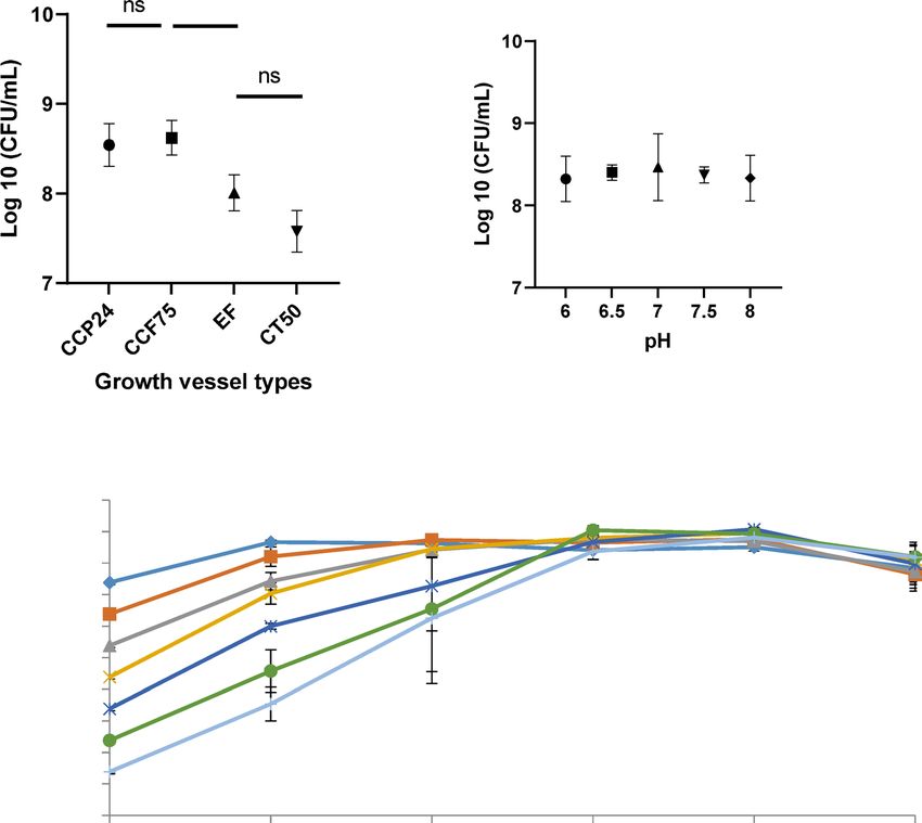
www.nature.com/scientificreports/
Figure 1. KEGG pathway of l-cysteine of C. hepaticus. Green boxes indicate the genes identified in C.
hepaticus while white boxes show absent genes. C. hepaticus lacks genes including cysE (EC 2.3.1.30, serine
acetyltransferase), cysK (EC 2.5.1.47, cysteine synthase A), cysM (EC 2.5.1.65, O-phosphoserine sulfhydrylase)
and cys3 (EC 4.4.1.1, cystathionine γ-lyase).
would aid in the design of reproducible assays to investigate stress resistance, virulence mechanisms, vaccine
development, and survival in the environment of C. hepaticus. This study applied genomic analysis and in vitro
experiments to develop an enhanced C. hepaticus liquid culture method.
Results
In silico pathway analysis of C. hepaticus. KEGG pathway analysis of three C. hepaticus strains showed
that this bacterium harbours a complete tricarboxylic acid (TCA) cycle. Therefore, C. hepaticus can use all sub-
strates in the citric pathways including pyruvate, succinate, oxaloacetate, fumarate, 2-oxoglutarate, malate and
citrate as energy sources. The genome of C. hepaticus also contains genes encoding the enzymes for complete
metabolic pathways for many amino acids, such as l-methionine, l-histidine, l-alanine, l-glycine, l-valine,
l-leucine and l-threonine. In contrast, C. hepaticus lacks a complete pathway for the biosynthesis of l-arginine
from l-glutamate. The argE gene, encoding acetylcitrulline deacetylase is absent. l-lysine cannot be biosyn-
thesised from l-aspartate as the dapA and dapB genes are not present. Similarly, it is predicted that l-cysteine
cannot be biosynthesised, as the cysE, cysM and cysK genes required to synthesise l-cysteine from l-serine
and l-methionine, are absent (Fig. 1). Consequently, C. hepaticus was predicted to be unable to biosynthesise
l-cysteine, l-lysine and l-arginine. Adding these amino acids to the culture media may improve the growth of C.
hepaticus. This prediction was tested in the following in vitro experiments. Sodium pyruvate was also added into
Brucella broth to check the growth of C. hepaticus, because it has been used as a carbon s ource14 and scavenges
hydrogen peroxide17, an oxidative stress factor that is generated via the use of oxygen of b
acteria18.
The effect of culture vessel type, pH, and inoculum level on the yield of C. hepaticus. Before
testing the effect of amino acids and sodium pyruvate on the growth of C. hepaticus, three factors, culture ves-
sel types, pH, and inoculum level were examined. Figure 2A shows that C. hepaticus grew to a higher final
density when growth in 24-well cell culture plates (CCP24) and 75 c m2 cell culture flasks (TCF75) (log10 CFU/
mL = 8.54–8.62 (P ≤ 0.05) compared to that seen in 50 mL tubes (CT50) and 250 mL Erlenmeyer flasks (EF)
(log CFU/mL = 7.75–8.0). The CCP24 culture plates (1 mL of culture added/well) were therefore used to test
the effect of amino acid and sodium pyruvate supplementation on the growth of C. hepaticus, and TCF75 flasks
(25 mL of media/flask) were used to scale-up the production of biomass of C. hepaticus.
C. hepaticus was monitored for growth in Brucella broth adjusted to a pH ranging from 6.0 to 8.0 to evaluate
the pH tolerance. There was no statistically significant difference in the growth of C. hepaticus, indicating little
or no effect of pH within this range (Fig. 2B). Therefore, Brucella broth without pH adjustment was used for the
further culturing of C. hepaticus, as it has a neutral pH value (7.0 ± 0.2).
The growth kinetics of C. hepaticus in Brucella broth was investigated using initial inocula ranging from
101 to 1 07 CFU/mL (Fig. 2C). Viable cells counts were carried out after 0, 24, 48, 60, 72 and 96 h of incubation.
The two highest inoculum levels ( 106 and 107 CFU/ml) produced maximum growth to 1 08 CFU/ml after 24 h
of incubation. With the lower inoculum levels (105 and 104 CFU/mL), C. hepaticus achieved maximum growth
after 48 h of incubation and for low inoculum levels of 1 01 to 1 03 CFU/ml, the maximum growth of C. hepaticus
was reached after 60 h.
The effect of amino acids and sodium pyruvate on the growth of C. hepaticus. C. hepaticus
cultured in Brucella broth supplemented with 0.4 mM l-cysteine, 4 mM l- glutamine, 0.8 mM l-valine, 0.4 mM
l-serine and 10 mM sodium pyruvate exhibited a significantly greater growth compared to a culture grown in
Scientific Reports | (2021) 11:20802 | https://doi.org/10.1038/s41598-021-00277-8 2
Vol:.(1234567890)www.nature.com/scientificreports/
✱
B
A
10
9
8
Log 10 (CFU/mL)
7
6
5
4
3
2
1
0
0h 24h 48h 60h 72h 96h
Incubaon me (hours)
C
Figure 2. (A) Effect of growth vessel type on the growth of C. hepaticus HV10T. CCP24 24 well cell culture
flask, TCF75 tissue culture flask 75cm2, EF Erlenmeyer flask, CT50 corning 50 mL tube. (B) Growth of C.
hepaticus in Brucella broth pH adjusted 6–8 after 48 h of incubation. (C) Kinetic growth of C. hepaticus in
Brucella broth. The initial inoculum was from 1 01 to 107 CFU/mL. Viable cells were counted after 0, 24, 48, 60,
72 and 96 h of incubation. P values were calculated using unpaired t-tests. Significant difference: * p ≤ 0.05, ns no
significance. Data points represent results of 4 biological replicates.
Brucella broth only (log10 = 8.66–9.26 vs 8.31 CFU/mL) (Fig. 3). l-cysteine (0.4 mM) showed highest growth
enhancement to log10 = 9.26 CFU/mL, significantly higher than the density obtained with the other supplements
(p ≤ 0.005). No significant difference was observed (p > 0.05, Fig. 3) when C. hepaticus was grown in Brucella
broth supplemented with other amino acids including l-lysine, l-methionine, l-histidine, l-glycine, l- arginine,
l-leucine, and l-threonine compared to a culture grown in Brucella broth only.
The growth of C. hepaticus was then investigated in Brucella broth supplemented with different combinations
of each compound that could enhance the growth of C. hepaticus, as demonstrated above, including l-cysteine,
l-glutamine, l-valine, l-serine, and sodium pyruvate. In general, all combinations of supplements in Brucella
broth significantly enhanced the growth of C. hepaticus compared to Brucella broth alone. The highest growth of
C. hepaticus observed was in Brucella broth supplemented with a mixture of l-cysteine (0.4 mM), l-glutamine
(4 mM) and sodium pyruvate (10 mM) (log10 = 9.34), followed by a combination of l-cysteine (0.4 mM) and
sodium pyruvate (10 mM) (log10 = 9.17), and only l-cysteine (0.4 mM) (log10 = 9.11) (Table 1). Brucella broth
supplemented with l-cysteine (0.4 mM), and l-glutamine (4 mM) and sodium pyruvate (10 mM) was then used
to grow C. hepaticus in TCF75 flasks in the following experiments.
Effect of temperature and incubating conditions on growth of C. hepaticus. Because the cul-
ture vessels that provided the largest surface to volume ratio produced the highest density cultures it was pro-
posed that gas exchange between the liquid medium and the microaerophilic atmosphere might be important.
Therefore, the effects of agitation of the cultures by shaking were investigated. In the modified Brucella broth
developed in this study, after 48 h of incubation C. hepaticus grew significantly better (p ≤ 0.05) in static condi-
tions, with log10 = 9.25 CFU/mL compared to log10 = 8.76 CFU/mL when shaken. It was also found that C.
Scientific Reports | (2021) 11:20802 | https://doi.org/10.1038/s41598-021-00277-8 3
Vol.:(0123456789)www.nature.com/scientificreports/
Figure 3. Effect of sodium pyruvate and amino acids on the growth of C. hepaticus HV10T in Brucella broth
(control). Cultures were inoculated with 106 CFU/ml and incubated for 48 h at 37 °C. P-values were calculated
using unpaired t-tests. Significant difference *: p < 0.05; **: p < 0.005.
Medium Log10 (CFU/mL) SEM P value Significant difference
Brucella broth (reference) 8.42 0.10
Brucella broth + l-cysteine 9.11 0.05 0.0008*** Yes
Brucella broth + l-cysteine + sodium pyruvate 9.17 0.07 0.0009*** Yes
Brucella broth + l-cysteine + l-glutamine 9.04 0.10 0.0049** Yes
Brucella broth + l-cysteine + l-glutamine + sodium pyruvate 9.34 0.08 0.0003*** Yes
Brucella broth + l-cysteine + Sodium pyruvate + l-valine 9.03 0.03 0.0012** Yes
Brucella broth + l-cysteine + Sodium Pyruate + l-serine 9.01 0.18 0.0278* Yes
Table 1. The effects of different supplements added to Brucella broth on the growth of C. hepaticus compared
to Brucella broth only. Cultures were inoculated with 106 CFU/ml and incubated for 48 h at 37 °C. One way
ANOVA followed by Dunnett’s multiple comparison test showed that the differences between all the additive
groups and the reference group (unsupplemented Brucella broth) were all highly significantly different. The
P-values presented are more conservative values calculated using unpaired t-tests. SEM standard error of mean,
CFU colony-forming unit; significant difference. *: p ≤ 0.05; **: p ≤ 0.01; ***: p ≤ 0.001.
hepaticus exhibited significantly better growth at 37 °C than 42 °C (p ≤ 0.05), log10 = 9.28 CFU/mL compared to
log10 = 8.09 CFU/mL.
Growth of different C. hepaticus strains in modified Brucella broth. The medium development
experiments described above were carried out on a single strain of C. hepaticus. To determine if the improved
media composition could also enhance the growth of other C. hepaticus isolates, the growth of different isolates
in the modified Brucella broth, at 37 °C, under microaerobic conditions, in static TCF75 flasks was examined.
All tested strains of C. hepaticus (HV10T, 19L. VICOCT18, WESTERN3, NSWJUNE19, SAJULY18 and DALE3)
reached densities of log10 = 9.25–9.49 CFU/mL after 48 h, significantly higher (p ≤ 0.05) than non-supplemented
Brucella broth (log10 = 8.24–8.66 CFU/mL) Fig. 4). No significant difference in growth was observed among all
C. hepaticus isolates in the modified Brucella broth.
Experimental infection of laying hens with C. hepaticus culture grown in modified Brucella
broth. One of the principal reasons to develop improved culturing conditions for C. hepaticus was to make
the preparation of challenge inocula easier and more reproducible for experimental infection trials used to
investigate the biology of infections and assess the efficacy of various SLD treatment protocols. Experience with
preparing challenge inocula using the previously described plate harvesting method had suggested that the state
of the inoculum was critical to the success of infection10. Therefore, it was important to establish that the cul-
tures grown in the modified Brucella liquid medium were capable of eliciting disease. Hens entering peak lay
were challenged with, either C. hepaticus HV10T harvested from Brucella agar plates, or bacteria grown in the
newly devised liquid media conditions and the induction of SLD lesions on the liver were scored. Birds in the
Scientific Reports | (2021) 11:20802 | https://doi.org/10.1038/s41598-021-00277-8 4
Vol:.(1234567890)www.nature.com/scientificreports/
Figure 4. Growth of C. hepaticus isolates in modified Brucella broth (Brucella broth supplemented with
0.4 mM l-cysteine, 4 mM l-glutamine and 10 mM sodium pyruvate) compared to non-supplemented Brucella
broth after 48 h of incubation under microaerobic conditions. Solid black bars illustrate the growth of C.
hepaticus strains in modified Brucella broth, grey bars in Brucella broth only. P values were calculated using
unpaired t-tests. Significant difference *: p ≤ 0.05. **: p ≤ 0.01.
control group, inoculated with fresh modified Brucella broth, showed no lesion on livers, whereas 12 out of 12
hens inoculated with the agar plate derived bacteria had lesions on the liver and 11 out of 12 hens inoculated
with bacteria grown in the modified Brucella liquid had liver lesions. Based on disease scores, there was no
significant difference between the degree of disease elicited by bacteria grown under the two conditions (Fig. 5),
demonstrating that C. hepaticus grown in the modified Brucella broth developed in this study had equal levels
of virulence as those grown by the previously described plate culture method. The group sizes were sufficient to
detect a one-point difference in mean scores with an alpha of 0.05 and 80% power.
Discussion
In silico pathway analysis of the C. hepaticus genome predicted that the bacterium lacks some of the genes encod-
ing enzymes required for the biosynthesis of l-cysteine, l-lysine and l-arginine. C. hepaticus lacks the genes cysE
and cysK, cysM to synthesise l-cysteine from l-serine and sulphur. cysE encodes serine acetyltransferase and cysK
encodes cysteine synthase. These enzymes are required for the synthesis of l-cysteine from l-serine in bacteria
and plants19. CysE synthesises l-cysteine from l-serine by catalysing an acyl transfer from acetyl-CoA. CysK
catalyses O-acetyl-l-serine combining with hydrogen sulphide to yield l-cysteine19. The addition of l-cysteine
to Brucella broth improved the growth of C. hepaticus significantly, to more than 1 09 CFU/mL compared to
Brucella broth only. This indicates that even though Brucella broth is a rich nutrient source the l-cysteine level
is insufficient to support maximal growth of C. hepaticus. Growth support of l-cysteine was also reported in
other Campylobacter species. l-cysteine was defined as a vital source of sulphur for C. jejuni20. C. jejuni grown
in minimal medium with l-cysteine added was better than their growth in the medium without c ysteine14,21.
l-cysteine has been identified as a chemotactic attractants of C. jejuni22,23. In contrast, although C. hepaticus was
also predicted to be unable to biosynthesise l-lysine and l-arginine, the addition of these amino acids to Brucella
broth did not significantly increase the culture densities that could be achieved for C. hepaticus compared to
densities supported by unmodified Brucella broth. Similarly, the addition of l-arginine and l-lysine to minimal
media did not enhance the growth of C. jejuni because C. jejuni may have genes that support the biosynthesis
of l-arginine and l-lysine14. Better growth of C. hepaticus in Brucella broth supplemented with l-glutamine,
l-valine or l-serine was observed although no missing genes that are responsible for the biosynthesis of these
amino acids by C. hepaticus were detected. It was demonstrated that l-glutamine and l-serine are chemoattract-
ants for C. jejuni24. In chickens, l-cysteine, l-glutamine, and l-serine are abundant in chicken liver. The chicken
gut also has sufficent quantities of necessary amino acids such as l-cysteine, l-glutamine, l-valine and l-serine
for the growth of many bacteria25 and this likely explains why, despite its inability to synthesise a number of
amino acids, C. hepaticus can colonise the intestinal tract of laying h ens10. It was found that the reduction of
amino acids such as l-cysteine, l-glutamine and l-serine in the chicken diet contributed to the reduction of
Campylobacters in c hickens26. The authors explained that these amino acid are involved in the formation of the
mucin production of the intestinal mucus layer and essential for the survival and growth of Campylobacters27.
Thus, an increase or decrease in the concentration of these amino acids results in changes in the number of
Campylobacters in the chicken g ut26. For other amino acids including l-methionine, l-histidine, l-glycine,
Scientific Reports | (2021) 11:20802 | https://doi.org/10.1038/s41598-021-00277-8 5
Vol.:(0123456789)www.nature.com/scientificreports/
Figure 5. SLD spot scores of SLD-positive laying hens after challenge with C. hepaticus HV10T grown from
modified Brucella broth (liquid method) and HBA plate method based on scores of spots. This score is based on
a logarithmic scale from 0 to 4, where 0 = no visible lesions, 1 = 1–9 lesions, 2 = 10–99 lesions, 3 = 100–999 lesions
and 4 = more than 1000 lesions. P values were calculated using unpaired t-tests.
l-leucine, and l-threonine, there was no significant difference in the growth of C. hepaticus. These results agree
with the bioinformatic pathway analysis.
This study found that the C. hepaticus showed maximum growth in Brucella broth supplemented with a
mixture of l-cysteine (0.4 mM), l-glutamine (4 mM) and sodium pyruvate (10 mM) and reached 1 09.34 CFU/
mL. Sodium pyruvate provides an additional carbon source to support the growth of C. hepaticus in Brucella
broth. C. hepaticus may heavily depend on the Kreb’s cycle to generate energy. Many studies have demonstrated
the important role of pyruvate in the growth of campylobacters. Sodium pyruvate is one of the compounds in
the Campylobacter growth supplement product (FBP supplement)28 and Campylobacter selective supplement29.
Pyruvate was found to promote the growth of C. jejuni NCTC 11168 in MEM medium14. This compound plays a
central role in C. jejuni metabolism and can be fermented to various products such as acetate, formate, lactate and
succinate. It is linked to carbohydrate and amino acid catabolism to produce energy. The synthesis of l-leucine,
l-valine, l-alanine and l-isoleucine by campylobacters use pathways in which pyruvate can play a key r ole30.
Pyruvate also acts as an electron acceptor and can decrease the concentration of hydrogen peroxide to reduce
the damage caused by oxygen to b acteria31.
C. hepaticus grew to higher densities in CCP24 and TCF75 than CT50 and EF, suggesting that the sur-
face-area-to-volume ratio (S:V) may be important, possibly for gas exchange, as mentioned in a study of improve-
ment of culturing of C. jejuni32. These authors demonstrated that TCF75 has more S:V ratio than EF and CT50
and therefore TCF75 provided better atmospheric exchange than EF and CT50. Also, tissue culture flasks were
recommended to grow and study standard growth curves of C. jejuni33. In a study of the growth of Helicobacter
pylori, Gas–Permeable Lifecell tissue culture flasks gave improved growth in Brucella broth supplemented with
fetal bovine s erum34. These authors also mentioned the effect of surface area on the growth of H. pylori and sug-
gested that a small surface area resulted in poorer growth of H. pylori. Tissue culture flasks were used early in
the history of the culture of C. jejuni and C. pylori35,36 and have been employed in many studies of Campylobacter
species. It may show that C. hepaticus requires a larger S:V ratio to grow due to better gas exchange, although it is
then unexpected that agitation had a negative effect. A low level of oxygen may be an obligatory requirement for
C. hepaticus growth. Under anaerobic conditions and higher oxygen tensions (21%), C. hepaticus failed to g row1.
Similarly, C. jejuni does not grow under anaerobic conditions37,38 and oxygen is considered a requirement for
Scientific Reports | (2021) 11:20802 | https://doi.org/10.1038/s41598-021-00277-8 6
Vol:.(1234567890)www.nature.com/scientificreports/
DNA synthesis in C. jejuni37. Campylobacter spp. are sensitive to high oxygen tensions, but still need an optimal
oxygen concentration (2%-10%) to g row38. C. hepaticus contains genes for oxidative p hosphorylation11 that need
oxygen as an acceptor to generate ATP.
C. hepaticus achieved maximum growth after 48–72 h, depending on the initial inoculum, showing slower
growth in comparison to other Campylobacters. The maximum growth of C. jejuni was at around 30 h in both
Nutrient Broth Number 2 and Mueller Hinton b roth39. C. jejuni reached the highest densities after 24 h of growth
in Brain Heart Infusion broth . C. hepaticus can grow in a range of pH from 6.0 to 8.0. It has been reported that
40
other Campylobacter species also grow well in this pH r ange41. Chickens normally have a pH of around 6.3 in
the liver42, 6.4 in the small and large i ntestine43, 6.6—6.7 for c aecum43,44, and 6.0 for b
ile45. These are all tissues
and environments in which it has been shown that C. hepaticus can survive and colonise. C. hepaticus showed
better growth at 37 °C than 42 °C while this microorganism has only been isolated from chickens, which have a
body temperature of 40–42°oC46. The growth of C. hepaticus was reported to be somewhat slow at 42 °C, taking
7 days to form colonies in sheep blood a gar3. Temperature differently affects Campylobacter species regarding the
growth, motility, and ability to invade the host cells. A study showed that C. jejuni grew at both 37 °C and 42 °C
but showed differences in motility and invasion. C. coli grew and moved better at 42 °C. C. fetus, a bacterium
that is frequently detected in poultry, showed greater growth and invasion at 37 °C47.
Using the modified Brucella broth developed in this study, together with growth conditions including the
use of a large surface area culture vessel, at 37 °C, in microaerophilic and static conditions, C. hepaticus cultures
could grow to 1 09 CFU/mL and showed virulence in laying hens. This culturing method is time-saving and
more cost-effective than the previously used plate harvesting method to obtain the large biomass required for
SLD animal induction experiments and bacterin vaccine production. It also reduces the amount of subcultur-
ing needed, possibly minimising the effect of repeated subculture on the virulence of C. hepaticus. A study has
shown that the repeated subculturing of the somewhat related organism, H. pylori, could result in a decrease of
adhesion, motility, gastric inflammation and cytotoxicity, and repeated culturing is a recognised way that bacteria
have been attenuated to produce live v accines48. Thus, the method described in this study can facilitate further
studies on C. hepaticus biology and SLD.
Materials and methods
In silico analysis of C. hepaticus metabolism. The metabolic pathways encoded within the genomes of
C. hepaticus strains HV10T, 19L and VICOCT18, were analysed using (1) RAST (Rapid Annotation using Sub-
system Technology)12 and the SEED49 (http://rast.nmpdr.org/rast.cgi) for annotating the genomes and metabolic
pathways prediction of C. hepaticus; and (2) B LAST50 (Basic Local Alignment Search Tool) (https://blast.ncbi.
nlm.nih.gov/Blast.cgi) for comparing nucleotides and protein sequences of C. hepaticus with available sequences
in the gene bank. C. hepaticus HV10T is the type strain for C. hepaticus, 19L is a representative of a clade that is
distinct form HV10T and VICOCT18 is a more recent isolate.
Bacterial strains and culture conditions. The C. hepaticus strains HV10T, 19L11, VICOCT18, WEST-
ERN3, NSWJUNE19, SAJULY18 and DALE351 were used in the study. These strains are representative isolates
from independent SLD outbreaks from widely separated geographical locations in Australia. All C. hepaticus
strains were stored at − 80 °C in 70% Brucella broth (BD BBL™) and 30% glycerol. C. hepaticus was routinely cul-
tured on Brucella agar plates (Brucella broth (BD BBL™) + 1.5% agar (BD BBL™)) supplemented with 5% defibri-
nated horse blood (Equicel) (HBA) and cultured at 37 °C under microaerobic conditions (created using Campy-
gen 3.5L gas generation packs (Oxoid)) in an anaerobic jar, for 96 h to recover C. hepaticus cells from − 80 °C
stock or for 72 h if they were subcultured from HBA plates.
Effect of type of culture vessel on the growth of C. hepaticus. Costar® 24-well cell culture plates
(CCP24), Corning® 50 mL centrifuge tubes (CT50) with a vented cap (0.2 μm pore size), Corning® 75cm2 cell
culture flask (TCF75) with a vented cap (0.2 μm pore size) and Erlenmeyer flasks (250 mL) (EF) were used to
compare the growth of C. hepaticus in Brucella broth (BD BBL™). The volume of culture media used in CCP24
was 1 mL, 25 mL (CT50 and TCF) and 40 mL (EF). All vessels were placed in BD GasPak™ EZ container, charged
with CampyGen 3.5 L (Oxoid) to produce microaerobic conditions and then incubated at 37 °C for 48 h. The
growth rate of C. hepaticus HV10 was determined by the plate count method on HBA plates.
Effect of pH and initial inoculum on C. hepaticus growth. The pH of Brucella broth was adjusted
from 6.0 to 8.0 in increments of 0.5 units using 1 M NaOH or 1 M HCl. C. hepaticus was cultured in CCP24
plates to examine growth at different pH levels. Each well of CCP24 was inoculated with 106 CFU/mL of C.
hepaticus HV10T. Plates were incubated under microaerobic conditions at 37 °C. Growth was enumerated after
48 h of incubation by plating serial dilution on HBA. The experiment was performed in triplicate.
The effect of initial inoculum size on the growth kinetics and final culture yields was analysed using seven
initial inoculum levels ( 101 to 1 07 CFU/ml) of C. hepaticus HV10T in a CCP24 plate. The plates were incubated
at 37 °C under microaerobic conditions. The growth of C. hepaticus HV10T was examined after 24, 48, 60, 72,
and 96 h of incubation using the plate count method on HBA.
Growth of C. hepaticus in static and shaking conditions. C. hepaticus HV10T was suspended into
Brucella broth to achieve acell density of 1 05 CFU/ml and then 1 mL of bacterial suspension was incubated into
each well of CCP24 at static condition. For shaking conditions, the anaerobic jars were shaken in a shaking incu-
bator with a speed of 100 rpm. After 48 h viable C. hepaticus cells were enumerated on HBA plates. The shaking
conditions were only used for this test; static growth conditions were used at all other times.
Scientific Reports | (2021) 11:20802 | https://doi.org/10.1038/s41598-021-00277-8 7
Vol.:(0123456789)www.nature.com/scientificreports/
Effects of sodium pyruvate and amino acids on the growth of C. hepaticus. Based on the results of
in silico pathway analysis of C. hepaticus and results from Alazzam et al.14, the following supplements were added
to Brucella broth to examine the growth of this bacterium: amino acids (l-cysteine, l-lysine, l-methionine, and
l-leucine, l-glutamine, l-valine, l-histidine); carbon source (sodium pyruvate) (Sigma). Each compound was
completely dissolved in Milli-Q water at a concentration recommended in MCLMAN, a new minimal medium
for C. jejuni14. The chemical solution was passed through a 0.22 µm syringe filter and stored at − 20 °C if not
used immediately. On the day of the experiment, each substrate was added into a bacterial culture and then
1 mL transferred to each well of CCP plates. The plates were incubated under microaerobic conditions at 37 °C
for 48 h.
Effect of temperature on growth of C. hepaticus. Growth of C. hepaticus was tested at 37 °C and
42 °C in Brucella broth. Briefly, C. hepaticus cells were harvested from an HBA plate and suspended into Bru-
cella broth to obtain an OD of 0.01–0.03. The bacterial suspension was supplemented with sodium pyruvate
(10 mM), l-cysteine (0.4 mM), and l-glutamine (4 mM) (based on the results from the experiments described
above). Bacterial culture (1 mL) was placed in CCP plates and incubated under microaerobic conditions for 48 h
at 37 °C and 42 °C.
Animal trial. A C. hepaticus chicken challenge trial was carried out to compare the virulence between C.
hepaticus HV10T cultures grown from modified Brucella broth developed in this study (liquid method) and the
HBA plates (plate method). The plate method was described by Van et al.10 in which C. hepaticus HV10T stored
at − 80 °C in glycerol stocks was first streaked on to HBA plates, then further subcultured to HBA plates and cells
were harvested and resuspended in Brucella broth to obtain 1 × 10⁹ CFU/mL. For the liquid method, C. hepati-
cus was grown in Brucella broth supplemented with l-cysteine (0.4 mM), and l-glutamine (4 mM) and sodium
pyruvate (10 mM) in TCF75 at 37 °C for 48 h in microaerophilic conditions and used directly for the challenge.
The animal experimentation was approved by the Wildlife and Small Institutions Animal Ethics Committee of
the Victorian Department of Economic Development, Jobs, Transport and Resources (approval number 14.16).
A total of 36 Hy-Line brown laying hens were used in the experiment. Only healthy birds laying eggs regularly
were included in the study. Chickens were housed in groups of 4 birds per pen, with 3 pens per group: a total of
12 birds in each group (n = 12). Birds were randomly allocated to groups and cages by stratified rank order based
on weight. Unchallenged control birds were orally inoculated with 1 ml of Brucella broth only whereas 1 ml of
Brucella broth containing 1 × 10⁹ CFU of C. hepaticus HV10T was orally administered to the birds for the plate
and broth methods groups. Birds were sacrificed after 5 days and SLD lesions on the surface of the liver were
counted to measure the severity of the induced disease. The experienced and trained scorers were blinded to
the treatment groups. Scores were based on a logarithmic scale from 0 to 4, where 0 = no visible lesions, 1 = 1–9
lesions, 2 = 10–99 lesions, 3 = 100–999 lesions and 4 = more than 1000 lesions.
Statistical analysis. All experiments to study the growth of C. hepaticus HV10 in different conditions and
supplements were repeated a minimum of three times with biological replicates. Campylobacter cell counts in all
tests were converted to log10 CFU/mL. Statistical comparison of all parameters was performed by t-test, one-way
ANOVA using Graphpad Prism version 8 for Windows, GraphPad Software (San Diego California USA, www.
graphpad.com). The significance level was set at 5% (p ≤ 0.05). Sample size calculation for the SLD animal trial
was performed using the online calculator at https://clincalc.com/stats/samplesize.aspx.
Declarations. All the methods were carried out in accordance with relevant guidelines and regulations and
the animal trial is reported in accordance with the ARRIVE Essential 10 guidelines.
Received: 5 July 2021; Accepted: 30 September 2021
References
1. Van, T.T.H., Elshagmani, E., Gor, M.C., Scott, P.C. & Moore, R.J. Campylobacter hepaticus sp. nov., isolated from chickens with
spotty liver disease. Int. J. Syst. Evol. Microbiol. 66, 4518–4524. https://doi.org/10.1099/ijsem.0.001383 (2016).
2. Crawshaw, T. R. et al. Isolation of a novel thermophilic Campylobacter from cases of spotty liver disease in laying hens and experi-
mental reproduction of infection and microscopic pathology. Vet. Microbiol. 179, 315–321. https://doi.org/10.1016/j.vetmic.2015.
06.008 (2015).
3. Gregory, M., Klein, B., Sahin, O. & Girgis, G. Isolation and characterization of Campylobacter hepaticus from layer chickens with
spotty liver disease in the United States. Avian. Dis. 62, 78–85. https://doi.org/10.1637/11752-092017-Reg.1 (2018).
4. Crawshaw, T. R. et al. Isolation of Campylobacter hepaticus from free-range poultry with spotty liver disease in New Zealand. N.
Z. Vet. J. 69, 58–64. https://doi.org/10.1080/00480169.2020.1801532 (2020).
5. Grimes, T. & Reece, R. Spotty liver disease—An emerging disease in free-range egg layers in Australia. in Proceedings of the Sixtieth
Western Poultry Disease Conference. 53–56 (2011).
6. Khan, I. U. H., Hill, S., Nowak, E. & Edge, T. A. Effect of incubation temperature on the detection of thermophilic Campylobacter
species from freshwater beaches, nearby wastewater effluents, and bird fecal droppings. Appl. Environ. Microbiol. 79, 7639–7645.
https://doi.org/10.1128/AEM.02324-13 (2013).
7. Kim, J. et al. An improved culture method for selective isolation of Campylobacter jejuni from wastewater. Front. Microbiol. 7,
1345. https://doi.org/10.3389/fmicb.2016.01345 (2016).
8. Ismail, Y., Lee, H., Riordan, S. M., Grimm, M. C. & Zhang, L. The effects of oral and enteric Campylobacter concisus strains on
expression of TLR4, MD-2, TLR2, TLR5 and COX-2 in HT-29 cells. PLoS ONE 8, e56888. https://doi.org/10.1371/journal.pone.
0056888 (2013).
9. Reilly, S. S. & Gilliand, S. E. Improved culturing techniques for Campylobacter. J. Food Sci. 68, 2752–2757 (2003).
Scientific Reports | (2021) 11:20802 | https://doi.org/10.1038/s41598-021-00277-8 8
Vol:.(1234567890)www.nature.com/scientificreports/
10. Van, T. T. H. et al. Induction of spotty liver disease in layer hens by infection with Campylobacter hepaticus. Vet. Microbiol. 199,
85–90. https://doi.org/10.1016/j.vetmic.2016.12.033 (2017).
11. Van, T. T. H. et al. Survival mechanisms of Campylobacter hepaticus identified by genomic analysis and comparative transcriptomic
analysis of in vivo and in vitro derived bacteria. Front. Microbiol. 10, 107–107. https://doi.org/10.3389/fmicb.2019.00107 (2019).
12. Aziz, R. K. et al. The RAST server: Rapid annotations using subsystems technology. BMC Genomics 9, 75. https://doi.org/10.1186/
1471-2164-9-75 (2008).
13. Kanehisa, M. & Goto, S. KEGG: Kyoto encyclopedia of genes and genomes. Nucleic Acids Res. 28, 27–30. https://doi.org/10.1093/
nar/28.1.27 (2000).
14. Alazzam, B., Bonnassie-Rouxin, S., Dufour, V. & Ermel, G. MCLMAN, a new minimal medium for Campylobacter jejuni NCTC
11168. Res. Microbiol 162, 173–179. https://doi.org/10.1016/j.resmic.2010.09.024 (2011).
15. Velayudhan, J. & Kelly, D. J. Analysis of gluconeogenic and anaplerotic enzymes in Campylobacter jejuni: an essential role for
phosphoenolpyruvate carboxykinase. Microbiology 148, 685–694. https://doi.org/10.1099/00221287-148-3-685 (2002).
16. Kawanishi, T. et al. New detection systems of bacteria using highly selective media designed by SMART: Selective medium-design
algorithm restricted by two constraints. PLoS ONE 6, e16512. https://doi.org/10.1371/journal.pone.0016512 (2011).
17. Salahudeen, A. K., Clark, E. C. & Nath, K. A. Hydrogen peroxide-induced renal injury. A protective role for pyruvate in vitro and
in vivo. J. Clin. Invest. 88, 1886–1893. https://doi.org/10.1172/jci115511 (1991).
18. Kim, J. C., Oh, E., Kim, J. & Jeon, B. Regulation of oxidative stress resistance in Campylobacter jejuni, a microaerophilic foodborne
pathogen. Front. Microbiol. 6, 751. https://doi.org/10.3389/fmicb.2015.00751 (2015).
19. Benoni, R. et al. Modulation of Escherichia coli serine acetyltransferase catalytic activity in the cysteine synthase complex. FEBS
Lett. 591, 1212–1224. https://doi.org/10.1002/1873-3468.12630 (2017).
20. Vorwerk, H. et al. Utilization of host-derived cysteine-containing peptides overcomes the restricted sulphur metabolism of Campy-
lobacter jejuni. Mol. Microbiol. 93, 1224–1245. https://doi.org/10.1111/mmi.12732 (2014).
21. Dickgiesser, N. & Czylwik, D. Chemically defined media for auxotyping of Campylobacter jejuni. Zentralbl. Bakteriol. Mikrobiol.
Hyg. A 260, 57–64. https://doi.org/10.1016/S0176-6724(85)80098-5 (1985).
22. Chandrashekhar, K., Kassem, I. I. & Rajashekara, G. Campylobacter jejuni transducer like proteins: Chemotaxis and beyond. Gut
Microbes 8, 323–334. https://doi.org/10.1080/19490976.2017.1279380 (2017).
23. Li, Z. et al. Methyl-accepting chemotaxis proteins 3 and 4 are responsible for Campylobacter jejuni chemotaxis and jejuna coloniza-
tion in mice in response to sodium deoxycholate. J. Med. Microbiol. 63, 343–354. https://doi.org/10.1099/jmm.0.068023-0 (2014).
24. Vegge, C. S., Brøndsted, L., Li, Y.-P., Bang, D. D. & Ingmer, H. Energy taxis drives Campylobacter jejuni toward the most favorable
conditions for growth. Appl. Environ. Microbiol. 75, 5308–5314. https://doi.org/10.1128/aem.00287-09 (2009).
25. Seong, P. N. et al. Characterization of chicken by-products by mean of proximate and nutritional compositions. Korean. J. Food.
Sci. Anim. Resour. 35, 179–188. https://doi.org/10.5851/kosfa.2015.35.2.179 (2015).
26. Visscher, C. et al. Influence of a specific amino acid pattern in the diet on the course of an experimental Campylobacter jejuni
infection in broilers. Poult. Sci. 97, 4020–4030. https://doi.org/10.3382/ps/pey276 (2018).
27. Adedokun, S. A., Adeola, O., Parsons, C. M., Lilburn, M. S. & Applegate, T. J. Factors affecting endogenous amino acid flow in
chickens and the need for consistency in methodology. Poult. Sci. 90, 1737–1748. https://doi.org/10.3382/ps.2010-01245 (2011).
28. Hoffman, P. S., George, H. A., Krieg, N. R. & Smibert, R. M. Studies of the microaerophilic nature of Campylobacter fetus subsp.
jejuni. II. Role of exogenous superoxide anions and hydrogen peroxide. Can. J. Microbiol. 25, 8–16. https://doi.org/10.1139/m79-
002 (1979).
29. Karmali, M. A. et al. Evaluation of a blood-free, charcoal-based, selective medium for the isolation of Campylobacter organisms
from feces. J. Clin. Microbiol. 23, 456–459. https://doi.org/10.1128/JCM.23.3.456-459.1986 (1986).
30. Mendz, G. L., Ball, G. E. & Meek, D. J. Pyruvate metabolism in Campylobacter spp. Biochim. Biophys. Acta 1334, 291–302. https://
doi.org/10.1016/S0304-4165(96)00107-9 (1997).
31. Verhoeff-Bakkenes, L., Arends, A. P., Snoep, J. L., Zwietering, M. H. & de Jonge, R. Pyruvate relieves the necessity of high induc-
tion levels of catalase and enables Campylobacter jejuni to grow under fully aerobic conditions. Lett. Appl. Microbiol. 46, 377–382.
https://doi.org/10.1111/j.1472-765X.2008.02326.x (2008).
32. Reilly, S. S. & Gilliland, S. E. Improved culturing techniques for Campylobacter. J. Food Sci. 68, 2752–2757. https://doi.org/10.
1111/j.1365-2621.2003.tb05800.x (2003).
33. Davis, L. & DiRita, V. Growth and laboratory maintenance of Campylobacter jejuni. Curr. Protoc. Microbiol. 8(8A), 1 1–8A 1 7.
https://doi.org/10.1002/9780471729259.mc08a01s10 (2008).
34. Secker, D., Tompkins, D. & Alderson, G. Gas-permeable lifecell tissue culture flasks give improved growth of Helicobacter pylori
in a liquid medium. J. Clin. Microbiol. 29, 1060–1061. https://doi.org/10.1128/JCM.29.5.1060-1061.1991 (1991).
35. Rollins, D. M., Coolbaugh, J. C., Walker, R. I. & Weiss, E. Biphasic culture system for rapid Campylobacter cultivation. Appl. Environ.
Microbiol. 45, 284–289. https://doi.org/10.1128/AEM.45.1.284-289.1983 (1983).
36. Shadowen, R. D. & Sciortino, C. V. Improved growth of Campylobacter pylori in a biphasic system. J. Clin. Microbiol. 27, 1744–1747.
https://doi.org/10.1128/JCM.27.8.1744-1747.1989 (1989).
37. Sellars, M. J., Hall, S. J. & Kelly, D. J. Growth of Campylobacter jejuni supported by respiration of fumarate, nitrate, nitrite,
trimethylamine-n-oxide, or dimethyl sulfoxide requires oxygen. J. Bacteriol. 184, 4187–4196. https://doi.org/10.1128/jb.184.15.
4187-4196.2002 (2002).
38. Kaakoush, N. O., Miller, W. G., De Reuse, H. & Mendz, G. L. Oxygen requirement and tolerance of Campylobacter jejuni. Res.
Microbiol. 158, 644–650. https://doi.org/10.1016/j.resmic.2007.07.009 (2007).
39. Ghaffar, N., Connerton, P. & Connerton, I. Filamentation of Campylobacter in broth cultures. Front. Microbiol. 6, 657. https://doi.
org/10.3389/fmicb.2015.00657 (2015).
40. Wright, J. et al. Metabolite and transcriptome analysis of Campylobacter jejuni in vitro growth reveals a stationary-phase physi-
ological switch. Microbiology 155, 80–94. https://doi.org/10.1099/mic.0.021790-0 (2009).
41. Skirrow, M. B. Encyclopedia of Food Sciences and Nutrition. 2nd Edn. (ed. Benjamin Caballero) 779–786 (Academic Press, 2003).
42. King, Y. T. & Chen, T. C. Chemical and physical characteristics of chicken livers following adrenocorticotropic hormone-induced
stress. J. Food. Sci. 63, 589–591. https://doi.org/10.1111/j.1365-2621.1998.tb15791.x (1998).
43. Mabelebele, M., John, A., Ng’ambi, J., Norris, D. & Ginindza, M. Comparison of gastrointestinal tracts and pH values of digestive
organs of Ross 308 broiler and indigenous Venda chickens fed the same diet. Asian. J. Anim. Vet. Adv. 9, 71–76. https://doi.org/
10.3923/ajava.2014.71.76 (2014).
44. Ciurescu, G., Vasilachi, A., Habeanu, M. & Dragomir, C. Effects of dietary lentil seeds inclusion on performance, carcass charac-
teristics and cecal pH of broiler chickens. Indian J. Anim. Sci. 87, 1130–1134 (2017).
45. Zaefarian, F., Abdollahi, M. R., Cowieson, A. & Ravindran, V. Avian liver: The forgotten organ. Animals (Basel) 9, 63. https://doi.
org/10.3390/ani9020063 (2019).
46. Bolzani, R., Ruggeri, F. & Olivo, O. M. Average normal temperature of the chicken in the morning and after 1–2 days of fasting.
Boll. Soc. Ital. Biol. Sper. 55, 1618–1622 (1979).
47. Aroori, S. V., Cogan, T. A. & Humphrey, T. J. The effect of growth temperature on the pathogenicity of Campylobacter. Curr.
Microbiol. 67, 333–340. https://doi.org/10.1007/s00284-013-0370-1 (2013).
48. Kim, S. S. et al. The effect of the repeated subcultures of Helicobacter pylori on adhesion, motility, cytotoxicity, and gastric inflam-
mation. J. Korean. Med. Sci. 17, 302–306. https://doi.org/10.3346/jkms.2002.17.3.302 (2002).
Scientific Reports | (2021) 11:20802 | https://doi.org/10.1038/s41598-021-00277-8 9
Vol.:(0123456789)www.nature.com/scientificreports/
49. Overbeek, R. et al. The SEED and the Rapid Annotation of microbial genomes using Subsystems Technology (RAST). Nucleic
Acids Res. 42, 206–214. https://doi.org/10.1093/nar/gkt1226 (2014).
50. Altschul, S. F. et al. Gapped BLAST and PSI-BLAST: A new generation of protein database search programs. Nucleic Acids Res. 25,
3389–3402. https://doi.org/10.1093/nar/25.17.3389 (1997).
51. Phung, C. et al. Campylobacter hepaticus, the cause of Spotty Liver Disease in chickens: Transmission and routes of infection. Front.
Vet. Sci. 6, 505. https://doi.org/10.3389/fvets.2019.00505 (2019).
Acknowledgements
CP was supported by a joint Vietnam International Education Development and RMIT Scholarship.
Author contributions
C.P. designed, conducted the study and carried out data analysis. R.J.M. and T.T.H.V. conceived and supervised
the work. T.B.W., J.A.Q. and P.C.S. facilitated and conducted the animal trial. C.P. wrote the first draft of the
manuscript and T.T.H.V. and R.J.M. critically reviewed the manuscript. All authors commented on the manu-
script and approved the final version of the manuscript.
Competing interests
The authors declare no competing interests.
Additional information
Correspondence and requests for materials should be addressed to R.J.M.
Reprints and permissions information is available at www.nature.com/reprints.
Publisher’s note Springer Nature remains neutral with regard to jurisdictional claims in published maps and
institutional affiliations.
Open Access This article is licensed under a Creative Commons Attribution 4.0 International
License, which permits use, sharing, adaptation, distribution and reproduction in any medium or
format, as long as you give appropriate credit to the original author(s) and the source, provide a link to the
Creative Commons licence, and indicate if changes were made. The images or other third party material in this
article are included in the article’s Creative Commons licence, unless indicated otherwise in a credit line to the
material. If material is not included in the article’s Creative Commons licence and your intended use is not
permitted by statutory regulation or exceeds the permitted use, you will need to obtain permission directly from
the copyright holder. To view a copy of this licence, visit http://creativecommons.org/licenses/by/4.0/.
© The Author(s) 2021
Scientific Reports | (2021) 11:20802 | https://doi.org/10.1038/s41598-021-00277-8 10
Vol:.(1234567890)You can also read