O antigen restricts lysogenization of non O157 Escherichia coli strains by Stx converting bacteriophage phi24B
←
→
Page content transcription
If your browser does not render page correctly, please read the page content below
www.nature.com/scientificreports
OPEN O antigen restricts lysogenization
of non‑O157 Escherichia coli strains
by Stx‑converting bacteriophage
phi24B
A. K. Golomidova1, A. D. Efimov1, E. E. Kulikov1,2, A. S. Kuznetsov1,3, I. Sh. Belalov1 &
A. V. Letarov1,3*
Acquisition of new prophages that are able to increase the bacterial fitness by the lysogenic
conversion is believed to be an important strategy of bacterial adaptation to the changing
environment. However, in contrast to the factors determining the range of bacteriophage lytic
activity, little is known about the factors that define the lysogenization host range. Bacteriophage
phi24B is the paradigmal model of Stx-converting phages, encoding the toxins of the Shiga-toxigenic
E. coli (STEC). This virus has been shown to lysogenize a wide range of E. coli strains that is much
broader than the range of the strains supporting its lytic growth. Therefore, phages produced by the
STEC population colonizing the small or large intestine are potentially able to lysogenize symbiotic
E. coli in the hindgut, and these secondary lysogens may contribute to the overall patient toxic load
and to lead to the emergence of new pathogenic STEC strains. We demonstrate, however, that O
antigen effectively limit the lysogenization of the wild E. coli strains by phi24B phage. The lysogens
are formed from the spontaneous rough mutants and therefore have increased sensitivity to other
bacteriophages and to the bactericidal activity of the serum if compared to their respective parental
strains.
Temperate bacteriophages affect many aspects of the life of lysogenic bacteria through multiple mechanisms
including direct or indirect influence on the host genome e xpression1, gene transduction including the recently
described highly effective lateral t ransduction2 and specific mobilization of some genomic islands3 and other
mechanisms4. The most known and, probably, the most ecologically significant mechanism of such influence
is the lysogenic c onversion1,5. Such a conversion occurs due to the expression in the lysogen of some prophage
encoded genes, that confer the bacteria new features potentially increasing their fitness in particular h abitats5,6.
Therefore, acquisition of new prophages is believed to be one of the important strategies of bacterial adaptation
in nature4. Bacterial antiviral systems are often expressed in phase-variable m
anner7–9. The adaptive value of such
variations may be in part due to the “opening of window” for acquisition of new potentially beneficial prophages.
However, the most important factor that determines the bacteriophage host range is not the activity of the
intracellular antiviral systems but the specificity of the bacteriophage adsorption. The major determinants of
adsorption specificity that define bacteriophage lytic activity host range are well characterized10. At the same
time data on the factors determining the lysogenization host range are largely missing.
The shigatoxigenic (STEC) Escherichia coli strains are associated with multiple foodborne diseases causing
morbidity and mortality in h umans1,4,5. STEC are zoonotic pathogens that may colonize livestock animals such as
cattle without causing symptoms in them but making the agricultural environments and products dangerous for
humans10. The majority of STEC strains belong to the O157:H7 s erotype11, although non-O157 STEC strains have
been identified and currently gain increased a ttention4 as, for example, the so-called “Big Six”—O26, O45, O1;
O111,O121 and O14512, as well as the O104:H4 serotype that caused the well-known 2011 outbreak in Germany13.
STEC strains possess a number of pathogenicity factors, the foremost being the production of the Shiga
toxins11,14 encoded by the Stx-converting prophages. Although the Stx-converting bacteriophages are quite
divergent morphologically and contain genomic modules divergent by their sequences, however the genome
1
Winogradsky Institute of Microbiology, RC Biotechnology RAS, Prospekt 60‑letiya Oktyabrya 7 bld. 2, Moscow,
Russia 117312. 2Phystech School of Biological and Medical Physics, Moscow Institute of Physics and Technology,
Moscow, Russia. 3Faculty of Biology, Lomonosov Moscow State University, Moscow, Russia. *email: letarov@
gmail.com
Scientific Reports | (2021) 11:3035 | https://doi.org/10.1038/s41598-021-82422-x 1
Vol.:(0123456789)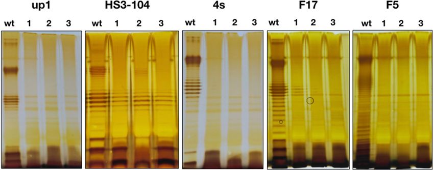
www.nature.com/scientificreports/
organization of these viruses is similar to that of the bacteriophage λ, therefore the Stx phages are considered as
lambdoid phages15,16. In these phages, the toxin gene stx is located downstream of the conserved gene Q encod-
ing the antiterminator of the late gene r egion15. Toxin expression is repressed in lysogenic bacterial cells, and
takes place only upon the prophage i nduction15,16. Toxin molecules lacking the signal peptide for secretion are
released upon cell l ysis6,15,16. The lysogeny in Stx-converting phages is less stable compared to the non-Stx lamb-
doid phages17–20 resulting in higher rate of spontaneous induction and in increased sensitivity to environmental
factors such as DNA-damaging agents, oxidative stress or increased salt concentrations. Many antibiotics also
increase the induction rate of Stx-converting prophages thus enhancing the toxin production. The increase of
the toxin production may worsen the patient’s conditions and provoke the haemolytic uremic syndrome often
leading to fatal outcome1. Therefore, the use of antibiotics to treat STEC infections remains c ontroversial21.
At the same time, the STEC infections are self-limiting, and the pathogen gets spontaneously eliminated in
ca. 2 weeks. The standard for the treatment of these infections relies on supportive care (symptomatic treatment,
plasma exchange, infusion therapy) aiming at stabilization of the patient condition during the time required
for self–curing of the i nfection22. Nevertheless in approximately 10% of the cases the hemato-uremic syndrome
(HUS) is developed leading to severe kidney damage resulting in long-term morbidity or death of the patient22.
Thus, it is possible to speculate that the severity of the symptoms and the outcome of the disease may also
depend on interaction of the Stx phage released by the STEC population with the resident E. coli population in
the hindgut. In case of active phage multiplication in this site, the released toxin may contribute to the overall
toxin load. However, Stx phages are seldom able to form plaques in vitro on isolated symbiotic gut E. coli strains23.
also known as phage ϕ24B20,24. The phage ϕ24B lysogenization host range was reported to be much broader
About 70% of Stx-converting bacteriophages are podoviruses related to the bacteriophage vb_EcoP_24B,
than its range of hosts that support plaque formation23. The same observations were also made for some other
Stx phages25,26. The establishment of the lysogenic E. coli population in the patient’s hindgut may also represent
a threat of inducible increased toxin load. The route of lateral toxin gene transmission to other (potentially)
enteropathogenic E. coli strains adapted to gut environment may lead to emergence of new highly virulent STEC
The secondary (terminal) receptor of bacteriophage ϕ24B has been identified as BamA protein, previously
lineages4,25.
referred to as Y aeT27, responsible for insertion of the newly synthesized beta-barrel outer membrane proteins
into the bacterial outer membrane28. BamA protein is essential for bacterial cell viability and is therefore highly
conserved. This circumstance allows speculating that a large variety of the non-Stx-producing or even non-
pathogenic E. coli strains can be potentially lysogenized in vivo and thus get involved in STEC evolution and/or
pathogenesis of the STEC-induced d iseases15.
The available data suggest that the presence of a suitable secondary receptor is not the only factor required for
successful phage adsorption and DNA delivery into the host cell. For E. coli, it has been shown that many O-anti-
gen types protect the cells nearly completely against the phages not able to recognize O-antigen specifically29–32.
ϕ24B and related viruses that encode only one potential tail spike protein, gp6120, may penetrate the O antigen
This is achieved by non-specific shielding of the intimate cell surface by this structure. It was unclear how phage
shield in diverse E. coli strains belonging to different O-serotypes.
Obviously, the threshold of infection efficacy required for plaque formation is much higher than for lysogeni-
lysogenization host range of ϕ24B.
zation of a small fraction of the host population. Therefore, several hypotheses can be raised to explain wide
(1) This phage may exploit some uncharacterized molecular mechanism to penetrate through diverse O anti-
gens albeit with the efficiency non-sufficient for plaque formation.
(2) It is also possible that the phage takes an advantage of local or temporal breaks in the O-antigen shield.
The existence of a temporary phenotypical sensitivity to a virulent bacteriophage in the population of the
phage resistant derivative of E. coli O157:H7 strain has been demonstrated previously33. In this system only
small fraction (0.4–8%) of the cells were able to adsorb the bacteriophage but the progeny of such cells was
resistant as the bulk of the population. Such phenotypical sensitivity window would be sufficient to form
lysogens as it was described by James et al.23.
(3) Alternatively, it is possible to speculate that, in the experimental conditions used by James et al.23, when
a massive amount of the phage is added to the host cell suspension, the phage may lysogenize the small
fraction of the mutant cells depleted of the O-antigen biosynthesis that is normally present in bacterial
cultures31.
In order to discriminate between these potential mechanisms we challenged with the phage ϕ24B:cat (phage
ϕ24B in which the toxin gene was disrupted by the chloramphenicol-acetyltransferase cassette) a series of envi-
ronmental E. coli strains producing different types of O antigens. For the majority of these strains the effective
non-specific protection of the cell surface by the O antigen was confirmed previously29,30,34–39.We compared the
status of the O antigen production the lysogens formed with their parental strains.
Results
evaluation of the O antigen production status of the ϕ24B lysogens generated in environmental E. coli isolates.
We decided to use LPS profiling and the sensitivity to the bacteriophages with known adsorption mechanisms for
of the rough strains 4sR and C600 were challenged with phage ϕ24B:cat as described by James23. The lysogens
To do so, liquid cultures of the O antigen producing strains 4s, HS1/2, HS3-104, F5, F17, UP1 and UP11 and
were then selected by plating the mixture on LB plates supplemented with 34 μg/ml of chloramphenicol. The
lysogens were obtained for strains 4s, HS1/2, HS3-104, F5 and F17. No lysogens were observed on strain UP11.
Scientific Reports | (2021) 11:3035 | https://doi.org/10.1038/s41598-021-82422-x 2
Vol:.(1234567890)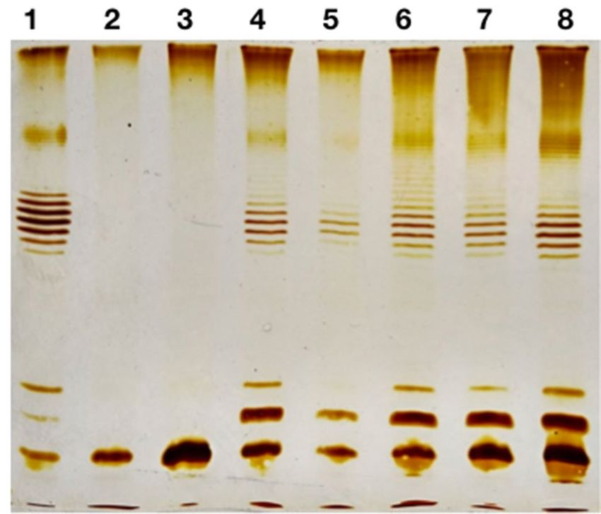
www.nature.com/scientificreports/
Figure 1. LPS profiles of the E. coli strains used and their derivative ϕ24B:cat lysogens. The wt lane on each of
the panels—the wild type cells, lanes 1–3—three lysogenic clones for each strain.
The lysogenization frequency was about 1 0−4 lysogen cfu/phage pfu for the rough strains and about 1 0−7–10−6
in O antigen-producing strains. The latter value is comparable to the frequency of spontaneous mutations by a
We selected 3 lysogen clones per strain and confirmed the ϕ24B prophage presence using PCR for gene 56
particular gene in E. coli (e.g. phage-resistant mutants).
sion electron microscopy that confirmed that a phage morphologically identical to ϕ24B was produced (Sup-
(the tail protein gene). For E. coli 4s lysogens we also performed mitomycin C induction followed by transmis-
plementary file: Fig. S1).
LPS profiling of the lysogens obtained indicated that in all cases these strains did not produce O-antigen at
all or the O-chain synthesis was greatly decreased compared to the parental strains (Fig. 1).
The bacteriophages that are potentially able to infect the strain but are restrained by its O-antigen can be
successfully used as a probe for testing the efficacy of the O-antigen-mediated p rotection31. Previously we have
developed the use of a T5-like bacteriophage DT571/2 mutant FimX lacking lateral tail fibers (LTFs) as such a
probe30. We tested the ability of phage FimX to grow on the lawns of the lysogens obtained. This phage was not
able to form plaques on the parental O-antigen—producing strains, except for F5 on which it formed plaques with
an efficiency of plating (EOP) of 10−4 compared to the C600 strain used for FimX propagation35. At the same time
the EOP of FimX phage on all the lysogenic cultures tested was in the range of 0.1–1.0 compared to the E. coli
C600 strain. The gain of sensitivity to the phage FimX observed after the lysogenization was undistinguishable
from other methods of rough mutant generation previously used by us in 4s or F17 strains29,31.
The other T5-like phages (DT57C, DT571/2, ABF and Gostya9) as well as the siphovirus 9 g demonstrated
the infectivity on the lysogens derivatives of some strains that were initially resistant to these phages (Table. 1).
Phage G7C that is dependent on the specific O antigen recognition for infection of E. coli 4s c ells30 was not able
to infect E. coli 4s (ϕ24B:cat) lysogenic strains in good agreement with O antigen production loss detected by
the LPS profiling (Fig. 1).
We concluded that the lysogenization of the wild O-antigen-producing strains of E. coli is associated with the
loss or reduced amount of the O antigen. The most probable explanation of this effect is that the phage infects
and lysogenizes naturally occurring rough mutants that are normally present in the E. coli cultures (see refs 29
and 31 and the literature cited therein). However, an alternative explanation that the prophage acquisition leads
to severe impairment of the O antigen production remains possible.
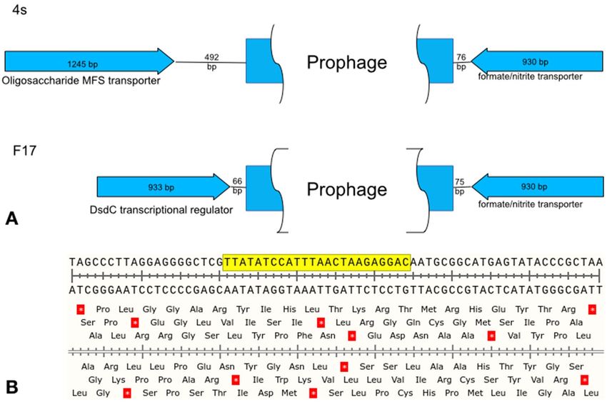
To further address this question, we extracted the DNA from the lysogens obtained on the strains 4s and F17
and submitted it for the whole genome using Oxforde Nanopore MiniIon sequencer. The contigs were assembled
and the prophage integration sites were determined. In both strains the prophage was integrated by a conserved
attB sequence located in the spacer between two oppositely directed genes (Fig. 2). These genes are not involved
into the O antigen biosynthesis pathway and are located at long distances (345 kbp and 336 kbp in 4s and F17
strains respectively) from the O antigen clusters previously described for these strains29,36. Thus the prophage
integration itself does not directly break the genes or transcription units responsible for the O antigen synthesis.
However, it is possible that the prophage interferes with this process at the metabolic level. To test such
possibility we used the complementation system previously developed by us in E. coli 4s strain. In this strain a
rough mutant, refered to as 4sR, was described. The rough phenotype of the 4sR strain was due to the gene wclH
inactivation by the insertion of an IS1-like mobile element, and the complementation by the expression of wclH
the phage ϕ24B and transformed the lysogen by the pWclH plasmid. The O antigen production status was then
gene from the plasmid was shown to restore O-antigen producing p henotype29. We lysogenized E. coli 4sR with
the parental 4sR strain and in all three lysogenic clones tested. We concluded that the ϕ24B prophage does not
assessed by LPS profiling (Fig. 3). The LPS profile, similar to the wild type E. coli 4s strain was restored both in
significantly interfere with the O antigen biosynthesis. Thus, the rough phenotype is not induced by the prophage
acquisition but but is selected by the lysogenization procedure.
Since the O antigen synthesis compromised strains are believed to be more vulnerable to immunity factors,
we decided to measure the susceptibility of the lysogens obtained to the bactericidal activity of the horse serum
Scientific Reports | (2021) 11:3035 | https://doi.org/10.1038/s41598-021-82422-x 3
Vol.:(0123456789)www.nature.com/scientificreports/
E. coli strains
HS3-104 4s F5 F17 UP1
Phages wt Lysogens wt Lysogens wt Lysogens wt Lysogens wt Lysogens
DT57C + + + + +/− + − + − +
DT571/2 + + − + +/− + − + − +
fimX − + − + +/− + − + − +
ABF + + − + +/− + − + − +
Gostya9 − + − + + + − + − −
G7C − − + − − − − − − −
T5 − + − + − − − + − +
9g − + − + + + + + − +
Table 1. Sensitivity of the E. coli strains and their derivative ϕ24B:cat lysogens to virulent coliphages. “+”—the
phage plaques are formed at the EOP > 0.1 in respect to plating on the optimal host strain, “−”—no plaque
formation observed, “+/−”—very small turbid plaques with the EOP < 10−3.
Figure 2. (A) The genetic organization of the chromosomal locus of phi24B prophage integration in the E. coli
strains 4s and F17. (B) The nucleotide sequence of the conserved integration site. The yellow box—the sequence
that is duplicated during the integration to flank the prophage in the lysogens.
(SBA). All the wild type strains were resistant to SBA in our conditions (Fig. 4). Their cultures grew in presence
of the serum as well or even slightly more rapidly than in the control experiment. In the absence of the serum the
lysogenic strains showed the growth rates close to their cognate wild type strains. At the same time the growth
of the lysogens was almost completely abolished in the presence of the serum (Fig. 4).
Only one of the lysogenic clones tested, the derivative of the strain HS3-104, was able to grow significantly
in presence of the horse serum, though the rise of the optical density was delayed and the growth rate was
significantly lower than in the parental strain (Fig. 2). This result can be explained by the fact that in HS3-104
lysogens the O antigen synthesis was strongly decreased but not completely abolished (Fig. 1). So, the actual
synthesis of O-polysaccharide could be upregulated in this particular clone in the conditions of the experiment
of SBA sensitivity measurement.
Scientific Reports | (2021) 11:3035 | https://doi.org/10.1038/s41598-021-82422-x 4
Vol:.(1234567890)www.nature.com/scientificreports/
Figure 3. Complementation of the wclH mutation from the plasmid on phi24B lysogens background. Lanes
1—E. coli 4s wild type, 2—4sR, 3—4sR (phi24B), 4—4sR:pWclH, 5—the same as lane 4 with less sample load,
6—8 three clones of 4sR(phi24B):pWclH.
The results obtained allow us to conclude that lysogenization by the phage ϕ24B of diverse E. coli strains produc-
Discussion
ing O antigens was not due to an unusual ability of this virus to penetrate the O antigen shield, but was mediated
by spontaneous formation of bacterial rough mutants or of mutants with significantly compromised O antigen
biosynthesis. It is not clear why the lysogenization was not effective for some strains. The activity of antiviral
systems, such as restriction-modification, avoiding the lysogenization at stages after the viral DNA penetration
into the cell46, cannot be excluded. Also, the effect may be due to point mutations present in BamA protein or
lower frequency of rough mutants in particular strains.
In the conditions of our experiment, the high concentration of bacteriophage used allowed almost all the cells
potentially susceptible to the phage to be infected. However, in vivo the populations of E. coli are very unlikely to
face such a massive viral attack. The fraction of rough mutants in natural habitats is hard to estimate, but we can
speculate that it should be lower than in in vitro conditions because such mutants have compromised protection
not only from the phage attack but also from immune system agents such as serum bactericidal a ctivity47–49 and
from other environmental f actors32 and therefore should be counter-selected. Moreover, if Stx-phage lysogens
were formed by infection of such rough mutants, their expected fitness and/or virulence would be significantly
lower than that of the parental strains. These strains, noteworthily, were highly sensitive to SBA of the horse
for the ϕ24B phage are expected to have reduced virulence. Thus, the factor of non-specific protection of the
serum to which the parental O-antigen producing strains were completely resistant. Therefore, the lysogens
bacterial cells by the O antigen should not be neglected during the evaluation of the potential significance of
We also should note that the lysogenization by ϕ24B:cat appears to be a simple and efficient procedure for
Stx-converting phage transmission in nature (as it currently is neglected in many studies25,26).
selection for mutants with compromised or completely abolished O antigen synthesis. This procedure may be
particularly valuable for the researchers working with field isolates of E. coli for which the genomic sequences are
not yet available and/or in which other rapid techniques such as recombination with PCR fragments for genes
knockout50 are frequently less effective than in laboratory E. coli.
Methods
phage ϕ24B:cat was a kind gift of Prof. G. Wegrzyn, University of Gdansk, Poland. Phage T5 was a gift of Dr. V.
E. coli and bacteriophage strains and their cultivation. The E. coli strain MG1655 lysogenized for
Ksenzenko (Institute of protein research RAS, Puschino-na-Oke, Russia). We previously described T5-like bac-
teriophages of DT57C species and their LTF mutants30. These include: phage DT57C, phage DT571/2, DT571/2
ltfA− mutant lacking the LTFs (hereafter FimX) and DT571/2 mutant ABF that carries LTF non-branched LTF
with only one receptor-binding domain (instead of two such domains on the branched LTFs of the phages
DT57C or DT571/2). Bacteriophage 9 g, a siphovirus representing the type strain of the genus Nonagvirus40.
Gostya9 is a T5-like bacteriophage that was shown to recognize a different secondary receptor distinct from
the receptors of the phages T5, DT57C and 9 g41. Bacteriophage G7C, a N4-related podovirus specifically rec-
ognizing O antigen of E. coli 4s strain was isolated and characterized by us previously42,43. We isolated all the
above-mentioned phages except for T5 and engineered phage mutants from horse feces as it described in the
corresponding publications cited above.
The wild E. coli strains were previously isolated by us from horse feces and characterized. These were
4s (O22)29, HS1/2 (O87)37,44, HS3-104 (O81)39, F5 (O28 ab)35, and F17 (new O-serotype)36. The clinical
Scientific Reports | (2021) 11:3035 | https://doi.org/10.1038/s41598-021-82422-x 5
Vol.:(0123456789)www.nature.com/scientificreports/
Figure 4. Sensitivity of the E. coli strains and their derivative ϕ24B:cat lysogens’ growth to the horse serum
bactericidal activity. Black lines—the wild type strain, grey lines—three lysogenic clones tested for each original
strain.
Scientific Reports | (2021) 11:3035 | https://doi.org/10.1038/s41598-021-82422-x 6
Vol:.(1234567890)www.nature.com/scientificreports/
uropathogenic E. coli isolates UP1 and UP11 were received from the clinical microbiological facility of the
Institute of Epidemiology (Moscow, Russia). UP11 strain was further identified as an O5 O-antigen p roducer34.
The ability of the strains to produce O antigens was controlled by LPS profiling as described in Kulikov et al.
(2019)31.
E. coli 4s and F17 rough variants 4sR (a wclH mutant of 4s)29 and F17 wbbL−36 were engineered by us
previously.
All the E. coli strains were cultured on LB medium (trypton 10 g, yeast extract 5 g, NaCl—10 g, distilled
H2O—up to 1 l). This medium was supplemented with 15 g of bacto-agar per 1 l for plates or with 6 g of bacto-
agar for top agar.
Bacteriophage FimX was propagated on E. coli 4sR and enumerated using the conventional double-layer
Bacteriophage ϕ24B:cat was obtained by mitomycin C induction of E. coli MG1655 (ϕ24B:cat) strain. For
plating technique.
this procedure, the overnight culture of the lysogen was grown in the presence of 34 μg/ml of chloramphenicol.
Then 300 ml of LB in 500 ml Erlenmeyer flask was inoculated with 3 ml of the overnight culture (N.B.—this
volume ratio gave a better phage yield than conventional conditions with better aeration). The culture was grown
in the orbital shaker at 220 rpm, 37 °C up to OD600 = 0.2. The mitomycin C was then added up to 1 μg/mL and
the incubation was continued overnight at the same conditions. After the incubation, lysis of the culture was
observed. The lysate was cleared by centrifugation at 15,000 × g for 15 min. The supernatant was collected, PEG-
precipitated45, and resuspended in 3 mL of SM buffer (Tris–HCl pH 7.5–10 mM, NaCl—50 mM, M gCl2—10 mM,
For titration of the phage ϕ24B:cat, a modified double-layer technique was used. The top-layer medium con-
gelatin—5 g/l). The phage stock was titered and used in these experiments.
tained 4 g/l of the bacto-agar (instead of 6 g/l) and was supplemented with C aCl2 up to 5 mM. The bottom layer
was supplemented with 2.5 μg/ ml of chloramphenicol. 300 μg of log-phase culture of E. coli C600 (OD600 = 0.6)
was used for the lawn inoculation.
Lysogenization of the E. coli strains. This procedure was performed as described in James et al.23 with
minor modifications. Briefly, a mid-log liquid culture of an appropriate strain was grown in LB medium, the
phage was added at a multiplicity of 5 pfu/host cfu, and the mixture was incubated at 37 °C for 30 min. After
the incubation, the cells were spun down in a table-top centrifuge (10,000×g, 1 min), the cells were resuspended
in LB, washed twice with LB to remove non-bound phage and plated on plates supplemented with 34 μg/ml of
chloramphenicol for lysogen selection.
Sequencing of the bacterial genomic DNA. DNA was extracted using the Wizard DNA extraction kit (Promega
Corporation, USA). The sequencing was performed with MinION sequencing (Oxford Nanopore Technologies,
UK). The sequencing libraries were prepared using the ligation sequencing kit (catalog number SQK-LSK109)
and native barcoding expansion kit (catalog number EXP-NBD114) and run in a FLO-MIN106 flow cell. Reads
were base called trimmed and demultiplexed using Guppy v. 3.2.5. The contigs were assembled with Flye 2.5.
LPS profiling. By SDS-PAGE electrophoresis was performed as recently d escribed31. The lysogens cultures
were grown for this prodecure on the LB medium without antibiotic to avoid possible chloramphenicol-induced
alterations of the LPS synthesis.
Serum bactericidal activity (SBA). Against different strains was measured as follows. The blood samples
obtained from clinically healthy horses were used. These samples were collected by a qualified veterinary doc-
tor for the the animal healthcare purposes unrelated to our study, and we used the excess of the serum in our
experiments. According to the local legislation this is not considered as the experiment on animals and does not
require specific ethical approval. Therefore, we formally confirm that all invasive or non-invasive experimental
protocols involving experimental animals were carried out with strict adherence to Russian legislation in this
area, and in complete accordance with the current regulation status. The samples were collected into the yellow-
cap vacuum tubes with the clot activator (Elamed, Moscow, Russia). The serum was separated by centrifugation
at 1600×g for 10 min. The serum was stored at + 4 °C and used for the tests within 24 h. For the SBA assessment,
the wells of 96-well plate containing 175 μl of LB medium and 25 μl of the serum were inoculated with 5 μl of the
corresponding strain mid-log phase culture ( OD600 = 0.6) and the plates were incubated at 37 °C in an automated
plate reader with agitation. The OD600 was recorded every 30 min. In the control experiment the same volume of
physiological saline replaced the serum. The whole experiment was triplicated.
Received: 21 April 2020; Accepted: 18 January 2021
References
1. Cody, E. M. & Dixon, B. P. Hemolytic uremic syndrome. Pediatr. Clin. North Am. 66, 235–246. https://doi.org/10.1016/j.
pcl.2018.09.011 (2019).
2. Chiang, Y. N., Penades, J. R. & Chen, J. Genetic transduction by phages and chromosomal islands: The new and noncanonical.
PLoS Pathog 15, e1007878. https://doi.org/10.1371/journal.ppat.1007878 (2019).
3. Penades, J. R. & Christie, G. E. The phage-inducible chromosomal islands: A family of highly evolved molecular parasites. Annu.
Rev. Virol. 2, 181–201. https://doi.org/10.1146/annurev-virology-031413-085446 (2015).
Scientific Reports | (2021) 11:3035 | https://doi.org/10.1038/s41598-021-82422-x 7
Vol.:(0123456789)www.nature.com/scientificreports/
4. Valilis, E., Ramsey, A., Sidiq, S. & DuPont, H. L. Non-O157 Shiga toxin-producing Escherichia coli-A poorly appreciated enteric
pathogen: Systematic review. Int. J. Infect Dis. 76, 82–87. https://doi.org/10.1016/j.ijid.2018.09.002 (2018).
5. Murinda, S. E. et al. Shiga toxin-producing Escherichia coli in mastitis: An international perspective. Foodborne Pathog. Dis. 16,
229–243. https://doi.org/10.1089/fpd.2018.2491 (2019).
6. Feiner, R. et al. A new perspective on lysogeny: prophages as active regulatory switches of bacteria. Nat. Rev. Microbiol. 13, 641–650.
https://doi.org/10.1038/nrmicro3527 (2015).
7. Bikard, D. & Marraffini, L. A. Innate and adaptive immunity in bacteria: mechanisms of programmed genetic variation to fight
bacteriophages. Curr. Opin. Immunol. 24, 15–20. https://doi.org/10.1016/j.coi.2011.10.005 (2012).
8. Hoskisson, P. A. & Smith, M. C. Hypervariation and phase variation in the bacteriophage “resistome”. Curr. Opin. Microbiol. 10,
396–400. https://doi.org/10.1016/j.mib.2007.04.003 (2007).
9. De Ste Croix, M. et al. Phase-variable methylation and epigenetic regulation by type I restriction-modification systems. FEMS
Microbiol. Rev. 41, S3–S15. https://doi.org/10.1093/femsre/fux025 (2017).
10. Heredia, N. & Garcia, S. Animals as sources of food-borne pathogens: A review. Anim. Nutr. 4, 250–255. https://doi.org/10.1016/j.
aninu.2018.04.006 (2018).
11. Fatima, R. & Aziz, M. in StatPearls (2019).
12. Mellor, G. E. et al. National Survey of Shiga Toxin-Producing Escherichia coli Serotypes O26, O45, O103, O111, O121, O145, and
O157 in Australian Beef Cattle Feces. J. Food Prot. 79, 1868–1874. https://doi.org/10.4315/0362-028X.JFP-15-507 (2016).
13. Kampmeier, S., Berger, M., Mellmann, A., Karch, H. & Berger, P. The 2011 German enterohemorrhagic Escherichia coli O104:H4
outbreak-the danger is still out there. Curr. Top. Microbiol. Immunol. 416, 117–148. https://doi.org/10.1007/82_2018_107 (2018).
14. Lee, M. S. & Tesh, V. L. Roles of shiga toxins in immunopathology. Toxins (Basel) https://doi.org/10.3390/toxins11040212 (2019).
15. Schmidt, H. Shiga-toxin-converting bacteriophages. Res. Microbiol. 152, 687–695. https://doi.org/10.1016/s0923-2508(01)01249
-9 (2001).
16. Herold, S., Karch, H. & Schmidt, H. Shiga toxin-encoding bacteriophages–genomes in motion. Int. J. Med. Microbiol. 294, 115–121.
https://doi.org/10.1016/j.ijmm.2004.06.023 (2004).
17. Chakraborty, D., Clark, E., Mauro, S. A. & Koudelka, G. B. Molecular mechanisms governing “hair-trigger” induction of shiga
toxin-encoding prophages. Viruses https://doi.org/10.3390/v10050228 (2018).
18. Bloch, S. et al. Inhibition of Shiga toxin-converting bacteriophage development by novel antioxidant compounds. J. Enzyme Inhib.
Med. Chem. 33, 639–650. https://doi.org/10.1080/14756366.2018.1444610 (2018).
19. Fang, Y., Mercer, R. G., McMullen, L. M. & Ganzle, M. G. Induction of Shiga Toxin-Encoding Prophage by Abiotic Environmental
Stress in Food. Appl. Environ. Microbiol. https://doi.org/10.1128/AEM.01378-17 (2017).
20. Smith, D. L. et al. Comparative genomics of Shiga toxin encoding bacteriophages. BMC Genomics 13, 311. https://doi.
org/10.1186/1471-2164-13-311 (2012).
21. Kakoullis, L., Papachristodoulou, E., Chra, P. & Panos, G. Shiga toxin-induced haemolytic uraemic syndrome and the role of
antibiotics: A global overview. J. Infect. 79, 75–94. https://doi.org/10.1016/j.jinf.2019.05.018 (2019).
22. Kavanagh, D., Raman, S. & Sheerin, N. S. Management of hemolytic uremic syndrome. F1000Prime Rep 6, 119. https://doi.
org/10.12703/P6-119 (2014).
23. James, C. E. et al. Lytic and lysogenic infection of diverse Escherichia coli and Shigella strains with a verocytotoxigenic bacterio-
phage. Appl. Environ. Microbiol. 67, 4335–4337. https://doi.org/10.1128/aem.67.9.4335-4337.2001 (2001).
24. Smith, D. L. et al. Multilocus characterization scheme for shiga toxin-encoding bacteriophages. Appl. Environ. Microbiol. 73,
8032–8040. https://doi.org/10.1128/AEM.01278-07 (2007).
25. Eichhorn, I. et al. Lysogenic conversion of atypical enteropathogenic Escherichia coli (aEPEC) from human, murine, and bovine
origin with bacteriophage Phi3538 Deltastx2::cat proves their enterohemorrhagic E. coli (EHEC) progeny. Int. J. Med. Microbiol.
308, 890–898. https://doi.org/10.1016/j.ijmm.2018.06.005 (2018).
26. Khalil, R. K., Skinner, C., Patfield, S. & He, X. Phage-mediated Shiga toxin (Stx) horizontal gene transfer and expression in non-
Shiga toxigenic Enterobacter and Escherichia coli strains. Pathog. Dis. https://doi.org/10.1093/femspd/ftw037 (2016).
27. Smith, D. L. et al. Short-tailed Stx phages exploit the conserved YaeT protein to disseminate Shiga toxin genes among enterobacteria.
J. Bacteriol. 189, 7223–7233. https://doi.org/10.1128/JB.00824-07 (2007).
28. Botos, I., Noinaj, N. & Buchanan, S. K. Insertion of proteins and lipopolysaccharide into the bacterial outer membrane. Philos.
Trans. R. Soc. Lond. Ser. B Biol. Sci. https://doi.org/10.1098/rstb.2016.0224 (2017).
29. Knirel, Y. A. et al. Variations in O-antigen biosynthesis and O-acetylation associated with altered phage sensitivity in Escherichia
coli 4s. J. Bacteriol. 197, 905–912. https://doi.org/10.1128/JB.02398-14 (2015).
30. Golomidova, A. K. et al. Branched lateral tail fiber organization in T5-like bacteriophages DT57C and DT571/2 is revealed by
genetic and functional analysis. Viruses https://doi.org/10.3390/v8010026 (2016).
31. Kulikov, E. E., Golomidova, A. K., Prokhorov, N. S., Ivanov, P. A. & Letarov, A. V. High-throughput LPS profiling as a tool for
revealing of bacteriophage infection strategies. Sci. Rep. 9, 2958. https://doi.org/10.1038/s41598-019-39590-8 (2019).
32. van der Ley, P., de Graaff, P. & Tommassen, J. Shielding of Escherichia coli outer membrane proteins as receptors for bacteriophages
and colicins by O-antigenic chains of lipopolysaccharide. J. Bacteriol. 168, 449–451. https: //doi.org/10.1128/jb.168.1.449-451.1986
(1986).
33. Kunisaki, H. & Tanji, Y. Intercrossing of phage genomes in a phage cocktail and stable coexistence with Escherichia coli O157:H7
in anaerobic continuous culture. Appl. Microbiol. Biotechnol. 85, 1533–1540. https://doi.org/10.1007/s00253-009-2230-2 (2010).
34. Golomidova, A. K., Kulikov, E. E., Babenko, V. V., Kostryukova, E. S. & Letarov, A. V. Complete genome sequence of bacterio-
phage St11Ph5, which Infects uropathogenic Escherichia coli strain up11. Genome Announcements https://doi.org/10.1128/genom
eA.01371-17 (2018).
35. Golomidova, A. K., Naumenko, O. I., Senchenkova, S. N., Knirel, Y. A. & Letarov, A. V. The O-polysaccharide of Escherichia coli
F5, which is structurally related to that of E. coli O28ab, provides cells only weak protection against bacteriophage attack. Arch.
Virol. 164, 2783–2787. https://doi.org/10.1007/s00705-019-04371-1 (2019).
36. Knirel, Y. A. et al. Structure and gene cluster of the O antigen of Escherichia coli F17, a candidate for a new O-serogroup. Int. J.
Biol. Macromol. 124, 389–395. https://doi.org/10.1016/j.ijbiomac.2018.11.149 (2019).
37. Zdorovenko, E. L. et al. Structure of the O-polysaccharide of Escherichia coli O87. Carbohyd. Res. 412, 15–18. https://doi.
org/10.1016/j.carres.2015.04.014 (2015).
38. Zdorovenko, E. L. et al. Corrigendum to “Structure of the O-polysaccharide of Escherichia coli O87” [Carbohydr. Res. 412 (2015)
15–18]. Carbohydrate Res. 464, 1. https://doi.org/10.1016/j.carres.2018.04.013 (2018).
39. Zdorovenko, E. L. et al. O-Antigens of Escherichia coli strains O81 and HS3–104 are structurally and genetically related, except
O-Antigen glucosylation in E. coli HS3–104. Biochemistry 83, 534–541. https://doi.org/10.1134/S0006297918050061 (2018).
40. Kulikov, E. E. et al. Genomic sequencing and biological characteristics of a novel Escherichia coli bacteriophage 9g, a putative
representative of a new Siphoviridae genus. Viruses 6, 5077–5092. https://doi.org/10.3390/v6125077 (2014).
41. Golomidova, A. K. et al. Escherichia coli bacteriophage Gostya9, representing a new species within the genus T5virus. Adv. Virol.
164, 879–884. https://doi.org/10.1007/s00705-018-4113-2 (2019).
42. Kulikov, E. et al. Isolation and characterization of a novel indigenous intestinal N4-related coliphage vB_EcoP_G7C. Virology 426,
93–99. https://doi.org/10.1016/j.virol.2012.01.027 (2012).
Scientific Reports | (2021) 11:3035 | https://doi.org/10.1038/s41598-021-82422-x 8
Vol:.(1234567890)www.nature.com/scientificreports/
43. Prokhorov, N. S. et al. Function of bacteriophage G7C esterase tailspike in host cell adsorption. Mol. Microbiol. 105, 385–398. https
://doi.org/10.1111/mmi.13710(2017).
44. Zdorovenko, E. L. et al. Corrigendum to “Structure of the O-polysaccharide of Escherichia coli O87”. Carbohydrate Res. 412, 15–18.
https://doi.org/10.1016/j.carres.2018.04.013 (2015).
45. Sambrook, J., Fritsch, E. F. & Maniatis, T. Molecular Cloning: A Laboratory Manual (Cold Spring Harbor Laboratory Press, New
York, 1989).
46. Samson, J. E., Magadan, A. H., Sabri, M. & Moineau, S. Revenge of the phages: Defeating bacterial defences. Nat. Rev. Microbiol.
11, 675–687. https://doi.org/10.1038/nrmicro3096 (2013).
47. Pawlak, A. et al. Salmonella O48 serum resistance is connected with the elongation of the lipopolysaccharide O-Antigen containing
sialic acid. Int. J. Mol. Sci. https://doi.org/10.3390/ijms18102022 (2017).
48. Coggon, C. F. et al. A novel method of serum resistance by Escherichia coli that causes urosepsis. mBio https://doi.org/10.1128/
mBio.00920-18 (2018).
49. Kintz, E. et al. Salmonella enterica Serovar Typhi lipopolysaccharide O-antigen modification impact on serum resistance and
antibody recognition. Infect. Immunity https://doi.org/10.1128/IAI.01021-16 (2017).
50. Datsenko, K. A. & Wanner, B. L. One-step inactivation of chromosomal genes in Escherichia coli K-12 using PCR products. Proc.
Natl. Acad. Sci. U.S.A. 97, 6640–6645. https://doi.org/10.1073/pnas.120163297 (2000).
Acknowledgements
Poland for their help in establishing the procedures of phage ϕ24B cultivation in our lab and to Dr. E. M. Kutter
We are grateful to Dr. S. Bloch, Dr. B. Nejman-Faleńczyk and Prof. G. Wegrzyn from the University of Gdansk,
from the Evergreen state college, Olympia, USA, for critical reading and linguistic correction of the early ver-
sion of the manuscript.
Author contributions
A.L. developed the concept of the study, analysed the data and drafted the manuscript, A.G., E.K., A.E, I.B. and A.
K. performed the experiments, A.G. prepared figure 1 and Table 1., I.B. prepared figure 2, A.K. prepared figure 3,
A.E. prepared figure 4., E.K. prepared figure S1, All the authors reviewed the manuscript.
Competing interests
The authors declare no competing interests.
Additional information
Supplementary Information The online version contains supplementary material available at https://doi.
org/10.1038/s41598-021-82422-x.
Correspondence and requests for materials should be addressed to A.V.L.
Reprints and permissions information is available at www.nature.com/reprints.
Publisher’s note Springer Nature remains neutral with regard to jurisdictional claims in published maps and
institutional affiliations.
Open Access This article is licensed under a Creative Commons Attribution 4.0 International
License, which permits use, sharing, adaptation, distribution and reproduction in any medium or
format, as long as you give appropriate credit to the original author(s) and the source, provide a link to the
Creative Commons licence, and indicate if changes were made. The images or other third party material in this
article are included in the article’s Creative Commons licence, unless indicated otherwise in a credit line to the
material. If material is not included in the article’s Creative Commons licence and your intended use is not
permitted by statutory regulation or exceeds the permitted use, you will need to obtain permission directly from
the copyright holder. To view a copy of this licence, visit http://creativecommons.org/licenses/by/4.0/.
© The Author(s) 2021
Scientific Reports | (2021) 11:3035 | https://doi.org/10.1038/s41598-021-82422-x 9
Vol.:(0123456789)You can also read