Signaling Pathways in Proton and Non-proton ASIC1a Activation
←
→
Page content transcription
If your browser does not render page correctly, please read the page content below
ORIGINAL RESEARCH
                                                                                                                                         published: 05 October 2021
                                                                                                                                    doi: 10.3389/fncel.2021.735414

                                           Signaling Pathways in Proton and
                                           Non-proton ASIC1a Activation
                                           Libia Catalina Salinas Castellanos, Osvaldo Daniel Uchitel and Carina Weissmann*

                                           Instituto de Fisiología, Biología Molecular y Neurociencias (IFIBYNE—UBA CONICET), Facultad de Ciencias, Exactas y
                                           Naturales de la Universidad de Buenos Aires, Buenos Aires, Argentina

                                           Acid-sensing ion channels (ASICs) regulate synaptic activities and play important roles in
                                           neurodegenerative diseases as well as pain conditions. Classically, ASICs are described
                                           as transiently activated by a reduced pH, followed by desensitization; the activation
                                           allows sodium influx, and in the case of ASIC1a-composed channels, also calcium to
                                           some degree. Several factors are emerging and extensively analyzed as modulators,
                                           activating, inhibiting, and potentiating specific channel subunits. However, the signaling
                                           pathways triggered by channel activation are only starting to be revealed. The channel
                                           has been recently shown to be activated through a mechanism other than proton-
                                           mediated. Indeed, the large extracellular loop of these channels opens the possibility
                                           that other non-proton ligands might exist. One such molecule discovered was a toxin
                            Edited by:
                 Josef Bischofberger,      present in the Texas coral snake venom. The finding was associated with the activation
      University of Basel, Switzerland     of the channel at neutral pH via the toxin and causing intense and unremitting pain. By
                     Reviewed by:          using different pharmacological tools, we analyzed the downstream signaling pathway
                      Eric Gonzales,
TCU & UNTHSC School of Medicine,
                                           triggered either by the proton and non-proton activation for human, mouse, and rat
                       United States       ASIC1a-composed channels in in vitro models. We show that for all species analyzed,
                      Lachlan Rash,        the non-protonic mode of activation determines the activation of the ERK signaling
      The University of Queensland,
                            Australia      cascade at a higher level and duration compared to the proton mode. This study adds to
                 *Correspondence:          the growing evidence of the important role ASIC1a channels play in different physiological
                  Carina Weissmann         and pathological conditions and also hints at a possible pathological mechanism for a
       carina.weissmann@gmail.com
                                           sustained effect.
                   Specialty section:
        This article was submitted to      Keywords: ASIC1a, proton activation, non-proton activation, ERK, MitTx, pain
             Cellular Neurophysiology,
                a section of the journal
    Frontiers in Cellular Neuroscience
                                           INTRODUCTION
                                           ASICs, also called proton-gated channels belong to the degenerin/epithelial Na+ channel gene
           Received: 02 July 2021
                                           family (Boscardin et al., 2016). Five genes encode at least seven ASIC subtypes in rodents and
     Accepted: 09 September 2021
       Published: 05 October 2021          humans, and three subunits constitute a functional unit in either homotrimeric or heterotrimeric
                                           structures (Boscardin et al., 2016). These channels are primarily expressed in the nervous system
                            Citation:
                                           (Zha, 2013) and linked to several physiological (Uchitel et al., 2019) and pathological conditions
  Salinas Castellanos LC, Uchitel OD
  and Weissmann C (2021) Signaling         (Chu and Xiong, 2013), thus different pharmacological tools have been developed as potential
 Pathways in Proton and Non-proton         therapeutic treatments.
                   ASIC1a Activation.
   Front. Cell. Neurosci. 15:735414.       Abbreviations: ASIC, Acid Sensing Ion Chanel; eASIC, eGFP-ASIC1a; eASICx1 or eASICx3, eGFP-ASIC1a expressed at
    doi: 10.3389/fncel.2021.735414         a single or triple-level; ERK, extracellular signal-regulated kinase; CaMKII, Calcium/Calmodulin kinase II.

Frontiers in Cellular Neuroscience | www.frontiersin.org                          1                                     October 2021 | Volume 15 | Article 735414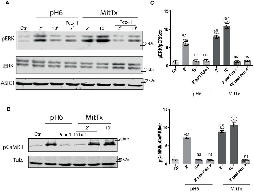
Salinas Castellanos et al.                                                                                 Downstream Signaling in ASIC1a Activation

    ASICs are Na+ -selective ion channels, and ASIC1a,—a key                     The ERK kinase belongs to the family of mitogen-activated
subunit in the central nervous system (Wang et al., 2016)—,                  protein kinases (MAPK) that operate within signaling cascades
show, in addition to its Na+ permeability, a small permeability              (Maik-Rachline et al., 2019). The activation of this pathway
to Ca2+ (Gründer and Chen, 2010). ASIC1a has been linked to                  and the duration of the activation (Marshall, 1995; Kriegsheim
neurodegenerative diseases (Friese et al., 2007; Wong et al., 2008;          et al., 2009) can lead to different biological responses that can
Sluka et al., 2009; Sun et al., 2011), ischemia (Xiong and Xu,               determine the fate of a cell. In addition, the activation of the
2012), and pain (Duan et al., 2007; Wemmie et al., 2013; Fan et al.,         pathway has been implicated in pain research. Activation of the
2018). The unique permeability to calcium compared with other                kinase via phosphorylation in the dorsal root ganglia has been
subunits makes ASIC1a a candidate to play a prominent role in                linked to different models of pain in animals (Cruz and Cruz,
neuronal death (Hoagland et al., 2010).                                      2007; Maruta et al., 2019). Different levels of activation of ERK
    Under experimental conditions, ASICs are activated only by               were distinguished in response to acute noxious stimulation or
rapid pH drops, and, particularly homomeric ASIC1a channels                  chronic noxious stimulation by Cruz and Cruz (2007), with more
desensitize rapidly in the continuous presence of acidic pH (Chu             intense levels of ERK phosphorylation and longer duration in
and Xiong, 2013). This fact remains puzzling, as to whether                  animals with chronic inflammation of the hind paw or joint. In
a significant amount of ASIC1a current can be activated in                   the study, spinal ERK activation was upregulated and became
pathological conditions, and as to whether the effect of its                 persistent (Cruz and Cruz, 2007).
activation could be long-lasting (Chu and Xiong, 2013; Tikhonov                  In this study, we aim to address aspects of the downstream
et al., 2019; Alijevic et al., 2020); thus the functional significance       effects triggered by non-proton activation of ASIC1a channels.
of these channels remains to be determined. As pointed out
by Zha (2013), although the canonical ligands for ASICs are                  MATERIALS AND METHODS
protons, the massive extracellular domain of ASICs has led
to the speculation that these receptors may also respond to                  Cellular and Molecular Biology
other ligands (Zha, 2013) like MitTx purified from the venom                 Human embryonic kidney 293 (HEK) cells [passage 18–26,
of the Texas coral (Kweon and Suh, 2013). The toxin has                      American Type Culture Collection (ATCC) number CRL-1573]
been instrumental to document ASIC1a channels in an open                     were maintained by serial passages. Primary striatal cultures
state (Baconguis et al., 2014). MitTx elicits robust pain-related            were prepared from mice of the C57BL/6 genetic background
behavior in mice via activation of ASIC1 channels on capsaicin-              as control and ASIC1a−/− mice (generated using mice of the
sensitive nerve fibers (Bohlen et al., 2012).                                C57BL/6 genetic background) were provided by the laboratory
    Gautschi et al. (2017) on the unresolved question of the                 of Dr. John A. Wemmie (University of Iowa, Iowa City, IA)
unphysiological pH values used to activate the channels and the              as used before (González-Inchauspe et al., 2017) and prepared
transient nature of the proton evoked ASIC current, described                according to the protocol used in Sodero et al. (2011). All
another type of activation other than acid, as a ‘‘non-proton’’              experiments involving mice were performed following national
mechanism (exemplified by MitTx), that activated a large                     guidelines for the humane treatment of laboratory animals from
sustained and non-desensitizing current at neutral pH and                    the University of Buenos Aires (CICUAL Protocol #112), which
exceeding in magnitude the maximal current evoked by the                     are comparable to those of the USA National Institutes of Health.
proton mode (Gautschi et al., 2017).                                         For biochemical analysis, six or 12-plates were coated with
    Many studies have focused on the mechanism regulating the                0.1 mg/mL of poly-L-lysine (PLL, Sigma, P2636), and dissociated
trafficking of the channel (Zeng et al., 2014; Boscardin et al., 2016;       neuronal and HEK cells were plated at a density of 2.2 × 10⁵ or
Wu et al., 2016), leading to changes in the amount of channel                1.4 × 10⁵ cells respectively. HEK cells were grown in Dulbecco’s
at the plasma membrane. The downstream signaling of ASIC                     Modified Eagle’s Medium containing 4 mM L-glutamine, 4.5 g/L
channels, however, is only starting to be documented.                        glucose, and 110 ml/L sodium pyruvate and supplemented with
    As an example, the activation of ERK via ASIC1a                          10% Fetal Calf Serum (NatoCor). Transfection of the cells was
(downstream ASIC1a activation) has been analyzed in different                performed with the calcium phosphate method as described
pathological conditions (Chen et al., 2016; Sun et al., 2018; Zhu            previously (Weissmann et al., 2013). The eGFP-ASIC1a encoding
et al., 2020, 2021). In addition, this pathway has also been linked          plasmid used was a gift of Dr. Stefan Gründer. Transfected cells
to inflammation (Yu et al., 2015). Conversely, the effect of MAP             were used 2 days after transfection. Neurons were grown in
kinases on ASIC1a (upstream of ASIC1a activation) has also                   Neurobasal mediumTM (Thermo Fisher) with B27 supplement
been analyzed in different scenarios (Duan et al., 2012; Aissouni            (Thermo Fisher) and used after 7–8 days in vitro. HEK and
et al., 2017; Peng and Kellenberger, 2021; Wei et al., 2021)                 neurons were both kept at 37◦ C and under 5% CO2 . For
especially in association to its effect on the insertion of channels,        microscopy experiments, cells were plated on glass coverslips
and thus increase of channels in the plasma membrane.                        (12 mm rounded Carolinar Assistant-Brand Cover), coated with
    Work by Yu et al in striatal neurons established a critical              1 mg/ml of PLL (Sigma, P2636). All materials were purchased
link between ASIC1a activity and CaMKII-ERK signaling                        from Sigma unless stated otherwise.
in the regulation of striatal synaptic remodeling (Yu et al.,
2018). In addition, they showed up-regulation of the                         Drugs and Treatments
ERK pathway in HEK cells via acid activation of ASIC1a                       Incubation of cells: ASIC inhibitors were used at the following
endogenous channels.                                                         concentrations before incubation with other reagents: Pctx-1

Frontiers in Cellular Neuroscience | www.frontiersin.org                 2                                October 2021 | Volume 15 | Article 735414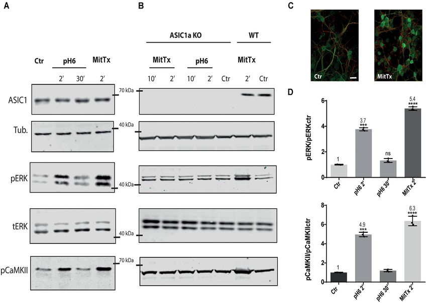
Salinas Castellanos et al.                                                                             Downstream Signaling in ASIC1a Activation

(Alomone, STP-200), 20 nM, 30 min before; as previously used            anti-phospho ERK (Phosphosolutions, p160–202, 1:100). Images
in Salinas et al. (2020). MitTx (Alomone, M-100) was used at            were taken using an Olympus FV300/BX61 microscope with a
a concentration of 20 nM for 2, 10 min according to Alomone             60× (1.4 NA) oil-immersion objective. Alexa-647 and Alexa-
Labs and (Bohlen et al., 2012). Solutions used for the different        488-conjugated secondary antibodies (ThermoFisher) were used.
incubations were prepared as follows: for incubation of cells
with the different reagents and controls were: solution at pH
7.3, containing the following (in mM): NaCl 128, KCl 2.5,               RESULTS
CaCl2 2, MgCl2 1, glucose 15, sucrose 15, HEPES 5, MES
5 adjusted to pH 7.4; and for treatments to activate through the
                                                                        Activation of pERK Through ASIC1a via
proton mechanism solution were adjusted to pH 6 with HCl                Non-proton Mechanisms
(‘‘pH6’’).                                                              The effect of MitTx on the activation of ERK on mouse striatum
                                                                        cells was studied since the toxin can activate ASIC1a channels
Western Blotting (WB)                                                   at neutral pH and for a longer duration (Bohlen et al., 2012).
Western blots were performed according to standard procedures.          The effect was studied at the mouse striatum neurons as these
In brief, cells were resuspended in a 1% SDS HEPES                      cells are enriched in ASIC1 channels composed predominantly of
pH 7.4 lysis buffer containing a protease inhibitor cocktail            ASIC1a subunits constituting homomeric channels (Jiang et al.,
(Roche, cOmpleteTM ); in the case of lysates used for detection         2009). Furthermore, Yu et al. (2018) showed at the striatum that
of phosphorylated ERK, the buffer included 50 mM sodium                 a decrease in pH triggered the activation of the CaMKII signaling
fluoride, 2 mM sodium orthovanadate. Proteins were resolved by          pathway leading to the activation of ERK kinases.
4–10% polyacrylamide gels and transferred onto Immobilonr -                 We decided to analyze whether the downstream effects of
FL PVDF membranes. Non-specific binding was blocked by 1%               MitTx activation would lead to the same signaling pathways as
non-fat powdered milk in TBS containing 0.2% Tween-20 for               those triggered by proton activation.
60 min at RT. Membranes were incubated overnight at 4◦ C with               For this purpose, mouse striatal cultures were treated with
primary antibodies in 1% BSA in TBS, followed by the addition           MitTx and compared to cultures treated with acidic solutions.
of secondary antibodies in 1% non-fat powdered milk in TBS.             As shown in Figures 1A–D, MitTx-treated cultures evidence an
    The following primary antibodies were used: rabbit polyclonal       increase in phospho ERK levels following the same pattern as
anti ASIC1 (Alomone ASC-014, 1:1,000); mouse monoclonal                 phospho CaMKII activation which is much stronger than that
anti-tubulin (DM1a; Cell signaling #3873, 1:5,000); rabbit              shown for pH6-treated cultures at 2 min (as documented by
polyclonal anti total ERK (Santa Cruz, C9, 1:500); rabbit               Yu et al., 2018) or 30 min. The signal ratio of pERK/tERK for
polyclonal anti phosphoERK (Cell Signaling, SC-7383, 1:500);            pH6 2’ treated cells is four times greater than control cells (pH6
pCaMKII (Phosphosolutions, p1005-286). Initially, each                  2’ 3.77 ± 0.07), and MitTx for 2’ leads to more than a 5-fold
antibody was detected in full membranes to verify that only             increase (MitTx 2’ 5.39 ± 0.08). This effect is not present in
the expected MW bands were present and the optimal dilution             striatal cultures obtained from ASIC1a knock-out cultures and
was decided upon. Accordingly, membranes were cut using                 treated either with pH6 solutions or MitTx (Figure 1B).
MW standards as a guide to detect different proteins in the
same membrane. No membrane stripping protocols were                     Proton and Non-proton Activation of
performed, thus bands of the same MW were obtained from                 ASIC1a Human Subunits
the same samples run on different membranes. Reactive bands             The effect of the non-proton activation of ASIC1a channels was
were detected by the LI-COR Odyssey system, using secondary             analyzed further with MitTx on HEK cells that endogenously
antibodies: 926-68073 IRDye 680RD Donkey anti-Rabbit IgG or             express ASIC1a subunits (Gunthorpe et al., 2001). Human,
926-32212 IRDye 800CW Donkey anti-Mouse.                                rat, and mice ASIC1a channels show differences, as shown
    Images were taken using the LI-COR Odyssey system and               for instance by a different degree of glycosylation that leads
quantified with ImageJ software (NIH, USA).                             to different surface channel levels (Kadurin et al., 2008) and
                                                                        levels of activation (Xu et al., 2018). Therefore, we also tested
Detection of Proteins by                                                the mechanism on this subunit. The activation of the pERK
Immunofluorescence (IF)                                                 pathway has also been shown through the treatment of HEK
Cells grown on PLL-coated glass coverslips were fixated with            cultures with pH6 solutions (Yu et al., 2018). Figure 2 shows
4% p- formaldehyde in PBS, permeabilized with 0.1% Triton               the effects of either pH6 or MitTx treatments of cultures for
x-100 (10 min), and treated with blocking solution (1% BSA,             different durations and also after incubation of cultures with
0.01% Triton x-100 in PBS) for an hour at RT. Coverslips                Psalmotoxin (Pctx-1) a toxin that stabilizes the desensitized state
were incubated then with the primary antibody for overnight             of the channel constituted by ASIC1a subunits (Chen et al., 2005).
in blocking buffer, washed in PBS, and incubated with the                   The degree of ERK phosphorylation is not only greater
secondary antibody for 60 min in blocking buffer. After a final         through the non-proton mechanism [compare pH6 2’ 6.10 ± 0.13
wash in PBS, coverslips, were placed onto a slide and covered           (6-fold increase) vs. MitTx 2’ 7.98 ± 0.10, 8-fold increase] but
with a mounting medium. The antibodies used were rabbit                 also, these levels increase in time compared to the transient
polyclonal antibody against mouse monoclonal anti-tubulin               activation of ERK via the proton mechanism (MitTx 10’
(DM1a; Cell Signaling, #3873, 1:2,000); rabbit polyclonal               10.90 ± 0.17, 10-fold increase to control levels). In both cases,

Frontiers in Cellular Neuroscience | www.frontiersin.org            3                                 October 2021 | Volume 15 | Article 735414
Salinas Castellanos et al. Downstream Signaling in ASIC1a Activation
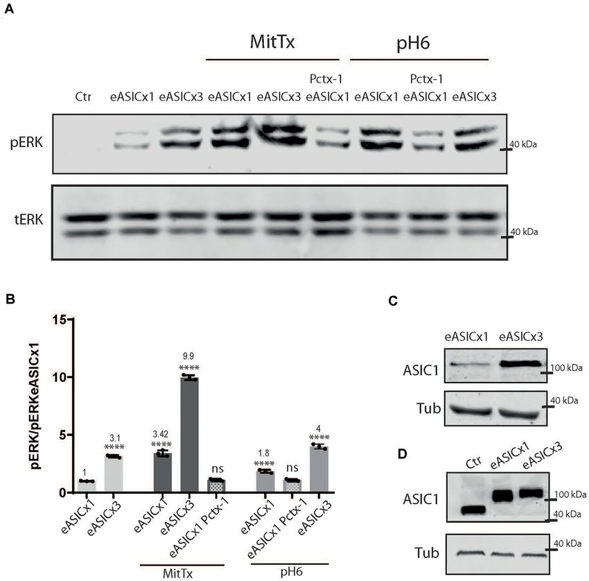
FIGURE 1 | Non-proton activation of ASIC1a in striatal neurons. Representative membranes of lysates from 7 DIV wild type (A), and ASIC1a knockout (B) C57 mice
striatal neuronal cultures were incubated with MitTx and compared to treatment with pH6 solutions for the time indicated (2, 10, or 30 min). The detection was
performed with anti ASIC1, tubulin (Tub), phospho ERK (pERK), total ERK (tERK), or phospho CaMKII (pCaMKII) antibodies, and Licor secondary antibodies. (C)
Examples of images of striatal cultures used and treated with MitTx and stained with tubulin (red) and phosphoERK (green) antibodies and secondary Alexa fluor
antibodies, 60× objective used. Scale bar 10 µm. (D) Result of the bands detected in (A) for pERK/ERKt levels relative to control samples showing an increase in
both, pH6 or MitTx treatments and the same pattern of increase for pCaMKII. Notice the lack of effect in ASIC1a knock-out derived cultures. (A) Notice that plots are
the result of the signal intensity of the bands detected for each antibody, and tERK and tubulin are used as loading controls between loaded samples. Data are
presented as the mean ± SEM ANOVA and Dunnet post hoc test for treatments against the control were performed, mean values above bars; n = 3 membranes,
****p < 0.0001; ***p 0.0001–0.001; ns: no significant differences. Mean values expressed relative to control (Ctr) levels ± SEM are as follows: for pERK/tERK: pH6
2’ 3.77 ± 0.07; pH6 30’ 1.32 ± 0.09; MitTx 2’ 5.39 ± 0.08. For pCaMKII/Tub: pH6 2’ 4.97 ± 0.14; pH6 30’ 1.20 ± 0.07; MitTx 2’ 6.37 ± 0.28.
Pctx-1 can inhibit the activation of ERK to control levels. (compare Ctr and eASICx1 bands, Figure 3A; and eASICx3
Phosphorylated ERK was detected even after 30 min incubation 3.16 ± 0.06, normalized to eASICx1 levels), and is activated
with MitTx (not shown). further via the proton or non-proton signaling mechanism.
But as the increase becomes greater (compare transfection
of plasmids to different levels, either ‘‘1x’’ or ‘‘3x’’), the
Effect of Proton and Non-proton Activation non-proton mechanism is still able to activate the channel
and Different Levels of ASIC Channels to greater levels (eASICx3 MitTx 9.96 ± 0.12), whereas the
Different pathological conditions show an increase in ASIC1a proton activation is no longer able to reflect this change
levels (Duan et al., 2007). To model this situation and analyze (Figures 3A,B). Interestingly, the pERK/tERK ratio of eASICx1-
the signaling pathway triggered by the non-proton activation of MitTx/eASICx1 and eASICx3-MitTx/eASICx3 remains about
the channel, we used HEK cells transfected with different levels the same (3-fold increase).
of ASIC1a channels using a plasmid encoding for the rat ASIC1a
subunit fused to eGFP (eASIC), thus distinctively detected in WB
via the different molecular weights (due to the eGFP tag added), DISCUSSION
as used before (Salinas et al., 2020).
As depicted in Figure 3, rat ASIC1a is also activated In this study, we analyzed the signaling pathway triggered
to a greater level when incubated with MitTx instead of by the activation of ASIC1a channels through a non-proton
pH6 (MitTx 3.42 ± 0.15 vs. pH6 1.85 ± 0.08). When mechanism.
HEK cells overexpress the channel (Figures 3B,C), the We show that this mechanism determines the activation of
activation of the ERK pathway is increased at basal levels the CaMKII-ERK pathway for a longer period than that resulting
Frontiers in Cellular Neuroscience | www.frontiersin.org 4 October 2021 | Volume 15 | Article 735414Salinas Castellanos et al. Downstream Signaling in ASIC1a Activation
FIGURE 2 | Proton and non-proton activation of ASIC1a in HEK cells. (A) Representative membranes of lysates of HEK cells treated with pH6 or MitTx for 2 or
10 min or preincubated with Pctx-1 compared to untreated cells (control, Ctr) and detected with phosphoERK (pERK), total ERK (tERK), and ASIC1 antibodies. (B)
Representative membrane of the same lysates to detect pCaMKII levels. (C) Plots showing detected levels of pERK (top panel) or pCaMKII (lower panel). Notice that
the increase in kinase levels goes further at a later time point (2 vs. 10 min) in MitTx treated cultures compared to pH6 treated ones that show an increase at 2 min
followed by a reversal to control levels consistent with the proton-activated desensitizing mechanism. (A) Notice that plots are the result of the signal intensity of the
bands—with tERK and tubulin used as loading controls between loaded samples—and expressed relative to control samples. Data are presented as the
mean ± SEM ANOVA and Dunnet post hoc test for treatments against the control were performed, mean values above bars; n = 3 membranes, ****p < 0.0001; ***p
0.0001–0.001; ns: no significant differences. Mean values expressed relative to control (Ctr) levels ± SEM are as follows: for pERK/tERK: pH6 2’ 6.10 ± 0.13; pH6
10’ 1.23 ± 0.11; pH6 2’ Pctx 1.39 ± 0.08; MitTx 2’ 7.98 ± 0.10; Mittx10’ 10.90 ± 0.17; MitTx 2’ Pctx 1.18 ± 0.05; MitTx 10’ Pctx 1.42 ± 0.05. For
pCaMKII/Tub: pH6 2’ 7.31 ± 0.21; pH6 10’ 1.16 ± 0.06; pH6 2’ Pctx1.14±0.07; Mittx 2’ 8.88 ± 0.15; Mittx 10’ 10.76 ± 0.32; Mittx 2’ Pctx 1.17 ± 0.07.
from proton activation. This mechanism was conserved for the ERK activation, and the activation of both has been shown for
different ASIC1a subunits analyzed (mouse, in Figure 1, human striatal cells through the proton-mediated activation of ASIC1a
in Figure 2, and rat in Figure 3) in the different in vitro models. channels (Yu et al., 2018). Nevertheless, whether the mechanism
Furthermore, the mechanism could even reach higher levels if requires the conducting channel is a matter of debate. We
ASIC1a subunits were expressed at higher levels (Figure 3). The showed that the presence of Pctx-1 prevents this mechanism
fact that mouse striatal KO cultures showed no evidence for and that CaMKII is activated, but whether the mechanism could
this mechanism reinforces the argument that these mechanisms rely on a conduction-independent pathway cannot be ruled
analyzed act via ASIC1a and no other pH-sensitive receptor. out. As an example, ASIC1a phosphorylation by RIP1 leading
The pathway (downstream ASIC1a activation) has been to necroptosis pathways does not rely on conducting channels
shown as signaling for different events relevant in physiological (Wang et al., 2015, 2020). Future experiments will reveal more
as well as pathological conditions (Kriegsheim et al., 2009). The details on the mechanism.
mechanism, which is dependent on a stimulus that leads to the A comparative analysis of both mechanisms analyzed in
three-tier activation cascade with sequential kinase activation, this work shows that the proton mechanism leads to transient
has also been shown to crosstalk with the CaMKII pathway in activation of ERK which can no longer be detected after
many cells (Illario et al., 2003; Salzano et al., 2012). Accordingly, 5 min. Increases or a decrease in pERK levels were detected
for some stimuli and cell models, CaMKII is necessary for in previous work via ASIC1a. Amiloride significantly decreased
Frontiers in Cellular Neuroscience | www.frontiersin.org 5 October 2021 | Volume 15 | Article 735414Salinas Castellanos et al. Downstream Signaling in ASIC1a Activation FIGURE 3 | Proton and non-proton activation of overexpressed ASIC1a channels. (A) Representative membranes of lysates of cells control (ctr) or transfected with eGFP-ASIC1a (eASIC) at two levels (1x or x3) to obtained different levels of expression of the protein (“eASICx1 or eASICx3”), and treated with pH6 or MitTx with or without pre-incubation of Pctx-1 or untreated. (B) Plots showing the increase in pERK and pCaMKII levels calculated from membranes as that shown in (A), consistent with the increase in eASIC expressed. Notice the level of increase achievable via MitTx incubation at the highest overexpressed level of eASIC, higher than that obtained via pH6. (C) Representative membrane showing the different levels of eASIC in cells overexpressing the channel (1x or 3x), detected with an ASIC1 antibody. (D) Comparison between the different ASIC1 proteins expressed (the endogenous human ASIC1a; of approx. 67 kDa) and the overexpressed eASIC (approx. 110 kDa, and expressed at different levels; x1 or x3). (A) Notice that plots are the result of the signal intensity of the band detected,—tERK and tubulin are used as loading controls between loaded samples—and expressed relative to eASICx1 levels. Data are presented as the mean ± SEM ANOVA and Dunnet post hoc test for treatments and conditions were performed, mean values above bars; n = 3 membranes, ∗∗∗∗ p < 0.0001; ns: no significant differences. Mean values expressed relative to eASICx1 levels ± SEM are as follows: eASICx3 3.16 ± 0.06; eASIC MitTx 3.42 ± 0.15; eASICx3 MitTx 9.96 ± 0.12; eASIC MitTx Pctx 1.10 ± 0.05; eASIC pH6 2’ 1.85 ± 0.08; eASIC pH6 2’ Pctx 1.08 ± 0.05; eASICx3 pH6 2’ 4.00 ± 0.12. (an approximately half-fold) the levels of CaMKKß and ERK in phosphorylation levels through pH 6 incubation reaching phosphorylation in a cell line of hepatic fibroblasts stimulated approximately 230% for ERK1 and 250% for ERK2 higher levels by high glucose and PDGF (Wang et al., 2019). Zhu et al. than control cells. In this work, we detected an increase in (2020) showed a contribution of ASIC1a to increased ERK total phosphorylated ERK levels via the proton mechanism. phosphorylation in the mechanism of liver fibrosis, as Pctx-1 In contrast, the non-proton activation of ASIC1a channels treatment decreased the approximate 2-fold increase (without leads to the phosphorylation of ERK to a greater extent and treatment) to a 1.5-fold increase. The same can be observed in for a longer period, as no desensitization is present (still ERK phosphorylated levels as the bands show a greater intensity active at 30 min). Thus, the activation of the channel in for ERK-mediated NF-κB activation through ASIC1 in response a non-proton mechanism (as in a Texas coral snake bite) to acidosis (although not quantified; Chen et al., 2016). In would trigger sustained phosphorylation of ERK that could striatal and HEK cells, Yu et al. (2018) showed an increase lead to further signaling. Additionally, we noted that the Frontiers in Cellular Neuroscience | www.frontiersin.org 6 October 2021 | Volume 15 | Article 735414
Salinas Castellanos et al.                                                                                                          Downstream Signaling in ASIC1a Activation

increased phosphorylation of ERK (measured as the signal                                     ASIC1a exist, however, remains to be determined. Nevertheless,
detected in pERK to tERK levels) reached the same level                                      the possibility that this pathway might explain aspects of pain
whether channels were expressed at higher levels as if the                                   warrants further analysis for potential therapies.
signaling could be the result of a fraction of occupied receptor
mechanism (Andrews et al., 2016) that should be analyzed in
the future.                                                                                  DATA AVAILABILITY STATEMENT
    The kinetics of ERK phosphorylation has been the subject
                                                                                             The original contributions presented in the study are included in
of various studies (Kriegsheim et al., 2009; Ahmed et al.,
                                                                                             the article, further inquiries can be directed to the corresponding
2014; Shindo et al., 2016; Maik-Rachline et al., 2019). These
                                                                                             author.
studies revealed different aspects of the complexity in the
regulation of ERK signaling, providing mathematical models
accounting for different levels of regulation. Among these,                                  ETHICS STATEMENT
negative feedback on ERK activation through upregulation of
phosphatases that dephosphorylate ERK, as well as depletion                                  The animal study was reviewed and approved by CICUAL,
of the stimulus (either through internalization or removal from                              University of Buenos Aires, Argentina.
the extracellular medium; Cirit et al., 2010) were shown to
play a role in the transient shape of the signal. Additionally,                              AUTHOR CONTRIBUTIONS
ERK translocation to the nucleus and binding to cytosolic and
nuclear substrates and dephosphorylation was also shown to                                   ODU and CW contributed to the conception and design of the
play a main role in the kinetics of ERK signaling (Ahmed                                     study. LCSC performed experiments and the statistical analysis.
et al., 2014). Indeed, the translocation of ERK was later                                    ODU and CW wrote the manuscript. All authors contributed to
interpreted as a key to transforming a graded response                                       the article and approved the submitted version.
(stimulus activating ERK) into a switch (ERK translocated to
the nucleus) that can determine the fate of a cell (Shindo
et al., 2016). Thus, ERK phosphorylated transiently (up to
                                                                                             FUNDING
5 min) or in a sustained manner determines a different                                       This     work     was     supported   by    Grant    01/Q666
biological response.                                                                         [20020130100666BA; Universidad de Buenos Aires Ciencia
    Nuclear ERK can determine the stabilization of immediate                                 y Tecnología (UBACYT)] from University of Buenos Aires
early gene products that can trigger further effects as observed                             (to ODU), PICT 2016 # 3642 (to ODU), and Investigator-
by c-fos-mediated signaling when ERK is activated in a sustained                             Initiated Research grant (no. IIR-AR-002659) funded by Takeda
manner (Murphy et al., 2002).                                                                Pharmaceuticals International AG Singapore Branch (to ODU).
    Our studies show that the proton-mediated activation of
ASIC1a channels acts transiently activating ERK, the channel is
desensitized and can no longer trigger the activation mechanism.                             ACKNOWLEDGMENTS
This could be comparable to depletion of the stimulus.
    The ERK pathway has been described as a network                                          We thank Dr. Stefan Gründer (RWTH-Aachen) for kindly
functioning as a potential switch, oscillator, or memory (Shindo                             providing us with the eGFP-ASIC1a plasmid, Valeria Buggiano
et al., 2016). All these mechanisms concerning pain could lead                               for her help with cell lines, and Zaira Naguila and Fernanda
to acute or persistent effects. Whether endogenous ligands for                               Toledo for their help in the breeding of mice.

REFERENCES                                                                                   Baconguis, I., Bohlen, C. J., Goehring, A., Julius, D., and Gouaux, E. (2014). X-
                                                                                                ray structure of acid-sensing ion channel 1-snake toxin complex reveals open
Ahmed, S., Grant, K. G., Edwards, L. E., Rahman, A., Cirit, M., Goshe, M. B.,                   state of a Na+-selective channel. Cell 156, 717–729. doi: 10.1016/j.cell.2014.
   et al. (2014). Data-driven modeling reconciles kinetics of ERK phosphorylation,              01.011
   localization and activity states. Mol. Syst. Biol. 10:718. doi: 10.1002/msb.              Bohlen, C. J., Chesler, A. T., Sharif-Naeini, R., Medzihradszky, K. F., Zhou, S.,
   134708                                                                                       King, D., et al. (2012). A heteromeric texas coral snake toxin targets
Aissouni, Y., El Guerrab, A., Mahdy Hamieh, A., Ferrier, J., Chalus, M.,                        acid-sensing ion channels to produce pain HHS public access. Nature 479,
   Lemaire, D., et al. (2017). Acid-sensing ion channel 1a in the amygdala is                   410–414. doi: 10.1038/nature10607
   involved in pain and anxiety-related behaviours associated with arthritis. Sci.           Boscardin, E., Alijevic, O., Hummler, E., Frateschi, S., and Kellenberger, S. (2016).
   Rep. 7:43617. doi: 10.1038/srep43617                                                         The Function and regulation of acid-sensing ion channels (ASICs) and the
Alijevic, O., Bignucolo, O., Hichri, E., Peng, Z., Kucera, J. P., and Kellenberger, S.          epithelial Na+channel (ENaC): IUPHAR review 19. Br. J. Pharmacol. 173,
   (2020). Slowing of the time course of acidification decreases the acid-sensing               2671–2701. doi: 10.1111/bph.13533
   ion channel 1a current amplitude and modulates action potential                           Chen, X., Kalbacher, H., and Gründer, S. (2005). The tarantula toxin psalmotoxin
   firing in neurons. Front. Cell. Neurosci. 14:41. doi: 10.3389/fncel.2020.                    1 inhibits acid-sensing ion channel (ASIC) 1a by increasing its apparent H+
   00041                                                                                        affinity. J. Gen. Physiol. 126, 71–79. doi: 10.1085/jgp.200509303
Andrews, S. S., Peria, W. J., Yu, R. C., Colman-Lerner, A., and Brent, R. (2016).            Chen, B., Liu, J., Ho, T. T., Ding, X., and Mo, Y. Y. (2016). ERK-mediated
   Push-pull and feedback mechanisms can align signaling system outputs with                    NF-κB activation through ASIC1 in response to acidosis. Oncogenesis 5:e279.
   inputs. Cell Syst. 3, 444–455. doi: 10.1016/j.cels.2016.10.002                               doi: 10.1038/oncsis.2016.81

Frontiers in Cellular Neuroscience | www.frontiersin.org                                 7                                         October 2021 | Volume 15 | Article 735414
Salinas Castellanos et al. Downstream Signaling in ASIC1a Activation
Chu, X. P., and Xiong, Z. G. (2013). Acid-sensing ion channels in pathological Marshall, C. J. (1995). Specificity of receptor tyrosine kinase signaling: transient
conditions. Adv. Exp. Med. Biol. 961, 419–431. doi: 10.1007/978-1-4614-4756- versus sustained extracellular signal-regulated kinase activation. Cell 80,
6_36 179–185. doi: 10.1016/0092-8674(95)90401-8
Cirit, M., Wang, C. C., and Haugh, J. M. (2010). Systematic quantification of Maruta, T., Nemoto, T., Hidaka, K., Koshida, T., Shirasaka, T., Yanagita, T.,
negative feedback mechanisms in the extracellular signal-regulated kinase et al. (2019). Upregulation of ERK phosphorylation in rat dorsal root ganglion
(ERK) signaling network. J. Biol. Chem. 285, 36736–36744. doi: 10.1074/jbc. neurons contributes to oxaliplatin-induced chronic neuropathic pain. PLoS
M110.148759 One 14:e0225586. doi: 10.1371/journal.pone.0225586
Cruz, C. D. C., and Cruz, F. (2007). The ERK 1 and 2 pathway in Murphy, L. O., Smith, S., Huei Chen, R., Fingar, D. C., and Blenis, J. (2002).
the nervous system: from basic aspects to possible clinical applications Molecular, interpretation of ERK signal duration by immediate early gene
in pain and visceral dysfunction. Curr. Neuropharmacol. 5, 244–252. products. Nat. Cell Biol. 4, 556–564. doi: 10.1038/ncb822
doi: 10.2174/157015907782793630 Peng, Z., and Kellenberger, S. (2021). Hydrogen sulfide upregulates acid-sensing
Duan, B., Liu, D.-S., Huang, Y., Zeng, W.-Z., Wang, X., Yu, H., et al. (2012). ion channels via the MAPK-Erk1/2 signaling pathway. Function 2:zqab007.
PI3-Kinase/Akt pathway-regulated membrane insertion of acid-sensing ion doi: 10.1093/function/zqab007
channel 1a underlies BDNF-induced pain hypersensitivity. J. Neurosci. 32, Salinas, L. C. C., Rozenfeld, P., Gabriel Gatto, R., Claudio Reisin, R., Daniel
6351–6363. doi: 10.1523/JNEUROSCI.4479-11.2012 Uchitel, O., Weissmann, C., et al. (2020). Upregulation of ASIC1a channels in
Duan, B., Wu, L.-J., Yu, Y.-Q., Ding, Y., Jing, L., Xu, L., et al. (2007). an in vitro model of fabry disease. Neurochem. Int. 140:104824. doi: 10.1016/j.
Upregulation of acid-sensing ion channel ASIC1a in spinal dorsal horn neuint.2020.104824
neurons contributes to inflammatory pain hypersensitivity. J. Neurosci. 27, Salzano, M., Rosaria Rusciano, M., Russo, E., Bifulco, M., Postiglione, L.,
11139–11148. doi: 10.1523/JNEUROSCI.3364-07.2007 and Vitale, M. (2012). Calcium/calmodulin-dependent protein kinase
Fan, S., Hao, Z.-Y., Zhang, L., Zhou, J., Zhang, Y.-F., Tai, S., et al. (2018). ASIC1a II (CaMKII) phosphorylates Raf-1 at serine 338 and mediates
contributes to the symptom of pain in a rat model of chronic prostatitis. Asian ras-stimulated Raf-1 activation. Cell Cycle 11, 2100–2106. doi: 10.4161/cc.
J. Androl. 20, 300–305. doi: 10.4103/aja.aja_55_17 20543
Friese, M. A., Craner, M. J., Etzensperger, R., Vergo, S., Wemmie, J. A., Shindo, Y., Iwamoto, K., Mouri, K., Hibino, K., Tomita, M., Kosako, H.,
Welsh, M. J., et al. (2007). Acid-sensing ion channel-1 contributes et al. (2016). Conversion of graded phosphorylation into switch-like nuclear
to axonal degeneration in autoimmune inflammation of the translocation via autoregulatory mechanisms in ERK signalling. Nat. Commun.
central nervous system. Nat. Med. 13, 1483–1489. doi: 10.1038/ 7:10485. doi: 10.1038/ncomms10485
nm1668 Sluka, K. A., Winter, O. C., and Wemmie, J. A. (2009). Acid-sensing ion channels:
Gautschi, I., Van Bemmelen, M. X., and Schild, L. (2017). Proton and non-proton a new target for pain and CNS diseases. Curr. Opin. Drug Discov. Devel. 12,
activation of ASIC channels. PLoS One 12:e0175293. doi: 10.1371/journal.pone. 693–704.
0175293 Sodero, A. O., Weissmann, C., Ledesma, M. D., and Dotti, C. G. (2011).
González-Inchauspe, C., Urbano, F. J., Di Guilmi, M. N., Uchitel, O. D., Cellular stress from excitatory neurotransmission contributes to cholesterol
Carlota Gonzalez-Inchauspe, F. J., Di Guilmi, U. M. N., et al. (2017). Acid- loss in hippocampal neurons aging in vitro. Neurobiol. Aging 32, 1043–1053.
sensing ion channels activated by evoked released protons modulate synaptic doi: 10.1016/j.neurobiolaging.2010.06.001
transmission at the mouse calyx of held synapse. J. Neurosci. 37, 2589–2599. Sun, X., Cao, Y.-B., Hu, L.-F., Yang, Y.-P., and Li, J. (2011). ASICs mediate the
doi: 10.1523/JNEUROSCI.2566-16.2017 modulatory effect by paeoniflorin on alpha-synuclein autophagic degradation.
Gründer, S., and Chen, X. (2010). Structure, function and pharmacology Brain Res. 1396, 77–87. doi: 10.1016/j.brainres.2011.04.011
of acid-sensing ion channels (ASICs): focus on ASIC1a. Int. J. Physiol. Sun, C., Wang, S., and Hu, W. (2018). Acid-sensing ion channel 1a
Pathophysiol. Pharmacol. 2, 73–94. mediates acid-induced inhibition of matrix metabolism of rat articular
Gunthorpe, M. J., Smith, G. D., Davis, J. B., and Randall, A. D. (2001). chondrocytes via the MAPK signaling pathway. Mol. Cell. Biochem. 443, 81–91.
Characterisation of a human acid-sensing ion channel (HASIC1a) doi: 10.1007/s11010-017-3212-9
endogenously expressed in HEK293 cells. Pflugers Arch. 442, 668–674. Tikhonov, D. B., Magazanik, L. G., and Nagaeva, E. I. (2019). Ligands of
doi: 10.1007/s004240100584 acid-sensing ion channel 1a: mechanisms of action and binding sites. Acta
Hoagland, E. N., Sherwood, T. W., Lee, K. G., Walker, C. J., and Askwith, C. C. Naturae 11, 4–13. doi: 10.32607/20758251-2019-11-1-4-13
(2010). Identification of a calcium permeable human acid-sensing ion channel Uchitel, O. D., González Inchauspe, C., and Weissmann, C. (2019). Synaptic
1 transcript variant. J. Biol. Chem. 285, 41852–41862. doi: 10.1074/jbc.M110. signals mediated by protons and acid-sensing ion channels. Synapse 73:e22120.
171330 doi: 10.1002/syn.22120
Illario, M., Cavallo, A. L., Ulrich Bayer, K., Di Matola, T., Fenzi, G., Rossi, G., Wang, J. J., Liu, F., Yang, F., Wang, Y. Z., Qi, X., Li, Y., et al. (2020).
et al. (2003). Calcium/calmodulin-dependent protein kinase II binds to Disruption of auto-inhibition underlies conformational signaling of ASIC1a to
Raf-1 and modulates integrin-stimulated ERK activation. J. Biol. Chem. 278, induce neuronal necroptosis. Nat. Commun. 11:475. doi: 10.1038/s41467-019
45101–45108. doi: 10.1074/jbc.M305355200 -13873-0
Jiang, Q., Li, M.-H., Papasian, C. J., Branigan, D., Xiong, Z.-G., Wang, J. Q., Wang, Y., O’Bryant, Z., Wang, H., and Huang, Y. (2016). Regulating factors
et al. (2009). Characterization of acid-sensing ion channels in medium in acid-sensing ion channel 1a function. Neurochem. Res. 41, 631–645.
spiny neurons of mouse striatum. Neuroscience 162, 55–66. doi: 10.1016/j. doi: 10.1007/s11064-015-1768-x
neuroscience.2009.04.029 Wang, Y., Sun, Y., Zuo, L., Wang, Y., and Huang, Y. (2019). ASIC1a promotes
Kadurin, I., Golubovic, A., Leisle, L., Schindelin, H., and Gründer, S. (2008). high glucose and PDGF-induced hepatic stellate cell activation by inducing
Differential effects of N-glycans on surface expression suggest structural autophagy through CaMKKβ/ERK signaling pathway. Toxicol. Lett. 300, 1–9.
differences between the acid-sensing ion channel (ASIC) 1a and ASIC1b. doi: 10.1016/j.toxlet.2018.10.003
Biochem. J. 412, 469–475. doi: 10.1042/BJ20071614 Wang, Y. Z., Wang, J. J., Huang, Y., Liu, F., Zeng, W. Z., Li, Y., et al. (2015). Tissue
Kriegsheim, A. V., Baiocchi, D., Birtwistle, M., Sumpton, D., Bienvenut, W., acidosis induces neuronal necroptosis via ASIC1a channel independent of its
Morrice, N., et al. (2009). Cell fate decisions are specified by the ionic conduction. eLife 4:e05682. doi: 10.7554/eLife.05682
dynamic ERK interactome. Nat. Cell Biol. 11, 1458–1464. doi: 10.1038/ Wei, S., Qiu, C. Y., Jin, Y., Liu, T. T., and Hu, W. P. (2021). TNF-α acutely enhances
ncb1994 acid-sensing ion channel currents in rat dorsal root ganglion neurons via a
Kweon, H.-J., and Suh, B.-C. (2013). Acid-sensing ion channels ( ASICs ): P38 MAPK pathway. J. Neuroinflammation 18:92. doi: 10.1186/s12974-021-
therapeutic targets for neurological diseases and their regulation. BMB Rep. 46, 02151-w
295–304. doi: 10.5483/bmbrep.2013.46.6.121 Weissmann, C., Di Guilmi, M. N., Urbano, F. J., and Uchitel, O. D.
Maik-Rachline, G., Hacohen-Lev-Ran, A., and Seger, R. (2019). Nuclear Erk: (2013). Acute effects of pregabalin on the function and cellular
mechanism of translocation, substrates and role in cancer. Int. J. Mol. Sci. distribution of Ca(V)2.1 in HEK293t cells. Brain Res. Bull. 90, 107–113.
20:1194. doi: 10.3390/ijms20051194 doi: 10.1016/j.brainresbull.2012.10.001
Frontiers in Cellular Neuroscience | www.frontiersin.org 8 October 2021 | Volume 15 | Article 735414Salinas Castellanos et al. Downstream Signaling in ASIC1a Activation Wemmie, J. A., Taugher, R. J., and Kreple, C. J. (2013). Acid-sensing ion Zhu, Y., Pan, X., Du, N., Li, K., Hu, Y., Wang, L., et al. (2020). ASIC1a regulates channels in pain and disease. Nat. Rev. Neurosci. 14, 461–471. doi: 10.1038/ miR-350/SPRY2 by N 6 -methyladenosine to promote liver fibrosis. FASEB J. nrn3529 34, 14371–14388. doi: 10.1096/fj.202001337R Wong, H. K., Bauer, P. O., Kurosawa, M., Goswami, A., Washizu, C., Machida, Y., Zhu, L., Yin, J., Zheng, F., Ji, L., Yu, Y., and Liu, H. (2021). ASIC1 inhibition et al. (2008). Blocking acid-sensing ion channel 1 alleviates Huntington’s impairs the proliferation and migration of pancreatic stellate disease pathology via an ubiquitin-proteasome system-dependent mechanism. cells induced by pancreatic cancer cells. Neoplasma 68, 174–179. Hum. Mol. Genet. 17, 3223–3235. doi: 10.1093/hmg/ddn218 doi: 10.4149/neo_2020_200803N811 Wu, J., Xu, Y., Jiang, Y.-Q., Xu, J., Hu, Y., and Zha, X.-M. (2016). ASIC subunit ratio and differential surface trafficking in the brain. Mol. Brain 9:4. Conflict of Interest: ODU, coauthor to this manuscript is also editor of this doi: 10.1186/s13041-016-0185-7 special topic. Xiong, Z. G., and Xu, T. L. (2012). The role of ASICs in cerebral ischemia. Wiley Interdiscip. Rev. Membr. Transport Signal. 1, 655–662. doi: 10.1002/wmts.57 The remaining authors declare that the research was conducted in the absence of Xu, Y., Jiang, Y.-Q., Li, C., He, M., George Rusyniak, W., Annamdevula, N., any commercial or financial relationships that could be construed as a potential et al. (2018). Human ASIC1a mediates stronger acid-induced responses as conflict of interest. compared with mouse ASIC1a. FASEB J. 32, 3832–3843. doi: 10.1096/fj.20170 1367R Publisher’s Note: All claims expressed in this article are solely those of the authors Yu, X.-W., Hu, Z.-L., Ni, M., Fang, P., Zhang, P.-W., Shu, Q., et al. (2015). Acid- and do not necessarily represent those of their affiliated organizations, or those of sensing ion channels promote the inflammation and migration of cultured rat the publisher, the editors and the reviewers. Any product that may be evaluated in microglia. Glia 63, 483–496. doi: 10.1002/glia.22766 this article, or claim that may be made by its manufacturer, is not guaranteed or Yu, Z., Jiao Wu, Y.-J., Zhi Wang, Y.-Z., Shi Liu, D.-S., Lei Song, X.-L., endorsed by the publisher. Jiang, Q., et al. (2018). The acid-sensing ion channel ASIC1a mediates striatal synapse remodeling and procedural motor learning. Sci. Signal. 11:eaar4481. Copyright © 2021 Salinas Castellanos, Uchitel and Weissmann. This is an doi: 10.1126/scisignal.aar4481 open-access article distributed under the terms of the Creative Commons Attribution Zeng, W. Z., Liu, D. S., and Xu, T. L. (2014). Acid-sensing ion channels: trafficking License (CC BY). The use, distribution or reproduction in other forums is permitted, and pathophysiology. Channels (Austin) 8, 481–487. doi: 10.4161/19336950. provided the original author(s) and the copyright owner(s) are credited and that the 2014.958382 original publication in this journal is cited, in accordance with accepted academic Zha, X.-M. (2013). Acid-sensing ion channels: trafficking and synaptic function. practice. No use, distribution or reproduction is permitted which does not comply Mol. Brain 6:1. doi: 10.1186/1756-6606-6-1 with these terms. Frontiers in Cellular Neuroscience | www.frontiersin.org 9 October 2021 | Volume 15 | Article 735414
You can also read