The Impact of Maternal High-Fat Diet on Bone Microarchitecture in Offspring - Frontiers
←
→
Page content transcription
If your browser does not render page correctly, please read the page content below
MINI REVIEW
                                                                                                                                             published: 30 August 2021
                                                                                                                                        doi: 10.3389/fnut.2021.730037

                                               The Impact of Maternal High-Fat Diet
                                               on Bone Microarchitecture in
                                               Offspring
                                               Emma J. Buckels 1 , Scott M. Bolam 2,3 , Mei Lin Tay 2 and Brya G. Matthews 1*
                                               1
                                                 Department of Molecular Medicine and Pathology, School of Medical Sciences, University of Auckland, Auckland,
                                               New Zealand, 2 Department of Surgery, School of Medicine, University of Auckland, Auckland, New Zealand, 3 Department of
                                               Orthopaedic Surgery, Auckland City Hospital, Auckland, New Zealand

                                               The incidence of obesity in women of reproductive age has significantly increased
                                               over the past 100 years. There is a well-established connection between maternal
                                               obesity during pregnancy and an increased risk of developing non-communicable
                                               cardiometabolic diseases in her offspring. This mini-review focuses on evidence
                                               examining the effect of maternal high-fat diet (HFD) on skeletal development and bone
                                               health in later life in offspring. The majority of rodent studies indicate that maternal HFD
                                               generally negatively affects both embryonic bone development and bone volume in adult
                                               animals. Details surrounding the mechanisms of action that drive changes in the skeleton
                                               in offspring remain unclear, although numerous studies suggest that some effects are
                                               sex-specific. Human studies in this area are limited but also suggest that HFD during
                            Edited by:         pregnancy may impair bone formation and increase fracture risk during childhood. Given
                 Clare Marie Reynolds,
      University College Dublin, Ireland       the consequences of low bone mass and deranged bone microarchitecture for offspring,
                       Reviewed by:            advances in our understanding of the developmental origins of bone health is critical in
                       Tania Romano,           the battle against osteoporosis.
          La Trobe University, Australia
                          Dian Teguh,          Keywords: early life nutrition, maternal obesity, developmental origins of health and disease (DOHaD),
Beth Israel Deaconess Medical Center           osteoporosis, osteoblast, osteoclast, bone marrow adipocytes, skeletal development
         and Harvard Medical School,
                        United States

                  *Correspondence:             INTRODUCTION
                    Brya G. Matthews
       brya.matthews@auckland.ac.nz            The prevalence of obesity over the past 100 years has dramatically increased, with obesity identified
                                               as the most common metabolic disorder. Globally, an estimated 600 million adults were obese
                    Specialty section:         (body mass index ≥ 30 kg/m2 ), and 1.9 billion adults were overweight in 2015 (body mass index
          This article was submitted to        25–30 kg/m2 ) (1). The prevalence of obesity is expected to reach 1.12 billion individuals by 2030
             Nutrition and Metabolism,         (2). Obese individuals have an increased risk of morbidity from type 2 diabetes mellitus (T2DM),
                a section of the journal
                                               cardiovascular disease, specific cancers, and osteoarthritis (3).
                    Frontiers in Nutrition
                                                  The incidence of obesity in women of reproductive age has also increased. Maternal obesity
            Received: 24 June 2021             is a significant risk factor for maternal, fetal, and neonatal morbidities, including miscarriage,
          Accepted: 02 August 2021
                                               preterm delivery, hypertension, pre-eclampsia, and gestational diabetes (4–6). Research in the field
          Published: 30 August 2021
                                               of developmental origins of health and disease (DOHaD) has highlighted that maternal obesity
                             Citation:         during pregnancy predisposes offspring to develop obesity and other non-communicable diseases,
  Buckels EJ, Bolam SM, Tay ML and
                                               including T2DM, hypertension, and cardiovascular disease, later in adulthood (7). Paternal
  Matthews BG (2021) The Impact of
     Maternal High-Fat Diet on Bone
                                               obesity also increases the risk of developing non-communicable diseases in offspring (8–10), and
      Microarchitecture in Offspring.          both maternal and paternal obesity have transgenerational effects on subsequent generations via
               Front. Nutr. 8:730037.          epigenetic effects on the germline (10–12). Thus, obesity and its related comorbidities represent an
     doi: 10.3389/fnut.2021.730037             increasing burden on healthcare systems.

Frontiers in Nutrition | www.frontiersin.org                                         1                                          August 2021 | Volume 8 | Article 730037
Buckels et al. Maternal High-Fat Diet and Bone
Although not fully understood, the effect of maternal obesity Anabolic treatments restore bone microarchitecture to some
on the development of various organs and tissues such as the degree, but are expensive biologics so not available to everyone.
brain, liver, kidney, endocrine pancreas, and skeletal muscle Understanding risk factors for low bone mass and identifying
and their structure and function have been well-researched with people at high risk of fracture are important for preventing
the aid of animal models. In these models, maternal obesity is fracture-related morbidity and mortality in aging populations.
induced via a high-fat diet (HFD) (13, 14). Recently the effects of
maternal HFD on bone mass and strength in offspring and the EFFECT OF MATERNAL HFD ON BONE IN
risk of developing osteoporosis later in life have been researched.
This mini-review will discuss evidence that maternal HFD-
RODENTS
induced obesity affects bone development and microarchitecture, Thirteen rodent studies considered the effects of maternal HFD
focusing on recent advancements using rodent models, and will on offspring bone development (Table 1). The majority of studies
discuss the potential mechanisms involved. implemented the maternal HFD regime before mating (4–15
weeks before conception) and continued through pregnancy and
lactation. However, in three studies, the maternal HFD-feeding
EARLY BONE DEVELOPMENT AND window was exclusively during pregnancy and lactation and
IMPACT LATER IN LIFE exclusively during lactation in one study.
The skeleton develops in utero from mesenchymal Fetal and Neonatal Offspring
condensations. Most of the skeleton forms via a process known Offspring of dams fed a maternal HFD before and during
as endochondral ossification: initially, a cartilaginous template pregnancy have evidence of skeletal developmental delay in late-
forms, which is later progressively replaced by mineralized gestation with decreased bone formation, bone volume, and
bone matrix. Cartilaginous growth plates continue to control BMD (18–21). Chen et al. have demonstrated that maternal HFD
the longitudinal growth of bones throughout neonatal and promotes cellular senescence in fetal calvarial osteoblasts cells,
childhood growth, while the overall bone shape, mineralization, potentially suppressing fetal bone formation (18–20).
and microarchitecture are determined by the balance of bone In both newborns and weanlings, exposure to maternal HFD
formation by osteoblasts and bone resorption by osteoclasts in resulted in increased total bone mass, BMD, and trabecular bone
different locations. Longitudinal skeletal growth continues until volume in long bones (22, 23). This phenotype is likely the result
the late teens in humans, ending when the growth plates fuse. of increased osteoblast activity, as bone modeling is most active
However, bone mineral density (BMD) continues to increase over this period of rapid growth (31). Increased bone mass in
slowly until peak bone mass is reached in the mid-20s to weanlings may be an indirect effect of maternal HFD, as these
early-30s. Rodents continue to grow slowly for a longer portion offspring consume more milk, and the milk consumed has a
of their life, but mice reach peak bone mass at ∼12-weeks of higher energy content compared to dams fed control diet (CD)
age. Bone mass begins to decline as bone resorption outpaces (22). Additionally, Miotto et al. found higher concentrations
bone formation as we age. In women, there is a dramatic period of monounsaturated fatty acids in the long bones of offspring
of bone loss following menopause when reductions in sex exposed to maternal HFD, which is likely to reflect the diet
hormone levels affect homeostasis. Skeletal size, BMD, and bone consumed by the dams; these lipid stores may have supported
microarchitecture are largely determined by genetics, with up to rapid bone growth (23).
85% of the variation in BMD explained by genetic factors (15).
However, various non-genetic factors also influence both bone Adult Offspring
accrual during growth and bone loss in later life. Nutrition is a From early adulthood, a pattern of sustained bone loss in
major factor influencing both growth and bone mass and can offspring of dams fed HFD is reported. In most studies, offspring
have effects at all life stages. Exercise, or the effect of loading on exposed to maternal HFD have reduced BMD and bone volume
the skeleton, plays a major role in bone accrual and retention. in long bones and vertebrae from as early as 8-weeks of age,
Osteoporosis is characterized by low BMD and a high risk of which persisted over their lifetime (18, 21–23, 25, 30). Hafner et
fracture, and affects one-half of elderly women and about one- al. found that maternal HFD during lactation alone was sufficient
fifth of elderly men. While osteoporosis is considered a disease of to increase bone marrow adiposity (28). Maternal HFD decreased
aging, early life events and a failure to achieve maximal peak bone trabecular bone parameters in offspring (18, 21–23, 25, 30).
mass determined by an individual’s genetics can significantly However, this effect was not observed in all studies; two studies
impact future osteoporosis risk. One line of evidence for long- found increased femoral bone trabecular volume and increased
term impacts of events earlier in life comes from studies in cortical thickness following maternal HFD (24, 27). Notably,
athletes. Baseball pitchers who develop larger, stronger bones these two studies analyzed offspring who were young adults, as
in their throwing arm during early adulthood can retain better opposed to the studies that found decreased bone volume in mice
bone structure in this arm for 50 years following retirement who were considerably older.
from the game (16). Various drugs are available that effectively Several studies demonstrated sex-specific variation in the
reduce fracture risk in people with osteoporosis; however, the effect of maternal HFD, with males more likely to exhibit a bone
majority are antiresorptive therapies that prevent further bone phenotype than females (18, 22, 24). Only one study exclusively
loss but do not enable the replacement of bone already lost (17). found changes in bones in females following maternal HFD (25).
Frontiers in Nutrition | www.frontiersin.org 2 August 2021 | Volume 8 | Article 730037Buckels et al. Maternal High-Fat Diet and Bone
TABLE 1 | Studies investigating the effect of maternal HFD on offspring bone properties.
Dietary Animal strain Dietary Offspring Main findings in maternal HFD vs. Proposed mechanism(s) of action References
detailsa intervention age CD of maternal HFD on offspring
period bone properties
Fetal
HFD (45% fat) C57BL/6J mice 8 weeks before E17.5 Decreased total bone volume and bone Maternal HFD promotes (18)
CD (17% fat) mating and mineralisation, increased senescence osteo-progenitor senescence and
pregnancy markers, pro-inflammatory cytokines, expression of pro-inflammatory
and chemokines in calvarial osteoblasts. factors, which could impair fetal
skeletal development.
HFD (45% fat) Sprague-Dawley 10 weeks before E18.5 Decreased bone formation and Demonstrate decreased osteogenic (19)
CD (17% fat) rats mating and ossification in calvaria and vertebrae, differentiation via hypermethylation
pregnancy and decreased potential for calvarial and decreased expression of
osteoblast differentiation. HoxA10.
HFD (42% fat) Sprague-Dawley 12 weeks before E18.5 Increased expression of Hypothesis: increased cell (20)
CD (17% fat) rats mating and p53/p21-mediated cell senescence senescence may result in decreased
pregnancy signaling-related genes and proteins in glucose metabolism and cell
calvarial osteoblasts. differentiation.
HFD (60% fat) C57BL/6J mice 4 weeks before E19 Decreased body length, total bone Hypothesis: increased oxidative (21)
CD (18% fat) mating and volume, long bone lengths, and BMD. stress leads to placental vascular
pregnancy Some effects are ameliorated by damage and impaired osteogenic
maternal antioxidant supplementation. fetal signaling pathways.
Postnatal
HFD (60% fat) Sprague-Dawley Pregnancy and P1 and P21 Increased Tb.BV/TV at P1 and P21. Hypothesis: increased bone volume is (22)
CD (10% fat) rats lactation only driven by increased osteoblast
activity.
5 and 15 Decreased femur length, Tb.BV/TV, at Demonstrate increased osteoclast (22)
weeks 15 weeks (males only). Increased activity in 15-week males.
osteoclast number and surface, and
osteoclastogenesis ex vivo.
HFD (41% fat) Wistar rats 10 weeks before P28 Increased BMD and fatty acid content in None. (23)
CD (17 % fat) mating, the femur at P28.
pregnancy, and
lactation
12 weeks BMD, femoral bone strength and fatty None. (23)
acid content not different.
HFD (45% fat) C57BL/6J mice 6 weeks before 14 and 26 Increased femoral Tb.BV/TV at 14 May be sexually dimorphic (24)
CD (18% fat) mating, weeks weeks, not different at 26 weeks. No mechanisms involved. Males had
pregnancy, and difference in bone strength. MAR higher MAR and lower osteoclast
lactation increased in males at 14 weeks. activity at 14 weeks.
HFD (60%) C57BL/6J mice 11–15 weeks 28 weeks (F1 Decreased Tb.BV/TV and BMD in tibia in None. (25)
CD (10%) before mating and and F2 F1 and F2 female offspring. No changes
during pregnancy offspring) in males.
and lactation
HFD (60% fat) C57BL/6J mice 4 weeks before 26 and 52 Decreased femoral BMD at 26 weeks, None. (26)
CD (18% fat) mating and during weeks increased Tb.Sp at 52 weeks.
pregnancy and (females only)
lactation
Post-weaning crossover diet studies
HFD (45% fat) C57BL/6J mice Pregnancy and 6 weeks Increased femoral length, bone volume, None. (27)
CD (7% fat) lactation. Four and cortical thickness (males only);
groups at changes were amplified HFD/HFD. MGP
weaning: CD/CD, expression negatively correlated with
CD/HFD, HFD/CD, bone volume.
HFD/HFD
HFD (45% fat) C57BL/6J mice 8 weeks before 17 weeks Decreased Tb.BV/TV in all male HFD Increased expression of (18)
CD (17 % fat) mating and during groups, increased CSA and medullary senescence-related proteins in both
pregnancy and area in HFD/CD males. ages. Early effects after maternal HFD
lactation. Four persist into adulthood.
groups at weaning
(as above)
(Continued)
Frontiers in Nutrition | www.frontiersin.org 3 August 2021 | Volume 8 | Article 730037Buckels et al. Maternal High-Fat Diet and Bone
TABLE 1 | Continued
Dietary Animal strain Dietary Offspring Main findings in maternal HFD vs. Proposed mechanism(s) of action References
detailsa intervention age CD of maternal HFD on offspring
period bone properties
HFD (60% fat) C57BL/6J mice Lactation only. 24 weeks Decreased Tb.BV/TV in HFD/HFD only Hypothesis: BMSCs are more (28)
CD (14% fat) Weaned onto CD, (males, females not analyzed). committed to a pro-adipogenic
4 groups at 12 Lactational HFD increased bone marrow lineage, resulting in greater bone
weeks (as above) adiposity, further amplified in HFD/HFD. marrow adiposity and decreased
bone mass.
HFD (43% fat) C57BL/6J mice 7 weeks before 30 weeks Femurs shorter in HFD/HFD, no None. (29)
CD (14% fat) mating and during difference in Tb.BV/TV. Increased
pregnancy and number of bone marrow adipocytes and
lactation. Three diameter of adipocytes (females only) in
groups at weaning HFD/HFD vs. CD/CD.
(no CD/HFD)
HFD (45% fat) C57BL/6J mice Pregnancy and 30 weeks Femoral Tb.BV/TV decreased in None. (30)
CD (7% fat) lactation. Four HFD/HFD males. Vertebral Tb.BV/TV
groups at weaning decreased in HFD/CD males. No
(as above) changes in females.
a Allshown as % kcal from fat.
BMSC, bone marrow stromal cell; BMD, bone mineral density; CD, control diet; CSA, cross section area; E, embryonic day; F1/2, first/second generation offspring; HFD, high-
fat diet; HoxA10, homeodomain-containing factor A10; MAR, mineral apposition rate; MGP, matrix gla protein; P, postnatal day; Tb.BV/TV, trabecular bone volume fraction; Tb.Sp,
trabecular separation.
Therefore, maternal HFD most likely has a sexually dimorphic dairy products (37, 38). Interestingly, offspring of mothers in the
effect on the skeleton of offspring. Danish National Birth Cohort who consumed a Western diet had
a significantly increased risk of fracture between birth and 16-
Multigenerational Effects of Maternal HFD years of age (39). None of these studies reported maternal BMI
Maternal HFD can have multigenerational effects on bone related to study groups. Due to the paucity of human data (33),
in offspring and grand-offspring. Harasymowicz et al. found it is unclear whether maternal obesity in the absence of HFD,
decreased trabecular bone volume and BMD in F1 and F2 maternal HFD in the absence of obesity, or any other dietary
generations, even with no additional exposure to HFD in either conditions of over-nutrition with or without maternal obesity
generation (25). affects the skeletal phenotype in human offspring.
Postnatal Exposure to HFD
A post-weaning HFD, or a “second hit,” has the potential to MECHANISMS OF ACTION
amplify the effects of maternal HFD (32). Three studies that
investigated continued feeding of HFD in offspring after weaning Research into the mechanisms involved in linking maternal
found that post-weaning HFD further decreased trabecular bone HFD with a bone phenotype in rodents remains in its infancy,
volume (28, 30) or increased bone-marrow adiposity (28, 29), and to our knowledge, no studies in humans have explored
compared to exposure to maternal HFD alone. any mechanisms of action. The following section discusses
some key mechanisms demonstrated in rodent maternal HFD
studies, linking the early life environment and the observed bone
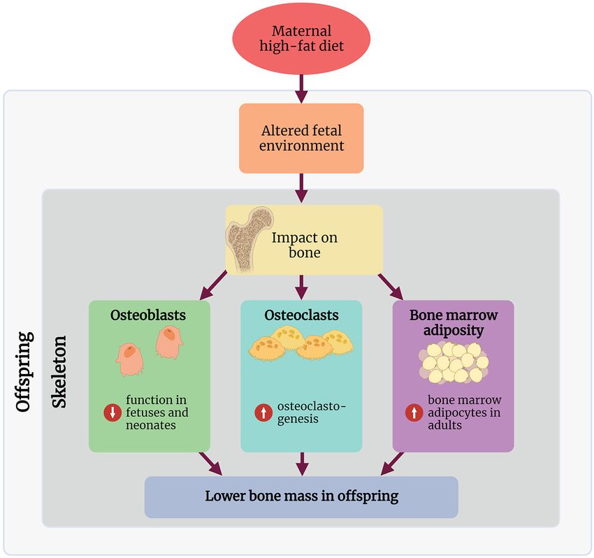
EFFECT OF MATERNAL OBESITY AND phenotype in these offspring (Figure 1).
HFD ON BONE IN HUMANS
Several studies have specifically addressed whether maternal Osteoblasts
obesity or maternal HFD during pregnancy affects bone Osteoblasts are derived from mesenchymal stem cells and are
development in offspring, both in utero and post-partum (33). responsible for the synthesis and mineralisation of bone. Whilst
Longitudinal studies show that obese mothers have babies with osteoblast number is unaffected (22, 24), there may be a
increased body length, whole-body bone area, and mineral negative relationship between osteoblast function in offspring
content (34–36), but maternal diet was not reported. Two studies and maternal HFD during pregnancy and lactation. However,
demonstrated that mothers consuming a high-fat “Western diet” this relationship with osteoblast function may be transient and
during pregnancy, defined as a diet high in meat, processed lost as offspring age. Whole-embryo skeletal ossification and total
food, and saturated fat, have children with lower whole-body bone volume are decreased following maternal HFD (18, 19).
bone area, bone mineral content, and BMD, compared with Rat calvarial osteoblasts from offspring exposed to maternal
children of mothers on low-fat “prudent diets” during pregnancy, HFD have decreased proliferation and osteoblastic differentiation
defined as a diet high in fruits, vegetables, grains and low-fat (19). Therefore, decreased differentiation of osteoblasts could
Frontiers in Nutrition | www.frontiersin.org 4 August 2021 | Volume 8 | Article 730037Buckels et al. Maternal High-Fat Diet and Bone
FIGURE 1 | Summary of the proposed mechanisms through which maternal high-fat diet affects the skeleton in the offspring. Created with BioRender.com.
directly be responsible for decreased or delayed bone formation offspring. Surprisingly, Devlin et al. also found no difference in
during development. concentrations of the bone formation marker type 1 procollagen
Although bone marrow stromal cells (BMSCs) are a N-terminal (P1NP) in circulation at either 14- or 26-weeks of age,
significant source of osteoblast progenitor cells contributing despite observed differences in MAR (24). Overall, there is no
to bone remodeling, only one study examined whether the consensus on whether maternal HFD affects osteoblast function
differentiation capacity of these cells into osteoblasts is influenced in adult offspring.
by maternal HFD. Kushwaha et al. assessed cellular activity
in 15-week old animals and found no difference in osteogenic Osteoclasts
differentiation of BMSCs exposed to maternal HFD. However, Osteoclasts are multinucleated phagocytic cells responsible for
compared to BMSCs derived from CD-fed mothers, these cells bone resorption and are derived from the macrophage-monocyte
have higher mRNA expression of RANKL, which will have cell lineage. Very few studies have considered the effects of
implications for osteoclastogenesis (22). maternal HFD on osteoclasts. One study broadly examined
Osteoblast function in adult rodents exposed to maternal HFD ex vivo osteoclastogenesis following maternal HFD during
is variable. Circulating osteocalcin concentrations are decreased pregnancy and lactation (22). Kushwaha et al. demonstrated
at 17-weeks of age in mice, indicating osteoblast function is via histomorphometry that osteoclast number, erosion surface,
decreased following maternal HFD. Mineral apposition rate and osteoclast surface were increased in 15-week old male
(MAR) is a reliable direct measurement of osteoblast function rats exposed to maternal HFD. Ex vivo cultures of osteoclast
(40); one rat study found no difference in MAR in 15-week old precursors isolated from these animals had increased potential
males following maternal HFD (22). Interestingly, Devlin et al. to differentiate into osteoclasts, with these osteoclasts more
found that MAR is increased in 14-week old male mice exposed numerous and larger. Interestingly, these osteoclasts were more
to maternal HFD (24). This relationship is no longer detected sensitized to the effects of RANKL and had increased RANK
in male mice at 26-weeks of age (24), indicating the rate of mRNA expression. Cultured osteoblasts from these same animals
mineralization has decreased to a level similar to maternal CD had increased RANKL mRNA expression, indicating that the
Frontiers in Nutrition | www.frontiersin.org 5 August 2021 | Volume 8 | Article 730037Buckels et al. Maternal High-Fat Diet and Bone
potential for osteoclastogenesis is increased following maternal to be through increased expression of p300/CBP, which
HFD. Alternatively, when mice were fed HFD for 6-weeks before increased H3K27 acetylation, which promoted p53/p21-
mating, pregnancy, and lactation, their offspring demonstrated mediated cell senescence signaling in pre-osteoblasts; increased
no significant difference in osteoclast number but decreased expression of p300/CPB persisted until adulthood (18).
osteoclast activity at 14- and 26-weeks of age (24). Increased bone Maternal HFD also promoted increases in methylated CpG
resorption is a major contributor to decreased bone volume that sites in the homeobox protein A10 (HoxA10) promoter.
develops when rodents are fed HFD, which is likely secondary HoxA10 is important for fetal osteoblastogenesis and adult
to increased inflammation (41). Maternal HFD is known to bone regeneration.
cause low-grade chronic inflammation in offspring; therefore,
it is feasible that this could contribute to bone loss in these CURRENT RESEARCH GAPS
animals (42). Chen et al. noted increased inflammatory cytokine
production in fetal calvarial osteoblasts exposed to maternal Many unanswered questions surrounding how HFD-
HFD, but this potential mechanism has not been addressed induced maternal obesity affects bone development and
in adult offspring (18). Given these conflicting data, further microarchitecture in offspring remain.
studies are needed to determine the effects of maternal HFD on One outstanding question is whether the detrimental effects
osteoclast number and function. on offspring skeleton are driven by maternal obesity, maternal
HFD, or both. In humans, most studies explore the effects of
Bone Marrow Adiposity obesity during pregnancy, commonly assessed by measuring
The balance between BMSCs giving rise to osteogenic or body mass index rather than dietary patterns (46). In rodents,
adipogenic precursors is critical for maintaining bone mass; if the majority of studies implemented a maternal HFD regime
this balance is shifted toward adipogenesis, this may come at at least 4 weeks before mating. This would have induced
the expense of osteoblastogenesis (43). Additionally, increased an obesity phenotype in these dams, as well as ongoing
bone-marrow adiposity can affect osteogenesis, with an apparent exposure to HFD. However, it is unlikely that maternal
negative relationship between bone marrow adiposity and bone obesity would have been induced in studies where HFD-
mass (44). Maternal HFD during pregnancy and lactation (29) feeding was restricted to pregnancy and lactation, or lactation
or lactation only (28) is associated with increased adipocyte alone. Therefore, these offspring likely experienced exposure
number and adipocyte size in the bone marrow cavity. However, to HFD in the absence of maternal obesity. It is challenging
interpretation of these studies is complicated by their study to tease out whether HFD-induced obesity before pregnancy
design; offspring were either weaned directly onto HFD or CD, or ongoing maternal HFD affects the skeleton in offspring
or onto CD followed by HFD between 12 and 24-weeks of age. using a rodent model. Unlike in humans, changing the diet
Both studies had conflicting results as to whether maternal HFD of a rodent from HFD to CD induces rapid weight loss
with post-weaning HFD affected bone microarchitecture. Hafner (47). This could be overcome using embryo transfer following
et al. found bone marrow adiposity was increased, and trabecular pre-conception maternal HFD, placing embryos into CD-fed
bone volume was decreased in the maternal HFD/post-weaning recipients (46).
HFD group at 24-weeks of age (28). However, Lanham et al. Another gap in our understanding is deciphering the effects of
found no difference in bone microarchitecture at 30-weeks of maternal HFD on other tissues in offspring and how these effects,
age (29). No studies performed ex vivo adipogenesis assays on in turn, modulate the skeleton. For instance, there is cross-talk
BMSCs. Further studies are required to confirm whether changes between the skeleton and skeletal muscle, adipose tissue, and the
in bone marrow adiposity contribute to the bone phenotype in endocrine pancreas (48–52). The structure and function of these
these offspring. tissues are affected by the early life environment (7). Therefore,
it would be interesting to determine whether this cross-talk
Epigenetic Modifications is affected by maternal HFD and the downstream effects on
Epigenetic modifications, including DNA methylation the skeleton.
and various post-translational histone modifications, In this mini-review, we exclusively discussed the effect
describe changes to gene expression that occur without of maternal HFD on the skeleton in offspring; however,
affecting the underlying DNA sequence. Epigenetic other paradigms of early life exposure to nutritional excess
modifications allow the individual to alter gene expression are also worthy of exploration. For instance, a maternal
in response to the environment and have long been high-protein diet (53) and a combination of high-fat and
considered a principal mechanism through which the high-sugar diet (54) also negatively impact the skeleton in
early-life environment affects offspring (7, 45). Despite offspring. Additionally, pre-conception paternal nutrition
this connection, there is a paucity of studies that have also has long-term effects on the metabolic health of
specifically measured epigenetic modifications in response offspring (55). We are unaware of any studies that have
to maternal HFD. addressed the paternal influence of skeletal development in
Chen et al. demonstrated that maternal HFD promotes offspring. Thus, understanding the impact of paternal health
cellular senescence in fetal calvarial osteoblasts, potentially will also be necessary for understanding the mechanisms
suppressing bone formation in the prenatal period in that link the early life environment with skeletal health
both mice and rats (18–20). These findings were shown in offspring.
Frontiers in Nutrition | www.frontiersin.org 6 August 2021 | Volume 8 | Article 730037Buckels et al. Maternal High-Fat Diet and Bone
Critically, we are unaware of any studies investigating AUTHOR CONTRIBUTIONS
the effect of nutritional, pharmacological, or behavioral
interventions on skeletal outcomes in the offspring. EB designed the review. EB, SB, and MT collected relevant
Whilst we do not yet fully understand the mechanisms articles. All authors have contributed to writing and revision of
that impact the developing skeleton in response to the manuscript, read, and approved the submitted version.
maternal HFD, a significant gap lies in the lack of
intervention studies. FUNDING
This research was funded by Health Research Council of
CONCLUSION
New Zealand Sir Charles Hercus Fellowship, American Society
There is growing evidence that exposure to maternal HFD during of Bone and Mineral Research Rising Star Award, Auckland
pregnancy has long-lasting adverse effects on the skeleton of Medical Research Foundation Project Grant 1118008 to BM.
offspring. However, many details surrounding these changes and The University of Auckland Doctoral Scholarship to MT. Health
the mechanisms of action that drive these effects remain unclear, Research Council Clinical Research Training Fellowship to SB.
and further basic studies are required. Given the consequences
of low bone mass and deranged bone microarchitecture for ACKNOWLEDGMENTS
offspring, advances in our understanding of the developmental
origins of bone health is critical in our battle against diseases The authors would like to thank Marcus Ground for assistance
like osteoporosis. with drawing the diagram.
REFERENCES 15. Boudin E, Fijalkowski I, Hendrickx G, Van Hul W. Genetic control of bone
mass. Mol Cell Endocrinol. (2016) 432:3–13. doi: 10.1016/j.mce.2015.12.021
1. Chooi YC, Ding C, Magkos F. The epidemiology of obesity. Metab Clin Exp. 16. Warden SJ, Roosa SMM, Kersh ME, Hurd AL, Fleisig GS, Pandy MG, et
(2019) 92:6–10. doi: 10.1016/j.metabol.2018.09.005 al. Physical activity when young provides lifelong benefits to cortical bone
2. Kelly T, Yang W, Chen C-S, Reynolds K, He J. Global burden of size and strength in men. Proc Natl Acad Sci USA. (2014) 111:5337–42.
obesity in 2005 and projections to 2030. Int J Obesity. (2008) 32:1431–7. doi: 10.1073/pnas.1321605111
doi: 10.1038/ijo.2008.102 17. Seeman E, Martin TJ. Antiresorptive and anabolic agents in the prevention
3. Apovia CM. Obesity: definition, comorbidities, causes, and burden. Am J and reversal of bone fragility. Nat Rev Rheumatol. (2019) 15:225–36.
Manag Care. (2016) 22.7:s176–85. doi: 10.1038/s41584-019-0172-3
4. Davies GA, Maxwell C, McLeod L, Gagnon R, Basso M, Bos H, et 18. Chen J-R, Lazarenko OP, Zhao H, Alund AW, Shankar K. Maternal obesity
al. Obesity in pregnancy. J Obstetr Gynaecol Canada. (2010) 32:165–73. impairs skeletal development in adult offspring. J Endocrinol. (2018) 239:33–
doi: 10.1016/S1701-2163(16)34432-2 47. doi: 10.1530/JOE-18-0244
5. Sirimi N, Goulis DG. Obesity in pregnancy. Hormones. (2010) 9:299–306. 19. Chen J-R, Zhang J, Lazarenko OP, Kang P, Blackburn ML, Ronis MJJ, et al.
doi: 10.14310/horm.2002.1280 Inhibition of fetal bone development through epigenetic down-regulation
6. Catalano PM. Obesity, insulin resistance and pregnancy outcome. of HoxA10 in obese rats fed high-fat diet. FASEB J. (2012) 26:1131–41.
Reproduction. (2010) 140:365–71. doi: 10.1530/REP-10-0088 doi: 10.1096/fj.11-197822
7. Warner MJ, Ozanne SE. Mechanisms involved in the developmental 20. Chen J-R, Lazarenko OP, Blackburn ML, Rose S, Frye RE, Badger TM, et al.
programming of adulthood disease. Biochem J. (2010) 427:333–47. Maternal obesity programs senescence signaling and glucose metabolism in
doi: 10.1042/BJ20091861 osteo-progenitors from rat and human. Endocrinology. (2016) 157:4172–83.
8. Fernandez-Twinn DS, Hjort L, Novakovic B, Ozanne SE, Saffery R. doi: 10.1210/en.2016-1408
Intrauterine programming of obesity and type 2 diabetes. Diabetologia. (2019) 21. Liang C, Oest ME, Jones JC, Prater MR. Gestational high saturated fat diet
62:1789–801. doi: 10.1007/s00125-019-4951-9 alters C57BL/6 mouse perinatal skeletal formation. Birth Defects Res. (2009)
9. Eriksson JG. Developmental pathways and programming of 86:362–9. doi: 10.1002/bdrb.20204
diabetes: epidemiological aspects. J Endocrinol. (2019) 242:T95–104. 22. Kushwaha P, Khambadkone SG, Li M, Goodman EJ, Aravindan N, Riddle RC,
doi: 10.1530/JOE-18-0680 et al. Maternal high-fat diet induces long-lasting defects in bone structure in
10. Hur SS, Cropley JE, Suter CM. Paternal epigenetic programming: rat offspring through enhanced osteoclastogenesis. Calcified Tissue Int. (2021)
evolving metabolic disease risk. J Mol Endocrinol. (2017) 58:R159–68. 108:680–92. doi: 10.1007/s00223-020-00801-4
doi: 10.1530/JME-16-0236 23. Miotto PM, M CL, Amoye F, LeBlanc PJ, Peters SJ, Roy BD, et al. Maternal high
11. Vickers MH. Developmental programming and transgenerational fat feeding does not have long-lasting effects on body composition and bone
transmission of obesity. Ann Nutr Metab. (2014) 64:26–34. health in female and male Wistar rat offspring at young adulthood. Molecules.
doi: 10.1159/000360506 (2013) 18:15094–109. doi: 10.3390/molecules181215094
12. Drake AJ, Liu L. Intergenerational transmission of programmed effects: 24. Devlin MJ, Grasemann C, Cloutier AM, Louis L, Alm C, Palmert MR, et
public health consequences. Trends Endocrinol Metab. (2010) 21:206–13. al. Maternal perinatal diet induces developmental programming of bone
doi: 10.1016/j.tem.2009.11.006 architecture. J Endocrinol. (2013) 217:69–81. doi: 10.1530/JOE-12-0403
13. Desai M, Jellyman JK, Han G, Beall M, Lane RH, Ross MG. Maternal 25. Harasymowicz NS, Choi Y-R, Wu C-L, Iannucci L, Tang R, Guilak
obesity and high-fat diet program offspring metabolic syndrome. F. Intergenerational transmission of diet-induced obesity, metabolic
Am J Obstetr Gynecol. (2014) 211:e1–13. doi: 10.1016/j.ajog.2014. imbalance, and osteoarthritis in mice. Arthrit Rheumatol. (2020) 72:632–44.
03.025 doi: 10.1002/art.41147
14. Williams L, Seki Y, Vuguin PM, Charron MJ. Animal models of in utero 26. Liang C, Oest ME, Prater MR. Intrauterine exposure to high saturated fat diet
exposure to a high fat diet: a review. Biochim Biophys Acta. (2014) 1842:507– elevates risk of adult-onset chronic diseases in C57BL/6 mice. Birth Defects
19. doi: 10.1016/j.bbadis.2013.07.006 Res. (2009) 86:377–84. doi: 10.1002/bdrb.20206
Frontiers in Nutrition | www.frontiersin.org 7 August 2021 | Volume 8 | Article 730037Buckels et al. Maternal High-Fat Diet and Bone
27. Lanham SA, Cagampang FR, Oreffo ROC. Maternal high-fat diet and 43. Rharass T, Lucas S. Mechanisms in endocrinology: bone marrow adiposity
offspring expression levels of vitamin K-dependent proteins. Endocrinology. and bone, a bad romance? Eur J Endocrinol. (2018) 179:R165–82.
(2014) 155:4749–61. doi: 10.1210/en.2014-1188 doi: 10.1530/EJE-18-0182
28. Hafner H, Chang E, Carlson Z, Zhu A, Varghese M, Clemente J, et 44. Devlin MJ, Rosen CJ. The bone-fat interface: basic and clinical implications
al. Lactational high-fat diet exposure programs metabolic inflammation of marrow adiposity. Lancet Diabetes Endocrinol. (2015) 3:141–7.
and bone marrow adiposity in male offspring. Nutrients. (2019) 11:1393. doi: 10.1016/S2213-8587(14)70007-5
doi: 10.3390/nu11061393 45. Bansal A, Simmons RA. Epigenetics and developmental origins of metabolic
29. Lanham SA, Roberts C, Hollingworth T, Sreekumar R, Elahi MM, Cagampang dysfunction: correlation or causation? Am J Physiol Endocrinol Metab. (2018)
FR, et al. Maternal high-fat diet: effects on offspring bone structure. Osteopor 315:E15–28. doi: 10.1152/ajpendo.00424.2017
Int. (2010) 21:1703–14. doi: 10.1007/s00198-009-1118-4 46. Christians JK, Lennie KI, Wild LK, Garcha R. Effects of high-fat diets on fetal
30. Lanham SA, Cagampang FR, Oreffo ROC. Maternal high fat diet affects growth in rodents: a systematic review. Reprod Biol Endocrinol. (2019) 17:39.
offspring’s vitamin K-dependent proteins expression levels. PLoS ONE. (2015) doi: 10.1186/s12958-019-0482-y
10:e0138730. doi: 10.1371/journal.pone.0138730 47. Matikainen-Ankney BA, Ali MA, Miyazaki NL, Fry SA, Licholai JA, Kravitz
31. Kimmel DB, Jee WSS. Bone cell kinetics during longitudinal bone growth in AV. Weight loss after obesity is associated with increased food motivation and
the rat. Calcified Tissue Int. (1980) 32:123–33. doi: 10.1007/BF02408531 faster weight regain in mice. Obesity. (2020) 28:851–6. doi: 10.1002/oby.22758
32. Dickinson H, Moss TJ, Gatford KL, Moritz KM, Akison L, Fullston T, 48. Ducy P. The role of osteocalcin in the endocrine cross-talk between
et al. A review of fundamental principles for animal models of DOHaD bone remodelling and energy metabolism. Diabetologia. (2011) 54:1291–7.
research: an Australian perspective. J Dev Origins Health Dis. (2016) 7:449–72. doi: 10.1007/s00125-011-2155-z
doi: 10.1017/S2040174416000477 49. Sims NA, Walsh NC. Intercellular cross-talk among bone cells:
33. Jensen KH, Riis KR, Abrahamsen B, Handel MN. Nutrients, diet, and other new factors and pathways. Curr Osteopor Rep. (2012) 10:109–17.
factors in prenatal life and bone health in young adults: a systematic review of doi: 10.1007/s11914-012-0096-1
longitudinal studies. Nutrients. (2020) 12:2866. doi: 10.3390/nu12092866 50. Argiles JM, Lopez-Soriano J, Almendro V, Busquets S, López-Soriano FJ.
34. Enstad S, Cheema S, Thomas R, Fichorova RN, Martin CR, O’Tierney-Ginn Cross-talk between skeletal muscle and adipose tissue: a link with obesity?
P, et al. The impact of maternal obesity and breast milk inflammation on Med Res Rev. (2004) 25:49–65. doi: 10.1002/med.20010
developmental programming of infant growth. Eur J Clin Endocrinol. (2021) 51. Brotto M, Johnson ML. Endocrine crosstalk between muscle and bone. Curr
75:180–8. doi: 10.1038/s41430-020-00720-5 Osteopor Rep. (2014) 12:135–41. doi: 10.1007/s11914-014-0209-0
35. Harvey NC, Javaid MK, Arden NK, Poole JR, Crozier SR, Robinson 52. Li F, Li Y, Duan Y, Hu C-AA, Tang Y, Yin Y. Myokines and adipokines:
SM, et al. Maternal predictors of neonatal bone size and geometry: the involvement in the crosstalk between skeletal muscle and adipose tissue. Cytok
Southampton Women’s Survey. J Dev Origins Health Dis. (2010) 1:35–41. Growth Fact Rev. (2017) 33:73–82. doi: 10.1016/j.cytogfr.2016.10.003
doi: 10.1017/S2040174409990055 53. Ellur G, Sukhdeo SV, Khan MT, Sharan K. Maternal high protein-
36. Zhang C, Hediger ML, Albert PS, Grewal J, Sciscione A, Grobman WA, et al. diet programs impairment of offspring’s bone mass through miR-24-1-5p
Association of maternal obesity with longitudinal ultrasonographic measures mediated targeting of SMAD5 in osteoblasts. Cell Mol Life Sci. (2021) 78:1729–
of fetal growth: findings from the NICHD fetal growth studies–singletons. 44. doi: 10.1007/s00018-020-03608-6
JAMA Pediatr. (2018) 172:24–31. doi: 10.1001/jamapediatrics.2017.3785 54. Shi Y, Saben JL, He G, Moley KH, Long F. Diet-induced metabolic
37. Cole ZA, Gale CR, Javaid MK, Robinson SM, Law C, Boucher BJ, et dysregulation in female mice causes osteopenia in adult offspring. J Endocr
al. Maternal dietary patterns during pregnancy and childhood bone Soc. (2020) 4:1–14. doi: 10.1210/jendso/bvaa028
mass: a longitudinal study. J Bone Mineral Res. (2009) 24:663–8. 55. Soubry A. POHaD: why we should study future fathers. Environ Epigenet.
doi: 10.1359/jbmr.081212 (2018) 4:dvy007. doi: 10.1093/eep/dvy007
38. Yin J, Dwyer T, Cochrane J, Jones G. The association between maternal diet
during pregnancy and bone mass of the children at age 16. Eur J Clin Nutr. Conflict of Interest: The authors declare that the research was conducted in the
(2010) 64:131–7. doi: 10.1038/ejcn.2009.117 absence of any commercial or financial relationships that could be construed as a
39. Petersen SB, Rasmussen MA, Olsen SF, Vestergaard P, Mølgaard C, potential conflict of interest.
Halldorsson TI, et al. Maternal dietary patterns during pregnancy in
relation to offspring forearm fractures: prospective study from the Danish Publisher’s Note: All claims expressed in this article are solely those of the authors
National Birth Cohort. Nutrients. (2015) 7:2382–400. doi: 10.3390/nu70 and do not necessarily represent those of their affiliated organizations, or those of
42382
the publisher, the editors and the reviewers. Any product that may be evaluated in
40. Recker RR, Kimmel DB, Dempster D, Weinstein RS, Wronski TJ, Burr
this article, or claim that may be made by its manufacturer, is not guaranteed or
DB. Issues in modern bone histomorphometry. Bone. (2011) 49:955–64.
doi: 10.1016/j.bone.2011.07.017 endorsed by the publisher.
41. Shu L, Beier E, Sheu T, Zhang H, Zuscik MJ, Puzas EJ, et al. High-fat diet causes
bone loss in young mice by promoting osteoclastogenesis through alteration Copyright © 2021 Buckels, Bolam, Tay and Matthews. This is an open-access article
of the bone marrow environment. Calcified Tissue Int. (2015) 96:313–23. distributed under the terms of the Creative Commons Attribution License (CC BY).
doi: 10.1007/s00223-015-9954-z The use, distribution or reproduction in other forums is permitted, provided the
42. Zhou D, Pan YX. Pathophysiological basis for compromised health original author(s) and the copyright owner(s) are credited and that the original
beyond generations: role of maternal high-fat diet and low-grade chronic publication in this journal is cited, in accordance with accepted academic practice.
inflammation. J Nutr Biochem. (2015) 26:1–8. doi: 10.1016/j.jnutbio.2014. No use, distribution or reproduction is permitted which does not comply with these
06.011 terms.
Frontiers in Nutrition | www.frontiersin.org 8 August 2021 | Volume 8 | Article 730037You can also read