THE KNOCKOUT FOR G PROTEIN-COUPLED RECEPTOR-LIKE PFSR25 INCREASES THE SUSCEPTIBILITY OF MALARIA PARASITES TO THE ANTIMALARIALS LUMEFANTRINE AND ...
←
→
Page content transcription
If your browser does not render page correctly, please read the page content below
ORIGINAL RESEARCH
                                                                                                                                                published: 15 March 2021
                                                                                                                                        doi: 10.3389/fmicb.2021.638869

                                              The Knockout for G Protein-Coupled
                                              Receptor-Like PfSR25 Increases the
                                              Susceptibility of Malaria Parasites to
                                              the Antimalarials Lumefantrine and
                                              Piperaquine but Not to Medicine for
                                              Malaria Venture Compounds
                                              Benedito M. Santos 1, Bárbara K. M. Dias 2, Myna Nakabashi 1 and Celia R. S. Garcia 1*
                                              1
                                               Department of Clinical and Toxicological Analysis, School of Pharmaceutical Sciences, University of São Paulo, São Paulo,
                                              Brazil, 2 Department of Parasitology, Institute of Biomedical Sciences, University of São Paulo, São Paulo, Brazil

                                              Previously we have reported that the G protein-coupled receptor (GPCR)-like PfSR25 in
                           Edited by:         Plasmodium falciparum is a potassium (K+) sensor linked to intracellular calcium signaling
            Annette Elizabeth Kaiser,
        University of Duisburg-Essen,
                                              and that knockout parasites (PfSR25-) are more susceptible to oxidative stress and
                             Germany          antimalarial compounds. Here, we explore the potential role of PfSR25 in susceptibility
                        Reviewed by:          to the antimalarial compounds atovaquone, chloroquine, dihydroartemisinin, lumefantrine,
               Vanessa Zuzarte-Luis,
                                              mefloquine, piperaquine, primaquine, and pyrimethamine and the Medicine for Malaria
        University of Lisbon, Portugal
                 Mirian A. F. Hayashi,        Venture (MMV) compounds previously described to act on egress/invasion (MMV006429,
      Federal University of São Paulo,        MMV396715, MMV019127, MMV665874, MMV665878, MMV665785, and MMV66583)
                                 Brazil
                                              through comparative assays with PfSR25- and 3D7 parasite strains, using flow cytometry
                  *Correspondence:
                    Celia R. S. Garcia
                                              assays. The IC50 and IC90 results show that lumefantrine and piperaquine have greater
                      cgarcia@usp.br          activity on the PfSR25- parasite strain when compared to 3D7. For MMV compounds,
                                              we found no differences between the strains except for the compound MMV665831,
                  Specialty section:
        This article was submitted to         which we used to investigate the store-operated calcium entry (SOCE) mechanism. The
       Antimicrobials, Resistance and         results suggest that PfSR25 may be involved in the mechanism of action of the antimalarials
                       Chemotherapy,
                                              lumefantrine and piperaquine. Our data clearly show that MMV665831 does not affect
               a section of the journal
             Frontiers in Microbiology        calcium entry in parasites after we depleted their internal calcium pools with thapsigargin.
       Received: 07 December 2020             The results demonstrated here shed light on new possibilities on the antimalarial
        Accepted: 17 February 2021            mechanism, bringing evidence of the involvement of the GPCR-like PfSR25.
         Published: 15 March 2021

                             Citation:        Keywords: antimalarials, G protein-coupled receptor-like, medicine for malaria venture compounds, PfSR25,
               Santos BM, Dias BKM,           Plasmodium falciparum
Nakabashi M and Garcia CRS (2021)
The Knockout for G Protein-Coupled
 Receptor-Like PfSR25 Increases the
 Susceptibility of Malaria Parasites to
                                              INTRODUCTION
  the Antimalarials Lumefantrine and
 Piperaquine but Not to Medicine for
                                              Present in more than 90 countries spread across different continents, malaria is still an infectious
        Malaria Venture Compounds.            disease with a high global impact. Epidemiological data released by the WHO estimate there
        Front. Microbiol. 12:638869.          are approximately 228 million cases of the disease, causing more than 400,000 deaths annually,
   doi: 10.3389/fmicb.2021.638869             with children under 5 years of age being the group that is most affected by the disease and

Frontiers in Microbiology | www.frontiersin.org                                      1                                            March 2021 | Volume 12 | Article 638869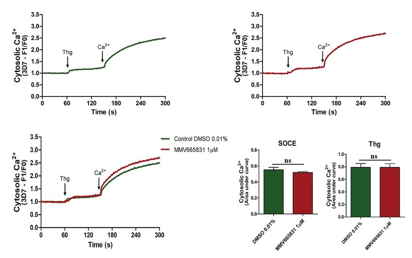
Santos et al.                                                                              Plasmodium Receptor PfSR25 and Resistance to Antimalarials

representing 67% of the total deaths from malaria globally                pyrimethamine. These compounds possess different mechanisms
(World malaria report, 2019).                                             of action to block the P. falciparum cell cycle.
    The complex biological cycle of malaria includes different                We next compared the response of the two P. falciparum
microenvironments in the Anopheles mosquito vector, the liver             strains, 3D7 and the GPCR-like PfSR25 knockout, to antimalarial
stage and the intraerythrocytic cycle, which helps to hamper              compounds from the Medicine for Malaria Venture (MMV)
the control and eradication of the disease (Koyama et al., 2009;          library (MMV006429, MMV396715, MMV019127, MMV665874,
Da Silva et al., 2013; Schuck et al., 2013; Josling and Llinás,           MMV665878, MMV665785, and MMV665831). For these studies,
2015). Another important factor that has also contributed to              we used flow cytometry and markers for parasite nucleic acid
the ineffectiveness of combating malaria is the emergence of              staining, namely, SYBR Green I and MitoTracker Deep Red,
strains resistant to the drugs used for the treatment (Khoury             to evaluate the mitochondrial membrane potential of the parasites.
et al., 2020; Wicht et al., 2020). The widespread use of                      In addition, we also investigated the involvement of the
antimalarials in recent decades has triggered a selection of              promising anti-malarial compound MMV665831, which acts
parasites, causing Plasmodium falciparum to develop drug                  to block the parasite’s egress/invasion (Subramanian et al.,
resistance mechanisms (Le Bras and Durand, 2003; White, 2004).            2018), regarding its involvement in store-operated calcium entry
    In an attempt to delay antimalarial resistance, the use of            (SOCE). For that experiment, wild-type parasites (3D7) were
combination therapies, such as the so-called artemisinin-based            loaded with Fluo-4/AM, and the calcium dynamics were
therapies (ACTs), which include an artemisinin analog in                  monitored in parasites that had previously been incubated with
conjunction with another longer-acting compound, has been                 1 μM of the MMV665831 compound. Our data show no
established (Kremsner and Krishna, 2004). In many cases,                  significant difference in the SOCE assays when the 3D7 parasites
resistance is associated with mutations in the genes for carrier          were preincubated in the presence of MMV665831 compared
proteins such as Plasmodium falciparum chloroquine resistance             with the control, in which using the solvent alone as the
transporter (PfCRT) and Plasmodium falciparum multidrug                   compound does not affect calcium entry in parasites. These
resistance 1 (PfMDR1; Reed et al., 2000; Le Bras and                      data reinforce the idea that the secondary messengers, such
Durand, 2003; Lee et al., 2018, Wicht et al., 2020).                      as cGMP and cAMP, are central to signaling aspects of parasite
    However, the mechanisms of action and the factors that                egress and invasion.
lead to drug resistance are not yet clear (Price et al., 2006).
The sequencing of the Plasmodium genome enabled the
acquisition of new information regarding this parasite’s biology,         MATERIALS AND METHODS
such as the identification of G protein-coupled receptor (GPCR)-
like (PfSR1, PfSR10, PfSR12, and PfSR25; Madeira et al., 2008).           All experiments were carried out following the standard biosafety
GPCRs are a large protein family of transmembrane receptors               and institutional safety procedures required by the School of
that are involved in the most diverse signal transduction                 Pharmaceutical Sciences at the University of São Paulo. The
pathways (Alberts et al., 2017; Alhadeff et al., 2018; Pereira            handling of P. falciparum cultures was carried out in a cell
et al., 2020). GPCRs represent almost 50% of pharmaceutical               culture facility with biosafety level 2.
company drug targets (Hauser et al., 2017; Sriram and Insel, 2018).
    During its complex life cycle, the parasite switches from
                                                                          Maintenance of Plasmodium falciparum
an environment with a high potassium (K+) concentration to
a low K+ concentration. Previous studies on the characterization
                                                                          Cultures
                                                                          Red blood cells infected with P. falciparum strain 3D7 and
of the GPCR-like PfSR25 by Moraes et al. (2017) indicated
                                                                          the PfSR25 receptor knockout strain (PfSR25-) were grown
that the parasite receptor (PfSR25) acts as a K+ sensor that
                                                                          according to (Trager and Jensen, 1976). The cultures were
is linked to increases in cytosolic calcium (Ca2+cyt). The Ca2+cyt
                                                                          kept in 175 cm2 culture bottles (Greiner Bio-One) containing
increase is triggered in assays performed on isolated parasites,
                                                                          RPMI 1640 (Gibco) with 0.5% NaHCO3, 0.04% gentamicin
and the observed Ca2+ increase could be blocked by either
                                                                          sulfate, and 0.05% hypoxanthine and supplemented with 0.5%
the phospholipase C (PLC) inhibition or by depleting the
                                                                          AlbuMAX I (Gibco). The culture bottles were maintained at
internal Ca2+ stores. However, when the pfsr25 gene was deleted,
                                                                          37°C under a mixture of gases consisting of 90% N2, 5% O2,
no rise in Ca2+cyt was observed in response to the change in
                                                                          and 5% CO2. The parasitemia was determined from a blood
K+ concentration. The knockout parasite has also been shown
                                                                          smear stained with Rapid Panotic (Laborclin). PfSR25- parasites
to be more susceptible to oxidative stress, amino acid deprivation,
                                                                          were selected with 2.5 μg/ml blasticidin (Sigma-Aldrich).
and some antimalarial compounds (Moraes et al., 2017). Recently,
Santos et al. (2020) reported the susceptibility of the PfSR25
knockout strain to synthetic compounds derived from 1H-and                Incubation Tests of Antimalarials in Plasmodium
2H-1,2,3-triazole ring stereoisomers.                                     falciparum Cultures
    To further characterize the role of PfSR25 in P. falciparum,          To assess the susceptibility of the P. falciparum 3D7 and PfSR25-
we evaluated the IC50 and IC90 for the wild-type strain (3D7)             strains to the classic antimalarials, asynchronous cultures of
and the SR25-knockout strain (PfSR25-), incubating them with              P. falciparum (3D7 and PfSR25-) with 0.3% parasitemia and
the antimalarials atovaquone, chloroquine, dihydroartemisinin,            1% hematocrit were incubated in 96-well plates for 72 h with
lumefantrine, mefloquine, piperaquine, primaquine, and                    antimalarial compounds at different concentrations at 37°C

Frontiers in Microbiology | www.frontiersin.org                       2                                        March 2021 | Volume 12 | Article 638869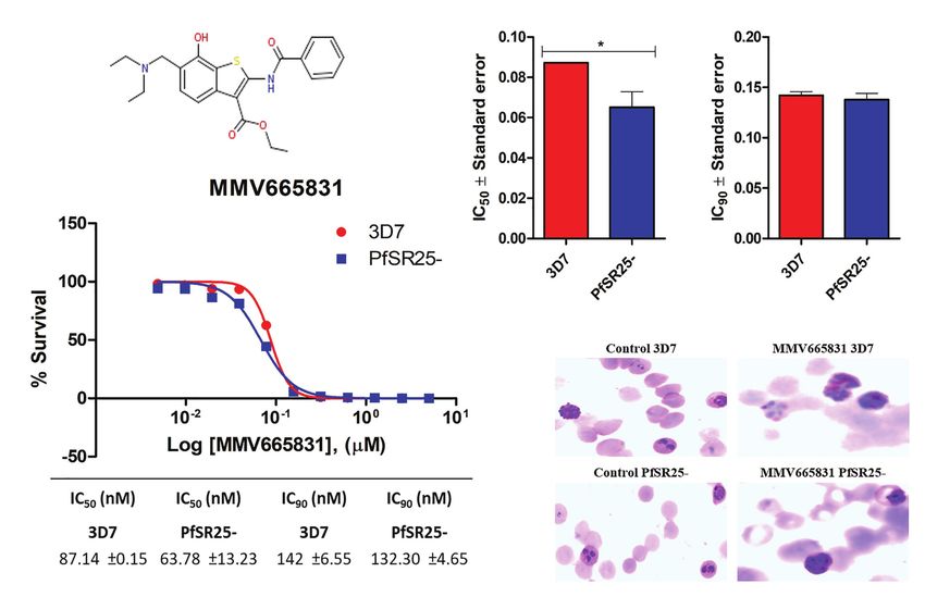
Santos et al.                                                                              Plasmodium Receptor PfSR25 and Resistance to Antimalarials

under an atmosphere consisting of 90% N2, 5% O2, and 5%                       The cDNA synthesis was performed using random primers
CO2. As a control, the parasite culture incubation was performed          and reverse transcriptase Superscript II (Invitrogen) according
with Dimethyl sulfoxide (DMSO) solvent at a concentration                 to the manufacturer’s protocol. SYBR Green (Applied Biosystems)
corresponding to the highest percentage present in the tests.             was used in quantitative real-time PCR on a 7300 Real-Time
   Incubation was performed with atovaquone (0.019–20 nM),                PCR System (Applied Biosystems). Amplification was carried
chloroquine (0.24–250 nM), dihydroartemisinin (0.03–40 nM),               out as follows: first step 50°C for 2 min and 95°C for 10 min
lumefantrine (0.12–128 nM), mefloquine (0.25–300 nM),                     for enzymatic activation followed by 40 cycles of 55°C for
piperaquine (0.15–160 nM), primaquine (39.06–40,000 nM),                  0.15 min for denaturation and 60°C for 1 min for annealing/
and pyrimethamine (0.24–250 nM).                                          extension. The primers used were as follows: PF3D7_0713400
                                                                          – Fwd GGGGATTCATACCGTTTTCACA and PF3D7_0713400
Incubation Assays With MMV Library Compounds                              – Rvs AACAAGGCTAGCAGTTCCCA. Changes in relative
The Malaria Box library received from the MMV (Geneva,                    expression were determined by 2(−ΔΔCt). We performed statistical
Switzerland) contained 390 compounds distributed in five                  analysis with ΔΔCt values with relative expression of the
96-well plates that were stored at −20°C until use. Ten microliters       normalized genes for the housekeeping gene seryl-
of cell culture-grade DMSO (Sigma-Aldrich) was added to the               tRNA synthetase.
plates to make 10 mM stocks that were eventually used to
prepare the assays. A selection of the compounds presents in              Spectrofluorometric Quantifications of
the library used in this study was based on the results obtained          Calcium (Ca2+)cyt in 3D7 and PfSR25-
from the work of Subramanian et al. (2018). The selected                  Strains
compounds (MMV006429, MMV396715, MMV019127,                               The calcium dynamics were evaluated as previously mentioned
MMV665874, MMV665878, MMV665785, and MMV665831)                           in (Pecenin et al., 2018). In brief, the parasites were synchronized
were incubated with P. falciparum cultures as mentioned                   with 5% sorbitol (Lambros and Vanderberg, 1979) and isolated
previously, in section “Incubation Tests of Antimalarials in              at the trophozoite stage (28–32 h). They were subsequently
Plasmodium falciparum Cultures.” To determine the IC50 values,            loaded with Fluo-4/AM in buffer M (116 mM NaCl, 5.4 mM
serial dilutions of the compounds at concentrations ranging               KCl, 0.8 mM MgSO4, 5.5 mM d-glucose, 50 mM MOPS,
from 0.0048 to 5 μM were performed in triplicates over three              and 2 mM CaCl2, pH 7.4) containing probenecid 40 μM
independent experiments.                                                  (Sigma), which was used to prevent the sequestration of Fluo-4/
                                                                          AM in the parasitic digestive vacuole. The parasites were
Parasite Marking and Flow Cytometry Assay                                 incubated for 1 h at 37°C. The cells were washed three times
After 72 h of incubation with the compounds, the parasites                in buffer M to remove extracellular Fluo4/AM and then
were dyed with nucleic acid staining SYBR Green I (1X) and                incubated with compound MMV665831 (1 μM) for 15 min
MitoTracker Deep Red (50 nM) as a membrane potential                      at 37°C before the agonist was added. Parasites incubated
marker that tends to deposit on the mitochondrial organelles,             with 0.01% DMSO were used as the control group, in free
and both markers were diluted in PBS (Ekland et al., 2011).               Ca2+ buffer M.
After 20 min of incubation at 37°C, the final parasitemia was                The cytosolic Ca2+ dynamics were monitored with excitation
obtained by reading the plates using a FACSCalibur flow                   at 488 nm and emission at 525 nm using a Shimadzu
cytometer (Becton Dickinson). The results obtained by cytometer           spectrofluorometer (RF5301PC, Japan), with the parasites
were converted into dot plots and analyzed using FlowJo-V10.7.1           (107 cells ml−1) being measured in a 1 ml stirred cuvette. All
software (FL4-H by FL1-H).                                                the tests were performed at 37°C, in triplicate, and three
    All the tests were performed in triplicates (with an N equal          independent experiments were performed.
to three independent experiments), and the parasitemia inhibition            Thapsigargin (5 μM) was added to the buffer (without Ca2+),
percentage was calculated in reference to the solvent (DMSO).             and fluctuations in the [Ca2+]cyt were normalized using the
The dose-response curves and inhibitory concentration values              baseline fluorescence (F1/F0). SOCE measurements were
IC50 and IC90 were calculated using GraphPad Prism-                       performed by adding 2 mM Ca2+ to the cuvette with free Ca2+
V5.01 software.                                                           buffer and calculated using the F1/F0 equation.

Real-Time Quantitative PCR                                                Statistical Analysis
Plasmodium falciparum 3D7 parasites were synchronized with                Three independent experiments were conducted in triplicates
5% sorbitol (Lambros and Vanderberg, 1979). Parasites were                for each of the tests mentioned. The GraphPad Prism 5.01
collected in the trophozoite phase (28–32 h) with approximately           program (GraphPad Software Inc., San Diego, CA,
4% parasitemia. Parasites were treated with 100 nM of piperaquine         United States) was applied to analyze the data. To evaluate
and incubated for 24 h before extraction of the total RNA                 the significant difference between the 3D7 wild-type strain
with Trizol (Invitrogen), 0.01% DMSO was used as control.                 of P. falciparum and the knockout strain for the GPCR-like
Purified RNA was quantified in a UV-Vis Nano Drop 2000c                   PfSR25 the student’s t-test was applied in all the tests performed
(Thermo Scientific). Three independent experiments were carried           in this study. The differences were considered statistically
out in triplicates.                                                       different at p ≤ 0.05.

Frontiers in Microbiology | www.frontiersin.org                       3                                        March 2021 | Volume 12 | Article 638869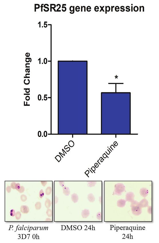
Santos et al.                                                                                                 Plasmodium Receptor PfSR25 and Resistance to Antimalarials

RESULTS                                                                                  SYBR-Green I and labeling the mitochondrial membrane potential
                                                                                         using MitoTracker Deep Red to guarantee the analysis of only
Plasmodium falciparum GPCR-Like                                                          viable parasites (Ekland et al., 2011). A typical parasitemia
PfSR25 Knockout Is More Susceptible to                                                   analysis is shown in Figures 1, 2. Thus, it was possible to
the Antimalarials Lumefantrine and                                                       construct survival curves (Supplementary Figure S1) and
Piperaquine                                                                              determine the IC50 and IC90 values for each compound (Table 1).
To understand the role of GPCR-like PfSR25 in parasite                                      Statistical analysis using Student’s t-test was performed to
susceptibility to antimalarial compounds, we assessed the                                compare the IC50 and IC90 (±SE) values obtained for the
parasitemia of PfSR25- compared to the wild-type strain 3D7                              knockout strain (PfSR25-) to those of the wild-type strain
relative to drugs belonging to the following classes of                                  (3D7). We observed a significant difference for the antimalarials
antimalarials: naphthoquinones (atovaquone), 4-aminoquinolines                           lumefantrine (Figure 1) and piperaquine (Figure 2), for both
(chloroquine        and      piperaquine),        endoperoxides                          the IC50 and the IC90 values in the assays comparing the
(dihydroartemisinin), aryl-amino alcohols (lumefantrine and                              knockout strain (PfSR25-) parasites with the wild-type
mefloquine), 8-aminoquinolines (primaquine), and antifolates                             strain (3D7).
(pyrimethamine), which are used in first-line malaria treatments.                           For the antimalarial lumefantrine (Figure 1), the IC50 and IC90
   Asynchronous parasites (3D7 and PfSR25-) were incubated                               values for the 3D7 strain were 2.65 ± 0.30 nM and 9.77 ± 1.27 nM,
with the drugs at different concentrations for 72 h. To assess                           respectively, while the knockout strain for PfSR25 showed an
the final parasitemia, we used flow cytometry and a                                      IC50 of 1.11 ± 0.04 nM and an IC90 of 2.66 ± 0.62 nM. Thus,
double-labeled methodology for staining the DNA with                                     the antimalarial drug displayed reductions of 58.12 and 72.78%

     A                                                                                      C

     B

                                                                                            D                                         E

 FIGURE 1 | Activity of the antimalarial drug lumefantrine against 3D7 and PfSR25- parasite strains. (A) Chemical structure of the antimalarial lumefantrine. (B) Dot plots
 are shown for different concentrations (128 μM, 8 μM, 0.5 μM and control) after 72 h of incubation. (C) Survival curves of Plasmodium falciparum 3D7 (red) and PfSR25-
 (blue) in an asynchronous blood stage for lumefantrine with concentrations ranging from 0.12 to 128 nM. The IC50 (D) and IC90 (E) values for lumefantrine were evaluated
 by Student’s t-test. The experiments were performed three times independently in triplicate. *Represents the significant difference found using the t-test (***p ≤ 0.001).

Frontiers in Microbiology | www.frontiersin.org                                      4                                              March 2021 | Volume 12 | Article 638869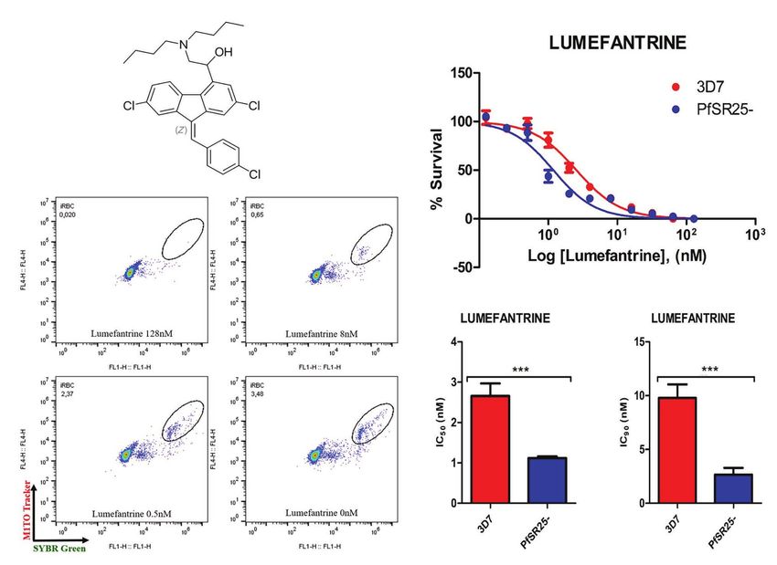
Santos et al. Plasmodium Receptor PfSR25 and Resistance to Antimalarials
A C
B
D E
FIGURE 2 | Activity of the antimalarial drug piperaquine in 3D7 and PfSR25- parasite strains (A) Chemical structure of the antimalarial piperaquine. (B) Dot plots
are shown for different concentrations (160, 10, 0.625 μM and control) after 72 h of incubation. (C) Survival curves of P. falciparum 3D7 (red) and PfSR25- (blue) in
an asynchronous blood stage for piperaquine with concentrations ranging from 0.15 to 160 nM. The IC50 (D) and IC90 (E) values for piperaquine were evaluated by
Student’s t-test. The experiments were performed three times independently, in triplicate. *Represents the significant difference found by t-test (***p ≤ 0.001).
TABLE 1 | Antimalarial drugs, location of the target(s) and IC50 and IC90 values for
the wild-type strain (3D7) and G protein-coupled receptor (GPCR)-like PfSR25
knockout (PfSR25-).
Antimalarial Target location (s) IC50 (nM) 3D7 IC50 (nM) PfSR25- IC90 (nM) 3D7 IC90 (nM) PfSR25-
Atovaquone Mitochondria (Staines 0.15 ± 0.03 0.13 ± 0.01 0.70 ± 0.03 0.98 ± 0.26
et al., 2018)
Chloroquine Digestive vacuole 11.35 ± 1.98 11.57 ± 0.69 19.10 ± 4.29 25.94 ± 1.14
(Olafson et al., 2015)
Dihydroartemisinin Cytosol and digestive 1.77 ± 0.11 1.57 ± 0.54 4.96 ± 1.09 5.17 ± 0.86
vacuole (Bridgford et al.,
2018)
Lumefantrine Cytosol and digestive 2.65 ± 0.30 1.11 ± 0.04 9.77 ± 1.27 2.66 ± 0.62
Mefloquine vacuole (Sullivan, 2017) 9.55 ± 0.44 10.90 ± 2.43 17.43 ± 1.01 22.25 ± 1.58
Piperaquine Digestive vacuole 13.07 ± 1.01 7.27 ± 0.13 18.02 ± 0.37 9.20 ± 0.33
(Dhingra et al., 2017)
Primaquine Unknown 2,310 ± 180 1,950 ± 330 13,730 ± 1,140 12,050 ± 1,370
Pyrimethamine Cytosol (Heinberg and 12.37 ± 1.11 12.06 ± 0.48 28.22 ± 1.48 25.47 ± 0.89
Kirkman, 2015)
for the IC50 and IC90 values, respectively, in the knockout strain For the antimalarial piperaquine (Figure 2), we obtained
(PfSR25) compared to the values of the wild-type strain (3D7), an IC50 value of 13.07 ± 1.01 nM and an IC90 of 18.02 ± 0.37
suggesting greater knockout strain susceptibility to lumefantrine. for the wild-type strain 3D7 parasites. When similar experiments
Frontiers in Microbiology | www.frontiersin.org 5 March 2021 | Volume 12 | Article 638869
Santos et al.                                                                                  Plasmodium Receptor PfSR25 and Resistance to Antimalarials

were performed with the PfSR25- knockout strain, we found                 allow the entry of Ca2+ into the cells (Broad et al., 2001;
an IC50 of 7.27 ± 0.13 nM and an IC90 of 9.20 ± 0.33 nM.                  Putney, 2003; Hewavitharana et al., 2007; Pecenin et al., 2018).
Thus, we observed reductions of 44.38 and 48.95% for the                     Thus, we investigated whether the promising antimalarial
IC50 and IC90 values, respectively, in the comparative assays             compound MMV665831 is interfering with the P. falciparum
on the SR25 knockout and 3D7 strains.                                     SOCE mechanism. For these experiments, parasites at the
                                                                          trophozoite stage were labeled with Fluo-4/AM, and
pfsr25 Expression Is Reduced in the                                       spectrofluorometric analyses were performed to evaluate the
Presence of Lumefantrine and Piperaquine                                  SOCE. Figure 5 (3D7 strain) and Figure 6 (PfSR25- strain)
Mwai et al. (2012) reported that the lumefantrine resistant               show the fluorescence of the isolated parasites in solution
strain generated in the laboratory under the drug pressure                within a cuvette, in the absence of external calcium. Adding
showed a downregulation of the pfsr25 gene in P. falciparum               thapsigargin (Thg; 5 μM) led to ER-calcium depletion and
the blood stage. Hence, we investigated whether piperaquine               raised the parasite cytosolic calcium in both parasite controls,
would be able to modulate the gene expression of the serpentine           and the cultures were treated with 1 μM MMV665831 for
receptor GPCR-like PfSR25 from P. falciparum.                             15 min. The data show that Thg addition led to the same
   Plasmodium falciparum 3D7 parasites synchronized in the                amount of calcium release under both conditions following
trophozoite stage (28–32 h) were treated with 100 nM piperaquine          Thg addition, and CaCl2 was added to reach a (2 mM) final
for 24 h. Figure 3 shows that the parasites submitted to
antimalarial high-pressure medium show a decrease in the
expression of pfsr25. In our results, we observed that the
antimalarial piperaquine (100 nM) presence leads to a 43.3%
decrease in the expression of pfsr25 compared to the control.
Thus, our data corroborate with Mwai et al. (2012) regarding
the role of lumefantrine and piperaquine to interfere in the
gene expression of PfSR25.

PfSR25 Knockout Strain Is Not Susceptible
to MMV Compounds That Act on Parasite
Egress/Invasion to Red Blood Cells
Following the investigation on the susceptibility of 3D7 and
PfSR25- parasites to the classical antimalarials, we evaluated the
IC50 and IC90 values for seven compounds from the MMV drug
library, as shown in Table 2. The compound selection was based
on the results by Subramanian et al. (2018), in which the authors
identified compounds that target the egress/invasion of the
intraerythrocytic cycle in the malaria parasite P. falciparum.
    The results obtained here demonstrated that the compounds
MMV665785 and MMV665831 have IC50 values below 100 nM
for both the 3D7 strain and the knockout strain. The data do
not point to a greater susceptibility to the compounds in the
knockout when compared to the wild-type parasites. However,
parasites treated with MMV665831 showed a slightly lower IC50
when comparing the IC50 value of the wild-type strain-with the
IC50 value of the PfSR25-. Morphological analysis of the parasites,
with Giemsa-stained blood smears, showed no difference in the
action of this compound in both strains (Figure 4).

Compound MMV665831 Does Not Interfere
in the SOCE Mechanism in Plasmodium
falciparum
Calcium signaling plays an essential role during the parasite              FIGURE 3 | Effect of the antimalarial piperaquine on the expression of the
                                                                           pfsr25 gene in P. falciparum 3D7. Erythrocytes infected with P. falciparum
cycle, primarily in the differentiation, motility, egress, and             parasites in the trophozoite stage were treated with 100 nM piperaquine for
invasion of Plasmodium in erythrocytes (Alves et al., 2011;                24 h. The expression of pfsr25 was analyzed and compared with the
Lourido and Moreno, 2015). SOCE is activated by the depletion              parasites that received only the solvent. The gene expression was normalized
of the intracellular calcium stores and is mediated by the                 for the housekeeping gene Seryl-tRNA synthase. Three independent
                                                                           experiments were performed in triplicates. *Indicates a significant difference in
grouping of STIM proteins in the endoplasmic reticulum (ER)
                                                                           gene expression by the Student’s t-test (*p ≤ 0.05).
adjacent to the Orai channels of the plasma membrane that

Frontiers in Microbiology | www.frontiersin.org                       6                                              March 2021 | Volume 12 | Article 638869
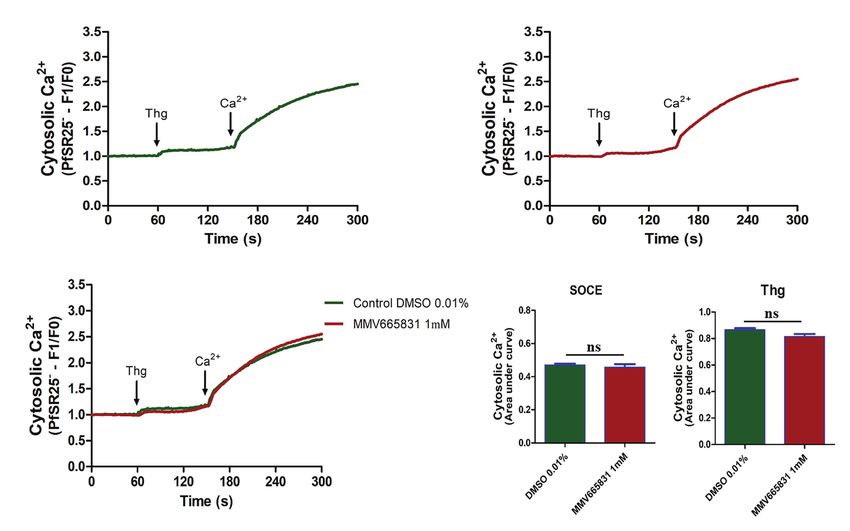
Santos et al.                                                                                                  Plasmodium Receptor PfSR25 and Resistance to Antimalarials

TABLE 2 | Compounds from the Medicine for Malaria Venture (MMV), with chemical structures and IC50 and IC90 values for the wild-type strain (3D7) and PfSR25-
parasitess.

Compound                       Chemical structure                IC50 (μM) 3D7              IC50 (μM) PfSR25-               IC90 (μM) 3D7                 IC90 (μM) PfSR25-

MMV006429                                                         2.34 ± 0.26                 2.13 ± 0.10                    3.30 ± 0.27                   3.14 ± 0.36

MMV396715                                                         1.48 ± 0.06                 1.30 ± 0.08                    2.25 ± 0.07                   2.23 ± 0.14

MMV019127                                                         0.86 ± 0.08                 0.77 ± 0.08                    1,73 ± 0.16                   1.78 ± 0.10

MMV665874                                                         0.33 ± 0.04                 0.33 ± 0.01                    0.70 ± 0.08                   0.71 ± 0.09

MMV665878                                                         0.17 ± 0.20                 0.19 ± 0.21                    0.26 ± 0.003                  0.29 ± 0.011

MMV665785                                                         0.082 ± 0.04                0.094 ± 0.06                   0.20 ± 0.0015                 0.19 ± 0.0047

MMV665831                                                         0.087 ± 0.001               0.065 ± 0.007                  0.14 ± 0.003                  0.13 ± 0.006

The images of the chemical structures for each compound were taken from the MMV database (About the Malaria Box | Medicines for Malaria Venture, n.d.).

concentration. This procedure is thought to induce SOCE in                                differences when compared to the control group, indicating
P. falciparum as described by Pecenin et al. (2018).                                      that this compound does not interfere with the SOCE mechanism
   We then tracked the calcium measurements with Fluo-4/                                  of the malaria parasite P. falciparum.
AM-loaded parasites that were previously treated with MMV665831,
which affects parasite egress/invasion to red blood cells (Subramanian
et al., 2018). For that purpose, we performed experiments similar                         DISCUSSION
to those described in Figures 5A, 6A. Therefore, the presence of
SOCE were accessed after incubating the parasites at the trophozoite                      The emergence of parasites resistant to classical antimalarial
stage (3D7 and PfSR25-), with 1 μM of MMV665831 compound                                  drugs from different compound classes (Ashley et al., 2014;
for 15 min, after they had previously been labeled with Fluo-4/                           Hanboonkunupakarn and White, 2016; Blasco et al., 2017)
AM. The spectrofluorometric data are shown in Figures 5B, 6B,                             demonstrates the urgent need to search for new targets in
the analysis used to evaluate the presence of SOCE are shown                              developing drugs with a different mechanism of action.
in Figures 5D, 6D, and the analysis on the calcium content                                   G protein-coupled receptors represent a class of receptors
depleted by Thg is reflected by the area of the peaks (Figures 5E, 6E).                   with seven transmembrane domains and are the targets for
   The data presented here showed that parasites at the                                   most drugs (Hauser et al., 2017). Four candidates presenting
trophozoite stage (3D7 and PfSR25-) and pretreated with                                   seven transmembrane domains were identified in the malaria
compound MMV665831 (1 μM) did not display any significant                                 parasite P. falciparum genome as serpentine-like receptors

Frontiers in Microbiology | www.frontiersin.org                                       7                                              March 2021 | Volume 12 | Article 638869
Santos et al.                                                                                         Plasmodium Receptor PfSR25 and Resistance to Antimalarials

                         A                                                  C                                D

                         B

                                                                                        E

  FIGURE 4 | Determination of MMV665831 antiplasmodial activity in strains 3D7 (red) and PfSR25- (blue). (A) Chemical structure of the MMV665831 compound.
  (B) Dose-response curve, with concentrations varying from 0.0048 to 5 μM. The IC50 (C) and IC90 (D) values were calculated using GraphPad Prism software and
  the values were compared between the 3D7 and PfSR25- parasite strains by Student’s t-test. (E) Microscopic image of Giemsa-stained blood smears, parasites
  (3D7 and PfSR25-) were incubated with compound MMV665831 (0.3125 μM) for 72 h, parasites in the control group were incubated with 0.01% Dimethyl sulfoxide
  (DMSO). The experiments were performed three independent times in triplicate. *Represents the significant difference found by t-test (*p ≤ 0.05).

(Madeira et al., 2008). Among the four candidates (PfSR1,                           and a 4–5-fold increase in the infectivity of Plasmodium yoelii
PfSR10, PfSR12, and PfSR25), PfSR25 was identified as a                             (Kumar et al., 2007). Singh et al. (2010) reported that exposing
potassium sensor, and parasites knockout were more susceptible                      merozoites to low K+ concentrations provide an external signal
to toxic agents such as oxidative stress induced by Nitric Oxide                    that leads to an increase in the Ca2+cyt, which triggers the
(NO) production and also to stress induced by the removal                           translocation of microneme proteins, such as the erythrocyte-
of albumax from the medium (Moraes et al., 2017).                                   binding antigen (EBA175 antigen) and apical membrane antigen-1
   Our results point that piperaquine and lumefantrine were                         (AMA1), which are expressed on the surface of the merozoite.
more active against parasites lacking PfSR25, with significant                         Kumar et al. (2007) and Singh et al. (2010) reported the
reductions of 58.12 and 44.38% in the IC50 values in PfSR25-,                       importance of K+ for the parasite mainly in its hepatic phase.
respectively. Piperaquine is concentrated in the digestive vacuole                  Although the knockout parasite for PfSR25 did not show
where it is protonated and cannot diffuse out of this compartment                   changes in its intra-erythrocyte cycle in vitro, the knockout
through the membrane. These protonated forms bind to the                            strain for SR25 in P. berghei (PbSR25) was unable to develop
growing hemozoin crystals (Wicht et al., 2020).                                     a successful infection in vivo (Moraes et al., 2017).
   The lumefantrine mechanism of action is still yet to                                To better understand the relationship between PfSR25 and
be completely elucidated; although it has been implicated in                        the events that govern egress/invasion in the intra-erythrocytic
the detoxification of products of heme degradation (Cowell and                      cycle, we used compounds from MMV (Malaria box) previously
Winzeler, 2019). Mwai et al. (2012) reported that the lumefantrine-                 described to impair the egress/invasion of the intra-erythrocytic
resistant strain has a negative regulation of the pfsr25 gene in                    cycle of P. falciparum (Subramanian et al., 2018). The results
the ring stage. Our results demonstrated that the antimalarial                      obtained here show that MMV665785 and MMV665831, which
piperaquine is also able to promote a downregulation in the                         were previously described as inhibitors of egress/invasion,
ring stage pfsr25 gene. Both works imply the involvement of                         presented IC50 values below 100 nM.
PfSR25 in the mechanism of action of these antimalarials.                              Previous studies showed that MMV665831 blocked the rupture
   Previous studies have shown the importance of the K+ shift                       of infected erythrocytes at nanomolar concentrations and
at the moment of parasite egress/invasion. During hepatic cell                      promoted almost a 90% decrease in ring formation at different
migration, sporozoites are exposed to major changes in the                          concentrations (0.3, 1, 3, and 10 μM). The MMV665785 compound
K+ concentration (Kumar et al., 2007). Kumar et al. (2007)                          was recently reported to impair the rupture of infected
reported that the incubation of sporozoites with K+ generated                       erythrocytes and prevent the infection of new ones. Nonetheless,
an 8–10-fold increase in the infectivity of Plasmodium berghei                      compound withdrawal after 2 h of incubation resulted in normal

Frontiers in Microbiology | www.frontiersin.org                                 8                                         March 2021 | Volume 12 | Article 638869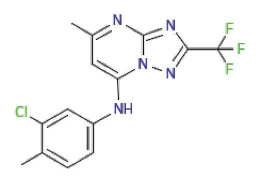
Santos et al.                                                                                                Plasmodium Receptor PfSR25 and Resistance to Antimalarials

      A                                                                                       B

      C                                                                                           D                                E

  FIGURE 5 | Spectrofluorometric evaluation of the store-operated calcium entry (SOCE) mechanism. Synchronized P. falciparum 3D7 in the trophozoite stage (28–32 h) were
  labeled with Ca2+ Fluo-4/AM probe. (A) The parasites were previously incubated with DMSO solvent (0.01%). (B) The parasites were previously treated with MMV665831
  compound (1 μM). After 15 min of treatment, parasites at 107 cells/ml were placed in free Ca2+ buffer. Thapsigargin (Thg; 5 μM) was used to deplete the Ca2+ from the
  endoplasmic reticulum (ER). CaCl2 (2 mM) was added and the increase in the [Ca2+]cyt was monitored. (C) Representative curve for SOCE measurement at the control and
  MMV665831 treated parasites. (D) F0 corresponds to the mean of the fluorescence obtained between 140 and 150 s, and F1 corresponds to the mean of the fluorescence
  obtained between 290 and 300 s. (E) F0 corresponds to the mean of the fluorescence obtained between 50 and 60 s, and F1 corresponds to the mean of the fluorescence
  obtained between 140 and 150 s. Three independent experiments were performed in triplicates. *Represents significant difference found by Student’s t-test (*p ≤ 0.05).

egress, but compound MMV665831 showed irreversible action,                              as protein kinases. Receptors for activated C kinases are scaffold
even after withdrawal (Subramanian et al., 2018).                                       proteins that anchor several signaling proteins and are involved
   Our results showed that among the tested compounds,                                  in modulating the cell cycle. Despite the absence of classical
MMV665785 and MMV665831 presented higher antimalarial                                   PKC from the Plasmodium database, PfRACK has been identified
activity in vitro with IC50 values of 0.082 ± 0.04 μM (82 nM)                           (Madeira et al., 2003). Moreover, Ca2+ mobilization regulates
and 0.087 ± 0.001 μM (87 nM), respectively. Compounds that                              important processes in protist biology (Cruz et al., 2012;
were previously described to impair egress/invasion did not                             Borges-Pereira et al., 2015).
present higher activity against parasites lacking PfSR25. Only                              The calcium-dependent protein kinase signaling pathway
the compound MMV665831 resulted in a lower IC50 value in                                has been identified in the Plasmodium database (Ward et al.,
the knockout strain. However, we observed no difference regarding                       2004; Doerig et al., 2008) and controls few processes in the
the concentration needed to eliminate 90% (IC90) of the parasite                        malaria parasite. One of these processes is male gamete formation
population between the two strains (3D7 and PfSR25).                                    and zygote development (Reininger et al., 2005), but the
   Calcium is an intracellular messenger that is present in                             molecular mechanisms of calcium release remain
malaria parasites, and it plays key roles in the parasite during                        poorly understood.
the asexual stages (Brochet and Billker, 2016; Singh et al.,                                In addition, Ca2+ participates in other important pathways
2020). The second messenger Ca2+ was found to participate                               such as the pathway triggered by tryptophan derivatives.
in erythrocyte invasion (Cowman and Crabb, 2006). Weiss                                 Melatonin activates phospholipase C (PLC), resulting in IP3
et al. (2015) observed that in Ca2+-free media, the invasion                            production and the release of Ca2+ from the ER (Passos and
rate decreases significantly, although the merozoites could attach                      Garcia, 1997; Alves et al., 2011; Garcia et al., 2017). Furthermore,
to new erythrocytes.                                                                    P. falciparum treated with melatonin presented an increase in
   Moreover, calcium also participates directly in the activation                       cAMP levels that was blocked by the PLC inhibitor U73122.
of proteins that participate in key signaling pathways, such                            Moreover, treating with cAMP analog 6-Bz-cAMP causes a

Frontiers in Microbiology | www.frontiersin.org                                     9                                             March 2021 | Volume 12 | Article 638869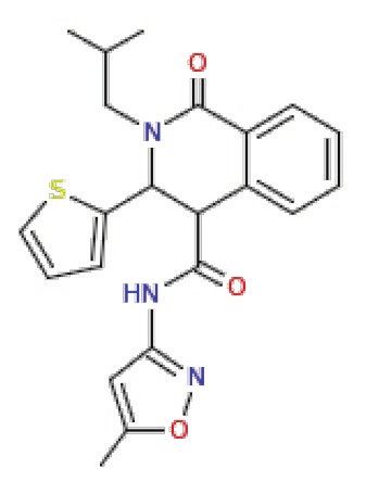
Santos et al.                                                                                        Plasmodium Receptor PfSR25 and Resistance to Antimalarials

      A                                                                                B

      C                                                                                    D                              E

 FIGURE 6 | Spectrofluorometric evaluation of the SOCE mechanism. Synchronized P. falciparum PfSR25- in the trophozoite stage (28–32 h) were labeled with
 Ca2+ Fluo-4/AM probe. (A) The parasites were previously incubated with DMSO solvent (0.01%). (B) The parasites were previously treated with MMV665831
 compound (1 μM). After 15 min of treatment, parasites at 107 cells/ml were placed in free Ca2+ buffer. Thg (5 μM) was used to deplete the Ca2+ from the ER.
 CaCl2 (2 mM) was added and the increase in the [Ca2+]cyt was monitored. (C) Representative curve for SOCE measurement at the control and MMV665831
 treated parasites. (D) F0 corresponds to the mean of the fluorescence obtained between 140 and 150 s, and F1 corresponds to the mean of the fluorescence
 obtained between 290 and 300 s. (E) F0 corresponds to the mean of the fluorescence obtained between 50 and 60 s, and F1 corresponds to the mean of the
 fluorescence obtained between 140 and 150 s. Two independent experiments were performed in triplicates. *Represents the significant difference found by
 t-test (*p ≤ 0.05).

Ca2+cyt increase that is dependent on the PKA, suggesting                          egress/invasion of the human’s intraerythrocytic cycle malaria
crosstalk between Ca2+ and cAMP (Beraldo et al., 2005; Gazarini                    parasite P. falciparum.
et al., 2011). The PLC-IP3 mechanism is thought to
be fundamental to the SOCE mechanism, the stored-operated
Ca2+ entry mechanism activated by the depletion of the                             CONCLUSION
intracellular calcium stores (Putney, 2003) that is present in
P. falciparum (Pecenin et al., 2018).                                              The results show that parasites lacking the PfSR25 receptor
   In this study, we have shown that compound MMV665831,                           are more susceptible to lumefantrine and piperaquine, classical
an inhibitor of egress/invasion, does not significantly interfere                  antimalarials that act on the digestive vacuole with heme
in the store-operated Ca2+ entry in P. falciparum 3D7 and                          metabolism. However, we found no difference between the
PfSR25- at the trophozoite stage. The same result was observed                     3D7 and PfSR25- strain after incubating parasites for 72-h
when the experiment was performed on the knockout strain                           with atovaquone, dihydroartemisinin, mefloquine, primaquine,
for the GPCR-like PfSR25. Our results suggest that this compound                   pyrimethamine, and chloroquine at the drug range concentration
action in the invasion process does not occur through the                          0.24–250 nM.
store operated Ca2+ entry and that PfSR25 is not necessary                            The studies on MMV compounds lead us to conclude
for the SOCE mechanism to occur.                                                   that PfSR25 is not involved in the action of compounds
   During egress/invasion, the parasite experiences a K+ shift                     that inhibit the egress/invasion process, although further
(Kumar et al., 2007), and the PfSR25 is involved in sensing                        efforts are necessary to elucidate the participation of the
the K+ alterations in the environment. The results here show                       receptor in these processes. Our data indicate the potential
that P. falciparum receptor PfSR25 is not involved in the                          involvement of GPCR-like PfSR25 in the mechanism of action
mechanism of action of MMV drugs known to block the                                of lumefantrine and piperaquine in the malaria parasites

Frontiers in Microbiology | www.frontiersin.org                               10                                         March 2021 | Volume 12 | Article 638869
Santos et al. Plasmodium Receptor PfSR25 and Resistance to Antimalarials
P. falciparum and points the GPCR-like as a novel tool to (process 17/08684-7 and 18/07177-7 Imaging 20). BS was a
study antimalarial drugs. recipient of a fellowship from the Fundação de Amparo à
Pesquisa do Estado de São Paulo (FAPESP Process 2020/08988-
9). BD was a recipient of a fellowship from the Conselho
DATA AVAILABILITY STATEMENT Nacional de Desenvolvimento Científico e Tecnologico (CNPq
process 142188/2017-4). The funders had no role in the study
The raw data supporting the conclusions of this article will design, data collection and analysis, decision to publish, or
be made available by the authors, without undue reservation. preparation of the manuscript.
AUTHOR CONTRIBUTIONS
ACKNOWLEDGMENTS
BS, MN, and BD conducted the experiments. BS and CG
designed the experiments. CG supervised all the work. BS, We thank Colsan for the blood supply and MMV for
BD, and CG wrote the manuscript. All authors contributed the compounds.
to the article and approved the submitted version.
SUPPLEMENTARY MATERIAL
FUNDING
The Supplementary Material for this article can be found online
This work was supported by a grant from the Fundação de at: https://www.frontiersin.org/articles/10.3389/fmicb.2021.638869/
Amparo à Pesquisa do Estado de São Paulo (FAPESP) to CG full#supplementary-material
REFERENCES Cowman, A. F., and Crabb, B. S. (2006). Invasion of red blood cells by malaria
parasites. Cell 124, 755–766. doi: 10.1016/j.cell.2006.02.006
About the Malaria Box | Medicines for Malaria Venture (n.d.). Available at: Cruz, L. N., Juliano, M. A., Budu, A., Juliano, L., Holder, A. A., Blackman, M. J.,
https://www.mmv.org/mmv-open/malaria-box/about-malaria-box (Accessed et al. (2012). Extracellular ATP triggers proteolysis and cytosolic Ca2+ rise
November 18, 2020). in Plasmodium berghei and Plasmodium yoelii malaria parasites. Malar. J.
Alberts, B., Johnson, A., Lewis, J., Morgan, D., Raff, M., Roberts, K., et al. 11:69. doi: 10.1186/1475-2875-11-69
(2017). Molecular biology of the cell. eds. J. Wilson and T. Hunt (Garland Da Silva, G. N. S., Maria, N. R. G., Schuck, D. C., Cruz, L. N., De Moraes, M. S.,
Science), 1227–1242. Nakabashi, M., et al. (2013). Two series of new semisynthetic triterpene
Alhadeff, R., Vorobyov, I., Yoon, H. W., and Warshel, A. (2018). Exploring the derivatives: differences in anti-malarial activity, cytotoxicity and mechanism
free-energy landscape of GPCR activation. Proc. Natl. Acad. Sci. U. S. A. of action. Malar. J. 12:89. doi: 10.1186/1475-2875-12-89
115, 10327–10332. doi: 10.1073/pnas.1810316115 Dhingra, S. K., Redhi, D., Combrinck, J. M., Yeo, T., Okombo, J., Henrich, P. P.,
Alves, E., Bartlett, P. J., Garcia, C. R. S., and Thomas, A. P. (2011). Melatonin et al. (2017). A variant pfcrt isoform can contribute to Plasmodium falciparum
and IP3-induced Ca2+ release from intracellular stores in the malaria parasite resistance to the first-line partner drug piperaquine. mBio 8:e00303–e00317.
Plasmodium falciparum within infected red blood cells. J. Biol. Chem. 286, doi: 10.1128/mBio.00303-17
5905–5912. doi: 10.1074/jbc.M110.188474 Doerig, C., Billker, O., Haystead, T., Sharma, P., Tobin, A. B., and Waters, N. C.
Ashley, E. A., Dhorda, M., Fairhurst, R. M., Amaratunga, C., Lim, P., S, S., (2008). Protein kinases of malaria parasites: an update. Trends Parasitol.
et al. (2014). Spread of artemisinin resistance in Plasmodium falciparum 24, 570–577. doi: 10.1016/j.pt.2008.08.007
malaria. N. Engl. J. Med. 371, 411–423. doi: 10.1056/NEJMoa1314981 Ekland, E. H., Schneider, J., and Fidock, D. A. (2011). Identifying apicoplast-
Beraldo, F. H., Almeida, F. M., Da Silva, A. M., and Garcia, C. R. S. (2005). targeting antimalarials using high-throughput compatible approaches. FASEB
Cyclic AMP and calcium interplay as second messengers in melatonin- J. 25, 3583–3593. doi: 10.1096/fj.11-187401
dependent regulation of Plasmodium falciparum cell cycle. J. Cell Biol. 170, Garcia, C. R. S., Alves, E., Pereira, P. H. S., Bartlett, P. J., Thomas, A. P.,
551–557. doi: 10.1083/jcb.200505117 Mikoshiba, K., et al. (2017). InsP3 signaling in apicomplexan parasites. Curr.
Blasco, B., Leroy, D., and Fidock, D. A. (2017). Antimalarial drug resistance: Top. Med. Chem. 17, 2158–2165. doi: 10.2174/1568026617666170130121042
linking Plasmodium falciparum parasite biology to the clinic. Nat. Med. 23, Gazarini, M. L., Beraldo, F. H., Almeida, F. M., Bootman, M., Da Silva, A. M.,
917–928. doi: 10.1038/nm.4381 and Garcia, C. R. S. (2011). Melatonin triggers PKA activation in the rodent
Borges-Pereira, L., Budu, A., McKnight, C. A., Moore, C. A., Vella, S. A., malaria parasite Plasmodium chabaudi. J. Pineal Res. 50, 64–70. doi: 10.1111/j.
Triana, M. A. H., et al. (2015). Calcium signaling throughout the toxoplasma 1600-079X.2010.00810.x
gondii lytic cycle a study using genetically encoded calcium indicators. Hanboonkunupakarn, B., and White, N. J. (2016). The threat of antimalarial drug
J. Biol. Chem. 290, 26914–26926. doi: 10.1074/jbc.M115.652511 resistance. Trop. Dis. Travel Med. Vaccines 2, 1–5. doi: 10.1186/s40794-016-0027-8
Bridgford, J. L., Xie, S. C., Cobbold, S. A., Pasaje, C. F. A., Herrmann, S., Hauser, A. S., Attwood, M. M., Rask-Andersen, M., Schiöth, H. B., and
Yang, T., et al. (2018). Artemisinin kills malaria parasites by damaging Gloriam, D. E. (2017). Trends in GPCR drug discovery: new agents, targets
proteins and inhibiting the proteasome. Nat. Commun. 9, 1–9. doi: 10.1038/ and indications. Nat. Rev. Drug Discov. 16, 829–842. doi: 10.1038/nrd.2017.178
s41467-018-06221-1 Heinberg, A., and Kirkman, L. (2015). The molecular basis of antifolate resistance
Broad, L. M., Braun, F. J., Lievremont, J. P., Bird, G. S. J., Kurosaki, T., and in Plasmodium falciparum: looking beyond point mutations. Ann. N. Y.
Putney, J. W. (2001). Role of the phospholipase C-inositol 1,4,5-trisphosphate Acad. Sci. 1342, 10–18. doi: 10.1111/nyas.12662
pathway in calcium release-activated calcium current and capacitative calcium Hewavitharana, T., Deng, X., Soboloff, J., and Gill, D. L. (2007). Role of STIM
entry. J. Biol. Chem. 276, 15945–15952. doi: 10.1074/jbc.M011571200 and Orai proteins in the store-operated calcium signaling pathway. Cell
Brochet, M., and Billker, O. (2016). Calcium signalling in malaria parasites. Calcium 42, 173–182. doi: 10.1016/j.ceca.2007.03.009
Mol. Microbiol. 100, 397–408. doi: 10.1111/mmi.13324 Josling, G. A., and Llinás, M. (2015). Sexual development in Plasmodium
Cowell, A. N., and Winzeler, E. A. (2019). The genomic architecture of antimalarial parasites: knowing when it’s time to commit. Nat. Rev. Microbiol. 13, 573–587.
drug resistance. Brief. Funct. Genomics 18, 314–328. doi: 10.1093/bfgp/elz008 doi: 10.1038/nrmicro3519
Frontiers in Microbiology | www.frontiersin.org 11 March 2021 | Volume 12 | Article 638869Santos et al. Plasmodium Receptor PfSR25 and Resistance to Antimalarials Khoury, D. S., Cao, P., Zaloumis, S. G., and Davenport, M. P. (2020). Artemisinin Reed, M. B., Saliba, K. J., Caruana, S. R., Kirk, K., and Cowman, A. F. (2000). resistance and the unique selection pressure of a short-acting antimalarial. Pgh1 modulates sensitivity and resistance to multiple antimalarials in Trends Parasitol. 36, 884–887. doi: 10.1016/j.pt.2020.07.004 Plasmodium falciparum. Nature 403, 906–909. doi: 10.1038/35002615 Koyama, F. C., Chakrabarti, D., and Garcia, C. R. S. (2009). Molecular machinery Reininger, L., Billker, O., Tewari, R., Mukhopadhyay, A., Fennell, C., of signal transduction and cell cycle regulation in Plasmodium. Mol. Biochem. Dorin-Semblat, D., et al. (2005). A NIMA-related protein kinase is essential Parasitol. 165, 1–7. doi: 10.1016/j.molbiopara.2009.01.003 for completion of the sexual cycle of malaria parasites. J. Biol. Chem. 280, Kremsner, P. P. G., and Krishna, P. S. (2004). Antimalarial combinations. Lancet 31957–31964. doi: 10.1074/jbc.M504523200 364, 285–294. doi: 10.1016/S0140-6736(04)16680-4 Santos, B. M. dos, Gonzaga, D. T. G., da Silva, F. C., Ferreira, V. F. , and Kumar, K. A., Garcia, C. R. S., Chandran, V. R., Van Rooijen, N., Zhou, Y., Garcia, C. R. S. (2020). Plasmodium falciparum knockout for the GPCR-like Winzeler, E., et al. (2007). Exposure of Plasmodium sporozoites to the PfSR25 receptor displays greater susceptibility to 1,2,3-triazole compounds intracellular concentration of potassium enhances infectivity and reduces that block malaria parasite development. Biomol. Ther. 10:1197. doi: 10.3390/ cell passage activity. Mol. Biochem. Parasitol. 156, 32–40. doi: 10.1016/j. biom10081197. molbiopara.2007.07.004 Schuck, D. C., Ferreira, S. B., Cruz, L. N., Da Rocha, D. R., Moraes, M. S., Lambros, C., and Vanderberg, J. P. (1979). Synchronization of Plasmodium Nakabashi, M., et al. (2013). Biological evaluation of hydroxynaphthoquinones falciparum erythrocytic stages in culture. J. Parasitol. 65, 418–420. doi: as anti-malarials. Malar. J. 12:234. doi: 10.1186/1475-2875-12-234 10.2307/3280287 Singh, S., Alam, M. M., Pal-Bhowmick, I., Brzostowski, J. A., and Chitnis, C. E. Le Bras, J., and Durand, R. (2003). The mechanisms of resistance to antimalarial (2010). Distinct external signals trigger sequential release of apical organelles drugs in Plasmodium falciparum. Fundam. Clin. Pharmacol. 17, 147–153. during erythrocyte invasion by malaria parasites. PLoS Pathog. 6:e1000746. doi: 10.1046/j.1472-8206.2003.00164.x doi: 10.1371/journal.ppat.1000746 Lee, A. H., Dhingra, S. K., Lewis, I. A., Singh, M. K., Siriwardana, A., Dalal, S., Singh, M. K., Dias, B. K., and Garcia, C. R. (2020). Role of melatonin in the et al. (2018). Evidence for regulation of hemoglobin metabolism and synchronization of asexual forms in the parasite Plasmodium falciparum. intracellular ionic flux by the Plasmodium falciparum chloroquine resistance Biomol. Ther. 10:1243. doi: 10.3390/biom10091243 transporter. Sci. Rep. 8, 1–13. doi: 10.1038/s41598-018-31715-9 Sriram, K., and Insel, P. A. (2018). G protein-coupled receptors as targets for Lourido, S., and Moreno, S. N. J. (2015). The calcium signaling toolkit of the approved drugs: how many targets and how many drugs? Mol. Pharmacol. apicomplexan parasites Toxoplasma gondii and Plasmodium spp. Cell Calcium 93, 251–258. doi: 10.1124/mol.117.111062 57, 186–193. doi: 10.1016/j.ceca.2014.12.010 Staines, H. M., Burrow, R., Teo, B. H. Y., Ster, I. C., Kremsner, P. G., and Madeira, L., DeMarco, R., Gazarini, M. L., Verjovski-Almeida, S., and Garcia, C. R. Krishna, S. (2018). Clinical implications of Plasmodium resistance to S. (2003). Human malaria parasites display a receptor for activated C kinase atovaquone/proguanil: a systematic review and meta-analysis. J. Antimicrob. ortholog. Biochem. Biophys. Res. Commun. 306, 995–1001. doi: 10.1016/ Chemother. 73, 581–595. doi: 10.1093/jac/dkx431 S0006-291X(03)01074-X Subramanian, G., Belekar, M. A., Shukla, A., Tong, J. X., Sinha, A., Chu, T. T. Madeira, L., Galante, P. A. F., Budu, A., Azevedo, M. F., Malnic, B., and Garcia, C. R. T., et al. (2018). Targeted phenotypic screening in Plasmodium falciparum S. (2008). Genome-wide detection of serpentine receptor-like proteins in malaria and Toxoplasma gondii reveals novel modes of action of medicines for parasites. PLoS One 3:e1889. doi: 10.1371/journal.pone.0001889 malaria venture malaria box molecules. mSphere 3, 534–551. doi: 10.1128/ Moraes, M. S., Budu, A., Singh, M. K., Borges-Pereira, L., Levano-Garcia, J., msphere.00534-17 Currà, C., et al. (2017). Plasmodium falciparum GPCR-like receptor SR25 Sullivan, D. J. (2017). Quinolines block every step of malaria heme crystal mediates extracellular K+ sensing coupled to Ca2+ signaling and stress survival. growth. Proc. Natl. Acad. Sci. U. S. A. 114, 7483–7485. doi: 10.1073/ Sci. Rep. 7, 1–13. doi: 10.1038/s41598-017-09959-8 pnas.1708153114 Mwai, L., Diriye, A., Masseno, V., Muriithi, S., Feltwell, T., Musyoki, J., et al. Trager, W., and Jensen, J. B. (1976). Human malaria parasites in continuous (2012). Genome wide adaptations of Plasmodium falciparum in response culture. Science 193, 673–675. doi: 10.1126/science.781840 to lumefantrine selective drug pressure. PLoS One 7:e31623. doi: 10.1371/ Ward, P., Equinet, L., Packer, J., and Doerig, C. (2004). Protein kinases of the journal.pone.0031623 human malaria parasite Plasmodium falciparum: the kinome of a divergent Olafson, K. N., Ketchum, M. A., Rimer, J. D., and Vekilov, P. G. (2015). eukaryote. BMC Genomics 5:79. doi: 10.1186/1471-2164-5-79 Mechanisms of hematin crystallization and inhibition by the antimalarial Weiss, G. E., Gilson, P. R., Taechalertpaisarn, T., Tham, W. H., de Jong, N. W. drug chloroquine. Proc. Natl. Acad. Sci. U. S. A. 112, 4946–4951. doi: 10.1073/ M., Harvey, K. L., et al. (2015). Revealing the sequence and resulting cellular pnas.1501023112 morphology of receptor-ligand interactions during Plasmodium falciparum Passos, A., and Garcia, C. (1997). Characterization of Ca2+ transport activity invasion of erythrocytes. PLoS Pathog. 11, 1–25. doi: 10.1371/journal. associated with a non-mitochondrial calcium pool in the rodent malaria ppat.1004670 parasite P. chabaudi. IUBMB Life 42, 919–925. doi: 10.1080/15216549700203361 White, N. J. (2004). Antimalarial drug resistance. J. Clin. Invest. 113, 1084–1092. Pecenin, M. F., Borges-Pereira, L., Levano-Garcia, J., Budu, A., Alves, E., doi: 10.1172/JCI21682 Mikoshiba, K., et al. (2018). Blocking IP3 signal transduction pathways Wicht, K. J., Mok, S., and Fidock, D. A. (2020). Molecular mechanisms of inhibits melatonin-induced Ca2+ signals and impairs P. falciparum development drug resistance in Plasmodium falciparum malaria. Annu. Rev. Microbiol. and proliferation in erythrocytes. Cell Calcium 72, 81–90. doi: 10.1016/j. 74, 431–454. doi: 10.1146/annurev-micro-020518-115546 ceca.2018.02.004 World malaria report (2019) Available at: https://www.who.int/publications/i/ Pereira, P. H. S., Borges-Pereira, L., and Garcia, C. R. S. (2020). Evidences of item/9789241565721 (Accessed October 23, 2020). G-Coupled Protein Receptor (GPCR) signaling in the human malaria parasite Plasmodium falciparum for sensing its microenvironment and the role of Conflict of Interest: The authors declare that the research was conducted in purinergic signaling in malaria parasites. Curr. Top. Med. Chem. 21, 171–180. the absence of any commercial or financial relationships that could be construed doi: 10.2174/1568026620666200826122716 as a potential conflict of interest. Price, R. N., Uhlemann, A. C., Van Vugt, M., Brockman, A., Hutagalung, R., Nair, S., et al. (2006). Molecular and pharmacological determinants of the Copyright © 2021 Santos, Dias, Nakabashi and Garcia. This is an open-access article therapeutic response to artemether-lumefentrine in multidrug-resistant distributed under the terms of the Creative Commons Attribution License (CC BY). Plasmodium falciparum malaria. Clin. Infect. Dis. 42, 1570–1577. doi: The use, distribution or reproduction in other forums is permitted, provided the original 10.1086/503423 author(s) and the copyright owner(s) are credited and that the original publication Putney, J. W. (2003). Capacitative calcium entry in the nervous system. Cell in this journal is cited, in accordance with accepted academic practice. No use, Calcium 34, 339–344. doi: 10.1016/S0143-4160(03)00143-X distribution or reproduction is permitted which does not comply with these terms. Frontiers in Microbiology | www.frontiersin.org 12 March 2021 | Volume 12 | Article 638869
You can also read