Melatonin prevents allergic airway inflammation in epicutaneously sensitized mice
←
→
Page content transcription
If your browser does not render page correctly, please read the page content below
Bioscience Reports (2021) 41 BSR20210398
https://doi.org/10.1042/BSR20210398

Research Article

Melatonin prevents allergic airway inflammation in
epicutaneously sensitized mice
Xudong Liu1 , Yuchao Zhang1 , Yaolin Ren2 and                                         Jinquan Li2
1 Department   of Food Science and Engineering, Moutai Institute, Renhuai 564507, China; 2 School of Medicine, Wuhan University of Science and Technology, Wuhan 430065, China
Correspondence: Jinquan Li (lijinquan@wust.edu.cn)

                                                                                                                                                                                           Downloaded from http://portlandpress.com/bioscirep/article-pdf/41/9/BSR20210398/921054/bsr-2021-0398.pdf by guest on 01 December 2021
                                           Purpose: The pathological process of atopic dermatitis (AD) progressing into other types of
                                           allergic diseases such as asthma and allergic rhinitis during the first several years of life is
                                           often referred to as the atopic march. Although the phenomenon of atopic march has been
                                           recognized for decades, how asthma stems from AD is still not fully understood, confound-
                                           ing a universal strategy to effectively protect people from the atopic march. Methods: We
                                           established experimental atopic march mice by first inducing allergic dermatitis with 0.5%
                                           fluorescein isothiocyante (FITC) applied to the skin, followed by an ovalbumin (OVA) air-
                                           way challenge. In addition, by examining serum immunoglobulin (Ig) concentrations, airway
                                           cytokines, the levels of oxidative stress markers, histopathological changes in lung tissue
                                           and airway hyperresponsiveness (AHR), we were able to validate the successful establish-
                                           ment of the model. Furthermore, by detecting the attenuating effects of melatonin (MT) and
                                           the levels of oxidative stress in the atopic march mice, we explored the potential molec-
                                           ular mechanisms involved in the development of atopic march. Results: By successfully
                                           establishing an experimental atopic march mouse model, we were able to demonstrate that
                                           overproduction of oxidative stress in the lung significantly up-regulated the activation of nu-
                                           clear factor-κB (NF-κB) signaling pathways causing thymic stromal lymphopoietin (TSLP)
                                           release, which further promotes the development of atopic march. Conclusions: To miti-
                                           gate the development of the atopic march, antioxidants such as MT may be imperative to
                                           inhibit NF-κB activation in the lung, especially after the onset of AD.

                                         Introduction
                                         Atopic diseases have been on the rise in recent decades, but no single risk factor can sufficiently explain
                                         the heightened prevalence across the globe [1]. There is robust evidence suggesting that the characteristic
                                         sequence of atopic manifestations typically occurs first with atopic dermatitis (AD) in infancy, followed
                                         by allergic rhinitis and/or asthma in later stages. In addition, the severity of AD appears to influence the
                                         course of respiratory allergy in adulthood [2]. The pathological process of AD progressing into other types
                                         of allergic diseases such as asthma and allergic rhinitis during the first several years of life is often referred
                                         to as the atopic march [3]. Both AD and asthma share similar atopy phenotypes, which include T helper
                                         type 2 inflammation with eosinophilia, and hyper-IgE immunoglobulinemia [4]. However, the molecular
                                         mechanisms underlying the atopic march, and how asthma stems from AD, remains obscure. Thus, fur-
Received: 09 March 2021
                                         ther research is needed to understand the development of these disorders and to find interventions for
Revised: 31 August 2021                  mediating the occurrence of atopic diseases.
Accepted: 02 September 2021                 Excessive levels of reactive oxygen species (ROS) and other oxidation products may result in oxidative
                                         damage, an important mechanism for the pathophysiology of allergic diseases including AD and asthma
Accepted Manuscript online:
15 September 2021                        [5]. In particular, alveolar epithelial type II cells, a type of lung cell, are especially susceptible to the effects
Version of Record published:             of oxidants [6]. Along with a significant increase in ROS, pro-inflammatory cytokines (tumor necrosis
22 September 2021                        factor-α (TNF-α) and IL-1β) were found in BEAS-2B cells treated with PM2.5 [7]. Moreover, excessive

© 2021 The Author(s). This is an open access article published by Portland Press Limited on behalf of the Biochemical Society and distributed under the Creative Commons Attribution   1
License 4.0 (CC BY).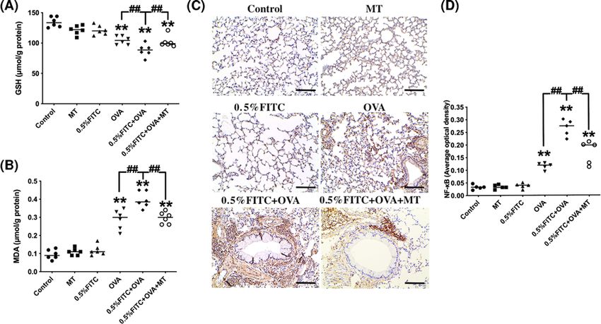
Bioscience Reports (2021) 41 BSR20210398
                                                                                                                                         https://doi.org/10.1042/BSR20210398

           production of ROS can initiate inflammatory responses by activating redox-sensitive transcription factors, such as
           nuclear factor-κB (NF-κB), activator protein-1 (AP-1), and hypoxia-inducible factor (HIF)-1 [8–10]. Various studies
           have reported the importance of the NF-κB pathway in promoting the production of cytokines during allergic disease
           pathogenesis [11]. These studies suggest that assessment of oxidative stress levels and activation of the NF-κB pathway,
           may be a promising approach for understanding the development of atopic diseases.
              Thymic stromal lymphopoietin (TSLP), an airway epithelium-derived cytokine, plays a central role in polarizing
           dendritic cells (DCs) and induces the differentiation of naive T cells into Th2 cells [12]. Lung fibroblasts and bronchial
           smooth muscle cells can also produce TSLP under inflammatory conditions initiated by IL-13 stimulation [13]. Pre-
           vious studies have suggested the critical role of keratinocytic TSLP in the development of AD into asthma [14,15].
           However, few studies have pinpointed the specific role of TSLP during the asthmatic phase of atopic march, let alone
           its mechanism of activation. In allergic asthma, TSLP activates DCs and promotes the homeostasis shift of Th1/Th2
           into Th2, which subsequently results in airway remodeling and sustained airway hyperresponsiveness (AHR) [16].

                                                                                                                                                                                           Downloaded from http://portlandpress.com/bioscirep/article-pdf/41/9/BSR20210398/921054/bsr-2021-0398.pdf by guest on 01 December 2021
           Blocking or knocking out TSLP or its receptor (TSLPR) in mice can prevent the development of airway inflammation
           and hyperactivity in response to exogenous antigens [17]. It has been reported that the human TSLP gene promoter
           contains binding sites for, and can be regulated by NF-κB [18]. In addition, ROS triggers the Th2 immune response
           by promoting the production of oxidized lipids, which are responsible for the Toll-like receptor 4 (TLR4)-mediated
           induction of TSLP in epithelial cells [19].
              Melatonin (MT), a major hormone released by the pineal gland mostly at night, is a receptor-independent
           free-radical scavenger with potent antioxidant properties in organs [20]. Previous studies showed that MT treat-
           ment not only suppresses the production of ROS but also decreases the level of lipid peroxidation. Furthermore, MT
           increases the release of several antioxidant enzymes, including glutathione peroxidase (GPx), glutathione reductase
           (GR) and superoxide dismutase (SOD), indicating that MT plays a protective role in redox balance and cell damage
           [21,22]. It has been shown that the administration of MT protects against exogenous substance-aggravated allergic
           asthma [23], however, the effect and mechanism underlying the protection by MT in an experimental atopic march
           model is poorly understood. Therefore, determining the levels of oxidative stress, NF-κB and TSLP in the lung tissue
           of the experimental atopic march mice after MT administration, may help to provide a better understanding of the
           development of the atopic march.
              In the present study, we established experimental atopic march mice by first inducing allergic dermatitis to achieve
           0.5% FITC, followed by an ovalbumin (OVA) airway challenge. In addition, by examining serum immunoglobulin
           concentrations, airway cytokines, histopathological changes in lung tissue and AHR, we were able to validate the
           successful establishment of the model. Furthermore, by determining the attenuating effects of MT and the levels of
           oxidative stress in atopic march mice, explored the potential molecular mechanisms involved in the development of
           atopic march.

           Materials and methods
           Animals
           Hubei Province Experimental Animal Center (Wuhan, China) provided male Balb/c mice (4–5 weeks, 20 +          − 1.5 g)
           and the commercial diet for the present study. All the mice were housed in the animal experimental center of Wuhan
           University of Science and Technology, subject to a 12-h light–dark cycle with ad libitum access to water and food. All
           animal experiments took place at the Animal Experimental Center of Wuhan University of Science and Technology
           following relevant guidelines and regulations. The Office of Scientific Research Management of Wuhan University of
           Science and Technology approved the experimental protocol, with approval ID: WUST-IACUC-201814. Mice were
           killed by cervical dislocation under pentobarbital sodium anesthesia.

           Main reagents and kits
           Acetone, dibutyl phthalate (DBP, 99%), fluorescein isothiocyante (FITC), MT and OVA were obtained from
           Sigma–Aldrich (St. Louis, MO, U.S.A.). All other chemicals were of analytical grade. Mouse enzyme-linked im-
           munosorbent assay (ELISA) kits for total IgE were obtained from Biolegend (San Diego, CA, U.S.A.), and OVA-IgE
           and OVA-IgG1 were purchased from BlueGene (Shanghai, China). ELISA kits for IL-1β, TNF-α, IL-4, IL-5, IL-13,
           interferon (IFN)-γ, IL-33 and TSLP were all obtained from eBioscience (San Diego, CA, U.S.A.). The glutathione
           (GSH) and malonaldehyde (MDA) test kits were obtained from Nanjing Jiancheng Bioengineering Institute (Nan-
           jing, Jiangsu, China). The protein test kit was provided by Sangon Biotech (Beijing, China).

2   © 2021 The Author(s). This is an open access article published by Portland Press Limited on behalf of the Biochemical Society and distributed under the Creative Commons Attribution
    License 4.0 (CC BY).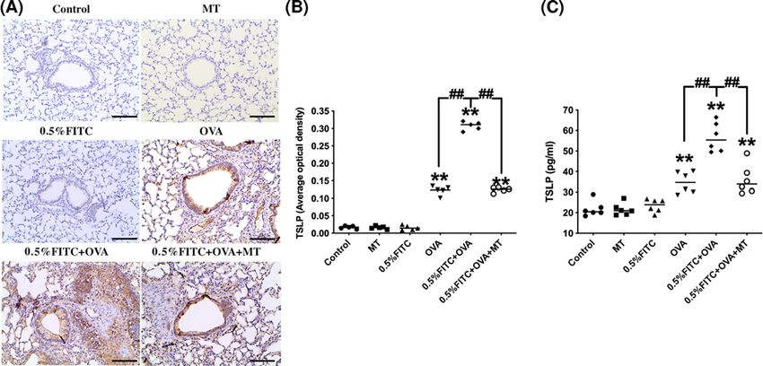
Bioscience Reports (2021) 41 BSR20210398
https://doi.org/10.1042/BSR20210398

Experimental design and procedure
A 0.5% FITC-mediated contact hypersensitivity (CHS) is a Th2-dominant immune system and elevated levels of IL-4
expression in the inflamed skin and increased IgE levels in the serum. Because AD shares many features with CHS
to FITC, this model of Th2-type CHS was used in the present study to induce AD-like skin lesions and then aerosol
challenge of OVA to establish the mice model of atopic march.
   There are six groups in this experiment with six mice per group. (1) Control group (Control) mice were treated with
120 μl of vehicle (1:1 DBP/acetone) on their shaven backs on days 5 and 6. On day 11, their shaven backs and left ears
were subjected to 40 μl of vehicle. In addition, the mice were exposed to an aerosol challenge of saline (30 min/day)
using an ultrasonic nebulizer (Yuyue 402A type I, China) on days 19 through 25. (2) MT control group (MT) mice
received the same treatment as the control group, but in addition the mice were treated with 5 mg/(kg.day) MT by
intratracheal instillation, from days 19 to 25. (3) A 0.5% FITC-induced AD group (0.5% FITC) mice were sensitized

                                                                                                                                                                                           Downloaded from http://portlandpress.com/bioscirep/article-pdf/41/9/BSR20210398/921054/bsr-2021-0398.pdf by guest on 01 December 2021
with 120 μl of 0.5% FITC on days 5 and 6 on their shaven backs, and on day 11 their shaven backs and left ear were
subjected to 40 μl of 0.5% FITC [24]. From days 19 to 25, the mice were exposed to saline (30 min/day) using an
ultrasonic nebulizer. (4) OVA exposure group (OVA) mice were exposed to an aerosol challenge of 1% OVA (30
min/day) using an ultrasonic nebulizer on days 19 through 25. (5) A 0.5% FITC and OVA co-treatment group (0.5%
FITC+OVA) mice were treated with 120 μl of 0.5% FITC on days 5 and 6 on their shaven backs, and on day 11, their
shaven backs and left ear were subjected to 40 μl of 0.5% FITC. The mice were then challenged with 1% OVA using
an ultrasonic nebulizer from days 19 to 25. (6) A 0.5% FITC and MT exposure combined with OVA challenged group
(0.5% FITC+OVA+MT). These mice were treated with 120 μl of 0.5% FITC on days 5 and 6 on their shaven backs,
and on day 11 their shaven backs and left ear were subjected to 40 μl of 0.5% FITC. From days 19 to 25, these mice
were challenged with 1% OVA using an ultrasonic nebulizer. In addition, 5 mg/(kg.day) MT was administered by
intratracheal instillation from days 19 to 25. The detailed protocol is shown in Figure 1.

Quantitative analyses of total serum IgE, OVA-IgE, and OVA-IgG1
Heart blood was collected after the mice were anesthetized with pentobarbital on day 18. Immediately after collection,
the blood serum was centrifuged at 3000 rpm for 10 min at room temperature. Serum total-IgE, OVA- IgE and OVA-
IgG1 levels were measured using commercial ELISA kits according to the manufacturer’s instructions.

Assessment of IL-1β and TNF-α
Serum levels of IL-1β and TNF-α were measured using commercial ELISA kits according to manufacturer’s protocols.
The sensitivity of the kit was 5 pg/ml.

Analysis of cytokines and inflammatory cells in bronchoalveolar lavage
fluid
After serum collection, the mice were killed. The trachea was exposed for alveolar lavage, by cutting the neck skin.
Sterile saline (0.9 ml) was injected through the trachea into the lungs and sucked out after a 1-min chest massage.
This procedure was repeated three times, and the bronchoalveolar lavage fluid (BALF) centrifuged at 1500 rpm at
4◦ C for 10 min. The supernatant was collected for measuring the levels of IL-4, IL-5, IL-13, IFN-γ, IL-33 and TSLP
in the lung using commercial ELISA kits. In addition, the cell sediment from the centrifuged BALF was suspended
in saline, and total inflammatory cells, neutrophils and eosinophils were counted using a Blood Cell Analysis system
(MTN-21, Matee3nu Technology Corp, Jinan, China).

Histology
The left lungs of the mice were removed and inflated with 4% paraformaldehyde, fixed overnight at 4◦ C and embedded
in paraffin. Sections (5 μm) were stained using Hematoxylin and Eosin (H&E). Briefly, the sections on slides were
deparaffinized and rehydrated with distilled water and kept in Hematoxylin for 1 min. After washing five-times, the
slides were counterstained in Alcoholic-Eosin for 1 min. The slides were then dehydrated with 95% EtOH and 100%
EtOH, followed by the application of Xylene to clear for 1 min before the coverslip was mounted. Masson’s Trichrome
staining was used to show the peribronchial collagen deposition. Lung slides were incubated with mordant (50 mg/ml
potassium bichromate + 50 mg/ml trichloroacetic acid) for 20 min and then washed for 1 min. Next, the slides were put
into Weigert’s iron Hematoxylin solution for 3 min, washed with water for 10 min and then incubated with mordant
(2.5% phosphomolybdic acid + 2.5% phosphotungstic acid) for 30 s. Following 1-min wash, the slides were incubated
in 1% acetic acid for 10 s and then 0.75% Orange G solution for 5 min. Before and after staining with Ponceau xylidine
(12 mg/ml) and Fuchsin S solution (8 mg/ml) for 30 min, the sections were washed with 1% acetic acid for 10 s, and

© 2021 The Author(s). This is an open access article published by Portland Press Limited on behalf of the Biochemical Society and distributed under the Creative Commons Attribution   3
License 4.0 (CC BY).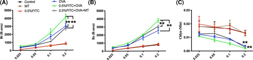
Bioscience Reports (2021) 41 BSR20210398
                                                                                                                                         https://doi.org/10.1042/BSR20210398

                                                                                                                                                                                           Downloaded from http://portlandpress.com/bioscirep/article-pdf/41/9/BSR20210398/921054/bsr-2021-0398.pdf by guest on 01 December 2021
           Figure 1. Exposure and sensitization protocol

           then incubated with 2.5% phosphotungstic acid followed by Aniline Blue solution for 3 min. After washing with 1%
           acetic acid for 10 s, the sections were dehydrated with a graded ethanol series (70, 80 90, 95, and 100%), defatted with
           xylene twice and mounted. Periodic Acid–Schiff (PAS) staining reveals the increased mucus production that occurs
           after allergen exposure due to goblet cell hyperplasia. The lung section slides were placed in a staining dish and fixed
           with Carnoy’s fixative for 10 min. After three washes, the slides were stained with periodic acid solution for 10 min.
           Before and after Shiff Reagent staining, the slides must be rinsed very carefully. The solutions were dehydrated in
           ascending alcohol and cleared with xylene in a staining dish, and then the coverslip was mounted. All sections of the
           lung tissue were assessed by pathologists in a blinded fashion using a DM4000B microscope.

           Assessment of AHR
           AHR was determined using a lung function system (AniRes2005 version 2.0, Beijing, China) on day 26. Briefly, exper-
           imental mice were anesthetized by intraperitoneal injection of pentobarbital sodium (100 mg/kg). The trachea was
           exposed and cannulated, and then attached to a computer-controlled ventilator. The ventilator set the standard pa-
           rameters to be 1:1.5 ratio of inspiration/breath and 90 breaths per minute. Four doses of methacholine (MCH; 0.025,
           0.05, 0.1 and 0.2 mg/kg), a constrictor agonist, were injected into the jugular vein at 5-min intervals. Any change in
           R-area of the inspiratory resistance (Ri) and expiratory resistance (Re) and in the value of pulmonary dynamic com-
           pliance (Cldyn) were recorded within 250 s after the MCH injection. The severity of AHR is represented by increased
           R-area of Ri, Re and a decrease in Cldyn.

           Lung tissue homogenate preparation and GSH and MDA determination
           After washing the lung tissue in ice-cold phosphate-buffered saline (PBS), 10% of the lung tissue homogenate samples
           were prepared by homogenizing on ice using 10 ml/g of ice-cold PBS (pH 7.5). The homogenate was centrifuged at

4   © 2021 The Author(s). This is an open access article published by Portland Press Limited on behalf of the Biochemical Society and distributed under the Creative Commons Attribution
    License 4.0 (CC BY).
Bioscience Reports (2021) 41 BSR20210398
https://doi.org/10.1042/BSR20210398
12000 rpm for 10 min at 4◦ C, and the supernatant collected for GSH and MDA testing. The total protein in each
sample was measured using a modified BCA protein assay kit. The concentrations of GSH and MDA were determined
in accordance with the kit’s manufacturer’s instructions.
Immunohistochemical staining of TSLP and NF-κB (phospho-S536)
Sections were blocked in 5% normal goat serum and incubated with the primary antibodies anti-TSLP (1:100, Abcam,
MA, U.S.A.) and anti-phospho-p65 (S536) (1:100, Abcam, MA, U.S.A.), respectively. The sections were then incubated
in an appropriate biotinylated immunoglobulin and avidin–biotin peroxidase complex. The negative control was
obtained by omitting the primary antibody. Stained sections were observed using a DM4000B Microscope. The results
of TSLP and NF-κB staining were estimated as an average of optical density (AOD) using ImagePro-Plus 6.0 software.
Downloaded from http://portlandpress.com/bioscirep/article-pdf/41/9/BSR20210398/921054/bsr-2021-0398.pdf by guest on 01 December 2021
Statistical analyses
The data are presented as the mean + − standard deviation. The statistical graphs were generated using GraphPad
Prism 5.0 (San Diego, CA, U.S.A.). Results were evaluated statistically using a one-way analysis of variance (ANOVA)
followed by Holm–Sidak’s multiple comparisons test. PBioscience Reports (2021) 41 BSR20210398
https://doi.org/10.1042/BSR20210398
Downloaded from http://portlandpress.com/bioscirep/article-pdf/41/9/BSR20210398/921054/bsr-2021-0398.pdf by guest on 01 December 2021
Figure 2. The changes in immunoglobulin levels and pro-inflammatory cytokines in the serum of atopic march mice, and
the attenuating effects of MT
Total IgE (A), OVA-IgE (B), OVA-IgG1 (C), TNF-α (D) and IL-1β (E) levels in serum. *: PBioscience Reports (2021) 41 BSR20210398
https://doi.org/10.1042/BSR20210398
Downloaded from http://portlandpress.com/bioscirep/article-pdf/41/9/BSR20210398/921054/bsr-2021-0398.pdf by guest on 01 December 2021
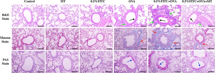
Figure 4. MT treatment reversed the pathological alterations in atopic march mouse model
Green arrow, lung tissue cell infiltration; black arrow, bronchial remodeling; red arrow, subepithelial collagen deposition (blue colored
stain); blue arrow, mucus hypersecretion (pale pink colored stain), scale bar = 50 μm.
Figure 5. MT treatment attenuated AHR in the atopic march mouse model
(A–C) Ri, Re and Cldyn values, respectively. **: PBioscience Reports (2021) 41 BSR20210398
https://doi.org/10.1042/BSR20210398
Downloaded from http://portlandpress.com/bioscirep/article-pdf/41/9/BSR20210398/921054/bsr-2021-0398.pdf by guest on 01 December 2021
Figure 6. Increased production of TSLP in the lungs of atopic march mice and the attenuating effects of MT
(A) Immunohistochemistry for TSLP in lung tissue, scale bar = 50 μm. (B) The optical density of immunohistochemistry for TSLP.
(C) Concentrations of TSLP in BALF as determined by ELISA. **: PBioscience Reports (2021) 41 BSR20210398 https://doi.org/10.1042/BSR20210398 staining are shown in Figure 7C, estimated as the average optical density using Image-Pro Plus 6.0 software (Figure 7D). The concentration of activated NF-κB was enhanced in the OVA group (P
Bioscience Reports (2021) 41 BSR20210398
                                                                                                                                     https://doi.org/10.1042/BSR20210398

         induction of TSLP in these cells [39,40]. Our results for NF-κB and TSLP, revealed similar trends to these other
         studies.
            MT, a free-radical scavenger, effectively blocks oxidative stress [41]. By examining the activities of alanine amino-
         transferase (ALT) and aspartate aminotransferase (AST) in the serum (Supplementary Figure S2) and inflammation
         levels, we confirmed in the present study and other studies [42], that a treatment of 5 mg/(kg.day) MT is safe because
         the liver enzymes AST and ALT were not affected. More future studies on its safety on vital organs and functions are
         important to be investigated for short and long terms. Interestingly, the asthma symptoms and oxidative stress in mice
         subjected to 0.5% FITC treatment and an OVA challenge can be effectively alleviated by MT treatment, suggesting
         that oxidative stress plays an important role in the development of the atopic march. Previous studies have already
         demonstrated the crucial role of TSLP in the atopic march, by promoting allergen sensitization in barrier-impaired
         skin [14,25]. However, not enough information is available on the role of TSLP in the lung during the atopic march,
         nor on the possible activation mechanisms. In this study, we found that OVA treatment activated the release of TSLP,

                                                                                                                                                                                Downloaded from http://portlandpress.com/bioscirep/article-pdf/41/9/BSR20210398/921054/bsr-2021-0398.pdf by guest on 01 December 2021
         and that Th2 inflammatory responses followed in the lung. TSLP is an important target protein for regulating the in-
         flammatory response in the asthmatic phase of atopic march. In addition, it had been proved that MT could alleviate
         inflammation by inhibiting the NF-κB signal in vivo [43]. We found that inhibition of oxidative stress by MT effec-
         tively alleviated the activation of NF-κB signaling pathways, and decreased the production of TSLP when compared
         with the OVA group, which further alleviated the development of atopic march. In addition to this, further studies for
         evaluating the effect of MT on 0.5% FITC or OVA-induced allergic inflammation are also important to be revealed.
            In conclusion, by successfully establishing an experimental atopic march mouse model, we were able to demon-
         strate that overproduction of oxidative stress in the lung significantly up-regulated the activation of NF-κB signaling
         pathways and the subsequent release of TSLP, which further promotes the development of the atopic march. From a
         therapeutic point of view, to mitigate the development of atopic march, antioxidants such as MT may be imperative
         for inhibiting NF-κB activation in the lung, especially after the occurrence of AD.

         Data Availability
         All the data generated or analyzed during the present study are included in this published article.

         Competing Interests
         The authors declare that there are no competing interests associated with the manuscript.

         Funding
         This work was supported by the Regular Undergraduate Institutions Nature Science Research Project of Guizhou Education De-
         partment (Technology top-notch talent support project QianjiaoheKYzi [2018] 083); Department of Education of Hubei Province
         Science Research Program [grant number Q20191103]; The Scientific and Technological Support Project ([2019] 2370) of Guizhou
         Province.

         CRediT Author Contribution
         Xudong Liu: Conceptualization, Writing—original draft. Yuchao Zhang: Investigation, Methodology. Yaolin Ren: Investigation,
         Methodology. Jinquan Li: Conceptualization, Writing—review & editing.

         Abbreviations
         AD, atopic dermatitis; AHR, airway hyperresponsiveness; ALT, alanine aminotransferase; AST, aspartate aminotransferase;
         BALF, bronchoalveolar lavage fluid; CHS, contact hypersensitivity; Cldyn, pulmonary dynamic compliance; DBP, dibutyl phtha-
         late; DC, dendritic cell; ELISA, enzyme-linked immunosorbent assay; FITC, fluorescein isothiocyante; GSH, glutathione; H&E,
         Hematoxylin and Eosin; IFN, interferon; Ig, immunoglobulin; MCH, methacholine; MDA, malonaldehyde; MT, melatonin; NF-κB,
         nuclear factor-κB; OVA, ovalbumin; PAS, Periodic Acid–Schiff; Re, inspiratory resistance; Ri, expiratory resistance; ROS, reac-
         tive oxygen species; TNF-alpha, tumor necrosis factor-α; TSLP, thymic stromal lymphopoietin.

         References
         1 Laughter, M.R., Maymone, M.B.C., Mashayekhi, S., Arents, B.W.M., Karimkhani, C., Langan, S.M. et al. (2021) The global burden of atopic dermatitis:
           lessons from the Global Burden of Disease Study 1990-2017. Br. J. Dermatol. 184, 304–309, https://doi.org/10.1111/bjd.19580
         2 Ravnborg, N., Ambikaibalan, D., Agnihotri, G., Price, S., Rastogi, S., Patel, K.R. et al. (2021) Prevalence of asthma in patients with atopic dermatitis: a
           systematic review and meta-analysis. J. Am. Acad. Dermatol. 84, 471–478, https://doi.org/10.1016/j.jaad.2020.02.055
         3 Matsumoto, K., Iikura, K., Morita, H. and Saito, H. (2020) Barrier dysfunction in the atopic march-how does atopic dermatitis lead to asthma in
           children? J. Allergy Clin. Immunol. 145, 1551–1553, https://doi.org/10.1016/j.jaci.2020.04.014

10   © 2021 The Author(s). This is an open access article published by Portland Press Limited on behalf of the Biochemical Society and distributed under the Creative Commons
     Attribution License 4.0 (CC BY).
Bioscience Reports (2021) 41 BSR20210398
https://doi.org/10.1042/BSR20210398
4 Leyva-Castillo, J.M., Yoon, J. and Geha, R.S. (2019) IL-22 promotes allergic airway inflammation in epicutaneously sensitized mice. J. Allergy Clin.
Immunol. 143, 619.e7–630.e7, PMCID: PMCPMC6298864, https://doi.org/10.1016/j.jaci.2018.05.032
5 Chen, P.Y., Chen, C.W., Su, Y.J., Chang, W.H., Kao, W.F., Yang, C.C. et al. (2020) Associations between levels of urinary oxidative stress of 8-OHdG and
risk of atopic diseases in children. Int. J. Environ. Res. Public Health 17, 8207–8220, PMCID: PMC7664398, https://doi.org/10.3390/ijerph17218207
6 Kim, S.J., Cheresh, P., Jablonski, R.P., Rachek, L., Yeldandi, A., Piseaux-Aillon, R. et al. (2020) Mitochondrial 8-oxoguanine DNA glycosylase mitigates
alveolar epithelial cell PINK1 deficiency, mitochondrial DNA damage, apoptosis, and lung fibrosis. Am. J. Physiol. Lung Cell. Mol. Physiol. 318,
L1084–L1096, PMCID: PMCPMC7272732, https://doi.org/10.1152/ajplung.00069.2019
7 Song, Y., Li, R., Zhang, Y., Wei, J., Chen, W., Chung, C.K.A. et al. (2019) Mass spectrometry-based metabolomics reveals the mechanism of ambient
fine particulate matter and its components on energy metabolic reprogramming in BEAS-2B cells. Sci. Total Environ. 651, 3139–3150,
https://doi.org/10.1016/j.scitotenv.2018.10.171
8 Liu, Z., Huang, Y., Jiao, Y., Chen, Q., Wu, D., Yu, P. et al. (2020) Polystyrene nanoplastic induces ROS production and affects the
MAPK-HIF-1/NFkB-mediated antioxidant system in Daphnia pulex. Aquat. Toxicol. 220, 105420, https://doi.org/10.1016/j.aquatox.2020.105420
9 Guo, H., Ji, J., Wei, K., Sun, J., Zhang, Y. and Sun, X. (2021) MAPK/AP-1 and ROS participated in ratio- and time-dependent interaction effects of
Downloaded from http://portlandpress.com/bioscirep/article-pdf/41/9/BSR20210398/921054/bsr-2021-0398.pdf by guest on 01 December 2021
deoxynivalenol and cadmium on HT-29 cells. Food Chem. Toxicol. 148, 111921, https://doi.org/10.1016/j.fct.2020.111921
10 Wu, Q., Wu, W. and Kuca, K. (2020) From hypoxia and hypoxia-inducible factors (HIF) to oxidative stress: a new understanding of the toxic mechanism
of mycotoxins. Food Chem. Toxicol. 135, 110968, https://doi.org/10.1016/j.fct.2019.110968
11 Gu, S.M., Yun, J., Son, D.J., Kim, H.Y., Nam, K.T., Kim, H.D. et al. (2017) Piperlongumine attenuates experimental autoimmune encephalomyelitis
through inhibition of NF-kappaB activity. Free Radic. Biol. Med. 103, 133–145, https://doi.org/10.1016/j.freeradbiomed.2016.12.027
12 Akdis, C.A., Arkwright, P.D., Bruggen, M.C., Busse, W., Gadina, M., Guttman-Yassky, E. et al. (2020) Type 2 immunity in the skin and lungs. Allergy 75,
1582–1605, https://doi.org/10.1111/all.14318
13 Mavissakalian, M. and Brady, S. (2020) The current state of biologic therapies for treatment of refractory asthma. Clin. Rev. Allergy Immunol. 59,
195–207, https://doi.org/10.1007/s12016-020-08776-8
14 Ziegler, S.F. (2021) Thymic stromal lymphopoietin, skin barrier dysfunction, and the atopic march. Ann. Allergy Asthma Immunol. 127, 306–311,
https://doi.org/10.1016/j.anai.2021.06.004
15 Han, H., Roan, F. and Ziegler, S.F. (2017) The atopic march: current insights into skin barrier dysfunction and epithelial cell-derived cytokines. Immunol.
Rev. 278, 116–130, PMCID: PMCPMC5492959, https://doi.org/10.1111/imr.12546
16 Porsbjerg, C.M., Sverrild, A., Lloyd, C.M., Menzies-Gow, A.N. and Bel, E.H. (2020) Anti-alarmins in asthma: targeting the airway epithelium with
next-generation biologics. Eur. Respir. J. 56, PMCID: PMCPMC7676874, https://doi.org/10.1183/13993003.00260-2020
17 An, G., Wang, W., Zhang, X., Huang, Q., Li, Q., Chen, S. et al. (2020) Combined blockade of IL-25, IL-33 and TSLP mediates amplified inhibition of
airway inflammation and remodelling in a murine model of asthma. Respirology 25, 603–612, https://doi.org/10.1111/resp.13711
18 Redhu, D., Franke, K., Kumari, V., Francuzik, W., Babina, M. and Worm, M. (2020) Thymic stromal lymphopoietin production induced by skin irritation
results from concomitant activation of protease-activated receptor 2 and interleukin 1 pathways. Br. J. Dermatol. 182, 119–129
19 Kumar, S., Khodoun, M., Kettleson, E.M., McKnight, C., Reponen, T., Grinshpun, S.A. et al. (2014) Glyphosate-rich air samples induce IL-33, TSLP and
generate IL-13 dependent airway inflammation. Toxicology 325, 42–51, PMCID: PMCPMC4195794, https://doi.org/10.1016/j.tox.2014.08.008
20 Wu, Q., Fang, L., Yang, Y., Wang, A., Chen, X., Sun, J. et al. (2021) Protection of melatonin against long-term radon exposure-caused lung injury.
Environ. Toxicol. 36, 472–483, https://doi.org/10.1002/tox.23052
21 Xalxo, R. and Keshavkant, S. (2019) Melatonin, glutathione and thiourea attenuates lead and acid rain-induced deleterious responses by regulating
gene expression of antioxidants in Trigonella foenum graecum L. Chemosphere 221, 1–10, https://doi.org/10.1016/j.chemosphere.2019.01.029
22 Gonzalez, A., Estaras, M., Martinez-Morcillo, S., Martinez, R., Garcia, A., Estevez, M. et al. (2020) Melatonin modulates red-ox state and decreases
viability of rat pancreatic stellate cells. Sci. Rep. 10, 6352, PMCID: PMCPMC7156707, https://doi.org/10.1038/s41598-020-63433-6
23 Wu, H.M., Zhao, C.C., Xie, Q.M., Xu, J. and Fei, G.H. (2020) TLR2-melatonin feedback loop regulates the activation of NLRP3 inflammasome in murine
allergic airway inflammation. Front. Immunol. 11, 172, PMCID: PMCPMC7025476, https://doi.org/10.3389/fimmu.2020.00172
24 Scuron, M.D., Fay, B.L., Connell, A.J., Peel, M.T. and Smith, P.A. (2020) Ruxolitinib cream has dual efficacy on pruritus and inflammation in experimental
dermatitis. Front. Immunol. 11, 620098, PMCID: PMCPMC7917252, https://doi.org/10.3389/fimmu.2020.620098
25 Leyva-Castillo, J.M., Hener, P., Jiang, H. and Li, M. (2013) TSLP produced by keratinocytes promotes allergen sensitization through skin and thereby
triggers atopic march in mice. J. Invest. Dermatol. 133, 154–163, https://doi.org/10.1038/jid.2012.239
26 Han, H., Xu, W., Headley, M.B., Jessup, H.K., Lee, K.S., Omori, M. et al. (2012) Thymic stromal lymphopoietin (TSLP)-mediated dermal inflammation
aggravates experimental asthma. Mucosal Immunol. 5, 342–351, PMCID: PMCPMC3328620, https://doi.org/10.1038/mi.2012.14
27 Celebi Sozener, Z., Cevhertas, L., Nadeau, K., Akdis, M. and Akdis, C.A. (2020) Environmental factors in epithelial barrier dysfunction. J. Allergy Clin.
Immunol. 145, 1517–1528, https://doi.org/10.1016/j.jaci.2020.04.024
28 Oyoshi, M.K., Larson, R.P., Ziegler, S.F. and Geha, R.S. (2010) Mechanical injury polarizes skin dendritic cells to elicit a T(H)2 response by inducing
cutaneous thymic stromal lymphopoietin expression. J. Allergy Clin. Immunol. 126, 976–984, 84.e1–e5; PMCID: PMCPMC3085022,
https://doi.org/10.1016/j.jaci.2010.08.041
29 Shen, S., Li, J., You, H., Wu, Z., Wu, Y., Zhao, Y. et al. (2017) Oral exposure to diisodecyl phthalate aggravates allergic dermatitis by oxidative stress and
enhancement of thymic stromal lymphopoietin. Food Chem. Toxicol. 99, 60–69, https://doi.org/10.1016/j.fct.2016.11.016
30 Hwang, Y.H., Paik, M.J. and Yee, S.T. (2017) Diisononyl phthalate induces asthma via modulation of Th1/Th2 equilibrium. Toxicol. Lett. 272, 49–59,
https://doi.org/10.1016/j.toxlet.2017.03.014
31 Chen, Y., Wu, X., Yang, X., Liu, X., Zeng, Y. and Li, J. (2021) Melatonin antagonizes ozone-exacerbated asthma by inhibiting the TRPV1 channel and
stabilizing the Nrf2 pathway. Environ. Sci. Pollut. Res. Int., https://doi.org/10.1007/s11356-021-14945-9
© 2021 The Author(s). This is an open access article published by Portland Press Limited on behalf of the Biochemical Society and distributed under the Creative Commons 11
Attribution License 4.0 (CC BY).Bioscience Reports (2021) 41 BSR20210398
https://doi.org/10.1042/BSR20210398
32 Hajimohammadi, B., Athari, S.M., Abdollahi, M., Vahedi, G. and Athari, S.S. (2020) Oral administration of acrylamide worsens the inflammatory
responses in the airways of asthmatic mice through agitation of oxidative stress in the lungs. Front. Immunol. 11, 1940, PMCID: PMCPMC7581680,
https://doi.org/10.3389/fimmu.2020.01940
33 Ma, Y., Deng, L., Ma, P., Wu, Y., Yang, X., Xiao, F. et al. (2021) In vivo respiratory toxicology of cooking oil fumes: Evidence, mechanisms and prevention.
J. Hazard. Mater. 402, 123455, https://doi.org/10.1016/j.jhazmat.2020.123455
34 Alharbi, K.S., Fuloria, N.K., Fuloria, S., Rahman, S.B., Al-Malki, W.H., Javed Shaikh, M.A. et al. (2021) Nuclear factor-kappa B and its role in
inflammatory lung disease. Chem. Biol. Interact. 345, 109568, https://doi.org/10.1016/j.cbi.2021.109568
35 Verstraete, K., Peelman, F., Braun, H., Lopez, J., Van Rompaey, D., Dansercoer, A. et al. (2017) Structure and antagonism of the receptor complex
mediated by human TSLP in allergy and asthma. Nat. Commun. 8, 14937, PMCID: PMCPMC5382266, https://doi.org/10.1038/ncomms14937
36 Kabata, H., Flamar, A.L., Mahlakoiv, T., Moriyama, S., Rodewald, H.R., Ziegler, S.F. et al. (2020) Targeted deletion of the TSLP receptor reveals cellular
mechanisms that promote type 2 airway inflammation. Mucosal Immunol. 13, 626–636, PMCID: PMCPMC7311324,
https://doi.org/10.1038/s41385-020-0266-x
37 Toki, S., Goleniewska, K., Zhang, J., Zhou, W., Newcomb, D.C., Zhou, B. et al. (2020) TSLP and IL-33 reciprocally promote each other’s lung protein
Downloaded from http://portlandpress.com/bioscirep/article-pdf/41/9/BSR20210398/921054/bsr-2021-0398.pdf by guest on 01 December 2021
expression and ILC2 receptor expression to enhance innate type-2 airway inflammation. Allergy 75, 1606–1617, PMCID: PMCPMC7354889,
https://doi.org/10.1111/all.14196
38 Lee, H.C., Headley, M.B., Iseki, M., Ikuta, K. and Ziegler, S.F. (2008) Cutting edge: inhibition of NF-kappaB-mediated TSLP expression by retinoid X
receptor. J. Immunol. 181, 5189–5193, PMCID: PMCPMC2878481, https://doi.org/10.4049/jimmunol.181.8.5189
39 Vu, A.T., Chen, X., Xie, Y., Kamijo, S., Ushio, H., Kawasaki, J. et al. (2011) Extracellular double-stranded RNA induces TSLP via an endosomal
acidification- and NF-kappaB-dependent pathway in human keratinocytes. J. Invest. Dermatol. 131, 2205–2212, https://doi.org/10.1038/jid.2011.185
40 Lin, T.H., Cheng, C.C., Su, H.H., Huang, N.C., Chen, J.J., Kang, H.Y. et al. (2016) Lipopolysaccharide attenuates induction of proallergic cytokines,
thymic stromal lymphopoietin, and interleukin 33 in respiratory epithelial cells stimulated with PolyI:C and human parechovirus. Front. Immunol. 7, 440,
PMCID: PMCPMC5078322, https://doi.org/10.3389/fimmu.2016.00440
41 Lan, M., Zhang, Y., Wan, X., Pan, M.H., Xu, Y. and Sun, S.C. (2020) Melatonin ameliorates ochratoxin A-induced oxidative stress and apoptosis in
porcine oocytes. Environ. Pollut. 256, 113374, https://doi.org/10.1016/j.envpol.2019.113374
42 Gonzaga, N.A., Awata, W.M.C., Ficher, S.P., Assis, V.O., Alves, J.V., Tostes, R.C. et al. (2021) Melatonin reverses the loss of the anticontractile effect of
perivascular adipose tissue in obese rats. J. Pineal Res. 70, e12710, https://doi.org/10.1111/jpi.12710
43 Liu, Z., Gan, L., Xu, Y., Luo, D., Ren, Q., Wu, S. et al. (2017) Melatonin alleviates inflammasome-induced pyroptosis through inhibiting
NF-kappaB/GSDMD signal in mice adipose tissue. J. Pineal Res. 63, e12414, https://doi.org/10.1111/jpi.12414
12 © 2021 The Author(s). This is an open access article published by Portland Press Limited on behalf of the Biochemical Society and distributed under the Creative Commons Attribution
License 4.0 (CC BY).You can also read