FRZB as a key molecule in abdominal aortic aneurysm progression affecting vascular integrity
←
→
Page content transcription
If your browser does not render page correctly, please read the page content below
Bioscience Reports (2021) 41 BSR20203204
https://doi.org/10.1042/BSR20203204

Research Article

FRZB as a key molecule in abdominal aortic
aneurysm progression affecting vascular integrity
Chang-Kyu Oh1,* , Yeji Ko2,* , Jeong Jun Park3 , Hye Jin Heo4 , Junho Kang5 , Eun Jung Kwon5 , Ji Wan Kang5 ,
Yoonsung Lee1 , Kyungjae Myung1 , Jin Mo Kang6 , Dai Sik Ko6 and Yun Hak Kim4,5
1 Center for Genomic Integrity, Institute for Basic Science (IBS), Ulsan 44919, Republic of Korea; 2 Department of Statistics, University of Michigan, Ann Arbor, MI, U.S.A.;
3 Department   of Anesthesiology and Pain Medicine, Asan Medical Center, University of Ulsan College of Medicine, Seoul, Republic of Korea; 4 Department of Anatomy, School of

                                                                                                                                                                                           Downloaded from http://portlandpress.com/bioscirep/article-pdf/41/1/BSR20203204/901282/bsr-2020-3204.pdf by guest on 21 January 2021
Medicine, Pusan National University, Yangsan, Republic of Korea; 5 Department of Biomedical Informatics, School of Medicine, Pusan National University, Yangsan, Republic of
Korea; 6 Division of Vascular Surgery, Department of Surgery, Gachon University Gil Medical Center, Incheon, Republic of Korea
Correspondence: Dai Sik Ko (daisik.ko@gilhospital.com) or Jin Mo Kang (calzevi@gmail.com) or Yun Hak Kim (yunhak10510@pusan.ac.kr)

                                           Abdominal aortic aneurysm (AAA), when ruptured, results in high mortality. The identifica-
                                           tion of molecular pathways involved in AAA progression is required to improve AAA prog-
                                           nosis. The aim of the present study was to assess the key genes for the progression of
                                           AAA and their functional role. Genomic and clinical data of three independent cohorts were
                                           downloaded from the National Center for Biotechnology Information (NCBI) Gene Expres-
                                           sion Omnibus (GEO) (GSE57691, GSE7084, and GSE98278). To develop AAA diagnosis and
                                           progression-related differentially expressed genes (DEGs), we used a significance analysis
                                           of microarray (SAM). Spearman correlation test and gene set analysis were performed to
                                           identify potential enriched pathways for DEGs. Only the Frizzled-related protein (FRZB) gene
                                           and chromosome 1 open reading frame 24 (C1orf24) exhibited significant down-regulation in
                                           all analyses. With FRZB, the pathways were associated with RHO GTPase and elastin fiber
                                           formation. With C1orf24, the pathways were elastic fiber formation, extracellular matrix or-
                                           ganization, and cell–cell communication. Since only FRZB was evolutionally conserved in
                                           the vertebrates, function of FRZB was validated using zebrafish embryos. Knockdown of
                                           frzb remarkably reduced vascular integrity in zebrafish embryos. We believe that FRZB is a
                                           key gene involved in AAA initiation and progression affecting vascular integrity.

                                         Introduction
                                         Abdominal aortic aneurysm (AAA) is a late age-of-onset disorder that affects approximately 5% of men
                                         aged 65–74 years; when ruptured, AAAs are associated with a mortality rate of up to 90% [1]. Ruptured
                                         aortic aneurysms are a major cause of death, and they were reported to be the 13th leading cause of mor-
                                         tality in 2017 [2]. Preemptive elective open surgical or endovascular repair is considered when the AAA
                                         diameter is ≥55 mm, when it is rapidly expanding (≥10 mm/year), or when it causes symptoms [3]. There
                                         are, however, large intrapatient and interpatient variations with regard to rates of expansion of small AAAs
                                         during follow-up [4]. Therefore, increasing demand to improve risk stratification in patients with AAA
* These authors contributed              has been claimed [5,6].
equally to this work.                       The pathophysiology of AAA has been extensively studied. AAA was previously believed to be a form
Received: 18 September 2020
                                         of atherosclerosis; however, it is now recognized as a distinct degenerative process involving all layers of
Revised: 10 November 2020                the vessel wall and is characterized by destruction of elastin and collagen in the media and adventitia and
Accepted: 26 November 2020               loss of smooth muscle cells with thinning of the media [1,7,8]. However, a lack of knowledge remains
                                         regarding the molecular mechanisms that initiate aneurysm formation and expansion. In current clinical
Accepted Manuscript online:
27 November 2020                         practice, the strategy to stratify patients at high risk for aneurysm progression is lacking.
Version of Record published:                Serial monitoring of biological activity of AAA would be necessary to lower mortality and morbidity
06 January 2021                          from AAA rupture; however, no biomarkers have yet proven to be of prognostic value additive to AAA

© 2021 The Author(s). This is an open access article published by Portland Press Limited on behalf of the Biochemical Society and distributed under the Creative Commons Attribution   1
License 4.0 (CC BY).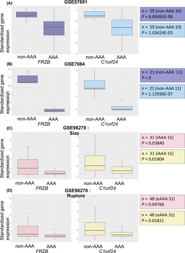
Bioscience Reports (2021) 41 BSR20203204
                                                                                                                                                     https://doi.org/10.1042/BSR20203204

Table 1 Patients’ characteristics
                     GSE57691                                  GSE7084                                    GSE98278
                     Non-AAA              AAA                  Non-AAA               AAA                  Size                                       Status
                                                                                                          Intermediate          Large                Stable               Rupture

 n                   10                   49                   11                    10                   15                    16                   31                   17
 Age (years)         68.4 (+
                           −4.5)          69.8 (+
                                                −7.0)*         64.5 (+
                                                                     −10.7)          66.4 (+
                                                                                           −5.7)          67.7(+
                                                                                                               −7.4)            72.3(+
                                                                                                                                     −9.2)           69.5 (+
                                                                                                                                                           −7.2)          73.5 (+
                                                                                                                                                                                −11.3)
 Women (%)           40.0%                4.1%                 27.3%                 20.0%                6.7%                  12.5%                3.2%                 23.5%
 Diameter (mm)       -                    62.3 (+
                                                −10.2)*        -                     -                    54.1 (+
                                                                                                                −1.8)           84.1 (+
                                                                                                                                      −12.6)         62.3 (+
                                                                                                                                                           −12.1)         77.0 (+
                                                                                                                                                                                −14.7)
 Hypertension        -                    81.6%                -                     -                    86.7%                 93.8%                93.5%                88.2%
 (%)
 Diabetes (%)        -                    22.4%                -                     -                    26.7%                 31.3%                32.3%                23.5%
 Dyslipidemia        -                    71.4%                -                     -                    86.7%                 81.3%                77.4%                82.3%

                                                                                                                                                                                                      Downloaded from http://portlandpress.com/bioscirep/article-pdf/41/1/BSR20203204/901282/bsr-2020-3204.pdf by guest on 21 January 2021
 (%)
 Coronary heart -                         51.0%                -                     -                    46.7%                 43.8%                58.1%                41.2%
 disease (%)
 Ever smoker         50.0%                57.1%                -                     -                    33.3%                 68.8%                58.1%                58.8%
 (%)

 Nominal variables are presented as percentages; continuous variables are presented as mean +
                                                                                            − standard deviation. Asterisk (*) means Cohen’s formula
 for pooled standard deviation.

                      diameter to predict AAA expansion [9,10]. AAA growth rate can only be acknowledged retrospectively. To be able
                      to find preventive strategies, the molecular mechanisms behind the disease and drug targets need to be evaluated in
                      detail.
                         Some studies have attempted to evaluate the candidate genes and pathways potentially associated with AAA, but a
                      large portion of their data was prone to bias and focused on pre-selected genes encoding matrix metalloproteinases,
                      components of the immune system, and known atherosclerosis-related factors [8,11–14]. Therefore, to understand
                      the molecular pathways controlling the progression of AAA without bias, we examined the statistical analysis of three
                      cohorts acquired from the National Center for Biotechnology Information (NCBI) Gene Expression Omnibus (GEO)
                      Series (GSE57691, GSE7084, and GSE98278).

                      Methods
                      Patient data
                      Microarray gene expression data and clinical information of three different AAA patient cohorts were obtained from
                      three independent sources: James Cook University, Wayne State University School of Medicine, and Ludwig Maxim-
                      ilians University Munich. They were accessed through the NCBI GEO Series (GSE57691, GSE7084, and GSE98278,
                      respectively). GSE57691 and GSE7084 contain both normal and AAA specimen data. Thus, they were evaluated to
                      find the differentially expressed genes (DEGs) among the AAA patient group. On the contrary, GSE98278, which
                      does not include the normal patient data, was used to determine genes that are linked to the further development of
                      AAA. More details of the patient data are shown below (Table 1). Since the data we used were publicly accessible, IRB
                      approval was not required.

                      Standardization
                      In microarray data, each gene has a different range of expression values. For instance, while some gene expression
                      levels range from 0 to 100, others can range from 500 to 1000. Thus, to facilitate comparison between them, we had
                      to standardize their values and to bring all the genes to the same range. Since microarray data from GSE57691 had
                      been distributed after standardization, only gene expression measurements from GSE7084 and GSE98278 needed to
                      be standardized. The standardization process has assigned a standard score (z-score) to each gene expression value
                      in the data. The ‘scale’ function in the ‘base’ package R (version 3.6.1) was used in this step.

                      Missing values
                      There were no missing values from GSE57691, GSE7084, and GSE98278. However, a huge portion of genes within the
                      three datasets appeared repetitively in different columns. In order to organize these recurring genes, we aggregated the
                      genes in the columns by mean, using the ‘aggregate’ function in the ‘stats’ package R (version 3.6.1). In consequence,

2              © 2021 The Author(s). This is an open access article published by Portland Press Limited on behalf of the Biochemical Society and distributed under the Creative Commons Attribution
               License 4.0 (CC BY).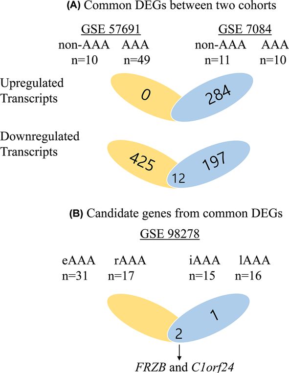
Bioscience Reports (2021) 41 BSR20203204
https://doi.org/10.1042/BSR20203204

a large number of columns (14190, 3023, and 15893) from each gene expression dataset were dropped, but the total
number of distinct gene symbols has not changed and genetic significance also remains the same.

 Significance analysis of microarray
Significance analysis of microarray (SAM) is a test method where the test statistic measures the difference in
gene-expression values between control and treatment groups divided by the standard deviation of repeated mea-
surements. It is specifically designed to address data from large microarrays, as SAM guarantees the probability of
error to not increase with the number of comparisons, in contrast with an increase observed with the number of
t tests. Once all test statistic scores are acquired, they are ranked, and genes with scores greater than an adjustable
threshold delta () are selected. Subsequently, the false discovery rate (FDR) is computed to identify the rate of genes
incorrectly identified as significant by using the permutations of the measurements [16]. In this study, we performed
the SAM tests in the ‘siggenes’ package R (version 3.6.1). Total permutations (1000) and FDR = 0.0005 were used for

                                                                                                                                                                                           Downloaded from http://portlandpress.com/bioscirep/article-pdf/41/1/BSR20203204/901282/bsr-2020-3204.pdf by guest on 21 January 2021
both cohorts.

Additional t test and correlation analysis
Using the GSE98278 dataset which contains stability (stable vs. ruptured) and size (intermediate vs. large) information
of AAA, we next aimed to find out the DEGs that are actively engaged in the development of AAA. Unpaired t tests
were implemented to 12 commonly significant genes from two previous SAM tests, and the resulting P-values were
corrected with Benjamini–Hochberg procedure (FDR = 0.25). Additionally, we conducted correlation tests between
two genes of our main focus, Frizzled-related protein (FRZB) and chromosome 1 open reading frame 24 (C1orf24),
and the other genes in each cohort (GSE7084, GSE57691) for gene set analysis. |R| > 0.7 and P
Bioscience Reports (2021) 41 BSR20203204
https://doi.org/10.1042/BSR20203204
Downloaded from http://portlandpress.com/bioscirep/article-pdf/41/1/BSR20203204/901282/bsr-2020-3204.pdf by guest on 21 January 2021
Figure 1. Flow charts of study design
(A) Up-regulated transcripts were 0 from GSE57691 and 284 from GSE7084. Down-regulated transcripts were 438 from 57691 and
208 from GSE7084. Twelve genes were significant in both cohorts. (B) Of these 12 genes, 3 were significant between intermediate
size of AAA and large size of AAA in GSE 98278; 2 were significant between stable AAA and rupture AAA in GSE 98278. FRZB
and C1orf24 were concomitantly significant. Abbreviations: eAAA, elective AAA; iAAA, intermediate AAA; lAAA, large AAA; rAAA,
ruptured AAA.
+
− 12.1 vs 77.0 +
− 14.7; PBioscience Reports (2021) 41 BSR20203204
https://doi.org/10.1042/BSR20203204
Downloaded from http://portlandpress.com/bioscirep/article-pdf/41/1/BSR20203204/901282/bsr-2020-3204.pdf by guest on 21 January 2021
Figure 2. Box plots for the expression values of FRZB and C1orf24
(A) The expression values of between FRZB and C1orf24 between AAA patients (n=49) and non-AAA patients (n=10) from GSE
57691. FRZB and C1orf24 were significantly down-expressed in AAA patients. (B) The expression values of FRZB and C1orf24
between AAA patients (n=10) and non-AAA patients (n=11) from GSE 7084. FRZB and C1orf24 were significantly down-expressed
in AAA patients. (C) The expression values of FRZB and C1orf24 between intermediate size AAA (n=15) and large size AAA (n=16)
from GSE 98278. FRZB and C1orf24 were significantly down-expressed in large size AAA. (D) The expression values of FRZB
and C1orf24 between eAAA (n=31) and rAAA (n=17) from GSE 98278. FRZB and C1orf24 were significantly down-expressed in
ruptured AAA. Abbreviations: eAAA, elective AAA; iAAA, intermediate AAA; lAAA, large AAA; rAAA, ruptured AAA.
© 2021 The Author(s). This is an open access article published by Portland Press Limited on behalf of the Biochemical Society and distributed under the Creative Commons Attribution 5
License 4.0 (CC BY).Bioscience Reports (2021) 41 BSR20203204
https://doi.org/10.1042/BSR20203204
Table 2 Top ten significantly enriched Reactome pathways with FRZB and correlated genes from GSE57691 and GSE7084
using Enrichr (https://amp.pharm.mssm.edu/Enrichr/)
Term Overlap P-value Odds ratio Combined score Genes
RHO GTPases activate 3/17 2.16E-05 54.29864253 583.4084401 PPP1CB; MYH11; MYH10
ROCKs
RHO GTPases activate 3/21 4.18E-05 43.95604396 443.2037946 PPP1CB; MYH11; MYH10
PAKs
Molecules associated 3/30 1.25E-04 30.76923077 276.5468079 MFAP4; ITGA8; FBLN5
with elastic fibers
Elastic fiber formation 3/41 3.20E-04 22.51407129 181.1923658 MFAP4; ITGA8; FBLN5
RHO GTPases activate 3/60 9.82E-04 15.38461538 106.5481366 PPP1CB; MYH11; MYH10
PKNs
Downloaded from http://portlandpress.com/bioscirep/article-pdf/41/1/BSR20203204/901282/bsr-2020-3204.pdf by guest on 21 January 2021
RHO GTPases activate 2/16 0.001211888 38.46153846 258.2913719 MYH11; MYH10
CIT
Semaphorin interactions 3/67 0.001353045 13.77726751 91.00432898 ITGA1; MYH11; MYH10
Integrin cell surface 3/67 0.001353045 13.77726751 91.00432898 ITGA1; ITGA8; JAM3
interactions
Extracellular matrix 5/283 0.002262817 5.436259853 33.11304623 MFAP4; ITGA1;
organization ITGA8;FBLN5; JAM3
Sema4D induced cell 2/24 0.00274105 25.64102564 151.2670332 MYH11; MYH10
migration and
growth-cone collapse
Table 3 Top ten significantly enriched Reactome pathways with C1orf24 and correlated genes from GSE57691 and
GSE7084 using Enrichr (https://amp.pharm.mssm.edu/Enrichr/)
Term Overlap P-value Odds ratio Combined score Genes
Elastic fiber formation 3/41 4.32E-04 20.32520325 157.4485645 MFAP4; EFEMP1; LOXL4
Extracellular matrix 6/283 5.44E-04 5.889281508 44.26820723 MFAP4; EFEMP1; ITGA1;
organization LOXL4; COL8A1; JAM3
Cell–cell communication 4/131 0.001281745 8.481764207 56.48458548 CLDN12; CTNNA1; CDH13;
WASL
Cell–cell junction 3/61 0.001386 13.6612 89.90722 CLDN12; CTNNA1; CDH13
organization
RHO GTPases activate 2/17 0.001678 32.67974 208.8211 PPP1R12A; MYH10
ROCKs
Semaphorin interactions 3/67 0.001817 12.43781 78.49063 DPYSL3; ITGA1; MYH10
RHO GTPases activate 2/21 0.002568 26.45503 157.7983 PPP1R12A; MYH10
PAKs
Cell junction 3/86 0.003697 9.689922 54.26647 CLDN12; CTNNA1; CDH13
organization
Molecules associated 2/30 0.005209 18.51852 97.35994 MFAP4; EFEMP1
with elastic fibers
Adherens junctions 2/31 0.005555 17.92115 93.06567 CTNNA1; CDH13
interactions
Correlation analysis
To identify the pathway correlated with FRZB and C1orf24, we performed Pearson’s correlation of FRZB and
C1orf24 with other genes GSE57691 and GSE7084. By applying |R| > 0.7 and PBioscience Reports (2021) 41 BSR20203204
https://doi.org/10.1042/BSR20203204
FRZB was conserved. Alignment between human FRZB and zebrafish frzb shows 70% exactly same residue, 12%
similar structure of residue, and 6% gaps in compositional matrix adjustment (Supplementary Figure S1). Consider-
ing these data, we validated our correlation data from FRZB by observing vascular formation of zebrafish embryos.
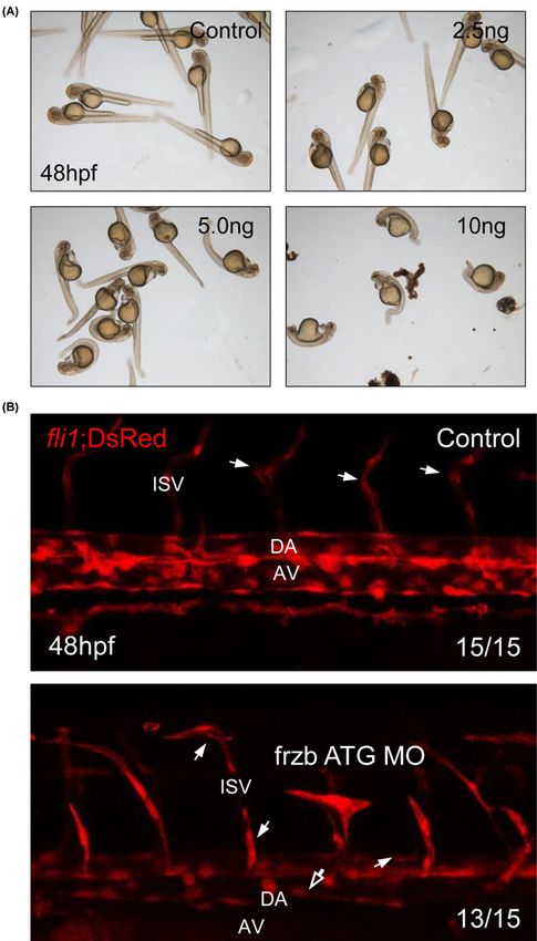
To investigate the function of frzb in vascular integrity, knockdown of frzb was processed using MO injection. De-
pendent on dosage of frzb ATG MO, development of zebrafish embryos was interrupted (Figure 3A). Since devel-
opment of embryos with 5 and 10 ng were highly interrupted and developmental defects were shown from 2.5 ng of
MO-njected group, we decided 2.5 ng is enough for knockdown.
Frzb is highly correlated with elastic fiber formation-related genes at patient database (Table 2), and elastic fiber
is important for the vascular integrity. To validate our informatics data, vascular formation of zebrafish embryos was
checked using fli1;DsRed transgenic zebrafish. As many zebrafish embryos with defects in vascular integrity showed
abnormal vascular formation [17,18], frzb ATG MO-injected embryos show disrupted vascular structure in trunk.
Lumen of dorsal aorta and axial vein became narrow, and abnormal branch of intersomite vessels were observed
Downloaded from http://portlandpress.com/bioscirep/article-pdf/41/1/BSR20203204/901282/bsr-2020-3204.pdf by guest on 21 January 2021
(Figure 3B). These data suggest that frzb is essential for the vascular integrity.
Discussion
Even though multiple etiological, genetic factors contributing to AAA, as well as its pathobiology, have been exten-
sively studied, the mechanisms of progression of AAA are still incompletely understood. Through analysis of three
gene expression studies, we tried to narrow down possible candidate genes that provide a comprehensive view of the
disease.
Through literature review, we had found three articles which analyzed DEGs of AAA from datasets in GEO
[19–21]. The datasets used from each articles were as follows; Shen et al. [19], GSE47472; Wan et al. [20], GSE7084,
GSE47472, and GSE57691; Chen et al. [21], GSE7084, GSE47472, and GSE57691. The tissue samples in GSE47472
were non-aneurysmal aortic neck tissues, so we considered the data in GSE47472 were not suitable for our study de-
sign. Because the data in GSE98278 included important clinical data such as size of AAA and rupture status of AAA,
we analyzed the data of GSE98278 with other two datasets, GSE7084 and GSE57691.
By applying very low FDR value (0.0005) in analysis of DEGs from two cohorts (GSE7084 and GSE57691), only
12 genes were identified. The major limitation of microarray-based mRNA expression profiling is that tissue sam-
ples from end-stage disease provide little information about initiation [22]. The only option for vascular surgeons to
retrieve tissue samples of AAA is through open surgeries of AAA, in which diameter is mostly over 50 mm. In our
study, the mean diameter of AAA from GSE57691 is 62.3 mm. By comparing disease-progressed tissue samples and
normal tissues, it is non-logical to develop DEGs involving initiation of AAA. Therefore, 12 DEGs from GSE 7084
and GSE 57691 were analyzed to identify which genes were involved in terminal AAA and rupture. Only two genes,
FRZB and C1orf24, were differentially expressed along size expansion and rupture. FRZB binds to both Wnt-8 and
Wnt-1 and acts as a functional inhibitor of Wnt-8 activity in Xenopus embryos [23]. FRZB has been reported as a key
factor of skeletal morphogenesis and osteoarthritis [24,25]. C1orf24 contains a DnaJ motif, a feature of heat shock
protein that was investigated as a factor associated with renal carcinogenesis [26]. It was also reported that C1orf24
was up-regulated in thyroid cancer and might increase proliferation and cell migration [27].
To evaluate which of the molecular pathways were related to FRZB and C1orf24, we performed correlation analysis
on GSE 7084 and GSE57691. Then, pathway analysis with correlated genes revealed correlated genes with FRZB and
C1orf24 genes are involved in the pathway of RHO GTPase and elastin fiber formation. In many experimental studies,
RHO GTPase played central roles in vascular smooth muscle cell proliferation, migration, and contractility, differenti-
ation, and ROS generation [28–32]. In rats with balloon-injured arteries, increased RhoA/ROCK activity contributes
to neointimal formation, and these detrimental effects are significantly suppressed by the ROCK-specific antagonist
Y27632 [33]. Regarding VSMC differentiation, through modulating serum response factor-dependent skeletal and
smooth muscle gene expression, the RhoA/ROCK signaling pathway regulates VSMC differentiation [34]. The im-
portant characteristics of aneurysmal wall are breakdown of elastin and collagen, VSMC apoptosis, and activation of
immune responses [35]. We showed FRZB and C1orf24 correlated with RHO GTPase and elastin fiber formation
pathways. Among them, we validated that FRZB is essential for the vascular integrity using zebrafish embryos. That
means low expression of FRZB is associated with the progression of AAA by diminishing integrity of abdominal
aorta.
Many studies have validated the candidate genes of AAA pathobiology through hypothesis-driven way. These
studies have been on a ‘best guess’ basis and do not fit with the studies of diseases for which underlying patholog-
ical processes are unclear [36]. With advances in genetic technology and bioinformatics, unbiased high throughput
genome-wide association studies (GWASs) have widely made. In meta-analysis of GWASs for AAA, four new AAA
© 2021 The Author(s). This is an open access article published by Portland Press Limited on behalf of the Biochemical Society and distributed under the Creative Commons Attribution 7
License 4.0 (CC BY).Bioscience Reports (2021) 41 BSR20203204
https://doi.org/10.1042/BSR20203204
Downloaded from http://portlandpress.com/bioscirep/article-pdf/41/1/BSR20203204/901282/bsr-2020-3204.pdf by guest on 21 January 2021
Figure 3. frzb is essential for the development of zebrafish embryo through regulation of vascular integrity
(A) Imaging of live embryos after morpholino injection (0, 2.5, 5, and 10 ng). (B) Confocal imaging of fli1; DsRed transgenic zebrafish
embryos from control embryos and frzb ATG MO-injected embryos. Abbreviations: AV, axial vein; DA, dorsal aorta; hollow arrow,
narrow dorsal aorta and axial vein; ISV, intersomite vessel. White arrow, abnormal branch of intersomite vessel.
8 © 2021 The Author(s). This is an open access article published by Portland Press Limited on behalf of the Biochemical Society and distributed under the Creative Commons Attribution
License 4.0 (CC BY).Bioscience Reports (2021) 41 BSR20203204
https://doi.org/10.1042/BSR20203204
risk loci, rs1795061 (SMYD2), rs9316871 (LINC00540), rs3827066 (PCIF1/MMP9/ZNF335), and rs2836411 (ERG),
were identified, as well as five of the six previous AAA genetic associations, rs602633 (PSRC1-CELSR2-SORT1),
rs4129267 (IL6R), rs10757274 (CDKN2BAS1/ANRIL), rs10985349 (DAB2IP), and rs6511720 (LDLR) [37]. One
study used VSMCs isolated from patients with AAA and controls to assess DNA methylation in the genes found within
the nine associated loci, and altered DNA methylation levels were found in three genes (ERG, IL6R, and SMYD2)
[38]. Despite their highly significant associations with AAA, these genetic loci explain only a small proportion of the
heritability of AAA.
Even though microarray-based mRNA expression profiling has limitations described before and difficulties, it
can a useful tool in dissecting disease pathogenesis, especially when pathogenesis of disease is not well established.
Among three cohorts we analyzed, Lenk et al. (GSE 7084) compared whole genome expression profile between AAA
(n=10) and normal (n=10) and showed broad coordinate gene expression in immunological pathways [39]. FRZB
was one of the top 100 most differential genes in Supplementary Table S2. Biros et al. (GSE 57691) used AAA tissue
Downloaded from http://portlandpress.com/bioscirep/article-pdf/41/1/BSR20203204/901282/bsr-2020-3204.pdf by guest on 21 January 2021
samples for analyzing expression difference between AAA and aortic occlusive disease [40]. Direct comparison of
our study with GSE57691 is impossible; however, FRZB and C1orf24 was down-regulated in AAA group. Gäbel et al.
(GSE98278) tried to validate the candidate genes relating to terminal AAA [41]. Based on the studies that molecular
mechanisms and genetic factors might differ in initiation, growth, and rupture of AAA [42], they developed ten DEGs
and showed that these genes converged at activation of HIF-1α network. They also showed histologic quantification
of angiogenesis in ruptured AAA. Even though it might provide important insight of terminal AAA, by not comparing
normal aortic tissue, genes involving process of AAA development might be neglected. Therefore, we tried to narrow
down the genes comparing AAA tissues and normal aortic tissue, then analyzed on two groups, size difference and
rupture. We considered this analytic strategy is more logical to develop candidate genes of AAA progression.
To prove the protein level of candidate genes in AAA patients, the optimal tissue for our purpose was aneurysmal
tissue retrieved from open surgery of AAA. In the Biobank of our country, however, there was no tissue sample of
AAA. This is our major limitation. Further investigations will be held to validate the correlation of these pathways
with our candidate genes.
Conclusions
The present study examined the candidate genes involved in the progression of AAA. We were able to identify two
gene expressions (FRZB and C1orf24) by analyzing three cohorts. In pathway analysis with correlated genes with
these two candidate genes (FRZB and C1orf24), we showed the pathways were related to VSMC and elastin fiber
formation, which might imply these candidate genes are involved in aneurysm integrity. Additionally, we validated
that FRZB was linked to the vascular integrity using zebrafish embryos. Considering our findings, we believe that
FRZB is a possible candidate gene involved in AAA progression.
Data Availability
The data that support the findings of the present study are available from the corresponding author upon reasonable request.
Competing Interests
The authors declare that there are no competing interests associated with the manuscript.
Funding
This work was supported by the National Research Foundation of Korea (NRF) funded by the Korea Government [grant num-
ber NRF-2020R1A2C1102433]; the Young Medical Scientist Research Grant through the Daewoong Foundation [grant number
DY20111P]; and the Korea Medical Institute (KMI); and the Institute for Basic Science [grant number IBS-R022-D1].
Author Contribution
D.S.K., J.M.K., and Y.H.K. made substantial contributions to the conception and design. J.J.P., H.J.H., J.K., and E.J.K. collected
the data and J.W.K. and Y.K. performed statistical analysis. C.-K.O., Y.L., and K.M. performed zebrafish experiments. C.-K.O. and
Y.K. analysed and interpreted the data. C.-K.O. and D.S.K. wrote the manuscript. J.M.K. and Y.H.K. made critical revision.
Ethics Approval
All experiments with zebrafish were performed in accordance with the guidelines of UNIST IACUC (IACUC approval number:
UNISTIACUC-15-14). All animal experiments have been performed in accordance with the ARRIVE/NC3R guidelines.
© 2021 The Author(s). This is an open access article published by Portland Press Limited on behalf of the Biochemical Society and distributed under the Creative Commons 9
Attribution License 4.0 (CC BY).Bioscience Reports (2021) 41 BSR20203204
https://doi.org/10.1042/BSR20203204
Abbreviations
AAA, abdominal aortic aneurysm; C1orf24, chromosome 1 open reading frame 24; DEG, differentially expressed gene; DEPC,
diethylpyrocarbonate; FDR, false discovery rate; FRZB, Frizzled-related protein; GEO, Gene Expression Omnibus; GWAS,
genome-wide association study; HIF-1α, hypoxia-inducible factor 1 alpha; IACUC, Institutional Animal Care and Use Committee;
MO, morpholino; NCBI, National Center for Biotechnology Information; ROCK, Rho-associated protein kinase; SAM, signifi-
cance analysis of microarray; UNIST, Ulsan National Institute of Science and Technology; VSMC, vascular smooth muscle cell.
References
1 Kent, K.C. (2014) Clinical practice. Abdominal aortic aneurysms. N. Engl. J. Med. 371, 2101–2108, https://doi.org/10.1056/NEJMcp1401430
2 GBD 2017 Causes of Death Collaborators (2018) Global, regional, and national age-sex-specific mortality for 282 causes of death in 195 countries and
Downloaded from http://portlandpress.com/bioscirep/article-pdf/41/1/BSR20203204/901282/bsr-2020-3204.pdf by guest on 21 January 2021
territories, 1980-2017: a systematic analysis for the Global Burden of Disease Study 2017. Lancet 392, 1736–1788,
https://doi.org/10.1016/S0140-6736(18)32203-7
3 Chaikof, E.L., Dalman, R.L., Eskandari, M.K., Jackson, B.M., Lee, W.A., Mansour, M.A. et al. (2018) The Society for Vascular Surgery practice guidelines
on the care of patients with an abdominal aortic aneurysm. J. Vasc. Surg. 67, 2.e2–77.e2, https://doi.org/10.1016/j.jvs.2017.10.044
4 Powell, J.T., Brady, A.R., Brown, L.C., Fowkes, F.G., Greenhalgh, R.M., Ruckley, C.V. et al. (2002) Long-term outcomes of immediate repair compared
with surveillance of small abdominal aortic aneurysms. N. Engl. J. Med. 346, 1445–1452, https://doi.org/10.1056/NEJMoa013527
5 Kontopodis, N., Pantidis, D., Dedes, A., Daskalakis, N. and Ioannou, C.V. (2016) The - Not So - Solid 5.5 cm threshold for abdominal aortic aneurysm
repair: facts, misinterpretations, and future directions. Front. Surg. 3, 1, https://doi.org/10.3389/fsurg.2016.00001
6 Forsythe, R.O., Newby, D.E. and Robson, J.M. (2016) Monitoring the biological activity of abdominal aortic aneurysms beyond ultrasound. Heart 102,
817–824, https://doi.org/10.1136/heartjnl-2015-308779
7 Ailawadi, G., Eliason, J.L. and Upchurch, Jr, G.R. (2003) Current concepts in the pathogenesis of abdominal aortic aneurysm. J. Vasc. Surg. 38,
584–588, https://doi.org/10.1016/S0741-5214(03)00324-0
8 Kim, Y.H., Lee, S.J., Seo, K.W., Bae, J.U., Park, S.Y., Kim, E.K. et al. (2013) PAF enhances MMP-2 production in rat aortic VSMCs via a
beta-arrestin2-dependent ERK signaling pathway. J. Lipid Res. 54, 2678–2686, https://doi.org/10.1194/jlr.M037176
9 Golledge, J., Tsao, P.S., Dalman, R.L. and Norman, P.E. (2008) Circulating markers of abdominal aortic aneurysm presence and progression. Circulation
118, 2382–2392, https://doi.org/10.1161/CIRCULATIONAHA.108.802074
10 Moris, D.N. and Georgopoulos, S.E. (2013) Circulating biomarkers for abdominal aortic aneurysm: what did we learn in the last decade? Int. Angiol. 32,
266–280
11 Ioannidis, J.P. (2005) Why most published research findings are false. PLoS Med. 2, e124, https://doi.org/10.1371/journal.pmed.0020124
12 Yoon, S., Tromp, G., Vongpunsawad, S., Ronkainen, A., Juvonen, T. and Kuivaniemi, H. (1999) Genetic analysis of MMP3, MMP9, and PAI-1 in Finnish
patients with abdominal aortic or intracranial aneurysms. Biochem. Biophys. Res. Commun. 265, 563–568, https://doi.org/10.1006/bbrc.1999.1721
13 Harrison, S.C., Smith, A.J., Jones, G.T., Swerdlow, D.I., Rampuri, R., Bown, M.J. et al. (2013) Interleukin-6 receptor pathways in abdominal aortic
aneurysm. Eur. Heart J. 34, 3707–3716, https://doi.org/10.1093/eurheartj/ehs354
14 Bown, M.J., Jones, G.T., Harrison, S.C., Wright, B.J., Bumpstead, S., Baas, A.F. et al. (2011) Abdominal aortic aneurysm is associated with a variant in
low-density lipoprotein receptor-related protein 1. Am. J. Hum. Genet. 89, 619–627, https://doi.org/10.1016/j.ajhg.2011.10.002
15 Kuleshov, M.V., Jones, M.R., Rouillard, A.D., Fernandez, N.F., Duan, Q., Wang, Z. et al. (2016) Enrichr: a comprehensive gene set enrichment analysis
web server 2016 update. Nucleic Acids Res. 44, W90–W97, https://doi.org/10.1093/nar/gkw377
16 Lawson, N.D. and Weinstein, B.M. (2002) In vivo imaging of embryonic vascular development using transgenic zebrafish. Dev. Biol. 248, 307–318,
https://doi.org/10.1006/dbio.2002.0711
17 Kwon, H.B., Choi, Y.K., Lim, J.J., Kwon, S.H., Her, S., Kim, H.J. et al. (2012) AKAP12 regulates vascular integrity in zebrafish. Exp. Mol. Med. 44,
225–235, https://doi.org/10.3858/emm.2012.44.3.017
18 Hall, C.J., Flores, M.V., Davidson, A.J., Crosier, K.E. and Crosier, P.S. (2002) Radar is required for the establishment of vascular integrity in the zebrafish.
Dev. Biol. 251, 105–117, https://doi.org/10.1006/dbio.2002.0794
19 Shen, Y., Zhang, F., Lan, H., Chen, K., Zhang, Q., Xie, G. et al. (2015) FRZB up-regulation is correlated with hepatic metastasis and poor prognosis in
colon carcinoma patients with hepatic metastasis. Int. J. Clin. Exp. Pathol. 8, 4083–4090
20 Wan, L., Huang, J., Ni, H. and Yu, G. (2018) Screening key genes for abdominal aortic aneurysm based on gene expression omnibus dataset. BMC
Cardiovasc. Disord. 18, 34, https://doi.org/10.1186/s12872-018-0766-8
21 Chen, S., Yang, D., Lei, C., Li, Y., Sun, X., Chen, M. et al. (2019) Identification of crucial genes in abdominal aortic aneurysm by WGCNA. PeerJ 7,
e7873, https://doi.org/10.7717/peerj.7873
22 Tromp, G. and Kuivaniemi, H. (2009) Developments in genomics to improve understanding, diagnosis and management of aneurysms and peripheral
artery disease. Eur. J. Vasc. Endovasc. Surg. 38, 676–682, https://doi.org/10.1016/j.ejvs.2009.08.010
23 Wang, S., Krinks, M., Lin, K., Luyten, F.P. and Moos, Jr, M. (1997) Frzb, a secreted protein expressed in the Spemann organizer, binds and inhibits
Wnt-8. Cell 88, 757–766, https://doi.org/10.1016/S0092-8674(00)81922-4
24 Hoang, B., Moos, M., Vukicevic, S. and Luyten, F.P. (1996) Primary structure and tissue distribution of FRZB, a novel protein related to Drosophila
frizzled, suggest a role in skeletal morphogenesis. J. Biol. Chem. 271, 26131–26137, https://doi.org/10.1074/jbc.271.42.26131
25 Thysen, S., Luyten, F.P. and Lories, R.J. (2015) Loss of Frzb and Sfrp1 differentially affects joint homeostasis in instability-induced osteoarthritis.
Osteoarthritis Cartilage 23, 275–279, https://doi.org/10.1016/j.joca.2014.10.010
10 © 2021 The Author(s). This is an open access article published by Portland Press Limited on behalf of the Biochemical Society and distributed under the Creative Commons
Attribution License 4.0 (CC BY).Bioscience Reports (2021) 41 BSR20203204
https://doi.org/10.1042/BSR20203204
26 Adachi, H., Majima, S., Kon, S., Kobayashi, T., Kajino, K., Mitani, H. et al. (2004) Niban gene is commonly expressed in the renal tumors: a new
candidate marker for renal carcinogenesis. Oncogene 23, 3495–3500, https://doi.org/10.1038/sj.onc.1207468
27 Carvalheira, G., Nozima, B.H. and Cerutti, J.M. (2015) microRNA-106b-mediated down-regulation of C1orf24 expression induces apoptosis and
suppresses invasion of thyroid cancer. Oncotarget 6, 28357–28370, https://doi.org/10.18632/oncotarget.4947
28 Cai, A., Zhou, Y. and Li, L. (2015) Rho-GTPase and atherosclerosis: pleiotropic effects of statins. J. Am. Heart Assoc. 4,
https://doi.org/10.1161/JAHA.115.002113
29 Laufs, U., Marra, D., Node, K. and Liao, J.K. (1999) 3-Hydroxy-3-methylglutaryl-CoA reductase inhibitors attenuate vascular smooth muscle
proliferation by preventing rho GTPase-induced down-regulation of p27(Kip1). J. Biol. Chem. 274, 21926–21931,
https://doi.org/10.1074/jbc.274.31.21926
30 Maruhashi, T., Noma, K., Iwamoto, Y., Iwamoto, A., Oda, N., Kajikawa, M. et al. (2014) Critical role of exogenous nitric oxide in ROCK activity in vascular
smooth muscle cells. PLoS ONE 9, e109017, https://doi.org/10.1371/journal.pone.0109017
31 Uehata, M., Ishizaki, T., Satoh, H., Ono, T., Kawahara, T., Morishita, T. et al. (1997) Calcium sensitization of smooth muscle mediated by a
Rho-associated protein kinase in hypertension. Nature 389, 990–994, https://doi.org/10.1038/40187
Downloaded from http://portlandpress.com/bioscirep/article-pdf/41/1/BSR20203204/901282/bsr-2020-3204.pdf by guest on 21 January 2021
32 Wassmann, S., Laufs, U., Baumer, A.T., Muller, K., Konkol, C., Sauer, H. et al. (2001) Inhibition of geranylgeranylation reduces angiotensin II-mediated
free radical production in vascular smooth muscle cells: involvement of angiotensin AT1 receptor expression and Rac1 GTPase. Mol. Pharmacol. 59,
646–654, https://doi.org/10.1124/mol.59.3.646
33 Sawada, N., Itoh, H., Ueyama, K., Yamashita, J., Doi, K., Chun, T.H. et al. (2000) Inhibition of rho-associated kinase results in suppression of neointimal
formation of balloon-injured arteries. Circulation 101, 2030–2033, https://doi.org/10.1161/01.CIR.101.17.2030
34 Liu, H.W., Halayko, A.J., Fernandes, D.J., Harmon, G.S., McCauley, J.A., Kocieniewski, P. et al. (2003) The RhoA/Rho kinase pathway regulates nuclear
localization of serum response factor. Am. J. Respir. Cell Mol. Biol. 29, 39–47, https://doi.org/10.1165/rcmb.2002-0206OC
35 Sakalihasan, N., Michel, J.B., Katsargyris, A., Kuivaniemi, H., Defraigne, J.O., Nchimi, A. et al. (2018) Abdominal aortic aneurysms. Nat. Rev. Dis.
Primers 4, 34, https://doi.org/10.1038/s41572-018-0030-7
36 Consortium, T.A. (2008) Genome Wide Association Studies: identifying the genes that determine the risk of abdominal aortic aneurysm. Eur. J. Vasc.
Endovasc. Surg. 36, 395–396, https://doi.org/10.1016/j.ejvs.2008.06.003
37 Jones, G.T., Tromp, G., Kuivaniemi, H., Gretarsdottir, S., Baas, A.F., Giusti, B. et al. (2017) Meta-analysis of genome-wide association studies for
abdominal aortic aneurysm identifies four new disease-specific risk loci. Circ. Res. 120, 341–353, https://doi.org/10.1161/CIRCRESAHA.116.308765
38 Toghill, B.J., Saratzis, A., Freeman, P.J., Sylvius, N. and Bown, M.J. (2018) SMYD2 promoter DNA methylation is associated with abdominal aortic
aneurysm (AAA) and SMYD2 expression in vascular smooth muscle cells. Clin. Epigenet. 10, 29, https://doi.org/10.1186/s13148-018-0460-9
39 Lenk, G.M., Tromp, G., Weinsheimer, S., Gatalica, Z., Berguer, R. and Kuivaniemi, H. (2007) Whole genome expression profiling reveals a significant role
for immune function in human abdominal aortic aneurysms. BMC Genomics 8, 237, https://doi.org/10.1186/1471-2164-8-237
40 Biros, E., Gabel, G., Moran, C.S., Schreurs, C., Lindeman, J.H., Walker, P.J. et al. (2015) Differential gene expression in human abdominal aortic
aneurysm and aortic occlusive disease. Oncotarget 6, 12984–12996, https://doi.org/10.18632/oncotarget.3848
41 Gabel, G., Northoff, B.H., Weinzierl, I., Ludwig, S., Hinterseher, I., Wilfert, W. et al. (2017) Molecular fingerprint for terminal abdominal aortic aneurysm
disease. J. Am. Heart Assoc. 6, e006798, https://doi.org/10.1161/JAHA.117.006798
42 Boddy, A.M., Lenk, G.M., Lillvis, J.H., Nischan, J., Kyo, Y. and Kuivaniemi, H. (2008) Basic research studies to understand aneurysm disease. Drug
News Perspect. 21, 142–148
© 2021 The Author(s). This is an open access article published by Portland Press Limited on behalf of the Biochemical Society and distributed under the Creative Commons Attribution 11
License 4.0 (CC BY).You can also read