Nuclear restriction of HIV 1 infection by SUN1 - Nature
←
→
Page content transcription
If your browser does not render page correctly, please read the page content below
www.nature.com/scientificreports
OPEN Nuclear restriction of HIV‑1
infection by SUN1
Mirjana Persaud1, Anastasia Selyutina1, Cindy Buffone1, Silvana Opp1, Daniel A. Donahue2,
Oliver Schwartz2 & Felipe Diaz‑Griffero1,3*
Overexpression of the human Sad-1-Unc-84 homology protein 2 (SUN2) blocks HIV-1 infection in a
capsid-dependent manner. In agreement, we showed that overexpression of SUN1 (Sad1 and UNC-
84a) also blocks HIV-1 infection in a capsid-dependent manner. SUN2 and the related protein SUN1
are transmembrane proteins located in the inner membrane of the nuclear envelope. The N-terminal
domains of SUN1/2 localizes to the nucleoplasm while the C-terminal domains are localized in the
nuclear lamina. Because the N-terminal domains of SUN1/2 are located in the nucleoplasm, we
hypothesized that SUN1/2 might be interacting with the HIV-1 replication complex in the nucleus
leading to HIV-1 inhibition. Our results demonstrated that SUN1/2 interacts with the HIV-1 capsid,
and in agreement with our hypothesis, the use of N-terminal deletion mutants showed that
SUN1/2 proteins bind to the viral capsid by using its N-terminal domain. SUN1/2 deletion mutants
correlated restriction of HIV-1 with capsid binding. Interestingly, the ability of SUN1/2 to restrict
HIV-1 also correlated with perinuclear localization of these proteins. In agreement with the notion
that SUN proteins interact with the HIV-1 capsid in the nucleus, we found that restriction of HIV-1 by
overexpression of SUN proteins do not block the entry of the HIV-1 core into the nucleus. Our results
showed that HIV-1 restriction is mediated by the interaction of SUN1/2N-terminal domains with the
HIV-1 core in the nuclear compartment.
Abbreviations
SUN1 Sad1 and UNC 84A
SUN2 Sad1 and UNC 84B
SUN1/2 SUN1 and SUN2
HIV-1-GFP HIV-1 pseudotyped with VSVG expressing GFP
SIVmac Simian immunodeficiency virus from macaque monkeys
FIV Feline immunodeficiency virus
BIV Bovine immunodeficiency virus
EIAV Equine infectious anemia virus
B-MLV B-Tropic murine leukemia virus
HAP-1 Huntington associated protein 1; haploid cells; human
HT1080 Human fibrosarcoma cell line; human
PF74 PF-3450074
PS Perinuclear staining
NP Non-perinuclear localization
Early steps of HIV-1 infection involved the delivery of the core into the cytoplasm. The HIV-1 core, which is a
supramolecular structure composed of ~ 1800 molecules of capsid, displays a patterned array in its surface that
allows the core to interact with multiple cellular factors. The interaction of the core with cellular factors deter-
mines the fate of infection. Several proteins that interact with the core are essential for wild type HIV-1 infection:
(1) Cyclophilin A promotes HIV-1 i nfectivity1–3, (2) TNPO3 is necessary for infection after nuclear import but
before integration4–10, (3) Nup153 and RanBP2 are required for nuclear i mport11–16, and (4) CPSF6 is essential
for wild type targeting of integration6,17–20. The incoming HIV-1 core interacts with all these cellular proteins
in order for the virus to achieve productive infection. Overall, the surface of the HIV-1 core is modulating the
early steps of HIV-1 infection through the interaction of known and unknown cellular factors. Therefore, finding
novel capsid interactors will shed light on the early steps of HIV-1 replication.
1
Department of Microbiology and Immunology, Albert Einstein College of Medicine, Bronx, NY, USA. 2Department
of Virology, Virus & Immunity Unit, Institut Pasteur, Paris, France. 3Albert Einstein College of Medicine, 1301 Morris
Park – Price Center 501, New York, NY 10461, USA. *email: Felipe.Diaz-Griffero@einsteinmed.org
Scientific Reports | (2021) 11:19128 | https://doi.org/10.1038/s41598-021-98541-4 1
Vol.:(0123456789)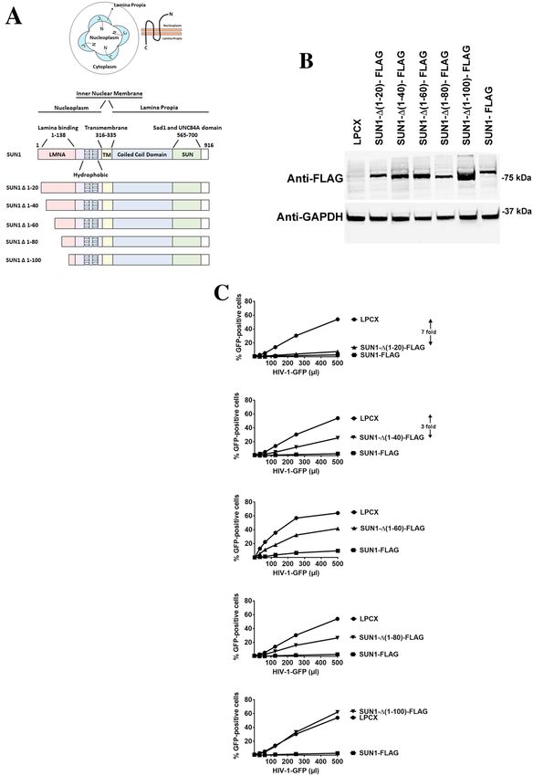
www.nature.com/scientificreports/
Overexpression of the human protein SUN2 was originally discovered to affect HIV-1 infection in a screen
that tested the ability of interferon-stimulated genes to block HIV-1 infection21. We have previously shown that
overexpression of the human SUN2 protein blocks HIV-1 infection in a capsid-dependent manner22. These
experiments suggested that SUN2 might be interacting with the HIV-1 capsid protein, and that restriction is
caused by this interaction.
SUN2 and the related protein SUN1 are transmembrane proteins located in the inner membrane of the nuclear
envelope. The nuclear envelope separates the nucleoplasm from the cytoplasm and is composed of inner and an
outer membrane. The outer nuclear membrane is contiguous with the endoplasmic reticulum. The two mem-
branes are separated by a thin lumen known as the nuclear lamina or lamina propia. The N-terminal domains
of SUN1 and SUN2 are located in the nucleoplasm whereas the C-terminal domains are located in the nuclear
lamina. The conserved SUN domains, in SUN 1 and SUN2, interact with KASH-domain containing proteins
inside the nuclear lamina to form bridges that span both nuclear membranes23. These protein bridges have been
postulated to be the “Velcro” that links the nucleoskeleton with the cytoskeleton23.
We have previously shown that overexpression of the human SUN2 protein blocks HIV-1 infection in a
capsid-dependent manner22. Here we tested whether overexpression of SUN1 and SUN2 proteins block HIV-1
and other retroviruses. To explore the role of capsid in the ability of SUN1 and SUN2 (SUN1/2) to block HIV-1
infection, we measured the ability of SUN1/2 to bind to the HIV-1 core. Our results demonstrated that SUN1/2
interacts with the HIV-1 capsid in cellular extracts. The use of deletion mutants showed that SUN1/2 proteins
bind to the viral capsid using its N-terminal domain, which is located in the nucleoplasm. Furthermore, we found
that the N-terminal domain of SUN1/2 governs the ability of the protein to bind HIV-1 capsid and to restrict
HIV-1. Interestingly, the ability of SUN1/2 to restrict HIV-1 also correlated with perinuclear localization of these
proteins. In addition, we found that restriction of HIV-1 by overexpression of SUN1/2 proteins do not block
the entry of the HIV-1 core into the nucleus suggesting that this restriction occurs in the nucleus, which is in
agreement with the cellular localization of the N-terminal domain of SUN1/2 proteins. Next, we tested whether
cells that do not endogenously expressed SUN1 and SUN2 protein plays a role on HIV-1 infection. To this end,
using the CRISPR/Cas9 system, we generated human HAP-1 cells that are knockout for the expression of SUN1,
SUN2, or SUN1/SUN2. We found that HAP1 SUN1, SUN2, or SUN1/SUN2 knockout cells were permissive to
HIV-1 infection, suggesting that this system is not providing evidence for a functional contribution of SUN1 or
SUN2 to HIV-1 infection.
Results
Ability of SUN1 to block HIV‑1 and other retroviruses. To test the ability of SUN1 to block HIV-1
infection, we challenged human HT1080 cells stably expressing SUN1 with increasing amounts of HIV-1-GFP.
As shown in Fig. 1, SUN1 potently blocks HIV-1 infection (eightfold). By contrast overexpression of SUN1
was not able to block HIV-1 viruses bearing the capsid mutation G208R. This is in agreement with previous
results suggesting that capsid is the viral determinant for the ability of the related protein SUN2 to block HIV-1
infection22. Next, we tested whether other retroviruses are restricted by SUN1 overexpression. We found that
other primate lentiviruses such as human immunodeficiency virus type 2 (HIV-2), and simian immunodefi-
ciency virus (SIVmac), were poorly restricted by SUN1 (Fig. 1). Similarly, feline immunodeficiency virus (FIV),
bovine immunodeficiency virus (BIV), equine infectious anemia virus (EIAV), and B-tropic murine leukemia
virus (B-MLV), were also poorly restricted by SUN-1 (Fig. 1). Overall, overexpression of SUN1 only restricted
HIV-1. Next we decided to study the SUN1 determinants for HIV-1 restriction.
SUN1 determinants for HIV‑1 restriction. A closer look at the topology of the SUN1 protein sug-
gested that residues 1–315 localized to the nucleoplasm whereas residues 336–916 localized to the nuclear
lamina (Fig. 2A). This topology implies that HIV-1 may be interacting with the N-terminal domain of SUN1
in the nucleoplasm (Fig. 2A). Therefore, as shown in Fig. 2A, we created several SUN1 N-terminal deletions.
Subsequently, the different SUN1 variants were stably expressed in human HT1080 cells and used to test HIV-1
restriction (Fig. 2B,C). Interestingly, SUN1-∆ (1–20) blocked HIV-1 infection (sevenfold) as potent as the wild
type protein (eightfold) suggesting that the first 20 amino acids of the protein are not important for HIV-1
restriction (Fig. 2C and Table 1). Deletions SUN1-∆ (1–40) (threefold), SUN1-∆ (1–60) (< twofold) and SUN1-∆
(1–80) (< twofold) lost potency against HIV-1 (Fig. 2C and Table 1). However, deletion of the first 100 amino
acids [SUN1-∆(1–100)] renders the protein inactive against HIV-1 (Fig. 2C and Table 1). Overall these experi-
ments suggested that SUN1 residues 20–100 are important for the ability of SUN1 to block HIV-1 infection. Next
we tested whether SUN1 interacts with HIV-1 capsids.
Capsid binding ability of SUN1. Our previous experiments suggested that the N-terminal domain of
SUN1 is required for its ability to block HIV-1 infection, which is in agreement with the fact that HIV-1 can
only interact with the N-terminus of SUN1 in the nucleoplasm. One hypothesis is that the N-terminal domain
of SUN1 is interacting with the HIV-1 replication complex in the nucleus consequentially affecting HIV-1 infec-
tion. Because capsid mutations such as G208R abolished the ability of SUN1 to block HIV-1, we tested whether
SUN1 binds to the HIV-1 capsid. To this end, we tested whether SUN1 binds to in vitro assembled HIV-1
CA-NC complexes, which recapitulates the surface of the HIV-1 core24. As shown in Fig. 3 and Table 1, SUN1
binds to in vitro assembled HIV-1 CA-NC complexes. Interestingly, deletions SUN1-∆(1–40), SUN1-∆(1–60)
and SUN1-∆(1–80) bind to in vitro assembled HIV-1 CA-NC complexes similar to wild type SUN1(Fig. 3 and
Table 1). By contrast, deletion SUN1-∆(1–100) lost its ability to bind in vitro assembled HIV-1 CA-NC com-
plexes (Fig. 3 and Table 1). These experiments showed that loss of binding correlates with loss of restriction.
Scientific Reports | (2021) 11:19128 | https://doi.org/10.1038/s41598-021-98541-4 2
Vol:.(1234567890)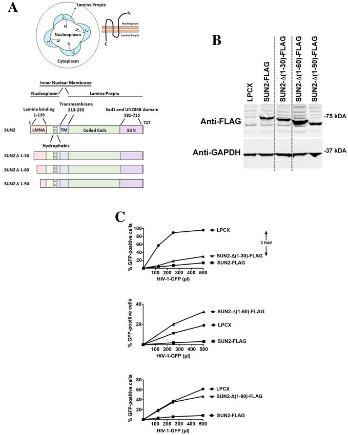
www.nature.com/scientificreports/
Figure 1. Ability of SUN1 to block HIV-1 and other retroviruses. Human HT1080 fibrosarcoma cells stably
expressing wild type SUN1-FLAG or containing the empty vector LPCX were challenged with increasing
amounts of HIV-1, HIV-1-G208R, HIV-2, S IVmac, FIV, BIV, EIAV, and B-MLV expressing GFP as a reporter.
Forty-eight hours post-infection, the percentage of GFP-positive cells was measured using a flow cytometer.
Experiments were repeated at least three times and a representative experiment is shown. Fold differences in
restriction are shown as the ratio of the area under the curve of the SUN variant to the empty vector pLPCX.
Fold values greater than 2 are shown.
This is in agreement with our hypothesis that the N-terminal domain of SUN1 is important for its ability to bind
capsid and restrict HIV-1 infection.
Ability of SUN2 to block HIV‑1 and other retroviruses. Next we tested the ability of SUN2 to block
HIV-1 infection. As we have previously shown22, SUN2 blocks HIV-1 infection (threefold). Although overex-
pression of SUN2 blocks HIV-1 infection (Fig. 4), we observed that this block is less potent when compared to
SUN1. Like SUN1, SUN2 poorly affected the infection of HIV-1 bearing the capsid mutation G208R, which is in
agreement with our previous results suggesting that capsid mutations overcome the HIV-1 restriction imposed
by overexpression of S UN222. Like in the case of SUN1, these experiments suggested that capsid is the viral
determinant for the restriction of SUN2. Next we tested whether other retroviruses are restricted by SUN2
overexpression. We found that SUN2 modestly restricts HIV-1 and HIV-2 (threefold), but not SIVmac (Fig. 4).
Overexpression of SUN2 poorly affected infection by FIV, BIV, EIAV, and B-MLV viruses.
Scientific Reports | (2021) 11:19128 | https://doi.org/10.1038/s41598-021-98541-4 3
Vol.:(0123456789)www.nature.com/scientificreports/
Figure 2. Ability of SUN1 variants to restrict HIV-1 infection. (A) SUN1 membrane topology is illustrated in a cell by
showing that the N-terminal domain of SUN1 faces the nucleoplasm while the C-terminal domain is in the nuclear lamina
or lamina propia. The SUN1 protein is also depicted with the numbers of the amino acid residues at the boundaries of
the domains. The SUN1 protein is depicted containing the lamina binding domain (LMNA) (1–138) on the N-terminus,
hydrophobic regions, a transmembrane region (316–335), coiled coil domain, and a SUN domain (565–700) on the
C-terminus. The different SUN1 N-terminal deletions variants are illustrated. (B) Human HT1080 cells stably expressing wild
type and mutant SUN1-FLAG proteins. Stable expression of wild type and mutant SUN1 proteins in human HT1080 cells
was analyzed by Western blotting using anti-FLAG antibodies. As loading control, extracts were also Western blotted using
anti-GAPDH antibodies. (C) HIV-1 restriction by wild type and mutant SUN1 proteins. HT1080 cells stably expressing wild
type and mutant SUN1 proteins were challenged with increasing amounts of HIV-1-GFP. Forty-eight hours post-infection,
the percentage of GFP-positive cells was measured using a flow cytometer. Experiments were repeated at least three times and
a representative experiment is shown. Fold differences in restriction are shown as the ratio of the area under the curve of the
SUN variant to the empty vector pLPCX. Fold values greater than 2 are shown.
Scientific Reports | (2021) 11:19128 | https://doi.org/10.1038/s41598-021-98541-4 4
Vol:.(1234567890)www.nature.com/scientificreports/
SUN1 Restriction of HIV-1a Binding to HIV-1 CA-NC complexesb Localizationc
WT-FLAG ++ + PS
∆(1–20)-FLAG ++ + PS
∆(1–40)-FLAG + + PS + NP
∆(1–60)-FLAG + + PS + NP
∆(1–80)-FLAG + + PS + NP
∆(1–100)-FLAG − − PS + NP
Table 1. Phenotypes of SUN1 variants. a Restriction was measured by infecting cells expressing the indicated
SUN1 variant with HIV-1-GFP. After 48 h, the percentage of GFP-positive cells (infected cells) was determined
by flow cytometry. “++”: indicates strong restriction, “+” indicates moderate restriction, “−”: indicates absence
of restriction. b Binding to the HIV-1 capsid complexes was determined for each SUN1 variant as described
in “Methods”. “+”: indicates binding, “−”: indicates no binding. c Wild type and mutant SUN1-FLAG proteins
were assayed to determine localization. “PS” indicates Perinuclear staining, “NP” indicates Non-Perinuclear
localization. “PS + NP” indicates both perinuclear and non-peri-nuclear staining is observed.
SUN2 determinants for HIV‑1 restriction. Similar to SUN1, the topology of SUN2 implies that HIV-1
may be interacting with the N-terminal domain of SUN2 in the nucleoplasm (Fig. 5A). Therefore, as shown in
Fig. 5A, we created several SUN2 N-terminal deletions. Subsequently, the different SUN2 variants were stably
expressed in human HT1080 cells and used to test HIV-1 restriction (Fig. 5B,C). Interestingly, SUN2-∆(1–30)
blocked HIV-1 infection (threefold) as potent as the wild type protein (threefold) suggesting that the first 30
amino acids of the protein are not important for HIV-1 restriction(Fig. 5C and Table 2). Deletions SUN2-∆(1–
60) and SUN2-∆(1–90) render the protein inactive against HIV-1 (Fig. 5C and Table 2). Overall these experi-
ments suggested that SUN2 residues 30–60 are required for its ability to block HIV-1 infection. On the next
section, we tested whether SUN2 interacts with HIV-1 capsids.
Capsid binding ability of SUN2. Our previous experiments suggested that the N-terminal domain of
SUN2 is required for its ability to block HIV-1 infection, which is in agreement with the fact that the N-termi-
nus of SUN2 is localized to the nucleoplasm. In the same manner as SUN1, we tested the hypothesis that the
N-terminal domain of SUN2 is interacting with the HIV-1 replication complex in the nucleus consequentially
affecting HIV-1 infection. Because capsid mutations such as G208R and P207S abolished the ability of SUN2
to block HIV-122, we also tested whether SUN2 binds to the HIV-1 capsid. As shown in Fig. 6, SUN2 binds to
in vitro assembled HIV-1 CA-NC complexes. Deletions SUN2-∆(1–30) retained binding to in vitro assembled
HIV-1 CA-NC complexes. However, deletions SUN1-∆(1–60) and SUN2-∆(1–90) decreased their ability to bind
in vitro assembled HIV-1 CA-NC complexes. In the case of the deletion SUN1-∆(1–60), we observed residual
binding when compared to SUN2-∆(1–90). These experiments are in agreement with our hypothesis that the
N-terminal domain is important for the ability of SUN2 to bind capsid and restrict HIV-1 infection.
Capsid Binding ability of SUN1 and SUN2 to mutants G208R and P207S. Next we tested the
ability of SUN1 and SUN2 to bind capsid mutants G208R and P207S. In agreement with the inability of SUN1
and SUN2 to block infection of HIV-1 viruses bearing the capsid changes G208R or P207S, we saw that SUN1
and SUN2 showed decreased binding to capsid bearing G208R or P207S changes when compared to wild type
(Fig. 7A). These experiments demonstrated that the inability of SUN1 and SUN2 to block HIV-1 viruses bear-
ing capsid changes G208R and P207S is due to the lose of capsid binding. To control for the bona fide folding
of the capsid mutants, we tested the ability of capsid mutants to interact with TRIMCyp in presence or absence
of cyclosporine A (CsA). As shown in Fig. 7B, TRIMCyp was able to interact with capsid mutants at wild type
levels. Overall these results correlate SUN1/2 binding with restriction.
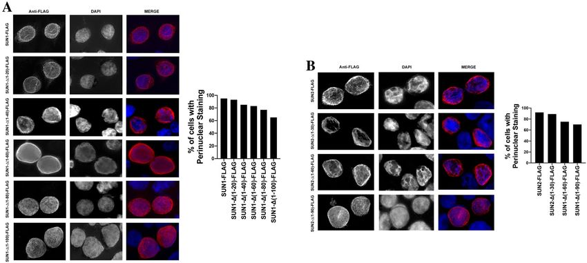
Subcellular localization of SUN1 and SUN2 variants. Previous experiments have defined that SUN1
and SUN2 localized to the lamina propia25. To understand weather the subcellular localization of SUN1 and
SUN2 are important for their ability to block HIV-1 infection, we analyzed subcellular localization of the dif-
ferent SUN1 and SUN2 variants. As shown in Fig. 8, SUN1 and SUN2 showed distinct perinuclear localization,
which according to the literature, corresponds to the lamina propia. Interestingly, the localization of the deletion
mutant SUN1-∆(1–20) resembles the subcellular localization of the wild type SUN1 protein (Fig. 8A), suggest-
ing that this deletion did not lose wild type localization. Although all other SUN1 deletions showed a perinu-
clear localization, they also displayed a more intense nucleoplasm staining (Fig. 8A). SUN2 deletion constructs
showed a perinuclear staining similar to wild type SUN2. In addition, deletion constructs showed a more intense
nucleoplasm staining (Fig. 8B). Interestingly, all study deletions localized to the perinuclear region suggesting
that these constructs were not majorly affected by the deletions, indirectly indicating that folding was not greatly
disrupted. Perinuclear staining was quantified by examining 50 cells for SUN1-FLAG, SUN2-FLAG and mutants
(Fig. 8A,B). Our results showed that for each sequential deletion mutant, there is some nucleoplasmic staining in
addition to the pre-dominant peri-nuclear staining.
Scientific Reports | (2021) 11:19128 | https://doi.org/10.1038/s41598-021-98541-4 5
Vol.:(0123456789)www.nature.com/scientificreports/
Figure 3. Capsid Binding ability of wild type and mutant SUN1 proteins. HEK293T cells were transiently
transfected with plasmids expressing wild type and mutant SUN1-FLAG proteins. Thirty-six hours after
transfection, cells were lysed. The lysates were incubated with in vitro assembled HIV-1 CA-NC complexes at
room temperature for 1 h. The mixtures were applied onto a 70% sucrose cushion and centrifuged as described
in methods. INPUT represents the lysates analyzed by Western blotting before being applied to the 70%
cushion. The INPUT fraction was analyzed by Western blotting using anti-FLAG antibodies. The pellet from the
70% cushion (BOUND) was analyzed by Western blotting using anti-FLAG and anti-p24 antibodies. The blots
for a representative experiment and the standard deviation for three independent experiments are shown.
Scientific Reports | (2021) 11:19128 | https://doi.org/10.1038/s41598-021-98541-4 6
Vol:.(1234567890)www.nature.com/scientificreports/
Figure 4. Ability of SUN2 to block HIV-1 and other retroviruses. Human HT1080 fibrosarcoma cells stably
expressing wild type SUN2-FLAG or containing the empty vector LPCX were challenged with increasing
amounts of HIV-1, HIV-1-G208R, HIV-2, S IVmac, FIV, BIV, EIAV, and B-MLV expressing GFP as a reporter.
Forty-eight hours post-infection, the percentage of GFP-positive cells was measured using a flow cytometer.
Experiments were repeated at least three times and a representative experiment is shown. Fold differences in
restriction are shown as the ratio of the area under the curve of the SUN variant to the empty vector pLPCX.
Fold values greater than 2 are shown.
Inhibition of HIV‑1 infection by SUN1 does prevent the entry of capsid into the nucleus. Over-
expression of SUN1 in human cells potently inhibit HIV-1 infection after reverse transcription but prior to
integration22. Because the occurrence of reverse transcription correlates with entry of capsid into the nucleus26–28,
and the SUN1 domain that interacts with capsid is in the nucleoplasm; we tested whether SUN1 inhibition
of HIV-1 allows the entry of capsid into the nucleus. For this purpose, using our published m ethodology26,
we tested whether capsid is imported into the nucleus of HIV-1 restricted cells by SUN1. To this end, human
HT1080 cells expressing SUN1 were challenged with HIV-1 using an MOI = 2. Eight hours post infection, cells
were separated into cytosolic (C) and nuclear (N) fractions. C and N fractions were analyzed for capsid content
using anti-P24 antibodies (Fig. 9). To ensure bona fide origin of the cellular fractions, we performed Western blot
analysis using anti-Nopp140 and anti-tubulin as nuclear and cytosolic markers, respectively. As shown on Fig. 9,
we found that expression of SUN1 does not inhibit the entry of capsid into the nucleus. As controls, we used the
small molecule PF74 that potently blocks the entry of capsid into the nucleus26. These experiments suggest that
HIV-1 restriction by SUN1 occurs in the nucleus, which is in agreement with findings showing that the region
of SUN1 that interact with capsid is in the nucleoplasm. Overall these experiments suggest that the restriction of
HIV-1 by overexpression of SUN1 occurs in the nuclear compartment.
Scientific Reports | (2021) 11:19128 | https://doi.org/10.1038/s41598-021-98541-4 7
Vol.:(0123456789)www.nature.com/scientificreports/
Figure 5. Ability of SUN2 variants to restrict HIV-1 infection. (A) SUN2 membrane topology is illustrated in
a cell by showing that the N-terminal domain of SUN2 faces the nucleoplasm while the C-terminal domain is
in the nuclear lamina or lamina propia. The SUN2 protein is also depicted with the numbers of the amino acid
residues at the boundaries of the domains. The SUN2 protein is depicted containing the lamina binding domain
(LMNA) (1–139) on the N-terminus, hydrophobic regions, a transmembrane region (213–233), coiled coil
domain, and a SUN domain (581–715) on the C-terminus. The different SUN2 N-terminal deletions variants are
illustrated. (B) Human HT1080 cells stably expressing wild type and mutant SUN2 proteins. Stable expression
of wild type and mutant SUN2 proteins in human HT1080 cells was analyzed by Western blotting using anti-
FLAG antibodies. As loading control, extracts were also Western blotted using anti-GAPDH antibodies. (C)
HIV-1 restriction by wild type and mutant SUN2 proteins. HT1080 cells stably expressing wild type and mutant
SUN2 proteins were challenged with increasing amounts of HIV-1-GFP. Forty-eight hours post-infection, the
percentage of GFP-positive cells was measured using a flow cytometer. Experiments were repeated at least three
times and a representative experiment is shown. Fold differences in restriction are shown as the ratio of the area
under the curve of the SUN variant to the empty vector pLPCX. Fold values greater than 2 are shown.
Scientific Reports | (2021) 11:19128 | https://doi.org/10.1038/s41598-021-98541-4 8
Vol:.(1234567890)www.nature.com/scientificreports/
SUN2 Restriction of HIV-1a Binding to HIV-1 CA-NC complexesb Localizationc
WT-FLAG ++ ++ PS
∆(1–30)-FLAG ++ + PS + NP
∆(1–60)-FLAG − +/− PS + NP
∆(1–90)-FLAG − − PS + NP
Table 2. Phenotypes of SUN2 variant. a Restriction was measured by infecting cells expressing the indicated
SUN2 variant with HIV-1-GFP. After 48 h, the percentage of GFP-positive cells (infected cells) was determined
by flow cytometry. “++”: indicates strong restriction, “+” indicates moderate restriction, “−”: indicates absence
of restriction. b Binding to the HIV-1 capsid complexes was determined for each SUN2 variant as described
in “Methods”. “+”: indicates binding, “−”: indicates no binding, “+/−” decreased binding. c Wild type and
mutant SUN2-FLAG proteins were assayed to determine localization. “PS” indicates Perinuclear staining, “NP”
indicates Non-Perinuclear localization. “PS + NP” indicates both perinuclear and non-peri-nuclear staining is
observed.
Endogenous expression of SUN1/SUN2 is not required for wild type HIV‑1 infection in human
haploid HAP‑1 cells. Although our work shows that overexpression of SUN1 and SUN2 block HIV-1 infec-
tion, we did not yet address whether endogenous expression of these proteins are important for HIV-1 infection.
To directly address this question, we knockout the expression of SUN1 and/or SUN2 in human haploid HAP-1
cells by using the CRISPR/Cas9 system. As shown in Fig. 10A, sequencing of the SUN1 and/or SUN2 alleles
revealed deletions that resulted in the generation of premature stop codons (Fig. 10A). Furthermore, we tested
expression of endogenous SUN1 and SUN2 in the KO HAP-1 cells. As shown in Fig. 10B, HAP-1 cell lines con-
taining the premature stop codons did not expressed SUN1/2 proteins. Next we tested whether HAP-1 cells that
do not express SUN1 and/or SUN2 were infected by HIV-1-GFP (Fig. 10B). As a control, we used a clone that
showed an intact sequence for SUN1 and SUN2 (HAP-1 CTRL). Interestingly, knocking out the expression of
these genes did not affect HIV-1 infection (Fig. 10B,C). These experiments suggested that expression of SUN1
and/or SUN2 is not important for HIV-1 infection of HAP-1 cells.
Discussion
This work and others have established that overexpression of SUN1 and SUN2 blocks HIV-1 infection, and this
process depends upon an intact capsid p rotein21,22,29. The present work showed that SUN1/2 is interacting with
capsid in the nuclear compartment to achieve HIV-1 restriction. To this end, we tested the ability of SUN1 and
SUN2 to bind to in vitro assembled HIV-1 CA-NC complexes. Our data showed that SUN1/2 proteins in cell
lysates interact with the HIV-1 core, in agreement with previous findings29. This interaction is likely to occur in
the nucleoplasm where the N-terminal of SUN1/2 is located. One possibility is that the interaction between the
N-terminal domain of SUN1 or SUN2 with the pre-integration complex negatively affects infection by changing
the localization of the core in the nuclear compartment.
Since the N-terminal of SUN1 or SUN2 is the only segment of the protein that might be interacting with HIV-
1, we performed SUN1/2N-terminal deletions and tested for restriction. In agreement with the hypothesis that
the N-terminal domain of SUN1/2 are important for restriction, we found that deletion of N-terminal residues
renders SUN1/2 inactive against HIV-1 infection. Our mapping experiments revealed that residues 20–100 and
30–60 are important for HIV-1 restriction of SUN1 and SUN2, respectively. These experiments demonstrated
that the N-terminal region of SUN1/SUN2 is important for the ability of the protein to block HIV-1. Similar
results for SUN1 were observed by o thers29,30.
Next, we investigated whether a correlation between restriction and capsid binding exists. Interestingly,
N-terminal deletion mutants that did not bind capsid did not restrict HIV-1. In addition, we demonstrated that
capsid mutations G208R and P207S prevent the ability of capsid to bind to SUN1/2 proteins; this is in agree-
ment with the inability of SUN1/2 to restrict HIV-1 viruses bearing changes G208R and P207S. These results
correlated binding to capsid with HIV-1 restriction. Overall this is in agreement with the hypothesis that the
N-terminal domain of SUN1/2 is interacting with capsid during infection and that this interaction is important
for restriction.
SUN1/2 proteins mainly localize to the lamina propia. To understand whether the localization of SUN1/2
correlates with restriction, we investigated the subcellular localization of SUN1/2N-terminal deletion mutants
and correlated this to restriction. Our observations revealed that intact restriction is only achieved by an intact
localization of SUN1/2. Although N-terminal deletion mutants remain in the lamina propia (perinuclear locali-
zation), we observed that HIV-1 restriction by SUN1 and SUN2 correlates with perinuclear localization. Dele-
tion mutants that exhibit perinuclear and nonperinuclear localization were less or none restrictive to HIV-1.
Therefore, SUN1/2 restriction correlates with capsid binding and perinuclear localization. Similar localization
for SUN1 deletion mutants was observed by Schaller et al.29.
Our results showed that the nuclear membrane protein SUN1/2 requires its N-terminal domain to interact
with HIV-1 capsid. This interaction is likely to take place in the nucleus and it is important for the ability of the
protein to block HIV-1 infectivity. Interestingly, our experiments demonstrated that the SUN1 block to HIV-1
infection is after the viral core has been transported into the nuclear compartment, which is in agreement with
the notion that SUN1 interacts with the HIV-1 core in the nucleus. Previous observations have shown that
Scientific Reports | (2021) 11:19128 | https://doi.org/10.1038/s41598-021-98541-4 9
Vol.:(0123456789)www.nature.com/scientificreports/
Figure 6. Capsid Binding ability of wild type and mutant SUN2 proteins. HEK293T cells were transiently
transfected with plasmids expressing wild type and mutant SUN2-FLAG proteins. Thirty-six hours after
transfection, cells were lysed. The lysates were incubated with in vitro assembled HIV-1 CA-NC complexes at
room temperature for 1 h. The mixtures were applied onto a 70% sucrose cushion and centrifuged as described
in methods. INPUT represents the lysates analyzed by Western blotting before being applied to the 70%
cushion. The INPUT fraction was analyzed by Western blotting using anti-FLAG antibodies. The pellet from the
70% cushion (BOUND) was analyzed by Western blotting using anti-FLAG and anti-p24 antibodies. The blots
for a representative experiment and the standard deviation for three independent experiments are shown.
Scientific Reports | (2021) 11:19128 | https://doi.org/10.1038/s41598-021-98541-4 10
Vol:.(1234567890)www.nature.com/scientificreports/
Figure 7. Binding of SUN1 and SUN2 to G208R and P207S capsid mutants. (A) HEK293T cells were
transiently transfected with plasmids expressing wild type SUN1-FLAG and SUN2-FLAG. Twenty-four hours
after transfection, cells were lysed. The lysates were incubated with in vitro assembled HIV-1-CA WT, HIV-
1-CA G208R, and HIV-1-CA P207S complexes at room temperature for 1 h. The mixture was centrifuged to
pellet the capsid as described in “Methods”. (B) HEK293T cells were transiently transfected with TRIMCyp-HA
as a control for binding. Twenty-four hours after transfection, cells were lysed. Lysates were incubated in the
presence of 10 μM cyclosporine A or DMSO vehicle control. In vitro assembled HIV-1-CA WT, HIV-1-CA
G208R, and HIV-1-CA P207S complexes were added to the pre-treated lysates for 1 h at room temperature.
INPUT represents the lysates analyzed by Western blotting prior to the addition of the capsid. The INPUT
fraction was analyzed by Western blotting using anti-FLAG or anti-HA antibodies respectively. The pellet
(BOUND) fraction was analyzed by Western blotting using anti-FLAG and anti-p24 antibodies. The blots for a
representative experiment and the standard deviations for three independent experiments are shown.
overexpression of SUN1/2 proteins blocks HIV-1 infection after reverse transcription but before integration22.
Because reverse transcription occurs in the nuclear compartment the interaction of SUN1 with the HIV-1 core
in the nucleus is likely to disrupt the subsequent steps of r eplication26–28. One possibility is that SUN1 binding
to capsid sequesters the viral complexes away from critical sites for replication such as open chromatin sites.
Overexpression of SUN1 or SUN2 blocks HIV-1 infection; however, the role of endogenously expressed
SUN1 and SUN2 in HIV-1 replication is not clear. For this purpose, we generated knockouts for the expression
of SUN1 and/or SUN2 in human haploid HAP-1 cells, and investigated whether HIV-1 infection is affected. To
our surprise, complete depletion of SUN1 and/or SUN2 by using CRISPR/Cas9 did not affect HIV-1 replication
suggesting that endogenous SUN1 and SUN2 does not have a role in HIV-1 replication of haploid HAP-1 cells.
In agreement with our results, knocking out the expression of SUN1 using CRISPR/Cas9 in THP-1 cells did not
affect HIV-1 infection29. However, depletion of SUN2 using CRIPSR/Cas9 in THP-1 cells mildly affected HIV-1
infection. More recent results, depleting SUN1/2 using siRNA in HEK293T cells dramatically decreased infectiv-
ity of HIV-130. To further complicate matters, depletion of SUN2 in human primary T cells affects proliferation
and viability31. Although several laboratories have attempted depletion of this protein further experiments will
be necessary to establish whether endogenous expression of SUN1/2 contribute to HIV-1 infection.
Overall our work showed that HIV-1 restriction by overexpression of SUN1/2 is mediated by the interaction
of the N-terminal domain of SUN1/2 with the HIV-1 core occurs in the nuclear compartment.
Conclusions
This work provides mechanistic understanding on how the overexpression of SUN1/2 proteins block HIV-1
infection in human cells. Our experiments showed that HIV-1 restriction is mediated by the interaction between
the N-terminal domain of SUN1/2 proteins and the HIV-1 core in the nuclear compartment.
Scientific Reports | (2021) 11:19128 | https://doi.org/10.1038/s41598-021-98541-4 11
Vol.:(0123456789)www.nature.com/scientificreports/
Figure 8. Subcellular localization of SUN1 and SUN2 variants. The subcellular localization of SUN1-FLAG (A)
and SUN2-FLAG (B) was studied in HeLa cells. Human HeLa cells transiently transfected with the indicated
constructs were fixed and permeabilized. Subsequently, SUN1/2 proteins were stained using anti-FLAG
antibodies conjugated to Alexa Fluor 594. The nuclei was stained by using DAPI (blue). Perinuclear staining was
quantified by examining 50 cells for SUN1-FLAG, SUN2-FLAG and mutants in three independent experiments;
results are shown as percentage of cells showing perinuclear staining.
Methods
Cells, viruses and compounds. HT1080 cells (ATCC CCL-121), CHME3 cells (ATCC CRL-3304), and
HAP-1 haploid cells (Horizon discovery) were grown in Dulbecco’s modified Eagle’s medium (DMEM), sup-
plemented with 10% fetal calf serum (FCS), 100 IU/mL of penicillin and 100 μg/mL of streptomycin at 37 °C in
5% CO2. Cell were seeded in 24-well plates (50,000 cells/well) 24 h prior to infection with HIV-1-GFP viruses
pseudotyped with the VSV-G envelope.
Generation of stable cell lines. Lentivirus for transduction was produced by transfection of HEK 293 T
cells with 3ug VSV-G, 7ug MLV-Gag Pol, and 7ug of plasmids expressing wild type and variant SUN1/2 proteins.
Transfections were performed using polyethylenimine (Polysciences) in a 10-cm dish. Viruses were harvested
48 h after transfection, filtered through a 0.45-μm filter (Millipore), and used to transduce HT1080 cells for five
days at a ratio of 1:1 with virus and complete media. Transduced cell lines were selected in 0.6 ug/ml of puromy-
cin for 5 days. Stably expressing cells were expanded and kept in 0.6ug/ml of puromycin to maintain selection.
Western Blot. Cellular proteins were extracted with RIPA buffer (1% NP40, 0.5% Deoxycholate, 0.05%
SDS, 1X Protease Inhibitor, 25 mM Tris pH 8, 150 mM NaCl), as previously described32. Detection of proteins
by Western blotting was performed using anti-FLAG (Sigma) and anti-GAPDH (Sigma) antibodies. Bands were
detected and scanned using a Li-Cor Odyssey Imaging System in the 700 channel (original blots are provided in
the supplementary file).
Infection with viruses expressing green fluorescent protein (GFP). Recombinant human immu-
nodeficiency virus type 1 (HIV-1), simian immunodeficiency virus (SIVmac), B-tropic murine leukemia virus
(BMLV), human immunodeficiency virus type 2 (HIV-2), HIV-1, bovine immunodeficiency virus (BIV), equine
infectious anemia virus (EIAV), Moloney murine leukemia virus (MMLV), feline immunodeficiency virus (FIV)
expressing GFP were prepared as previously described33. All recombinant viruses were pseudotyped with the
VSV-G glycoprotein. For infections, human HT1080 fibrosarcoma cell line or microglial CHME3 cell line stably
expressing SUN1/2 constructs were challenged with increasing amounts of the indicated virus for 24 h. Cells
were washed and returned to culture for 48 h, and the percentage of GFP-positive cells was determined by flow
cytometry (Becton Dickinson). Viral stocks were titrated by serial dilution on Cf2Th cells to determine the con-
centration of infectious viruses. As control, cells stably transduced with the empty vector LPCX were challenged
with the same viruses.
Subcellular fractionation to detect HIV‑1 capsid in the nucleus. HT1080 cells overexpressed for
SUN1-FLAG or LPCX vector control were challenged with HIV-1 viruses at a MOI = 2 for the indicated times.
Protocol was used as o utlined26. Cells were harvested using trypsin and were washed twice with cold PBS by
centrifugation at 4 °C. Supernatant was discarded and cell pellet was re-suspended in PBS. A fraction of the cell
Scientific Reports | (2021) 11:19128 | https://doi.org/10.1038/s41598-021-98541-4 12
Vol:.(1234567890)www.nature.com/scientificreports/
Figure 9. Nuclear Import of capsid in SUN1 overexpressed cells. Human HT1080 cells, stably expressing
SUN1-FLAG or containing the LPCX empty vector control, were infected with wild-type HIV-1-GFP at an
MOI of 2 in the presence of 10uM PF74, or DMSO vehicle control for 8 h. Cells were separated into nuclear and
cytosolic fractions and analyze for capsid content by western blotting using anti-p24, anti-Nopp140 and anti-
α-Tubulin antibodies. The ratio of nuclear to cytosolic capsid for three independent experiments with standard
deviations is shown. **p < 0.001; ***p < 0.0005; NS, not significant (as determined by unpaired t test).
suspension was centrifuged and cell pellet was re-suspended in lysis buffer and incubated for 1 h on ice then
centrifuged for 1 h at 4 °C. The resulting supernatant was mixed with Laemmli buffer and used to measure the
total amount of capsid. The remaining aliquot of the cell suspension was pelleted and re-suspended in lysis buffer
and incubated on ice. Subsequently, the sample was centrifuged at 4 °C. The resulting supernatant and pellet
correspond to cytosolic and nuclear fractions, respectively. Next, a fraction of the supernatant was mixed with
Laemmli buffer and used as cytosolic fraction. The nuclear pellet was washed twice using lysis buffer without
NP-40 by gently inverting the tube several times. The sample was then centrifuged and the nuclear pellet was
re-suspended in extraction buffer, and incubated on ice. Subsequently, the sample was centrifuged at 4 °C. A
fraction of the supernatant was mixed with Laemmli buffer and used as nuclear fraction. Proportional amounts
of total, cytosolic, and nuclear fractions were analyzed by western blot using anti-p24, anti-Nopp140, anti-a-
tubulin, or anti-GAPDH antibodies detailed below.
Generation of SUN1, SUN2, and SUN1/SUN2 double KO. Hap-1 SUN1, SUN2, and SUN1/SUN2
double knockout (KO) cell lines were generated by using the clustered regularly interspaced short palindromic
repeat (CRISPR)-Cas9 gene system. The CRISPR genomic guide RNA sequences for human monoploid fibro-
blast HAP-1 cells SUN1 CRISPR KO was created using a 1 bp (GRNA sequence: TGTCTCCCTGAAGAACCG
AG) deletion in exon 2. CRISPR KO of SUN2 in HAP-1 cells was designed to target exon 4. Sanger sequencing
Scientific Reports | (2021) 11:19128 | https://doi.org/10.1038/s41598-021-98541-4 13
Vol.:(0123456789)www.nature.com/scientificreports/
Figure 10. Endogenous SUN2 is not required for wild type HIV-1 infection in human HAP-1 cells. (A)
Construction of human HAP-1 cells that do not express SUN1, SUN2, or both (SUN1/SUN2). HAP-1 cells were
transiently transfected with CRISPR-Cas9 system using a specific RNA guide that targets the exon 2 of SUN1
and/or exon 4 of SUN2. Sequencing of SUN1, SUN2, and SUN1/2 KO clones revealed frame shifts that resulted
in the creation of early stop codons. (B) HAP-1 KO cells were tested for expression of endogenous SUN1 and
SUN2 by Western blotting using anti-SUN1 and SUN2 antibodies. To test for the bona fide recognition of SUN1
and SUN2 by anti-SUN1/2 antibodies, we performed similar Western blots using lysates from human HEK293T
cells that were transiently transfected with SUN1-FLAG or SUN2-FLAG proteins. As loading control, extracts
were also Western blotted using anti-GAPDH antibodies. (C) HIV-1 infection of HAP-1 SUN1 KO (clone 10),
SUN2 KO (clone 3), and SUN1/SUN2 KO (clone 9) cell lines. HAP-1 control cells and HAP-1 KO cell lines
were challenged with increasing amounts of HIV-1-GFP. Forty-eight hours post-infection, the percentage
of GFP-positive cells was measured using a flow cytometer. Experiments were repeated at three times and a
representative experiment is shown. Fold differences in restriction are shown as the ratio of the area under the
curve of the SUN variant to the empty vector pLPCX. Fold values greater than 2 are shown.
confirmed the 8 bp (GRNA sequence: CTTGCGCCCCACAAGCCCGC) deletion in knockout “3”. The resultant
deletions caused a frameshifts that inserted several stop codons in the open reading frame of SUN2. The SUN1/
SUN2 CRISPR double knockout generated included a 1 bp (GRNA sequence: TGTCTCCCTGAAGAACCGAG)
deletion in exon 2 of SUN1 and 1 bp (GRNA sequence: CTTGCGCCCCACAAGCCCGC) insertion in exon 4
of SUN2.
Capsid binding assay. HEK293T cells were transiently transfected with LPCX vector expressing mutant
or wild type SUN1/2 proteins. After 24 h, cells were lysed in RIPA buffer for 30 min on ice and spun down for
30 min at 4 °C. Lysates were then diluted in capsid binding buffer and incubated with capsid for 1 h at room
temperature. Subsequently, samples were layered on top of a sucrose cushion and spun down in a pre-chilled
ultracentrifuge for 1 h at 100,000×g. Soup (INPUT) and pellet (BOUND) fractions were analyzed by Western
blotting using anti-FLAG and anti-p24 antibodies.
Antibodies. Rabbit Monoclonal clone EPR6557 antibody against SUN2 was obtained from Millipore. Rab-
bit polyclonal antibody against SUN1 was obtained from Genetex. Anti-FLAG rabbit polyclonal, Anti-GAPDH
rabbit polyclonal, anti-mouse Alexa 594, and anti-rabbit Alexa 488 were obtained from Sigma.
Immunofluorescence. Transfections of cell monolayers were performed using PIE reagent. Transfec-
tions were incubated at 37 °C for 24 h. Indirect immunofluorescence microscopy was performed as previously
described34,35. Transfected monolayers grown on coverslips were washed twice with PBS1X (NaCl 137 mM, KCl
2.7 mM, Na2HPO4·2H2O 10 mM, KH2PO4 mM) and fixed for 15 min in 3.9% paraformaldehyde in PBS1X.
Fixed cells were washed twice in PBS1X, permeabilize for 4 min in permeabilizing buffer (0.5% Triton X-100
in PBS), and then blocked in PBS1X containing 2% bovine serum albumin (blocking buffer) for 1 h at room
temperature. Cells were then incubated for 1 h at room temperature with M2-Flag primary antibodies diluted
in blocking buffer. After three washes with PBS, cells were incubated for 30 min in anti-mouse secondary anti-
body. Cells were subsequently washed with PBS and stained with 1 μg/ml of 49,69-diamidino-2-phenylindole
(DAPI). Samples were mounted for fluorescence microscopy by using the ProLong Antifade Kit (Molecular
Scientific Reports | (2021) 11:19128 | https://doi.org/10.1038/s41598-021-98541-4 14
Vol:.(1234567890)www.nature.com/scientificreports/
Probes, Eugene, OR). Images were obtained with a Zeiss Observer.Z1 microscope using a 63 × objective, and
deconvolution was performed using the software AxioVision V4.8.1.0 (Carl Zeiss Imaging Solutions).
Data availability
Source data available on request.
Received: 2 April 2021; Accepted: 2 September 2021
References
1. Nigro, P., Pompilio, G. & Capogrossi, M. C. Cyclophilin A: A key player for human disease. Cell Death Dis. 4, e888. https://doi.
org/10.1038/cddis.2013.410 (2013).
2. Sokolskaja, E. & Luban, J. Cyclophilin, TRIM5, and innate immunity to HIV-1. Curr. Opin. Microbiol. 9, 404–408. https://doi.org/
10.1016/j.mib.2006.06.011 (2006).
3. Braaten, D. & Luban, J. Cyclophilin A regulates HIV-1 infectivity, as demonstrated by gene targeting in human T cells. EMBO J.
20, 1300–1309. https://doi.org/10.1093/emboj/20.6.1300 (2001).
4. De Iaco, A. et al. TNPO3 protects HIV-1 replication from CPSF6-mediated capsid stabilization in the host cell cytoplasm. Retro-
virology 10, 20. https://doi.org/10.1186/1742-4690-10-20 (2013).
5. Krishnan, L. et al. Structure-based modeling of the functional HIV-1 intasome and its inhibition. Proc. Natl. Acad. Sci. USA 107,
15910–15915. https://doi.org/10.1073/pnas.1002346107 (2010).
6. Fricke, T., Brandariz-Nuñez, A., Wang, X., Smith, A. B. & Diaz-Griffero, F. Human cytosolic extracts stabilize the HIV-1 core. J.
Virol. 87, 10587–10597. https://doi.org/10.1128/jvi.01705-13 (2013).
7. Luban, J. HIV-1 infection: Going nuclear with TNPO3/transportin-SR2 and integrase. Curr. Biol. 18, R710–R713. https://doi.org/
10.1016/j.cub.2008.07.037 (2008).
8. Christ, F. et al. Transportin-SR2 imports HIV into the nucleus. Curr. Biol. 18, 1192–1202. https://doi.org/10.1016/j.cub.2008.07.
079 (2008).
9. Diaz-Griffero, F. The role of TNPO3 in HIV-1 replication. Mol. Biol. Int. https://doi.org/10.1155/2012/868597 (2012).
10. Valle-Casuso, J. C. et al. TNPO3 is required for HIV-1 replication after nuclear import but prior to integration and binds the HIV-1
core. J. Virol. 86, 5931–5936. https://doi.org/10.1128/JVI.00451-12 (2012).
11. Di Nunzio, F. et al. Human nucleoporins promote HIV-1 docking at the nuclear pore, nuclear import and integration. PLoS ONE
7, e46037. https://doi.org/10.1371/journal.pone.0046037 (2012).
12. Di Nunzio, F. et al. Nup153 and Nup98 bind the HIV-1 core and contribute to the early steps of HIV-1 replication. Virology 440,
8–18. https://doi.org/10.1016/j.virol.2013.02.008 (2013).
13. Matreyek, K. A., Yucel, S. S., Li, X. & Engelman, A. Nucleoporin NUP153 phenylalanine-glycine motifs engage a common binding
pocket within the HIV-1 capsid protein to mediate lentiviral infectivity. PLoS Pathog. 9, e1003693. https://doi.org/10.1371/journ
al.ppat.1003693 (2013).
14. Ocwieja, K. E. et al. HIV integration targeting: A pathway involving transportin-3 and the nuclear pore protein RanBP2. PLoS
Pathog. 7, e1001313. https://doi.org/10.1371/journal.ppat.1001313 (2011).
15. Zhang, R., Mehla, R. & Chauhan, A. Perturbation of host nuclear membrane component RanBP2 impairs the nuclear import of
human immunodeficiency virus-1 preintegration complex (DNA). PLoS ONE 5, e15620. https://doi.org/10.1371/journal.pone.
0015620 (2010).
16. Schaller, T. et al. HIV-1 capsid-cyclophilin interactions determine nuclear import pathway, integration targeting and replication
efficiency. PLoS Pathog. 7, e1002439. https://doi.org/10.1371/journal.ppat.1002439 (2011).
17. Price, A. J. et al. CPSF6 defines a conserved capsid interface that modulates HIV-1 replication. PLoS Pathog. 8, e1002896. https://
doi.org/10.1371/journal.ppat.1002896 (2012).
18. Lee, K. et al. HIV-1 capsid-targeting domain of cleavage and polyadenylation specificity factor 6. J. Virol. 86, 3851–3860. https://
doi.org/10.1128/JVI.06607-11 (2012).
19. Sowd, G. A. et al. A critical role for alternative polyadenylation factor CPSF6 in targeting HIV-1 integration to transcriptionally
active chromatin. Proc. Natl. Acad. Sci. 113, E1054–E1063. https://doi.org/10.1073/pnas.1524213113 (2016).
20. Lee, K. E. et al. Flexible use of nuclear import pathways by HIV-1. Cell Host Microbe 7, 221–233. https://doi.org/10.1016/j.chom.
2010.02.007 (2010).
21. Schoggins, J. W. et al. A diverse range of gene products are effectors of the type I interferon antiviral response. Nature 472, 481–485.
https://doi.org/10.1038/nature09907 (2011).
22. Donahue, D. A. et al. SUN2 overexpression deforms nuclear shape and inhibits HIV. J. Virol. 90, 4199–4214. https://doi.org/10.
1128/jvi.03202-15 (2016).
23. Tzur, Y. B., Wilson, K. L. & Gruenbaum, Y. SUN-domain proteins: “Velcro” that links the nucleoskeleton to the cytoskeleton. Nat.
Rev. Mol. Cell Biol. 7, 782–788. https://doi.org/10.1038/nrm2003 (2006).
24. Ganser, B. K., Li, S., Klishko, V. Y., Finch, J. T. & Sundquist, W. I. Assembly and analysis of conical models for the HIV-1 core.
Science 283, 80–83 (1999).
25. Hodzic, D. M., Yeater, D. B., Bengtsson, L., Otto, H. & Stahl, P. D. Sun2 is a novel mammalian inner nuclear membrane protein. J.
Biol. Chem. 279, 25805–25812. https://doi.org/10.1074/jbc.M313157200 (2004).
26. Selyutina, A., Persaud, M., Lee, K., KewalRamani, V. & Diaz-Griffero, F. Nuclear import of the HIV-1 core precedes reverse tran-
scription and uncoating. Cell. Rep. 32, 108201. https://doi.org/10.1016/j.celrep.2020.108201 (2020).
27. Burdick, R. C. et al. HIV-1 uncoats in the nucleus near sites of integration. Proc. Natl. Acad. Sci. USA 117, 5486–5493. https://doi.
org/10.1073/pnas.1920631117 (2020).
28. Dharan, A., Bachmann, N., Talley, S., Zwikelmaier, V. & Campbell, E. M. Nuclear pore blockade reveals that HIV-1 completes
reverse transcription and uncoating in the nucleus. Nat. Microbiol. 5, 1088–1095. https://doi.org/10.1038/s41564-020-0735-8
(2020).
29. Schaller, T. et al. Effects of inner nuclear membrane proteins SUN1/UNC-84A and SUN2/UNC-84B on the early steps of HIV-1
infection. J. Virol. https://doi.org/10.1128/JVI.00463-17 (2017).
30. Luo, X., Yang, W. & Gao, G. SUN1 regulates HIV-1 nuclear import in a manner dependent on the interaction between the viral
capsid and cellular cyclophilin A. J. Virol. https://doi.org/10.1128/JVI.00229-18 (2018).
31. Donahue, D. A., Porrot, F., Couespel, N. & Schwartz, O. SUN2 silencing impairs CD4 T cell proliferation and alters sensitivity to
HIV-1 infection independently of cyclophilin A. J. Virol. https://doi.org/10.1128/JVI.02303-16 (2017).
32. Lienlaf, M. et al. Contribution of E3-ubiquitin ligase activity to HIV-1 restriction by TRIM5α(rh): Structure of the RING domain
of TRIM5α. J. Virol. 85, 8725–8737. https://doi.org/10.1128/jvi.00497-11 (2011).
33. Diaz-Griffero, F. et al. A human TRIM5α B30.2/SPRY domain mutant gains the ability to restrict and prematurely uncoat B-tropic
murine leukemia virus. Virology 378, 233–242. https://doi.org/10.1016/j.virol.2008.05.008 (2008).
Scientific Reports | (2021) 11:19128 | https://doi.org/10.1038/s41598-021-98541-4 15
Vol.:(0123456789)www.nature.com/scientificreports/
34. White, T. E. et al. Effects of human SAMHD1 polymorphisms on HIV-1 susceptibility. Virology 460–461, 34–44. https://doi.org/
10.1016/j.virol.2014.04.023 (2014).
35. Diaz-Griffero, F., Hoschander, S. A. & Brojatsch, J. Endocytosis is a critical step in entry of subgroup B avian leukosis viruses. J.
Virol. 76, 12866–12876. https://doi.org/10.1128/jvi.76.24.12866-12876.2002 (2002).
Acknowledgements
We would like to thank the NIH AIDS repository for providing essential reagents for the development of this
work. The work was supported by an R01 grant from the NIH AI087390 to F.D.-G.
Author contribution
M.P., A.S., C.B., S.O., D.A.D. performed experiments. O.S. and F.D.G. designed experiments and analyzed results.
F.D.G. wrote manuscript. All authors read and approve the manuscript.
Funding
The work was supported by a grant from the National Institute of Health R01 AI087390 to F.D.-G.
Competing interests
The authors declare no competing interests.
Additional information
Supplementary Information The online version contains supplementary material available at https://doi.org/
10.1038/s41598-021-98541-4.
Correspondence and requests for materials should be addressed to F.D.-G.
Reprints and permissions information is available at www.nature.com/reprints.
Publisher’s note Springer Nature remains neutral with regard to jurisdictional claims in published maps and
institutional affiliations.
Open Access This article is licensed under a Creative Commons Attribution 4.0 International
License, which permits use, sharing, adaptation, distribution and reproduction in any medium or
format, as long as you give appropriate credit to the original author(s) and the source, provide a link to the
Creative Commons licence, and indicate if changes were made. The images or other third party material in this
article are included in the article’s Creative Commons licence, unless indicated otherwise in a credit line to the
material. If material is not included in the article’s Creative Commons licence and your intended use is not
permitted by statutory regulation or exceeds the permitted use, you will need to obtain permission directly from
the copyright holder. To view a copy of this licence, visit http://creativecommons.org/licenses/by/4.0/.
© The Author(s) 2021
Scientific Reports | (2021) 11:19128 | https://doi.org/10.1038/s41598-021-98541-4 16
Vol:.(1234567890)You can also read