Long noncoding RNA XIST knockdown relieves the injury of microglia cells after spinal cord injury by sponging miR-219-5p
←
→
Page content transcription
If your browser does not render page correctly, please read the page content below
Open Medicine 2021; 16: 1090–1100
Research Article
Xueren Zhong#, Yongzheng Bao#, Qiang Wu*, Xinhua Xi, Wengang Zhu, Sanmei Chen,
Junjian Liao
Long noncoding RNA XIST knockdown relieves
the injury of microglia cells after spinal cord
injury by sponging miR-219-5p
https://doi.org/10.1515/med-2021-0292 the miR-219-5p level was declined in the SCI mice model.
received June 2, 2020; accepted April 27, 2021 XIST was also upregulated in LPS-induced BV2 cells. LPS
Abstract: Long noncoding RNAs have been demonstrated treatment restrained BV2 cell viability and accelerated
to play crucial roles in the pathogenesis of spinal cord apoptosis and inflammatory response. XIST knockdown
injury (SCI). In this study, we aimed to explore the roles and effectively weakened LPS-induced BV2 cell injury. miR-
underlying mechanisms of lncRNA X-inactive specific tran- 219-5p was identified as a target of XIST. Moreover, inhibi-
script (XIST) in SCI progression. SCI mice model was con- tion of miR-219-5p restored the impacts of XIST knockdown
structed and evaluated by the Basso–Beattie–Bresnahan on cell viability, apoptosis, and inflammation in LPS-
method. The SCI cell model was constructed by treating treated BV2 cells. In addition, LPS-induced XIST pro-
BV2 cells with lipopolysaccharide (LPS). The levels of moted the activation of the nuclear factor-κB (NF-κB)
XIST and miR-219-5p were determined by the reverse tran- pathway by sponging miR-219-5p. In conclusion, XIST
scription quantitative polymerase chain reaction. The con- silencing promoted microglial cell viability and repressed
centrations of inflammatory cytokines were measured by apoptosis and inflammation by sponging miR-219-5p,
enzyme-linked immunosorbent assay. Protein levels were thus promoting the recovery of SCI.
measured via western blot assay. Cell viability and apop- Keywords: XIST, miR-219-5p, NF-κB pathway, SCI, LPS,
tosis were evaluated by cell counting kit-8 assay and flow BV2 cells
cytometry analysis, respectively. The relationship between
XIST and miR-219-5p was analyzed by online tool starBase,
dual-luciferase reporter assay, and RNA immunoprecipita-
tion assay. As a result, the XIST level was enhanced and 1 Introduction
Spinal cord injury (SCI) is one of the most serious types of
nerve injury caused by external direct or indirect factors
[8,18]. The prognosis of patients with SCI is extremely
# These authors contributed equally to this paper.
dismal, causing limb movement disorders, loss of cogni-
tive function, and even paralysis, which seriously affect
* Corresponding author: Qiang Wu, Department of Spine Surgery, people’s quality of life [9,21]. Currently, although a large
Yue Bei People’s Hospital, No. 133 Huimin South Road, Wujiang number of studies have explored the treatment strategies
District, Shaoguan City 512026, Guangdong Province, China, for SCI, the effects remain unsatisfactory [1,4]. Thus, it is
e-mail: moon232462961@163.com, tel: +86-0751-6913420, crucial to explore the potential mechanisms of SCI devel-
fax: +86-0751-6913420
opment and develop novel therapeutic targets for SCI.
Xueren Zhong, Yongzheng Bao, Xinhua Xi: Department of Spine
Surgery, Yue Bei People’s Hospital, Shaoguan City 512026, Long noncoding RNAs (lncRNAs) are a series of non-
Guangdong Province, China coding RNAs (ncRNAs) containing >200 nucleotides (nts)
Wengang Zhu: Department of Arthrology, Yue Bei People’s Hospital, in length, exerting their functions mainly by sponging
Shaoguan City, Guangdong Province, China microRNAs (miRNAs) [19,25]. It has been demonstrated
Sanmei Chen: Department of Emergency, Yue Bei People’s Hospital,
that lncRNAs regulate a variety of physiological func-
Shaoguan City, Guangdong Province, China
Junjian Liao: Department of Trauma Orthopedic, Yuebei People’s
tions, and neurological diseases, including SCI, have
Hospital Affiliated to Shantou University Medical College, Shaoguan been demonstrated [24]. For example, Zheng et al. dis-
City, Guangdong Province, China closed that the elevation of taurine upregulated gene type 1
Open Access. © 2021 Xueren Zhong et al., published by De Gruyter. This work is licensed under the Creative Commons Attribution 4.0
International License.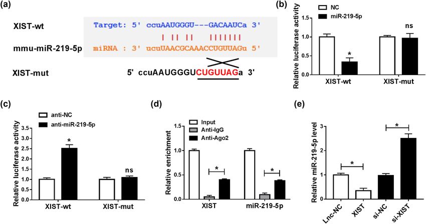
XIST knockdown attenuates spinal cord injury by targeting miR-219-5p 1091
(TUG1) repressed lipopolysaccharide (LPS)-stimulated anesthesia, and then C5 lamina was excised to exhibit
PC-12 cell damage, as demonstrated by the promotion the dural sac. Next, adjust the hammer position of the
in cell viability and the suppression in cell apoptosis spinal impactor on the C5 spinal cord. After that, bleed-
and inflammation, by decreasing miR-127 and inacti- ing was stopped and the incision layer by layer was
vating nuclear factor-κB (NF-κB) pathway [30]. Zhou sutured. In the sham group, C5 lamina was removed
et al. claimed that metastasis-associated lung adenocar- in the mice and C5 spinal cord was not impinged. The
cinoma transcript 1 (MALAT1) level was conspicuously hindlimb locomotor activity of the mice was evaluated by
raised in SCI mice and LPS-activated microglial cells, Basso–Beattie–Bresnahan (BBB) Locomotor Rating Scale
and MALAT1 knockdown relieved LPS-stimulated inflam- score. Subsequently, after 7 days of SCI, the mice were
matory injury by regulating miR-199b and IκB kinase β anesthetized and the spinal cord tissue specimens were
(IKKβ)/NF-κB pathway [31]. These reports indicated the collected for the following experiments. The study was
vital roles of lncRNAs in SCI development. As for X-inac- allowed by the Ethics Committee of Animal Research of
tive specific transcript (XIST), Zhao et al. uncovered that Yue Bei People’s Hospital and conducted according to the
XIST deficiency facilitated the recovery of SCI by reducing Guidelines for Care and Use of Laboratory Animals of
miR-27a and elevating Smurf1 [29]. Nevertheless, the “National Institutes of Health.”
molecular mechanisms of XIST in regulating SCI progres-
sion are not well understood.
miRNAs are small ncRNAs consisting of ∼22 nts and
exert vital regulatory roles at the posttranscriptional level 2.2 Cell culture and treatment
[6]. An increasing number of miRNAs have been identi-
fied to be closely linked to the progression of SCI. For The murine microglial cells (BV2) were bought from
instance, miR-27a-3p inhibited the inflammatory injury Procell (Wuhan, China) and cultured in Dulbecco’s modi-
of SCI by interacting with toll-like receptor 4 (TLR4) fied Eagle’s medium (DMEM; Gibco, Grand Island, NY,
[28]. miR-129-5p repressed the inflammatory response USA) added with 10% fetal bovine serum (FBS; Gibco)
and apoptosis in LPS-stimulated BV2 cells via the mod- and 1% penicillin–streptomycin (Gibco) in an incubator
ulation of high-mobility group protein B1 (HMGB1)/TLR4/ containing 5% CO2 at 37°C.
NF-κB pathway [22]. More importantly, a previous study For LPS treatment, BV2 cells were cultured with LPS
by Zhu et al. showed that miR-219-5p was able to promote (1, 10, 100, or 1,000 ng/mL; Solarbio, Beijing, China) for
the recovery of SCI and motor function by regulating 24 h. For the SCI cell model, BV2 cells were activated with
inflammation and oxidative stress [32]. However, the 100 ng/mL LPS for 24 h.
exact roles of miR-219-5p in SCI are largely unknown.
Through online tool starBase, we found that the XIST
contained potential binding sites with miR-219-5p. How-
ever, the relationship between XIST and miR-219-5p in 2.3 Cell transfection
regulating SCI progression has not been explored.
In this research, we established SCI mice and cell XIST small-interfering RNA (si-XIST) and scrambled siRNA
models and explored the effects of XIST on BV2 cell via- control (si-NC), the overexpression vector of XIST and
bility, apoptosis, and inflammation after SCI. Moreover, its control (lnc-NC), miR-219-5p mimics (miR-219-5p) and
the possible mechanism and signaling pathway of XIST its control (NC), miR-219-5p inhibitors (anti-miR-219-5p),
in SCI progression were further investigated. and anti-NC were synthesized by GeneCopoeia (Guangzhou,
China) and then transfected into BV2 cells with Lipofectamine
2000 (Invitrogen, Carlsbad, CA, USA). After 6 h of transfec-
tion, BV2 cells were triggered with 100 ng/mL LPS (Solarbio).
2 Materials and methods
2.1 Construction of SCI mice model
2.4 Reverse transcription quantitative
Adult C57bl/6J mice (female; 20–25 g) were purchased polymerase chain reaction (RT-qPCR)
from the Vital River (Beijing, China) and divided into
two groups: SCI group (n = 10) and sham group (n = 10). Total RNA in spinal cord tissues and BV2 cells was iso-
In SCI groups, the mice were incised along the neck after lated utilizing TRIzol (Invitrogen). Next, the RNAs were1092 Xueren Zhong et al.
reversely transcribed into complementary DNAs (cDNAs) ab86299; Abcam), B-cell lymphoma-2 (Bcl-2, ab196495;
utilizing miRNA 1st Strand cDNA Synthesis Kit (Vazyme, Abcam), BCL2-associated X (Bax, ab180733; Abcam), cleaved-
Nanjing, China) or avian myeloblastosis virus (AMV) caspase 3 (C-caspase 3, ab49822; Abcam), or total-caspase 3
reverse transcriptase (Promega, Madison, WI, USA). (t-caspase 3, ab90437; Abcam) overnight at 4°C and then
Afterward, RT-qPCR reaction was manipulated utilizing incubated with corresponding secondary antibody (ab6789;
AceQ Universal SYBR qPCR Master Mix (Vazyme) and Abcam) for 1.5 h at indoor temperature. Finally, the protein
specific primers (GeneCopoeia) on an ABI 7500 PCR bands were exposed through an enhanced chemiluminescence
system (Applied Biosystems, Foster City, CA, USA). The reagent (Vazyme) and analyzed by software Image J.
primers were as follows: XIST: (F: 5′-CGGGTCTCTTCAAG
GACATTTAGCC-3′ and R: 5′-GCACCAATACAGAGGAATGG
AGGG-3′); miR-219-5p: (F: 5′-ACACTCCAGCTGGGTGATTG
TCCAAACGCAAT-3′ and R: 5′-CTCAACTGGTGTCGTGGAG 2.7 Cell counting kit-8 (CCK-8) assay
TCGGC-3′); glyceraldehyde 3-phosphate dehydrogenase
(GAPDH): (F: 5′-GAAGATGGTGATGGGATTTC-3′ and R: After treatment with LPS and transfection, BV2 cells were
5′-GAAGGTGAAGGTCGGAGT-3′); U6: (F: 5′-CTCGCTTCGG harvested to assess cell viability through CCK-8 assay.
CAGCACA-3′ and R: 5′-AACGCTTCACGAATTTGCGT-3′). Briefly, BV2 cells were seeded into 96-well plates and
Glyceraldehyde 3-phosphate dehydrogenase (GAPDH) cultivated for 24 h. Next, 10 μL CCK-8 (Beyotime, Shanghai,
or U6 served as the internal reference. The expression China) was added into each well with incubation for another
was computed via the 2−ΔΔCt strategy. 4 h at room temperature. The optical density (OD) value (at
450 nm) was recorded using a microplate reader (Bio-Rad).
2.5 Enzyme-linked immunosorbent assay 2.8 Flow cytometry analysis
(ELISA)
After treated with 100 ng/mL LPS for 24 h or transfected
The concentrations of inflammatory cytokines (TNF-α, IL-1β, with indicated synthetic plasmids or oligonucleotides
IL-6, and IL-10) in the spinal cord tissue extracts or BV2 cell followed by LPS treatment for 24 h, BV2 cells were har-
supernatants were measured by ELISA kits (ab208348; vested and Annexin V-fluorescein isothiocyanate (FITC)
ab197742; ab100713; ab108870; Abcam, Cambridge, MA, Apoptosis Detection Kit (Vazyme) was utilized for the
USA) based on the guidelines of manufacturers. The absor- analysis of cell apoptosis. In brief, the harvested BV2 cells
bance was measured at 450 nm utilizing a microplate reader were rinsed with cold PBS (Sangon, Shanghai, China)
(Bio-Rad, Hercules, CA, USA), and the concentrations were and resuspended in binding buffer. BV2 cells were mixed
calculated based on the standard curve. with 5 µL Annexin V-FITC and 5 µL propidium iodide (PI)
for 15 min in the dark at indoor temperature. Finally,
the apoptotic cells were estimated with flow cytometry
(Beckman Coulter, Atlanta, GA, USA).
2.6 Western blot assay
Protein isolation was done using radioimmunoprecipita-
tion assay buffer (CWBio, Beijing, China), and protein 2.9 Dual-luciferase reporter assay
concentration was detected using a bicinchoninic acid
protein assay kit (Tiangen, Beijing, China). Then, an The fragments of XIST including the wild-type (wt) or
equal amount of proteins was split by sodium dodecyl mutant (mut) binding sites of miR-219-5p were cloned
sulfonate-polyacrylamide gel (Solarbio) electrophoresis into psi-CHECK2 plasmid (Promega), generating XIST-
and blotted onto polyvinylidene difluoride membranes wt and XIST-mut, respectively. BV2 cells were subse-
(Millipore, Billerica, MA, USA). The membranes were quently plated into six-well plates and transfected with
blocked using 5% nonfat milk for 1 h at indoor tempera- XIST-wt (or XIST-mut) and miR-219-5p (or NC, anti-miR-
ture. Next, the membranes were immunoblotted with pri- 219-5p, anti-NC). The renilla and firefly luciferase activ-
mary antibodies against GAPDH (ab181602; Abcam), total ities were detected using a Dual-Luciferase Reporter Assay
p65 (t-p65; ab16502; Abcam), phosphorylated p65 (p-p65; System (Promega) after 48 h of co-transfection.XIST knockdown attenuates spinal cord injury by targeting miR-219-5p 1093
Figure 1: XIST was elevated and miR-219-5p was reduced in SCI mice model. (a) The BBB score method was used to evaluate the locomotor
function changes of mice in SCI and sham groups. (b and c) RT-qPCR analysis was performed for the expression levels of XIST and miR-219-
5p in the spinal cord tissues from SCI and sham groups. (d–g) ELISA assay was conducted to determine the levels of TNF-α, IL-1β, IL-6, and
IL-10 in the spinal cord tissue extracts of SCI groups and sham groups. (h) Western blot assay was conducted for the protein levels of p-p65
and t-p65 in the spinal cord tissues from SCI and sham groups. *P < 0.05.
2.10 RNA immunoprecipitation (RIP) assay were estimated by one-way analysis of variance followed
by Tukey’s test. It was defined as significant if P < 0.05.
Magna RNA-binding protein immunoprecipitation kit
(Millipore) was exploited for RIP assay. Briefly, BV2 cells Ethics approval and consent to participate: The hospital’s
were disrupted in RIP buffer, and cell extracts were culti- Institutional Review Board approved the current study.
vated with magnetic beads, which were conjugated with
anti-immunoglobulin G (anti-IgG) or anti-argonaute-2
(anti-Ago2). Then, the immunoprecipitated RNAs were
purified, and the enrichment of XIST and miR-219-5p
3 Results
was examined via RT-qPCR analysis.
3.1 XIST was upregulated and miR-219-5p
was downregulated in SCI mice model
2.11 Statistical analysis
After the mice model of SCI was established, the recovery
All experiments were manipulated in triplicate. Data ana- of motor function in mice was evaluated by the BBB
lysis was executed using the software GraphPad Prism 7, method. The results showed that the hindlimb locomotor
and the results were exhibited as mean ± standard devia- activity was markedly decreased after spinal cord contu-
tion. The differences between two sets were estimated by sions, as indicated by the decreased BBB score in SCI
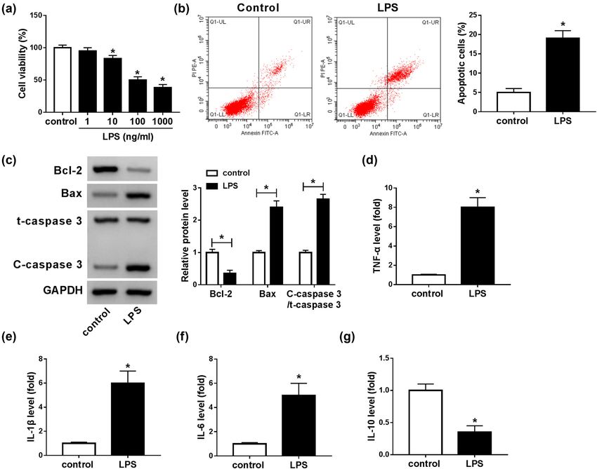
Student’s t-test, whereas differences among three groups groups compared with sham operation groups (Figure 1a).1094 Xueren Zhong et al. The results suggested that the SCI mice model was suc- Collectively, XIST was abnormally increased and miR-219- cessfully established. Then, we determined the expression 5p was abnormally decreased in the SCI mice model. levels of XIST and miR-219-5p in the spinal cord tissues from SCI groups and sham groups by RT-qPCR analysis. The results showed that the XIST level was notably ele- vated and miR-219-5p was conspicuously reduced in the 3.2 LPS repressed cell viability and induced spinal cord tissues from SCI groups compared with sham apoptosis and inflammatory response groups (Figure 1b and c). Next, we detected the levels of in BV2 cells inflammatory cytokines (including TNF-α, IL-1β, IL-6, and IL-10) in SCI mice through ELISA. As shown in LPS-induced microglial cell is a commonly used SCI model Figure 1d–g, the levels of TNF-α, IL-1β, and IL-6 were in vitro. To establish the SCI model in vitro, microglial cells drastically elevated and the level of IL-10 was distinctly (BV2) were exposed to different concentrations of LPS (1, 10, declined in SCI groups compared to sham operation 100, and 1,000 ng/mL) for 24 h. As illustrated by the CCK-8 groups. Herein, SCI also caused a noteworthy elevation in assay, the viability of BV2 cells was markedly repressed by p-p65 protein level compared to sham groups (Figure 1h). LPS in a dose-dependent manner (Figure 2a). There was no Figure 2: LPS treatment suppressed BV2 cell viability and promoted apoptosis and inflammatory damage. (a) BV2 cells were treated with LPS (1, 10, 100, and 1,000 ng/mL) for 24 h, and then, the viability of BV2 cells was evaluated by CCK-8 assay. (b) The apoptosis of BV2 cells treated with 100 ng/mL LPS was investigated by flow cytometry analysis. (c) The protein levels of Bcl-2, Bax, t-caspase 3, and C-caspase 3 in 100 ng/mL LPS-treated BV2 cells were measured through western blot assay. (d–g) The levels of TNF-α, IL-1β, IL-6, and IL-10 were measured by ELISA in LPS-stimulated BV2 cells. *P < 0.05.
XIST knockdown attenuates spinal cord injury by targeting miR-219-5p 1095 significant difference in BV cell viability between 100 ng/mL the level of IL-10 in BV cells compared to control groups groups and 1,000 ng/mL groups; thus, 100 ng/mL LPS was (Figure 2d–g). These observations suggested that LPS- utilized in the following study. Flow cytometry analysis induced SCI cell model was successfully constructed in vitro. showed that the apoptosis of BV2 cells was promoted by 100 ng/mL LPS treatment (Figure 2b). Meanwhile, we determined the levels of apoptosis-related proteins (Bcl-2, Bax, and C-caspase 3) in 100 ng/mL LPS-stimulated BV2 cells by western blot assay. The results indicated that LPS 3.3 XIST knockdown abrogated LPS- treatment led to an apparent reduction in Bcl-2 expression mediated BV2 cell viability, apoptosis, and an obvious elevation in Bax and C-caspase 3 expres- and inflammatory response sion in BV2 cells compared to control groups (Figure 2c). In addition, ELISA results showed that LPS distinctly As shown in Figure 3a, the XIST level was increased in enhanced the levels of TNF-α, IL-1β, and IL-6 and reduced LPS-stimulated BV2 cells, indicating that XIST might be Figure 3: XIST silencing restored the impacts of LPS on cell viability, apoptosis, and inflammation in BV2 cells. BV2 cells were transfected with si-XIST or si-NC and then treated with 100 ng/mL LPS. (a) The expression level of XIST in BV2 cells was determined by the RT-qPCR assay. (b and c) The viability and apoptosis of BV2 cells were evaluated through CCK-8 assay and flow cytometry analysis. (d) The protein levels of Bcl-2, Bax, t-caspase 3, and C-caspase 3 in BV2 cells were examined by western blot assay. (e–h) The secretion of TNF-α, IL-1β, IL-6, and IL-10 in BV2 cells was detected by ELISA. *P < 0.05.
1096 Xueren Zhong et al. involved in the regulation of LPS-mediated microglial 3.4 XIST negatively regulated miR-219-5p cell viability, apoptosis, and inflammatory cytokine pro- expression by directly targeting duction. Thus, we explored the function of XIST in LPS- stimulated microglial cell progression by transfecting To explore the potential mechanism of XIST regulating si-XIST or si-NC into BV2 cells and then treating the trans- BV2 cell progression, we analyzed the targets of XIST fected cells with LPS. As demonstrated by the RT-qPCR through online tool starBase (http://starbase.sysu.edu. assay, XIST silencing markedly suppressed the XIST cn/agoClipRNA.php?source=lncRNA&flag=target&clade= level in LPS-induced BV2 cells (Figure 3a). CCK-8 assay mammal&genome=mouse&assembly=mm10&miRNA= showed that the inhibitory effect on cell viability mediated all&clipNum=1&deNum=0&target=Xist). The results dis- by LPS was restored by decreasing XIST expression in played that miR-219-5p contained the complementary BV2 cells (Figure 3b). Flow cytometry analysis indicated sequences of XIST (Figure 4a). Then dual-luciferase that LPS-induced cell apoptosis was repressed by XIST reporter assay and RIP assay were carried out to confirm knockdown in BV2 cells (Figure 3c). Moreover, western the interaction between miR-219-5p and XIST. As sug- blot assay results showed that LPS treatment suppressed gested by the dual-luciferase reporter assay, miR-219-5p Bcl-2 expression and promoted Bax and C-caspase 3 transfection markedly inhibited the luciferase activity of expression in BV2 cells, while XIST knockdown effec- XIST-wt and anti-miR-219-5p transfection conspicuously tively restored the impacts (Figure 3d). In addition, we elevated the luciferase activity of XIST-wt in BV2 cells, observed that the upregulation of TNF-α, IL-1β, and IL-6 while the luciferase activity of XIST-mut was not affected and the downregulation of IL-10 mediated by LPS by miR-219-5p or anti-miR-219-5p (Figure 4b and c). The were restored by reducing XIST expression in BV2 cells results of the RIP assay showed that the levels of XIST (Figure 3e–h). To sum up, XIST knockdown could relieve and miR-219-5p were all enriched in anti-Ago2 protein LPS-induced injury in BV2 cells. complexes in BV2 cells compared to anti-IgG control Figure 4: XIST sponged miR-219-5p to suppress miR-219-5p expression. (a) The potential binding sites between XIST and miR-219-5p were predicted by starBase. (b and c) XIST-wt (or XIST-mut) and miR-219-5p (or NC, anti-miR-219-5p, anti-NC) were co-transfected into BV2 cells, and then, dual-luciferase reporter assay was conducted to measure the luciferase activity in BV2 cells. (d) After RIP assay, the abundance of XIST and miR-219-5p in anti-IgG or anti-Ago2 immunoprecipitates in BV2 cells was measured by RT-qPCR analysis. (e) The expression level of miR-219-5p in BV2 cells transfected with lnc-NC, XIST, si-NC, or si-XIST was determined using the RT-qPCR assay. *P < 0.05.
XIST knockdown attenuates spinal cord injury by targeting miR-219-5p 1097
groups, further confirming the interaction between XIST of XIST on LPS-mediated NF-κB signaling pathway acti-
and miR-219-5p (Figure 4d). Thereafter, we explored the vation in BV2 cells. As demonstrated by western blot
effect of XIST on miR-219-5p expression by transfecting assay, LPS treatment enhanced the protein level of
XIST or si-XIST into BV2 cells. Our results showed that p-p65 in BV2 cells, indicating the activation of the NF-
XIST transfection apparently decreased miR-219-5p level κB pathway. Moreover, we found that XIST deficiency
in BV2 cells, while si-XIST transfection exhibited the suppressed LPS-induced NF-κB pathway activation, as
opposite results (Figure 4e). These observations indicated shown by downregulation of p-p65 protein level, while
that XIST could negatively modulate miR-219-5p expres- the effect was alleviated by the inhibition of miR-219-5p
sion by direct interaction. (Figure 6a and b). Taken together, XIST knockdown could
inhibit LPS-activated NF-κB signaling pathway by mod-
ulating miR-219-5p expression in BV2 cells.
3.5 miR-219-5p inhibition reversed the
effects of XIST knockdown on cell
viability, apoptosis, and inflammation 4 Discussion
in LPS-stimulated BV2 cells
lncRNAs have been proved as essential mediators in the
Subsequently, we further explored whether XIST could development of SCI. After SCI, countless cytokines and
alter LPS-induced BV2 cell injury by targeting miR-219- signaling pathways have been demonstrated to mediate
5p. First, anti-miR-219-5p transfection evidently reduced the apoptosis and inflammatory response [14,15]. LPS-
the level of miR-219-5p in BV2 cells compared to anti-NC stimulated microglial cells are widely utilized to explore
and control groups, indicating that anti-miR-219-5p was the pathogenesis of SCI. In this study, we successfully
successfully transfected into BV2 cells (Figure 5a). Next, constructed the SCI mice model and found that XIST
BV2 cells were assigned to control, LPS, LPS + si-NC, LPS + was drastically increased in the spinal cord tissues of
si-XIST, LPS + si-XIST + anti-NC, and LPS + si-XIST + SCI mice. Moreover, the SCI cell model was constructed
anti-miR-219-5p groups. The results of the CCK-8 assay by stimulating BV2 cells with LPS. Then, we tested cell
and flow cytometry analysis indicated that XIST knock- viability, apoptosis, and the levels of inflammatory cyto-
down promoted cell viability and inhibited apoptosis in kines in LPS-triggered BV2 cells. We found that cell via-
LPS-stimulated BV2 cells, while the impacts were par- bility was repressed and cell apoptosis and inflammatory
tially overturned by decreasing miR-219-5p (Figure 5b response were induced, indicating the successful con-
and c). Western blot assay showed that the promotional struction of the SCI cell model. Thereafter, we explored
role in Bcl-2 level and the suppressive roles in Bax and the functions and mechanisms of XIST in SCI develop-
C-caspase 3 levels mediated by XIST silencing in LPS- ment. As a result, XIST knockdown recovered LPS-stimu-
treated BV2 cells were ameliorated following the suppres- lated BV2 cell injury by regulating miR-219-5p and NF-κB
sion of miR-219-5p (Figure 5d). In addition, ELISA results signaling pathway.
showed that the impacts of XIST deficiency on TNF-α, IL- In the past decades, the potential functions of XIST in
1β, IL-6, and IL-10 levels were all restored by decreasing SCI have been gradually studied. For example, Kwon et
miR-219-5p expression in LPS-activated BV2 cells (Figure al. revealed that the XIST level was enhanced in the SCI
5e–h). These outcomes suggested that XIST knockdown rat model [11]. Gu et al. manifested that XIST knockdown
attenuated LPS-induced BV2 cell injury by targeting miR- effectively limited the apoptosis of neuronal in SCI rats by
219-5p. modulating miR-494/phosphatase and tensin homolog
deleted on chromosome ten (PTEN)/phosphoinositide
3-kinase (PI3K)/AKT [5]. Moreover, Zhao et al. reported that
XIST silencing restored the suppressive role in cell viabi-
3.6 LPS-induced XIST promoted the lity and the promotional role in apoptosis and inflamma-
activation of NF-κB pathway by tion mediated by LPS in microglial cells by regulating
regulating miR-219-5p miR-27a/smad ubiquitination regulatory factor 1 (Smurf1)
axis [29]. Correspondingly, our results showed that XIST
Finally, BV2 cells were divided into six groups: control, was conspicuously increased in LPS-triggered BV2 cells.
LPS, LPS + si-NC, LPS + si-XIST, LPS + si-XIST + anti-NC, XIST interference enhanced cell viability and impeded
and LPS + si-XIST + anti-miR-219-5p to explore the effect apoptosis, concomitant with upregulation in Bcl-2 level1098 Xueren Zhong et al. Figure 5: XIST knockdown regulated LPS-induced BV2 cell viability, apoptosis, and inflammatory response by interacting with miR-219-5p. (a) The expression of miR-219-5p in untransfected BV2 cells and anti-NC or anti-miR-219-5p transfected BV2 cells was determined using RT- qPCR assay. (b–h) BV2 cells were divided into six groups: control, LPS, LPS + si-NC, LPS + si-XIST, LPS + si-XIST + anti-NC, and LPS + si-XIST + anti-miR-219-5p. (b and c) BV2 cell viability and apoptosis were determined by CCK-8 assay and flow cytometry analysis, respectively. (d) The protein level of Bcl-2, Bax, t-caspase 3, and C-caspase 3 in BV2 cells were measured by western blot assay. (e–h) The levels of TNF-α, IL-1β, IL-6, and IL-10 in BV2 cells were detected by ELISA kits. *P < 0.05. and downregulation in Bax and C-caspase-3 levels in XIST knockdown could accelerate the recovery of SCI LPS-triggered BV2 cells. In addition, our results exhibited through promoting microglial cell viability and impeding that XIST knockdown reduced TNF-α, IL-1β, and IL-6 apoptosis and inflammation. levels and enhanced IL-10 level in LPS-activated BV2 For mechanism analysis, the downstream target of cells, suggesting that XIST deficiency attenuated LPS- XIST was investigated. XIST has been identified as the induced inflammatory response in BV2 cells. Overall, sponge for multiple miRNAs, such as miR-152 [26], miR-
XIST knockdown attenuates spinal cord injury by targeting miR-219-5p 1099
Figure 6: XIST silencing inactivated LPS-stimulated NF-κB pathway by targeting miR-219-5p. (a and b) BV2 cells were assigned to control,
LPS, LPS + si-NC, LPS + si-XIST, LPS + si-XIST + anti-NC, and LPS + si-XIST + anti-miR-219-5p groups, and then, the protein levels of p-p65
and t-p65 were measured by western blot assay. *P < 0.05.
101 [3], miR-367 [16], and miR-137 [27]. While in our deepened our understanding on the molecular basis in the
study, miR-219-5p was proved to be a target of XIST. It management of SCI and might provide a novel direction for
has been reported that miR-219-5p was downregulated in SCI therapy.
SCI mice [17]. Moreover, miR-219-5p was found to amelio-
rate inflammatory injury in the SCI mice model [32]. Funding information: No funds.
Herein, we observed that miR-219-5p inhibition abro-
gated the impacts of XIST knockdown on cell viability, Conflict of interest: The authors declare that they have no
apoptosis, and inflammation in LPS-activated BV2 cells, conflicts of interest.
indicating XIST knockdown could attenuate SCI by tar-
geting miR-219-5p. Data availability statement: The available datasets gen-
It has been documented that the activation of the erated during and/or analysed during the current study
NF-κB pathway can trigger the production of pro-inflamma- are available from the corresponding author on reason-
tory cytokines, thereby inducing inflammatory response able request.
and apoptotic response [12,13,20]. Moreover, NF-κB pathway
activation plays a positive role in the SCI development [2,10].
For example, GRB1 relieved SCI via altering miR-130b-5p/ Reference
TLR4/NF-κB pathway [23]. circ_0000962 inhibited the
inflammation in the SCI cell model via activating PI3K/Akt [1] Ahuja CS, Nori S, Tetreault L, Wilson J, Kwon B, Harrop J, et al.
and inactivating NF-κB by sponging miR-302b-3p [7]. Thus, Traumatic spinal cord injury-repair and regeneration.
we explored the impact of LPS-induced XIST in the NF-κB Neurosurgery. 2017;80:S9–22. doi: 10.1093/neuros/nyw080.
pathway. Our results showed that the knockdown of XIST [2] Brambilla R, Bracchi-Ricard V, Hu WH, Frydel B, Bramwell A,
decreased LPS-induced p-p65 level in BV2 cells, while miR- Karmally S, et al. Inhibition of astroglial nuclear factor kappaB
reduces inflammation and improves functional recovery after
219-5p suppression restored the effect, suggesting that XIST
spinal cord injury. J Exp Med. 2005;202:145–56. doi: 10.1084/
silencing might block LPS-stimulated NF-κB pathway by jem.20041918.
targeting miR-219-5p. [3] Chen DL, Ju HQ, Lu YX, Chen LZ, Zeng ZL, Zhang DS, et al. Long non-
In conclusion, this study uncovered that XIST was coding RNA XIST regulates gastric cancer progression by acting as a
upregulated in SCI mice and LPS-activated BV2 cells. molecular sponge of miR-101 to modulate EZH2 expression. J Exp
Clin Cancer Res. 2016;35:142. doi: 10.1186/s13046-016-0420-1.
XIST knockdown ameliorated LPS-induced microglial cell
[4] Edgerton VR, Harkema S. Epidural stimulation of the spinal
apoptosis and inflammatory injury after SCI by sponging cord in spinal cord injury: current status and future chal-
miR-219-5p and inactivating NF-κB pathway. Our study lenges. Expert Rev Neurother. 2011;11:1351–3. doi: 10.1586/
revealed the protective effect of XIST silencing in SCI, which ern.11.129.1100 Xueren Zhong et al.
[5] Gu S, Xie R, Liu X, Shou J, Gu W, Che X. Long coding RNA XIST induced inflammatory and apoptotic responses in neuronal
contributes to neuronal apoptosis through the downregulation cells. PLoS One. 2016;11:e0160314. doi: 10.1371/
of AKT phosphorylation and is negatively regulated by miR- journal.pone.0160314.
494 in rat spinal cord injury. Int J Mol Sci. 2017;18:732. [21] Tran AP, Warren PM, Silver J. The biology of regeneration
doi: 10.3390/ijms18040732. failure and success after spinal cord injury. Physiol Rev.
[6] Hammond SM. An overview of microRNAs. Adv Drug Deliv Rev. 2018;98:881–917. doi: 10.1152/physrev.00017.2017.
2015;87:3–14. doi: 10.1016/j.addr.2015.05.001. [22] Wan G, An Y, Tao J, Wang Y, Zhou Q, Yang R, et al. MicroRNA-
[7] He R, Tang GL, Niu L, Ge C, Zhang XQ, Ji XF, et al. Quietness 129-5p alleviates spinal cord injury in mice via suppressing the
circ_0000962 promoted nerve cell inflammation through PIK3CA/ apoptosis and inflammatory response through HMGB1/TLR4/
Akt/NF-kappaB signaling by miR-302b-3p in spinal cord injury. NF-kappaB pathway. Biosci Rep. 2020;40:BSR20193315.
Ann Palliat Med. 2020;9:190–8. doi: 10.21037/apm.2020.02.13. doi: 10.1042/BSR20193315.
[8] Held KS, Lane TE. Spinal cord injury, immunodepression, [23] Wang D, Zhao S, Pan J, Wang Z, Li Y, Xu X, et al. Ginsenoside
and antigenic challenge. Semin Immunol. 2014;26:415–20. Rb1 attenuates microglia activation to improve spinal cord
doi: 10.1016/j.smim.2014.03.003. injury via microRNA-130b-5p/TLR4/NF-kappaB axis. J Cell
[9] Hossain MS, Islam MS, Rahman MA, Glinsky JV, Herbert RD, Physiol. 2021;236:2144–55. doi: 10.1002/jcp.30001.
Ducharme S, et al. Health status, quality of life and socio- [24] Wang F, Liu J, Wang X, Chen J, Kong Q, Ye B, et al. The emerging
economic situation of people with spinal cord injuries six role of lncRNAs in spinal cord injury. Biomed Res Int.
years after discharge from a hospital in Bangladesh. Spinal 2019;2019:3467121. doi: 10.1155/2019/3467121.
Cord. 2019;57:652–61. doi: 10.1038/s41393-019-0261-9. [25] Wang P, Ning S, Zhang Y, Li R, Ye J, Zhao Z, et al. Identification
[10] Kasuya Y, Umezawa H, Hatano M. Stress-activated protein of lncRNA-associated competing triplets reveals global pat-
kinases in spinal cord injury: focus on roles of p38. Int J Mol terns and prognostic markers for cancer. Nucleic acids
Sci. 2018;19:867. doi: 10.3390/ijms19030867. research. 2015;43:3478–89.
[11] Kwon BK, Oxland TR, Tetzlaff W. Animal models used in spinal [26] Yao Y, Ma J, Xue Y, Wang P, Li Z, Liu J, et al. Knockdown of long
cord regeneration research. Spine (Phila Pa 1976). non-coding RNA XIST exerts tumor-suppressive functions in
2002;27:1504–10. doi: 10.1097/00007632-200207150-00005. human glioblastoma stem cells by up-regulating miR-152.
[12] Lawrence T. The nuclear factor NF-kappaB pathway in inflam- Cancer Lett. 2015;359:75–86. doi: 10.1016/
mation. Cold Spring Harb Perspect Biol. 2009;1:a001651. j.canlet.2014.12.051.
doi: 10.1101/cshperspect.a001651. [27] Yu H, Xue Y, Wang P, Liu X, Ma J, Zheng J, et al. Knockdown of
[13] Lawrence T, Fong C. The resolution of inflammation: anti- long non-coding RNA XIST increases blood-tumor barrier per-
inflammatory roles for NF-kappaB. Int J Biochem Cell Biol. meability and inhibits glioma angiogenesis by targeting miR-
2010;42:519–23. doi: 10.1016/j.biocel.2009.12.016. 137. Oncogenesis. 2017;6:e303. doi: 10.1038/oncsis.2017.7.
[14] Lazaridis C, Andrews CM. Traumatic spinal cord injury: [28] Zhang P, Li LQ, Zhang D, Shen Y. Over-expressed miR-27a-3p
learn from the brain!*. Crit Care Med. 2014;42:749–50. inhibits inflammatory response to spinal cord injury by
doi: 10.1097/CCM.0000000000000077. decreasing TLR4. Eur Rev Med Pharmacol Sci.
[15] Leal-Filho MB. Spinal cord injury: From inflammation to glial scar. 2018;22:5416–23. doi: 10.26355/eurrev_201809_15800.
Surg Neurol Int. 2011;2:112. doi: 10.4103/2152-7806.83732. [29] Zhao Q, Lu F, Su Q, Liu Z, Xia X, Yan Z, et al. Knockdown of long
[16] Li C, Wan L, Liu Z, Xu G, Wang S, Su Z, et al. Long non-coding noncoding RNA XIST mitigates the apoptosis and inflammatory
RNA XIST promotes TGF-beta-induced epithelial-mesenchymal injury of microglia cells after spinal cord injury through miR-
transition by regulating miR-367/141-ZEB2 axis in non-small- 27a/Smurf1 axis. Neurosci Lett. 2020;715:134649.
cell lung cancer. Cancer Lett. 2018;418:185–95. doi: 10.1016/ doi: 10.1016/j.neulet.2019.134649.
j.canlet.2018.01.036. [30] Zheng H, Hu S, Cao J, Yao L, Zhang N. Long non-coding RNA
[17] Liu NK, Wang XF, Lu QB, Xu XM. Altered microRNA expression TUG1 alleviates LPS-induced injury of PC-12 cells by down-
following traumatic spinal cord injury. Exp Neurol. regulating microRNA-127. Exp Mol Pathol. 2019;110:104287.
2009;219:424–9. doi: 10.1016/j.expneurol.2009.06.015. doi: 10.1016/j.yexmp.2019.104287.
[18] Majdan M, Plancikova D, Nemcovska E, Krajcovicova L, [31] Zhou HJ, Wang LQ, Wang DB, Yu JB, Zhu Y, Xu QS, et al. Long
Brazinova A, Rusnak M. Mortality due to traumatic spinal cord noncoding RNA MALAT1 contributes to inflammatory response
injuries in Europe: a cross-sectional and pooled analysis of of microglia following spinal cord injury via the modulation of
population-wide data from 22 countries. Scand J Trauma Resusc a miR-199b/IKKbeta/NF-kappaB signaling pathway. Am J
Emerg Med. 2017;25:64. doi: 10.1186/s13049-017-0410-0. Physiol Cell Physiol. 2018;315:C52–61. doi: 10.1152/
[19] Mercer TR, Dinger ME, Mattick JS. Long non-coding RNAs: ajpcell.00278.2017.
insights into functions. Nat Rev Genet. 2009;10:155–9. [32] Zhu Y, Xu Q, Sha WP, Zhao KP, Wang LM. miR-219-5p promotes
doi: 10.1038/nrg2521. spinal cord injury recovery by inhibiting NEUROD2-regulated
[20] Srinivasan M, Bayon B, Chopra N, Lahiri DK. Novel nuclear inflammation and oxidative stress. Eur Rev Med Pharmacol Sci.
factor-kappaB targeting peptide suppresses beta-amyloid 2019;23:37–43. doi: 10.26355/eurrev_201901_16745.You can also read