LncRNA HIF1A AS2: A potential oncogene in human cancers (Review)
←
→
Page content transcription
If your browser does not render page correctly, please read the page content below
BIOMEDICAL REPORTS 15: 85, 2021

                        lncRNA HIF1A‑AS2: A potential oncogene
                              in human cancers (Review)
                            YANG LIU1, YUNYAN ZHANG2, CHA CHEN1 and YOUQIANG LI3

          1
         The Second Clinical College of Guangzhou University of Chinese Medicine, Guangzhou, Guangdong 510006;
      2
       Department of Stomatology, Guangzhou Women and Children's Medical Center, Guangzhou, Guangdong 510000;
       3
        Department of Laboratory Medicine, The Affiliated Hexian Memorial Hospital of Southern Medical University,
                                        Guangzhou, Guangdong 511400, P.R. China

                                          Received March 9, 2021; Accepted July 28, 2021

                                                      DOI: 10.3892/br.2021.1461

Abstract. Long non‑coding RNAs (lncRNAs) are transcripts              Contents
that are >200 nucleotides, but with no open reading frame. An
increasing number of lncRNAs have been identified following           1. Introduction
the development of second‑generation sequencing technologies,         2. Expression and function of lncRNA HIF1A‑AS2 in several
and they have since become a research hotspot. Functionally,             types of cancer
they play a vital role in tumor progression, including in tumor       3. Conclusions and future perspective
proliferation, migration, invasion, apoptosis and acquisition
of drug resistance. They regulate gene expression primarily
through interaction with DNA, RNA and proteins at the                 1. Introduction
epigenetic, transcriptional and post‑transcriptional levels.
Endogenous hypoxia‑inducible factor 1α antisense RNA 2                Technological advances have driven an improved under‑
(lncRNA HIF1A‑AS2) is aberrantly expressed and involved               standing of protein‑coding genes; however, the functional
the development/progression of various types of tumors, such          roles of non‑coding (nc)RNAs are relatively less well under‑
as bladder cancer, glioblastoma, breast cancer and osteo‑             stood. ncRNAs account for >90% of the human genome,
sarcoma. It plays a vital role in the proliferation, apoptosis,       whereas protein‑coding genes account for only 1.5% (1,2).
migration, invasion and epithelial‑mesenchymal transfor‑              Based on transcript size, ncRNAs are divided into two
mation of various tumor cells. This review summarizes the             groups: Small ncRNAs with transcripts 200 nucleotides
related molecular mechanisms of lncRNA HIF1A‑AS2 in the               in length (3). lncRNAs, first discovered in the sequencing
development/progression of human tumors and other diseases.           of cDNA libraries in mouse cells (4), are mRNA‑like tran‑
                                                                      scripts that are likely transcribed by RNA polymerase II
                                                                      (RNA pol II), but which lack a stable open reading frame (5).
                                                                      Initially, these non‑coding RNAs were viewed as by‑products
                                                                      and noise of the transcription process (4). However, with the
                                                                      continuous development of gene technologies, a large number
                                                                      of studies have found that lncRNAs are involved in various
Correspondence to: Dr Youqiang Li, Department of Laboratory           physiological and pathological processes.
Medicine, The Affiliated Hexian Memorial Hospital of                      lncRNA HIF1A‑AS2, also known as HIF1A‑AS2, is
Southern Medical University, 2 Qinghe East Road, Guangzhou,           the endogenous antisense transcript of hypoxia‑inducible
Guangdong 511400, P.R. China                                          factor 1α (HIF1α), and 3'aHIF, termed HIF1α antisense
E‑mail: liyouqiang21@126.com                                          RNA 2 (HIF1A‑AS2), is localized at chromosome 14q23.2,
                                                                      and is 2,052 nucleotides in length. In 1999, it was first discov‑
Abbreviations: PHLDA1, pleckstrin homology like domain,               ered to be abnormally expressed in clear cell renal carcinoma
family A, member 1; LSD1, lysine‑specific demethylase 1; EMT,
                                                                      by Thrash‑Bingham and Tartof (6), and was identified as the
epithelial‑mesenchymal transformation; IGF2BP2, insulin‑like growth
factor 2; DHX9, ATP‑dependent RNA helicase A; HMGA1, high             endogenous antisense transcript, which could bind to the
mobility group AT‑hook 1; lncRNA, long non‑coding RNA;                3' untranslated region (3'UTR) of HIF1α mRNA in a comple‑
HIF1A‑AS2, hypoxia‑inducible factor 1α antisense RNA 2                mentary manner (Fig. 1), and this bound form is referred to as
                                                                      aHIF. In 2002, Rossignol et al (7) reported that HIF1A‑AS2
Key words: lncRNA, HIF1A‑AS2, cancer, microRNA, biomarker             was expressed in several human tissues, both physiologically,
                                                                      and when the tissues had become cancerous. These findings
                                                                      attracted increased focus on HIF1A‑AS2. Further studies
                                                                      demonstrated that HIF1A‑AS2 was aberrantly expressed in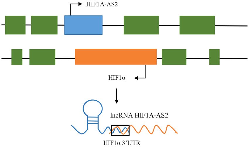
2                                          LIU et al: lncRNA HIF1A-AS2 AND HUMAN CANCER

       Figure 1. Location of HIF1A‑AS2 and HIF1α in chromosome 14. HIF1α, hypoxia‑inducible factor 1α; HIF1A‑AS2, HIF1α antisense RNA.

various human diseases, including preeclampsia (PE), epithe‑            inhibited proliferation, migration and invasion, as well as
lial ovarian cancer (EOC), colorectal cancer (CRC), gastric             inducing G0����������������������������������������������
                                                                                      /���������������������������������������������
                                                                                       G1 cell cycle arrest and increased cell apop‑
cancer (GC), breast cancer (BC), bladder cancer, osteosar‑              tosis in two trophoblast cell lines (HTR/SVneo and JAR). In
coma (OS), renal cell carcinoma, non‑small cell lung cancer             contrast, overexpression of HIF1A‑AS2 exerted the opposite
(NSCLC) and glioblastoma (GBM). Chen et al (8) reported                 effect. Mechanistically, a subcellular localization assay indi‑
that the expression levels of HIF1A‑AS2 were upregulated                cated that HIF1A‑AS2 was primarily localized in the cell
in GC tissues and cells, and this upregulated expression was            nucleus; thus, it may play a role in regulation of transcription.
correlated with Tumor‑Node‑Metastasis stage, tumor invasion,            Further experiments showed that HIF1A‑AS2 inhibited the
lymph node metastasis and a poor prognosis. Lin et al (9) also          transcription of pleckstrin homology like domain, family A,
demonstrated upregulated expression of HIF1A‑AS2 in 60 OS               member 1 (PHLDA1), which plays a significant role in the acti‑
tissues compared with the adjacent healthy tissues. Thus,               vation‑induced apoptosis following binding to lysine‑specific
HIF1A‑AS2 may serve as a promising target for treatment of              demethylase (LSD1) at the epigenetic level. Furthermore,
several types of cancer.                                                chromatin immunoprecipitation assays showed LSD1 and
    However, several studies demonstrated that the expres‑              H3K4 me2 enrichment in the promoter region of the PHLDA1
sion levels of HIF1A‑AS2 in tumor tissues was abnormal,                 gene (Fig. 2A) after transfection with small‑interfering
indicating the potential correlation between HIF1A‑AS2                  (si)‑HIF1A‑AS2. Thus, HIF1A‑AS2 may be a useful diagnostic
and cancer. Therefore, this review summarizes the current               biomarker for PE.
body of knowledge regarding the aberrant expression of this
lncRNA (Table I), its function and the regulatory mechanisms            EOC. Ovarian cancer (OC) is one of the most common types
of HIF1A‑AS2 in several types of cancer.                                of malignant tumors in females, with EOC being the most
                                                                        common, accounting for 80‑90% of OC cases (15,16) . Although
2. Expression and function of lncRNA HIF1A‑AS2 in sev‑                  EOC treatments have improved notably, even in developed
eral types of cancer                                                    countries, such as the United States and Canada, the overall
                                                                        survival remains at only 47% 5 years after diagnosis (17).
PE. PE is one of the leading causes of maternal death and a             Therefore, investigating the molecular mechanism and finding
pregnancy‑specific disease, affecting 3‑14% of parturients              effective therapeutic targets for management of EOC is of
worldwide (10). Although PE has been extensively studied (11),          great importance. Qiu et al (18) reported that the expression of
the underlying pathogenesis of PE remains elusive. However,             HIF1A‑AS2 in EOC tissues was significantly higher compared
it is hypothesized that inadequate trophoblastic invasion may           with the normal controls, and HIF1A‑AS2 was a lncRNA that
cause PE (12,13). Wu et al (14) reported that HIF1A‑AS2                 was upregulated under hypoxic conditions. Thus, the following
expression was significantly downregulated in the tissues               assays were performed under hypoxic conditions. Functional
of 52 patients with PE compared with the adjacent normal                assays revealed that knockdown of HIF1A‑AS2 promoted
samples. Knockdown of HIF1A‑AS2 expression significantly                cell apoptosis and weakened tumorigenesis in nude mice. In
BIOMEDICAL REPORTS 15: 85, 2021 3
Table I. Expression and function of long non-coding RNA HIF1α antisense RNA in PE and various types of cancer.
Change in
Disease expression Role Biological function Related genes Refs.
PE Down Pathogenic Proliferation, migration, invasion, LSD1, (14)
pro-apoptosis, cell cycle arrest PHLDA1
Epithelial ovarian Up Oncogenic Proliferation, migration Bax, caspase‑7, caspase‑9, (18)
cancer and invasion BCL-2, caspase‑3
Colorectal cancer Up Oncogenic Proliferation, migration miR-129-5p, miR-33b-5p (23)
and invasion DNMT3A
Gastric cancer Up Oncogenic Proliferation, migration - (8)
and invasion
Breast cancer Up Oncogenic Proliferation, migration miR-548c-3p, (30)
and invasion HIF1α, VEGF
Bladder cancer Up Oncogenic Proliferation, migration, - (35)
invasion and anti-apoptosis
Osteosarcoma Up Oncogenic Proliferation, migration, miR-33b-5p, SIRT6, (9,40)
invasion and anti-apoptosis miR-129-5p
Glioblastoma Up Oncogenic Neurosphere formation IGF2BP2, DHX9, (43)
HMGA1
Renal cancer Up Oncogenic Proliferation, migration, HIF1α, miR-130-5p (6,50)
invasion and anti-apoptosis
Non-small cell Up Oncogenic Proliferation, migration, miR-153b-5p, S100A14 (54)
lung cancer invasion and anti-apoptosis
HIF1α, hypoxia-inducible factor 1α; LSD1, lysine-specific demethylase 1; PHLDA1, pleckstrin homology like domain, family A, member 1;
IGF2BP2, insulin-like growth factor 2; DHX9, ATP-dependent RNA helicase A; HMGA1, high mobility group AT-hook 1; VEGF, vascular
endothelial growth factor; BCL-2, B-cell lymphoma 2; BAX, Bcl-2-associated X protein; miRNA, microRNA; PE, preeclampsia.
contrast, overexpression of HIF1A‑AS2 inhibited EOC cell (miR)‑129‑5p (Fig. 2C), a tumor suppressor. Consistent with
apoptosis and enhanced cell proliferation. this, DNMT3A was identified to be a target of miR‑129–5p.
Further mechanistic experiments showed that HIF1A‑AS2 The critical role of the HIF1A‑AS2/miR‑129‑5p/DNMT3A
functions by regulating the mitochondrial apoptosis axis in the proliferation, invasion and EMT of CRC cells was
pathway‑related genes (Fig. 2B). Briefly, HIF1A‑AS2 knock‑ further confirmed by reverse transcription‑quantitative PCR
down resulted in increased expression of Bax, Bcl‑2, caspase‑7, and dual‑ luciferase reporter assays. Thus, due to its oncogenic
and caspase‑9 at the mRNA level under hypoxic conditions. role and clinical significance in colorectal cancer, HIF1A‑AS2
Thus, overexpression of HIF1A‑AS2 may serve as a diagnostic may be considered a diagnostic biomarker and prognostic
biomarker for EOC. indicator for CRC.
CRC. CRC is the third most common type of cancer and GC. GC is the third leading cause of cancer‑associated death
the fourth leading cause of cancer‑associated death glob‑ worldwide, with ~1,000,000 newly diagnosed cases each year,
ally (19,20). At present, chemotherapy is an essential treatment and a higher rate of occurrence in East Asia (24,25). The
for CRC; however, both the incidence and death rate of CRC is majority of patients are diagnosed with advanced stage GC,
increasing rapidly (21,22). Thus, it is crucial to identify novel and thus, GC has a high mortality rate (26). Therefore, it has
critical genes involved in the pathogenesis of CRC to develop been a central issue to study the pathogenic mechanisms of GC
effective treatments. Lin et al (23) observed upregulated and identify effective tumor markers to improve early diag‑
expression of HIF1A‑AS2 in CRC tissues and cells compared nosis. Chen et al (8) reported that HIF1A‑AS2 was upregulated
with the healthy controls. Moreover, high expression of in 38 GC samples and four human GC cell lines compared
HIF1A‑AS2 was strongly associated with a poor prognosis with the matched paracarcinoma tissues or a normal GC cell
and advanced TNM stages in patients with CRC. Functionally, line (GES‑1), respectively. The high expression of HIF1A‑AS2
knockdown of HIF1A‑AS2 inhibited the proliferation, inva‑ was significantly associated with a more advanced TNM stage,
sion and epithelial‑mesenchymal transformation (EMT) of tumor invasion, lymph node metastasis and a poor prognosis.
CRC cells in‑vitro. HIF1A‑AS2 mechanistically functioned Functionally, knockdown of HIF1A‑AS2 suppressed the
as a competing endogenous (ce)RNA binding to microRNA proliferative ability of GC cells in‑vitro and restrained tumor4 LIU et al: lncRNA HIF1A-AS2 AND HUMAN CANCER
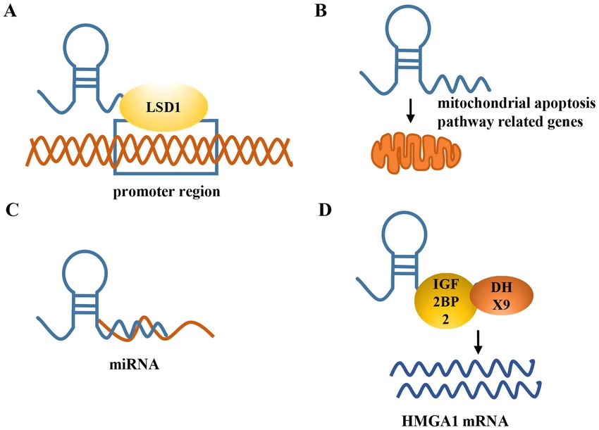
Figure 2. Mechanistic model of HIF1A‑AS2 in cancer. (A) HIF1A‑AS2 recruits LSD1 to the promoter of the target gene and regulates gene transcription
at the epigenetic level. (B) HIF1A‑AS2 can regulate expression of mitochondrial apoptosis pathway‑related genes. (C) HIF1A‑AS2 can function as a com‑
peting endogenous RNA to sponge miRNA in cancer. (D) HIF1A‑AS2 can bind to IGF2BP2 and DHX9 to modulate the expression of HMGA1. HIF1A‑AS,
hypoxia‑inducible factor 1α antisense RNA; LSD1, lysine‑specific demethylase 1; IGF2BP2, insulin‑like growth factor 2; DHX9, ATP‑dependent RNA
helicase A; HMGA1, high mobility group AT‑hook 1; miRNA, microRNA.
weight and volume in nude mice. In addition, it was found that that HIF1A‑AS2 functions as an oncogene. Mechanistically, a
HIF1A‑AS2 had value in the early diagnosis of GC and could HIF1A‑AS2/miR‑548c‑3p/HIF1a/VEGF axis was confirmed to
be used as a potential diagnostic marker for detection of GC. regulate the proliferation, invasion, migration and EMT of BC
Therefore, HIF1A‑AS2 is a potential tumorigenic gene in GC, cells. Jiang et al (31) also reported that expression of HIF1A‑AS2
but its molecular mechanisms have not been studied, to the was increased in 33 TNBC tissues compared with the adjacent
best of our knowledge. normal breast tissues. Knockdown of HIF1A‑AS2 functionally
suppressed TNBC cell proliferation. These results indicated
BC. BC is the most common malignancy and the leading cause of that HIF1A‑AS2 was involved in the pathogenesis of TNBC,
cancer‑related death in women (27). Breast cancer tumors usually suggesting that it could be a prognostic indicator or therapeutic
express a combination of the following receptors: Estrogen target for TNBC.
receptor (ER), progesterone receptor (PR) and human epidermal
growth factor receptor (HER2). Cases that lack expression of Bladder cancer. Bladder cancer is one of the most common
these three receptors are termed triple‑negative breast cancer malignancies of the urinary system worldwide, posing a severe
(TNBC). TNBC accounts for ~20% of all breast cancer cases, ad threat to human health (32). Surgery, radiotherapy and chemo‑
is most common in women >40 (28). TNBC is highly invasive, therapy are the primary modes of treatment for bladder cancer;
with high mortality and recurrence rates. Current treatments for however, the 5‑year overall survival rate is only 50‑60% (33).
TNBC include surgery, chemotherapy, radiotherapy and targeted Although several studies have demonstrated novel biomarkers
therapy. However, the median overall survival rarely extends for the early detection and diagnosis of bladder cancer, the
beyond 18 months in patients with advanced BC (29). Therefore, survival rate of patients with bladder cancer remains very
it is essential to study the molecular mechanism and identify low (34). Therefore, it is necessary to identify novel biomarkers
novel biomarkers for management of TNBC. Guo et al (30) to improve the early diagnosis and prognosis of bladder cancer.
showed that HIF1A‑AS2 was significantly overexpressed in Chen et al (35) revealed that the expression of HIF1A‑AS2 was
four BC cell lines compared with a normal mammary epithelial significantly upregulated in 44 bladder cancer samples and
cell line. Knockdown of HIF1A‑AS2 levels effectively suppress cancer cell lines (5637 and T24) compared with the matched
proliferation, invasion, EMT and senescence of MCF‑7 cell normal peritumoral tissues or the SVHUC‑1 normal bladder
lines in‑vitro. In vivo studies also showed that tumor growth cell line. In addition, the upregulated HIF1A‑AS2 expres‑
was reduced after the knockdown of HIF1A‑AS2 by short sion was closely related to histological grade, tumor invasion
hairpin (sh)RNA targeting HIF1A‑AS2 in‑vivo, thus indicating depth and TNM stage. These results indicated that lncRNABIOMEDICAL REPORTS 15: 85, 2021 5
HIF1A‑AS2 may function as an oncogene in bladder cancer. new therapeutic methods and targets are required. lncRNAs are
Functionally, knockdown of HIF1A‑AS2 significantly inhibited involved in the development of GBM. Mineo et al (43) reported
bladder cancer cell proliferation and migration, and increased that HIF1A‑AS2 contributes to the formation of stem‑like
apoptosis. Conversely, overexpression of HIF1A‑AS2 had the glioma cells (GSCs) in the tumor microenvironment and their
opposite effect. adaptation to hypoxia. Based on characterization of the GBM
Furthermore, a tetracycline‑induced shRNA using medical genome and transcriptome, GBM can be divided into several
synthetic biology techniques was designed, which could effec‑ cellular subtypes, including mesenchymal (M), proneural (P),
tively inhibit the expression of HIF1A‑AS2 in a dose‑dependent neural (N), and classical (C) (44). Patients with the aggres‑
manner, and in turn inhibited cell growth and migration, and sive and predominant M subtype exhibit a particularly high
induced apoptosis in bladder cancer cells. It also indicated degree of tumor necrosis (45). It was observed that HIF1A‑AS2
that tetracycline‑induced shRNA may be a novel approach for expression was significantly increased in the GSCs of patients
quantitatively controlling specific targets in human cancers, with the M subtype. Moreover, knockdown of HIF1A‑AS2 led
and may be an effective treatment method for bladder cancer. to reduced growth, decreased cellular activity and decreased
Thus, HIF1A‑AS2 may serve as a target for the treatment neurosphere‑forming capacity of M GSC cells (43).
of bladder cancer; however, the exact molecular regulatory Furthermore, the HIF1A‑AS2 expression is increased under
mechanisms in bladder cancer require further study. hypoxic conditions. In order to clarify the pro‑oncogenic func‑
tion of HIF1A‑AS2, researchers revealed that knockdown of
OS. OS is a skeletal system primary malignant tumor, common HIF1A‑AS2 by shRNA resulted in smaller tumor sizes in nude
amongst the younger population, particularly children and mice. Mechanistic experiments showed that HIF1A‑AS2 could
adolescents (36,37). OS accounts for 60% of all sarcoma cases, bind to IGF2BP2 and DHX9 to directly modulate the expression
characterized by early metastasis, high aggressiveness, a high of HMGA1 (Fig. 2D) and maintain the growth of M GSCs under
rate of disability and a high recurrence rate (38). Despite hypoxic conditions (43). In addition, Liao et al (46) showed that
advances in OS treatment, the overall survival of patients has the upregulated HIF1A‑AS2 expression could mediate radiation
not substantially increased, the 5‑year overall survival still resistance of the glioma, leading to tumor recurrence following
remains only 20% over the past 30 years (39). Thus, under‑ radiotherapy by regulating expression of apoptotic proteins.
standing the molecular mechanism of OS and identifying Knockdown of HIF1A‑AS2 increased the expression of the
novel therapeutic targets is of great clinical significance to pro‑apoptotic protein caspase 7 and the number of apoptotic
improve early diagnosis and survival rates of patients with cells. Thus, HIF1A‑AS2 may be a novel diagnostic indicator and
OS. Lin et al (9) observed increased HIF1A‑AS2 expression potential therapeutic target for the management of GBM.
in 60 OS samples and four OS cell lines when compared
with the 60 adjacent normal samples or the hFOB 1.19 cells, RCC. RCC is one of the most common malignancies of the urinary
respectively. In addition, high expression of HIF1A‑AS2 was system, and accounts for 2‑3% of all malignancies (47,48). The
significantly associated with a larger tumor size, higher tumor estimated number of new cases and deaths worldwide in 2018
grade, advanced stage disease and distance of metastasis. were 403,262 and 175,098, respectively (49). Relatively fewer
Furthermore, Kaplan‑Meier survival analysis showed that biomarkers for RCC have been identified when compared with
the 5‑year survival rate of the high HIF1A‑AS2 expression other types of cancer. Thus, it is essential to identify novel and
group was lower than the low HIF1A‑AS2 expression group. sensitive biomarkers to predict the progress and prognosis of the
Knockdown of HIF1A‑AS2 resulted in decreased cell prolif‑ disease. In 1999, Thrash‑Bingham and Tartof (6) first discov‑
eration, migration and invasion, increased cell cycle arrest in ered a natural antisense transcript that could bind to the 3'UTR
the G0/G1‑phase and an increased percentage of apoptotic of HIF1α mRNA in non‑papillary kidney cancer and termed it
cells. In in‑vivo experiments, knockdown of HIF1A‑AS2 aHIF, for which the official gene symbol is now HIF1A‑AS2.
resulted in reduced tumor size in nude mice. Mechanistically, Expression of HIF1A‑AS2 is increased in non‑papillary
HIF1A‑AS2/miR‑33b‑5p/SIRT6 was confirmed to regulate renal carcinoma cells compared with the control cells, but
OS cell proliferation, migration and apoptosis. Wang et al (40) not in papillary renal carcinoma cells. It is hypothesized that
also confirmed increased expression of HIF1A‑AS2 in 30 OS decreased HIF1a mRNA expression through HIF1A‑AS2 may
samples and four OS cell lines compared with the adjacent serve an important role in regulating P53 to regulate progression
normal tissues and osteoblast cell lines, respectively. Moreover, of cancer, but this mechanism requires further investigation (6).
high HIF1A‑AS2 expression was associated with poor survival Zhu et al (50) also reported increased expression of HIF1A‑AS2
rates. Functional assays revealed that HIF1A‑AS2 overexpres‑ in kidney cancer tissues and RCC cells compared with the
sion promoted osteosarcoma cell proliferation, cell cycle non‑cancerous tissues. In addition, knockdown of HIF1A‑AS2
progression and invasion. HIF1A‑AS2 mechanistically served inhibited renal cancer cell proliferation, invasion and migration,
as a ceRNA to negatively regulate miR‑129–5p (Fig. 2C). whilst accelerating cell apoptosis. Overexpression of HIF1A‑AS2
Thus, HIF1A‑AS2 may be an effective diagnostic and prog‑ resulted in the opposite effect. HIF1A‑AS2 mechanistically
nostic indicator of OS. functions as a ceRNA, binding to miR‑130a‑5p (Fig. 2C) to
modulate renal carcinoma progression. Thus, HIF1A‑AS2 may
GBM. GBM is the most common and aggressive primary be a promising diagnostic biomarker and a potential therapeutic
malignant brain tumor, with a median patient survival time of target for management of renal cancer.
14‑16 months (41). GBM is a highly proliferative and invasive
tumor with a poor prognosis. Despite advances in GBM treatment, NSCLC. Lung cancer is the most common type of cancer and
patients are still likely to face a poor prognosis (42). Therefore, the leading cause of cancer‑associated death worldwide. The6 LIU et al: lncRNA HIF1A-AS2 AND HUMAN CANCER
majority of patients are diagnosed with advanced stage disease Availability of data and materials
in the first instance, and NSCLC accounts for nearly 85% of
patients with lung cancer (51). Despite advances in cancer Not applicable.
treatment, lung cancer has a high mortality rate, accounting
for 18.4% of all cancer deaths (52,53). Thus, understanding the Authors' contributions
molecular mechanism of NSCLC and identifying novel thera‑
peutic targets is of great clinical significance. Zhang et al (54) YLi conceived and designed the study. YLiu, YZ and CC
reported elevated expression levels of HIF1A‑AS2 in NSCLC participated in drafting and revising the article. All authors
tissues and cell lines, and this increased expression was have read and approved the final manuscript. Data authentica‑
associated with a poor prognosis. However, knockdown of tion is not applicable.
HIF1A‑AS2 resulted in decreased cell proliferation, migration
and invasion, and an increased percentage of apoptotic cells. Ethics approval and consent to participate
Mechanistically, a HIF1A‑AS2/miR‑153–5p/S100A14 axis
was confirmed to regulate NSCLC cell proliferation, migra‑ Not applicable.
tion and apoptosis (Fig. 2C). Thus, HIF1A‑AS2 may be an
effective diagnostic and prognostic indicator for NSCLC. Patient consent for publication
3. Conclusions and future perspective Not applicable.
A wealth of studies have shown that lncRNAs exert their Competing interests
functions through various mechanisms, such as associating
with transcription factors, chromatin modifiers, signaling The authors declare that they have no competing interests.
adapters, enzymes and miRNAs, to influence gene expression,
post‑translational modifications and protein activities (55). References
HIF1A‑AS2 has been reported to regulate cellular
pathological processes, but is primarily focused on tumors. 1. Slack FJ and Chinnaiyan AM: The role of non‑coding RNAs in
HIF1A‑AS2 is primarily functions as a protein scaffold, protein oncology. Cell 179: 1033‑1055, 2019.
2. Esteller M: Non‑coding RNAs in human disease. Nat Rev
decoy and a ceRNA. Mineo et al (43) reported that HIF1A‑AS2 Genet 12: 861‑874, 2011.
acts as a protein scaffold to bind both IGF2BP2 and DHX9 to 3. Khandelwal A, Bacolla A, Vasquez KM and Jain A: Long non‑coding
RNA: A new paradigm for lung cancer. Mol Carcinog 54: 1235‑1251,
modulate the expression of HMGA1. Wu et al (14) reported 2015.
that HIF1A‑AS2 functions as a protein decoy to inhibit the 4. Mercer TR, Dinger ME and Mattick JS: Long non‑coding RNAs:
transcription of PHLDA1 by binding to LSD1, a histone Insights into functions. Nat Rev Genet 10: 155‑159, 2009.
5. Bhat SA, Ahmad SM, Mumtaz PT, Malik AA, Dar MA, Urwat U,
demethylase. Additionally, HIF1A‑AS2 acts as a molecular Shah RA and Ganai NA: Long non‑coding RNAs: Mechanism of
sponge to bind miRNAs to further affect expression of other action and functional utility. Noncoding RNA Res 1: 43‑50, 2016.
genes (23,40,50,54,56). Although significant achievements 6. Thrash‑Bingham CA and Tartof KD: aHIF: A natural antisense
transcript overexpressed in human renal cancer and during
have been obtained with regard to understanding the role of hypoxia. J Natl Cancer Inst 91: 143‑151, 1999.
HIF1A‑AS2 in various types of cancer, further studies are still 7. Rossignol F, Vaché C and Clottes E: Natural antisense transcripts
required with regard to its regulatory function, as lncRNAs of hypoxia‑inducible factor 1alpha are detected in different
normal and tumour human tissues. Gene 299: 135‑140, 2002.
often exhibit several complex regulatory functions/mechanisms. 8. Chen WM, Huang MD, Kong R, Xu TP, Zhang EB, Xia R,
Studies have shown that HIF1A‑AS2 may serve as a Sun M, De W and Shu YQ: Antisense long noncoding RNA
novel biomarker for the clinical diagnosis of several types of HIF1A‑AS2 is upregulated in gastric cancer and associated with
poor prognosis. Dig Dis Sci 60: 1655‑1662, 2015.
cancer. These data demonstrate that upregulated expression of 9. Lin H, Zhao Z, Hao Y, He J and He J: Long noncoding RNA
HIF1A‑AS2 is associated with poor overall survival and an unfa‑ HIF1A‑AS2 facilitates cell survival and migration by sponging
vorable prognosis, such as in TNBC, OS and CRC. Nevertheless, miR‑33b‑5p to modulate SIRT6 expression in osteosarcoma.
Biochem Cell Biol 98: 284‑292, 2020.
the clinical diagnostic value of HIF1A‑AS2 in these types of 10. Wu P, Haththotuwa R, Kwok CS, Babu A, Kotronias RA, Rushton C,
cancer needs to be validated using large‑scale multicenter cohorts. Zaman A, Fryer AA, Kadam U, Chew‑Graham CA, et al:
Preeclampsia and future cardiovascular health: A systematic
review and meta‑analysis. Circ Cardiovasc Qual Outcomes 10:
Acknowledgements e003497, 2017.
11. Phipps EA, Thadhani R, Benzing T and Karumanchi SA:
Pre‑eclampsia: Pathogenesis, novel diagnostics and therapies
Not applicable. (vol 15, pg 275, 2019). Nat Rev Nephrol 15: 386‑386, 2019.
12. Irminger‑Finger I, Jastrow N and Irion O: Preeclampsia: A danger
Funding growing in disguise. Int J Biochem Cell Biol 40: 1979‑1983, 2008.
13. Ma Y, Liang X, Wu H, Zhang C and Ma Y: Long non coding
RNA NR_002794 is upregulated in pre eclampsia and regulates
This study was supported by grants from The National Natural the proliferation, apoptosis and invasion of trophoblast cells. Mol
Science Fund of China (grant no. 81601736); Science and Med Rep 20: 4567‑4575, 2019.
14. Wu D, Yang N, Xu Y, Wang S, Zhang Y, Sagnelli M, Hui B,
Technology Planning Project of Panyu District, China (grant Huang Z and Sun L: lncRNA HIF1A antisense RNA 2 modulates
no. 2019-Z04-85); Medical Scientific Research Foundation of trophoblast cell invasion and proliferation through upregulating
Guangdong Province, China (grant no. A2020560); Basic and PHLDA1 expression. Mol Ther Nucleic Acids 16: 605‑615, 2019.
15. Tang X, Liu S, Liu Y, Lin X, Zheng T, Liu X, Qiu J and Hua K:
Applied Research Project of Guangzhou Research Program, Circulating serum exosomal aHIF is a novel prognostic predictor for
China ( grant no. 202102080539). epithelial ovarian cancer. Onco Targets Ther 12: 7699‑7711, 2019.BIOMEDICAL REPORTS 15: 85, 2021 7
16. Guler E, Smith DA, Somarouthu B, Gujrathi R, Ramaiya NH 37. Jiang N, Wang X, Xie X, Liao Y, Liu N, Liu J, Miao N, Shen J
and Tirumani SH: Overview of imaging findings associated with and Peng T: lncRNA DANCR promotes tumor progression and
systemic therapies in advanced epithelial ovarian cancer. Abdom cancer stemness features in osteosarcoma by upregulating AXL
Radiol (NY) 45: 828‑841, 2020. via miR‑33a‑5p inhibition. Cancer Lett 405: 46‑55, 2017.
17. Lheureux S, Braunstein M and Oza AM: Epithelial ovarian 38. Zhang CL, Zhu KP and Ma XL: Antisense lncRNA FOXC2‑AS1
cancer: Evolution of management in the era of precision promotes doxorubicin resistance in osteosarcoma by increasing
medicine. CA Cancer J Clin 69: 280‑304, 2019. the expression of FOXC2. Cancer Lett 396: 66‑75, 2017.
18. Qiu JJ, Lin XJ, Zheng TT, Tang XY and Hua KQ: Natural 39. Zhong L, Liao D, Li JJ, Liu WQ, Wang JX, Zeng CL, Wang X,
antisense transcript of hypoxia‑inducible factor 1 regulates Cao ZL, Zhang RH, Li M, et al: Rab22a‑NeoF1 fusion protein
hypoxic cell apoptosis in epithelial ovarian cancer. Onco Targets promotes osteosarcoma lung metastasis through its secretion into
Ther 11: 9101‑9110, 2018. exosomes. Signal Transduct Target Ther 6: 59, 2021
19. Yang J, McDowell A, Kim EK, Seo H, Lee WH, Moon CM, 40. Wang X, Peng L, Gong X, Zhang X and Sun R: lncRNA
Kym SM, Lee DH, Park YS, Jee YK, et al: Development of a HIF1A‑AS2 promotes osteosarcoma progression by acting as a
colorectal cancer diagnostic model and dietary risk assessment sponge of miR‑129‑5p. Aging (Albany NY) 11: 11803‑11813, 2019.
through gut microbiome analysis. Exp Mol Med 51: 1‑15, 2019.
20. Arnold M, Sierra MS, Laversanne M, Soerjomataram I, Jemal A 41. Tang C, Wang Y, Zhang L, Wang J, Wang W, Han X, Mu C and
and Bray F: Global patterns and trends in colorectal cancer Gao D: Identification of novel lncRNA targeting Smad2/PKCα
incidence and mortality. Gut 66: 683‑691, 2017. signal pathway to negatively regulate malignant progression of
21. Yuan W, Peng S, Wang J, Wei C, Ye Z, Wang Y, Wang M, Xu H, glioblastoma. J Cell Physiol 235: 3835‑3848, 2020.
Jiang S, Sun D, et al: Identification and characterization of 42. Ji J, Xu R, Ding K, Bao G, Zhang X, Huang B, Wang X,
circRNAs as competing endogenous RNAs for miRNA‑mRNA Martinez A, Wang X, Li G, et al: Long Noncoding RNA
in colorectal cancer. PeerJ 7: e7602, 2019. SChLAP1 forms a growth‑promoting complex with HNRNPL
22. Dong Y, Zhang Y, Kang W, Wang G, Chen H, Higashimori A, in human glioblastoma through stabilization of ACTN4 and acti‑
Nakatsu G, Go M, Tong JH, Zheng S, et al: VSTM2A suppresses vation of NF‑κ B signaling. Clin Cancer Res 25: 6868‑6881, 2019.
colorectal cancer and antagonizes Wnt signaling receptor LRP6. 43. Mineo M, Ricklefs F, Rooj AK, Lyons SM, Ivanov P, Ansari KI,
Theranostics 9: 6517‑6531, 2019. Nakano I, Chiocca EA, Godlewski J and Bronisz A: The Long
23. Lin J, Shi Z, Yu Z and He Z: lncRNA HIF1A‑AS2 positively Non‑coding RNA HIF1A‑AS2 facilitates the maintenance of
affects the progression and EMT formation of colorectal mesenchymal glioblastoma stem‑like cells in hypoxic niches.
cancer through regulating miR‑129‑5p and DNMT3A. Biomed Cell Rep 15: 2500‑2509, 2016.
Pharmacother 98: 433‑439, 2018. 44. Wang L, Babikir H, Müller S, Yagnik G, Shamardani K, Catalan F,
24. Fan H, Jin X, Liao C, Qiao L and Zhao W: MicroRNA‑301b‑3p Kohanbash G, Alvarado B, Di Lullo E, Kriegstein A, et al: The
accelerates the growth of gastric cancer cells by targeting zinc phenotypes of proliferating glioblastoma cells reside on a single
finger and BTB domain containing 4. Pathol Res Pract 215: axis of variation. Cancer Discov 9: 1708‑1719, 2019.
152667, 2019. 45. Bhat KPL, Balasubramaniyan V, Vaillant B, Ezhilarasan R,
25. Wan P, Bai X, Yang C, He T, Luo L, Wang Y, Fan M, Wang Z, Hummelink K, Hollingsworth F, Wani K, Heathcock L, James JD,
Lu L, Yin Y, et al: miR‑129‑5p inhibits proliferation, migration, Goodman LD, et al: Mesenchymal differentiation mediated by
and invasion in rectal adenocarcinoma cells through targeting NF‑κ B promotes radiation resistance in glioblastoma. Cancer
E2F7. J Cell Physiol 235: 5689‑5701, 2020. Cell 24: 331‑346, 2013.
26. Wang LL, Zhang L and Cui XF: Downregulation of long 46. Liao K, Ma X, Chen B, Lu X, Hu Y, Lin Y, Huang R and Qiu Y:
noncoding RNA LINC01419 inhibits cell migration, invasion, Upregulated AHIF‑mediated radioresistance in glioblastoma.
and tumor growth and promotes autophagy via inactivation of
the PI3K/Akt1/mTOR pathway in gastric cancer. Ther Adv Med Biochem Biophys Res Commun 509: 617‑623, 2019.
Oncol 11: 1758835919874651, 2019. 47. Kovacs G, Wilkens L, Papp T and de Riese W: Differentiation
27. Lee KL, Kuo YC, Ho YS and Huang YH: Triple‑negative breast between papillary and nonpapillary renal cell carcinomas by
cancer: Current understanding and future therapeutic break‑ DNA analysis. J Natl Cancer Inst 81: 527‑530, 1989.
through targeting cancer Stemness. Cancers (Basel) 11: 1334, 2019. 48. Korenaga Y, Naito K, Okayama N, Hirata H, Suehiro Y,
28. Khan MA, Jain VK, Rizwanullah M, Ahmad J and Jain K: Hamanaka Y, Matsuyama H and Hinoda Y: Association of
PI3K/AKT/mTOR pathway inhibitors in triple‑negative breast the BCRP C421A polymorphism with nonpapillary renal cell
cancer: A review on drug discovery and future challenges. Drug carcinoma. Int J Cancer 117: 431‑434, 2005.
Discov Today 24: 2181‑2191, 2019. 49. Zhang C, Huang D, Liu A, Xu Y, Na R and Xu D: Genome‑wide
29. Song N, Zhao L, Zhu M and Zhao J: 99mTc‑Labeled LyP‑1 for screening and cohorts validation identifying novel lncRNAs as
SPECT Imaging of Triple Negative Breast Cancer. Contrast prognostic biomarkers for clear cell renal cell carcinoma. J Cell
Media Mol Imaging 2019: 9502712, 2019. Biochem 121: 2559‑2570, 2020.
30. Guo X, Lee S and Cao P: The inhibitive effect of sh‑HIF1A‑AS2 50. Zhu Y, Yang Z, Chen H, Pan Y, Gong L, Chen F, Jin X, Wen S, Li Y
on the proliferation, invasion, and pathological damage of and Chen G: lncRNAHIF1A‑AS2 promotes renal carcinoma cell
breast cancer via targeting miR‑548c‑3p through regulating proliferation and migration via miR‑130a‑5p/ERBB2 pathway.
HIF‑1α/VEGF pathway in vitro and vivo. Onco Targets Ther 12: Onco Targets Ther 13: 9807‑9820, 2020.
825‑834, 2019. 51. The Lancet: Lung cancer: Some progress, but still a lot more to
31. Jiang YZ, Liu YR, Xu XE, Jin X, Hu X, Yu KD and Shao ZM: do. The Lancet 394: 1880, 2019.
Transcriptome Analysis of triple‑negative breast cancer reveals 52. Ramalingam SS, Owonikoko TK and Khuri FR: Lung cancer:
an integrated mRNA‑lncRNA signature with predictive and New biological insights and recent therapeutic advances. CA
prognostic value. Cancer Res 76: 2105‑2114, 2016. Cancer J Clin 61: 91‑112, 2011.
32. Yu G, Zhou H, Yao W, Meng L and Lang B: lncRNA TUG1 53. Dawson Q: NELSON trial: Reduced lung‑cancer mortality with
promotes cisplatin resistance by regulating CCND2 via epige‑ volume CT screening. Lancet Respir Med 8: 236‑236, 2020.
netically silencing miR‑194‑5p in bladder cancer. Mol Ther 54. Zhang W, Liu K, Pei Y, Tan J, Ma J and Zhao J: Long noncoding
Nucleic Acids 16: 257‑271, 2019. RNA HIF1A‑AS2 promotes non‑small cell lung cancer progression
33. Miao L, Liu HY, Zhou C and He X: LINC00612 enhances the by the miR‑153‑5p/S100A14 axis. Onco Targets Ther 13: 8715‑8722,
proliferation and invasion ability of bladder cancer cells as
ceRNA by sponging miR‑590 to elevate expression of PHF14. 2020.
J Exp Clin Cancer Res 38: 143, 2019. 55. Wang P: The Opening of Pandora's Box: An emerging role of long
34. Zhuang C, Ma Q, Zhuang C, Ye J, Zhang F and Gui Y: lncRNA noncoding RNA in viral infections. Front Immunol 9: 3138, 2019.
GClnc1 promotes proliferation and invasion of bladder cancer 56. Mu L, Wang Y, Su H, Lin Y, Sui W, Yu X and Lv Z: HIF1A‑AS2
through activation of MYC. FASEB J 33: 11045‑11059, 2019. promotes the proliferation and metastasis of gastric cancer cells
35. Chen M, Zhuang C, Liu Y, Li J, Dai F, Xia M, Zhan Y, Lin J, through miR‑429/PD‑L1 axis. Dig Dis Sci Feb 8, 2021 (Epub
Chen Z, He A, et al: Tetracycline‑inducible shRNA targeting ahead of print). doi: 10.1007/s10620-020-06819-w.
antisense long non‑coding RNA HIF1A‑AS2 represses the
malignant phenotypes of bladder cancer. Cancer Lett 376:
155‑164, 2016.
36. Qi X, Yu XJ, Wang XM, Song TN, Zhang J, Guo XZ, Li GJ This work is licensed under a Creative Commons
and Shao M: Knockdown of KCNQ1OT1 suppresses cell Attribution-NonCommercial-NoDerivatives 4.0
invasion and sensitizes osteosarcoma cells to CDDP by upregu‑ International (CC BY-NC-ND 4.0) License.
lating DNMT1‑mediated Kcnq1 expression. Mol Ther Nucleic
Acids 17: 804‑818, 2019.You can also read