Tea Tree Oil Prevents Mastitis-Associated Inflammation in Lipopolysaccharide-Stimulated Bovine Mammary Epithelial Cells
←
→
Page content transcription
If your browser does not render page correctly, please read the page content below
ORIGINAL RESEARCH
                                                                                                                                             published: 07 August 2020
                                                                                                                                        doi: 10.3389/fvets.2020.00496

                                             Tea Tree Oil Prevents
                                             Mastitis-Associated Inflammation in
                                             Lipopolysaccharide-Stimulated
                                             Bovine Mammary Epithelial Cells
                                             Zhi Chen 1,2† , Yi Zhang 1† , Jingpeng Zhou 1† , Lu Lu 1 , Xiaolong Wang 1,2 , Yusheng Liang 3 ,
                                             Juan J. Loor 3 , Deming Gou 4 , Huifen Xu 5* and Zhangping Yang 1,2*
                                             1
                                              College of Animal Science and Technology, Yangzhou University, Yangzhou, China, 2 Joint International Research Laboratory
                                             of Agriculture & Agri-Product Safety, Ministry of Education, Yangzhou University, Yangzhou, China, 3 Mammalian Nutrition
                                             Physiology Genomics, Division of Nutritional Sciences, Department of Animal Sciences, University of Illinois, Urbana, IL,
                                             United States, 4 College of Life Sciences, Shenzhen University, Shenzhen, Guangzhou, China, 5 College of Animal Science
                                             and Veterinary Medicine, Henan Agricultural University, Zhengzhou, Henan, China

                                             The main purpose of this study was to explore the effect of tea tree oil (TTO)
                          Edited by:
                           Haoyu Liu,        on lipopolysaccharide (LPS)-induced mastitis model using isolated bovine mammary
          Uppsala University, Sweden         epithelial cells (BMEC). This mastitis model was used to determine cellular responses
                        Reviewed by:         to TTO and LPS on cellular cytotoxicity, mRNA abundance and cytokine production.
                                Ying Yu,
                                             High-throughput sequencing was used to select candidate genes, followed by functional
   China Agricultural University, China
                          Runjun Yang,       evaluation of those genes. In the first experiment, LPS at a concentration of 200 µg/mL
                 Jilin University, China     reduced cell proliferation, induced apoptosis and upregulated protein concentrations of
                   *Correspondence:          tumor necrosis factor-α (TNF-α), interleukin 6 (IL-6), and signal transducer and activator
                             Huifen Xu
                   huifen221@126.com         of transcription 1 (STAT1). Addition of TTO led to reduced cellular apoptosis along
                        Zhangping Yang       with downregulated protein concentrations of nuclear factor kappa B, mitogen-activated
                        yzp@yzu.edu.cn
                                             protein kinase 4 (MAPK4) and caspase-3. In the second experiment, BMEC challenged
     † These   authors have contributed      with LPS had a total of 1,270 differentially expressed genes of which 787 were
                    equally to this work
                                             upregulated and 483 were downregulated. Differentially expressed genes included
                    Specialty section:
                                             TNF-α, IL6, STAT1, and MAPK4. Overall, results showed that TTO (at least in vitro)
         This article was submitted to       has a protective effect against LPS-induced mastitis. Further in vivo research should be
     Animal Nutrition and Metabolism,
                                             performed to determine strategies for using TTO for prevention and treatment of mastitis
                a section of the journal
        Frontiers in Veterinary Science      and improvement of milk quality.
             Received: 24 April 2020         Keywords: TTO, BMEC, LPS, mastitis, transcriptome sequencing
            Accepted: 30 June 2020
          Published: 07 August 2020

                            Citation:        INTRODUCTION
      Chen Z, Zhang Y, Zhou J, Lu L,
    Wang X, Liang Y, Loor JJ, Gou D,         Lipopolysaccharide (LPS) is one of the main components of the cell wall of gram-negative
  Xu H and Yang Z (2020) Tea Tree Oil        bacteria including Escherichia coli (E. coli) and other mastitis-inducing pathogenic bacteria such as
         Prevents Mastitis-Associated
                                             Staphylococcus aureus, Streptococcus agalactis, and Streptococcus lactis (1). In dairy cows, mastitis
                      Inflammation in
Lipopolysaccharide-Stimulated Bovine
                                             caused by E. coli results in increased concentrations of acute-phase proteins in milk (2, 3), and
            Mammary Epithelial Cells.        can be treated with antibiotics (4). However, with increasing concerns about drug resistance it has
               Front. Vet. Sci. 7:496.       become imperative to prevent usage of antibiotics and develop alternatives and treat cow mastitis
      doi: 10.3389/fvets.2020.00496          using alternative therapies.

Frontiers in Veterinary Science | www.frontiersin.org                              1                                               August 2020 | Volume 7 | Article 496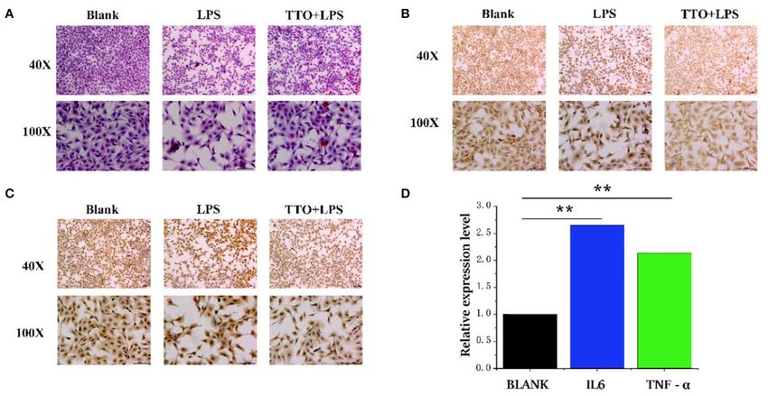
Chen et al.                                                                                              TTO Prevents Mastitis-Associated Inflammation

   Tea tree oil (TTO; terpinen-4-ol type), also known as M.                   CCK-8 Detection of Cell Proliferation
alternifolia oil, is an essential oil from several plants of Melaleuca,       Activity Induced by LPS
of which the main one is M. alternifolia (5). TTO is widely used              The density of BMEC was adjusted to 1 × 10⁴ in a 96-well
in many over-the-counter health products and cosmetics. With                  plate. After 24 h incubation, the culture medium was discarded.
the vigorous development of natural and alternative medicinals,               The BMEC were treated with LPS (50, 100, 200, 500, and
an increasing number of people are using products containing                  1,000 µg/mL). In addition, there was a control (BMEC without
TTO (6). TTO has a broad antibacterial spectrum and strong                    LPS) and a blank group (only culture medium without cells).
antibacterial activity, which explains its use to treat diseases              After 4, 8, 12, and 24 h incubation, cell proliferation activity
caused by fungi, bacteria, or viruses (7). Therefore, its potential           was detected using a CCK-8 kit (Watson Technology Co., Ltd.,
use as a natural antibacterial agent to replace antibiotics as a              Beijing, China) according to the manufacturer’s protocols.
component of mastitis therapy is of interest.
   With the development of sequencing and histochemistry
technology, analysis of the complex pathogenesis of mastitis                  Detection of Apoptosis Rate Induced by
in dairy cows from multiple perspectives can be performed.                    LPS via Flow Cytometry
More importantly, an integrative approach aids in effective                   The BMEC were plated in a 6-well plate and incubated for
biomarkers for timely and accurate prevention (8). Although                   24 h. Cells were then washed and collected with PBS, and cell
numerous studies have reported alterations of mRNA abundance                  concentration adjusted with buffer to 1 × 10⁶ /100 µL/test. Then,
in the mammary gland in response to mastitis, the role of                     5 µL annexin V-FITC and 5 µL PI were added, and cell apoptosis
gene transcription along with the complex networks and how                    determined in a dark room.
they respond to therapeutic agents is still unclear. For instance,
microRNA expression was first confirmed during mastitis in
                                                                              Effect of TTO on Apoptosis Rate During
2007 (9). Naeem et al. detected changes in 14 miRNA in
mammary tissue 12 h after infection with Streptococcus uberis.                LPS Challenge via Flow Cytometry
Compared with healthy tissue, expression of miR-15b, miR-                     The BMEC were plated in a 6-well plate and cultured for 24 h.
16a, miR-21, miR-145, and miR-181a was lower, and only miR-                   LPS and various concentrations of TTO (Yuanye biology Co.,
223 was greater in infected mammary tissue. The miR-16a was                   Ltd., Shanghai, China) were added to the culture (0.0002, 0.0004,
decreased of some interleukins (IL-6, IL-8, and IL-10). The                   0.0006, 0.0008, 0.001, 0.002, 0.004, 0.006, 0.008, and 0.01%,
present study aimed to use transcriptome technology to uncover                vol/vol). Annexin V-FITC and PI were added for detection
the response of bovine mammary epithelial cells (BMEC) to                     of apoptosis.
LPS as a way to identify key candidate genes that could be
target for functional verification. Along with other assays, a                Abundance of Inflammation- and
combined technological approach can provide precise targets
                                                                              Apoptosis-Related Proteins via ELISA
for research and development of effective therapeutic drugs,
                                                                              After washing with PBS, RIPA buffer was added to the cell
ultimately achieving positive effects in terms of prevention and
                                                                              lysate. Bovine nuclear factor kappa B (NF-κB), mitogen-activated
treatment (10).
                                                                              protein kinase 4 (MAPK4), tumor necrosis factor-α (TNF-
                                                                              α), interleukin 6 (IL-6), signal transducer and activator of
MATERIALS AND METHODS                                                         transcription 1 (STAT1), and apoptosis-related caspase-3 were
                                                                              determined according to protocols supplied with the ELISA kits
Ethics Statement                                                              (Qiaoshe Co., Shanghai, China).
The animal use protocol was approved by the Institutional
Animal Care and Use Committee in the College of Animal
Science and Technology, Yang Zhou University, Yang                            Transcriptome Sequencing
Zhou, China.                                                                  Library Construction
                                                                              Total RNA was extracted from BMEC (number of cells is 1
Culture of BMEC                                                               × 107 ) treated with 200 µg/mL LPS for 12 h. After total RNA
Three peak lactation dairy cows were selected for mammary                     was extracted and digested with DNase, eukaryotic mRNA
gland biopsy (11). After PBS washing, fat tissue and connective               was enriched with oligo (dT) using magnetic beads. A strand
tissue were peeled off. The BMEC were separated by the                        of cDNA was synthesized with random hexamers using the
tissue block method followed purification by differential                     interrupted mRNA as template. Double-stranded cDNA was
digestion and cryopreservation after subculturing (11). Cells                 synthesized using the two-stranded synthesis system and purified
were cultured in Dulbecco’s modified Eagle medium/F12                         followed by poly-(A) addition and sequencing. The library was
(DMEM/F12) supplemented with 10% (vol/vol) fetal                              inspected for quality using the Agilent 2100 Bioanalyzer, and
bovine serum in a humidified incubator at 37◦ C with                          eventually sequenced with the Illumina hiseqtm 2500 sequencer
5% CO2 . Medium was replaced every 48 h. The BMEC                             (12). The raw data generated by high-throughput sequencing
were digested with 0.25% trypsin for passaging, and                           was in FASTQ format. To obtain high-quality reads, we first
the growth of cells was observed using an inverted                            used NGS QC Toolkit software to conduct quality control and
microscope (11).                                                              remove joints.

Frontiers in Veterinary Science | www.frontiersin.org                     2                                        August 2020 | Volume 7 | Article 496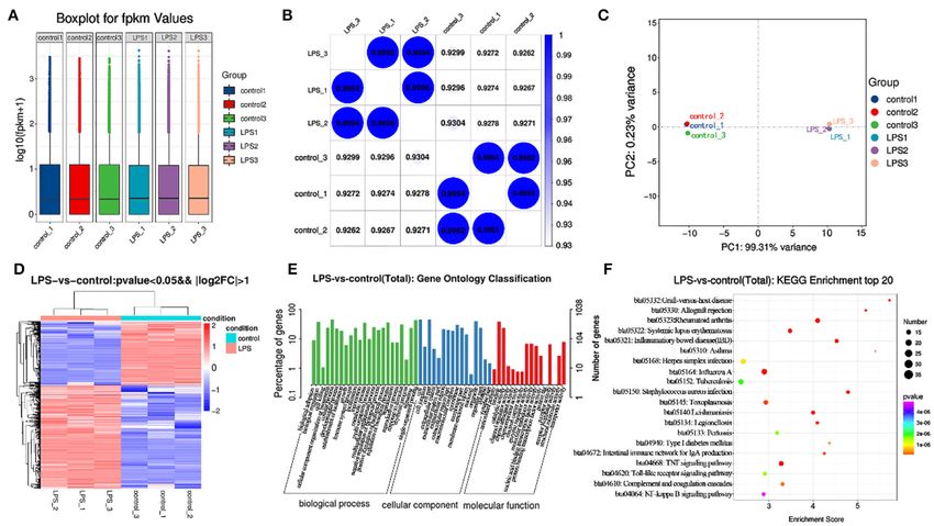
Chen et al.                                                                                                               TTO Prevents Mastitis-Associated Inflammation

Gene Quantification, Differential Gene Screening,                                       95, 85, and 75% ethanol for 5–10 min. The sections were soaked
Functional Enrichment, and Cluster Analysis                                             in distilled water for 5 min. For antigen retrieval, sections were
The comparison between clean reads and the reference genome                             incubated in citrate buffer (pH 6.0) and heated in a microwave
were stored in a binary file (BAM file). Genes were quantified                          at high heat for 8 min. Cells were then washed with 1× PBS (pH
to obtain the FPKM value using cufflinks. When calculating                              7.2∼7.6) three times for 3 min each time followed by addition
differences in gene expression, we used Htseq-count software                            of 3% H2 O2 at room temperature for 10 min to inactivate
to determine the number of gene reads in each sample. The                               endogenous peroxidases. Slides were rinsed with 1× PBS three
estimate SizeFactors function in the DESeq R package was                                times, 3 min each. Slides were incubated with primary antibodies
used to standardize the data, and the nbinomTest function                               (BV20932, Qiaoshe company, Shanghai, China) followed by
was used to calculate the P-value and fold-change values in                             a secondary antibody (BV30796, Qiaoshe company, Shanghai,
the difference comparisons (13). The condition used to screen                           China) in a box at 37◦ C for 1.5 h. Slides were washed 3 times
differentially expressed genes was a P < 0.05 and multiple                              with 1× PBS for 5 min each. A streptavidin-HRP antibody was
differences was more than 2 times. Gene ontology (GO) and                               incubated with the slides at 37◦ C for 20 min. Slides were then
Kyoto Encyclopedia of Genes and Genomes (KEGG) enrichment                               covered with 100 µL of the previously prepared color developer
analysis of differentially expressed genes were carried out to                          DAB working solution; the reaction time was monitored under
determine main biological functions or pathways.                                        the microscope.

H&E Staining of Cells                                                                   Statistical Analysis
Cell sections were deparaffinized in xylene, and slides hydrated                        Statistical analyses were performed by SPSS 19.0 (SPSS Inc.,
in 95% ethanol for 5 min, 85% ethanol for 5 min; slides were then                       Chicago, USA). The data are presented as mean values ±
hydrated in distilled water. Hematoxylin staining was performed                         s.d. from three independent experiments, duplicates. Statistical
for 3 min, and slides rinsed with distilled water for 2 min; 1%                         analysis was conducted using two-tailed unpaired Student’s t-test
hydrochloric acid alcohol was used for 2 s to differentiate the                         or one-way ANOVA with Bonferroni’s multiple comparisons test.
stain. The sections were rinsed with tap water for 15 min followed                      P < 0.05 was considered significant.
by 1–2 s of distilled water. Slides were stained with eosin for
30 s. Differentiation was determined according to the color, and
80% ethanol was used to differentiate stains. Slides were further                       RESULTS
dehydrated with 85% ethanol for 5 min, followed by 95% ethanol
for 5 min. Then, the slides were dehydrated with anhydrous                              CCK-8 Detection of Cell Proliferation
ethanol for 10 min. After the run off was transparent, slides                           Activity Induced by LPS
were sealed by adding a drop of neutral gum. Observation and                            As shown in Figure 1, cell proliferation activity of BMEC began
photography were performed with a microscope (Dmi4000b                                  to decline to varying degrees with 100 µg/mL LPS treatment
inverted fluorescence microscope, Leica, Germany).                                      for 12 h. As the activity of cells induced by LPS of 500 µg/mL
                                                                                        and 1,000 µg/mL was too low, we chose the challenge of LPS
Immunohistochemistry                                                                    concentration at 200 µg/mL for 12 h as the optimal treatment
Sections were dewaxed and hydrated followed by washing in                               condition for further analysis. Biological repeat is three times,
xylene twice for 10 min each. Slides were then incubated with 100,                      and technical repeat is two times.

 FIGURE 1 | Cell proliferation activity induced by LPS at different concentrations (0, 50, 100, 200, 500, and 1,000 µg/ml) and time points (0, 4, 8, 12, 24 h). Data were
 presented as means ± s.d. of at least three independent experiments.

Frontiers in Veterinary Science | www.frontiersin.org                               3                                                August 2020 | Volume 7 | Article 496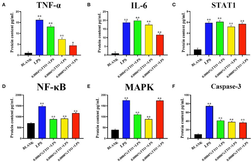
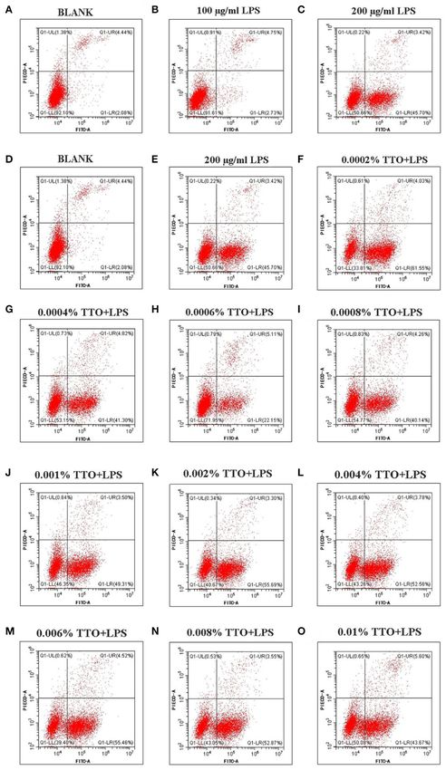
Chen et al.                                                                                                        TTO Prevents Mastitis-Associated Inflammation

 FIGURE 2 | Effect of different concentrations of TTO on apoptosis in BMEC with LPS challenge. (A) BLANK; (B) 100 µg/ml LPS; (C) 200 µg/ml LPS; (D) BLANK; (E)
 200 µg/mL LPS; (F) 0.0002% TTO+LPS; (G) 0.0004% TTO+LPS; (H) 0.0006% TTO+LPS; (I) 0.0008% TTO+LPS; (J) 0.001% TTO+LPS; (K) 0.002% TTO+LPS;
 (L) 0.004% TTO+LPS; (M) 0.006% TTO+LPS; (N) 0.008% TTO+LPS; (O) 0.01% TTO+LPS. Data were presented as means ± s.d. of at least three
 independent experiments.

Frontiers in Veterinary Science | www.frontiersin.org                          4                                             August 2020 | Volume 7 | Article 496
Chen et al.                                                                                                         TTO Prevents Mastitis-Associated Inflammation

Apoptosis of LPS-Induced BMEC                                                       0.0002% TTO+LPS, 0.0004% TTO+LPS, 0.0006% TTO+LPS,
Approximately 4.44% (4.44 ± 0.01) early apoptosis and late                          0.0008% TTO+LPS, 0.001% TTO+LPS, 0.002% TTO+LPS,
apoptosis were observed without LPS (Figure 2A). Upon                               0.004% TTO+LPS, 0.006% TTO+LPS, 0.008% TTO+LPS, and
addition of 100 µg/mL LPS, the whole image shifted to the                           0.01% TTO+LPS was 61.55% (61.55 ± 0.04), 41.30%(41.30 ±
right, and ∼7.48% (7.48 ± 0.02) [early apoptosis 2.73 (2.73 ±                       0.03, P < 0.05), 22.15% (22.15 ± 0.05, P
Chen et al.                                                                                                            TTO Prevents Mastitis-Associated Inflammation

 FIGURE 4 | Transcriptome analysis of control and LPS group. (A) Box line diagram in control and LPS group. The abscissa is the sample name and the ordinate is
 log10 (fpkm + 1). (B) Thermal diagram of the correlation coefficient between samples. The abscissa represents the name of the sample, and the ordinate represents
 the name of the corresponding sample. The color represents the size of the correlation coefficient. (C) PCA diagram in control and LPS group. (D) Screening for
 differentially expressed genes between control and LPS-induced BMEC. (E) GO enrichment. Horizontal axis is the GO entry name and the vertical axis is the –log10
 p-value. (F) KEGG enrichment, top 20 genes.

of TNF-α (P < 0.01) and IL-6 (P < 0.01), with a more                                 and dispersion also was deemed appropriate (Figure 4A,
pronounced effect on TNF-α. Expression of STAT1 increased                            GEO databases: SRR11862300, SRR11862301, SRR11862299,
slightly upon addition of 0.0004% TTO (P < 0.01). Protein                            SRR11862298, SRR11862297, SRR11862296).
concentrations of TNF-α, IL-6 and STAT1 were significantly                               The similarity of the LPS group was close to 1 (Figure 4),
downregulated with 0.0006% (P < 0.01) and 0.0008% (P <                               and that of the control was close to 1 (Figure 4B). Principal
0.01) TTO supplementation (Figures 3A–C). After addition of                          component analysis (PCA) indicated close concordance among
200 µg/mL LPS, the LPS group had a significant increase in                           samples in the LPS and control groups, underscoring the validity
protein concentrations of NF-κB (P < 0.01), MAPK4 (P < 0.01),                        of the data generated (Figure 4C).
and caspase-3 (P < 0.01) (Figures 3D–F). The protein expression                          A total of 1270 mRNAs were identified as differentially
levels of NF-κB (P < 0.01), MAPK4 (P < 0.01), and caspase-                           expressed, of which 787 genes were upregulated and 483
3 (P < 0.01) were significantly reduced in the groups treated                        downregulated. The differentially expressed genes included TNF-
with TTO.                                                                            α, IL6, STAT1, and MAPK4. Among these genes, TNF-α and IL6
                                                                                     were significantly upregulated. The difference multiples were 4.41
Transcriptome Analysis                                                               and 6.28 times, respectively (Figure 4D, Table S3).
After building LPS induced mastitis model, we want to study                              The GO annotation results indicated that differentially
its transcriptome level. Different genes were obtained by                            expressed mRNAs participate in biological adhesion, biological
high-throughput sequencing analysis to provide data support                          regulation, cell killing, cellular component organization or
for subsequent research. RNA-seq was used to sequence the                            biogenesis, cellular process, developmental process, growth,
LPS (200 µg/ml) induced model for 12 h. Considering the                              immune system process, negative regulation of biological
potential impact of the data error rate on the results, we used                      process, positive regulation of biological process, and cell
trimmatomatic software to preprocess the quality of the original                     junction among others (Figure 4E).
data and to generate a statistical summary of the number of                              Among the top 20 KEGG pathway entries, the differentially
reads in the whole quality control process (Table S1). Fpkm is                       expressed mRNAs participate in TNF signaling, rheumatoid
one of the most commonly-used methods to estimate expression                         arthritis, inflammatory, Staphylococcus aureus infection,
level of protein-coding genes (Table S2). The degree of symmetry                     systemic lupus erythematosus, graft-vs-host disease, allograft

Frontiers in Veterinary Science | www.frontiersin.org                            6                                               August 2020 | Volume 7 | Article 496
Chen et al. TTO Prevents Mastitis-Associated Inflammation
FIGURE 5 | Tea tree oil induced the expression changes of TNF-α and IL 6 in BMEC. (A) HE staining of BMEC in LPS (200 µg/ml) and TTO (0.008%) + LPS
(200 µg/ml) of 12 h; (B) TNF-α immunohistochemical of BMEC in LPS (200 µg/ml) and TTO (0.008%) + LPS (200 µg/ml) of 12 h; (C) IL-6 immunohistochemical of
BMEC in LPS (200 µg/ml) and TTO (0.008%) + LPS (200 µg/ml) of 12 h. (D) Sequencing results of TNF-α and IL 6 expression level. Black bar represents BLANK; blue
bar represents IL6; green bar represents TNF-α. Data were presented as means ± s.d. of at least three independent experiments, **P < 0.01 using two tailed
student t-test.
rejection, intestinal immune network for IgA production, type concentration of LPS was >100 µg/mL, which is consistent with
I diabetes mellitus, herpes simplex infection, toll-like receptor previous studies (17). Of particular interest was the improvement
signaling pathway, and NF-κB signaling pathway among of immune system activity and increased proliferation activity
others (Figure 4F). of cells at the low concentration of LPS; whereas, a high
concentration of LPS led to a serious inflammatory reaction
Physiological Gene Function Evaluation followed by apoptosis. These responses suggested that there is
Compared with BLANK, cells treated with LPS showed a a dose-effect of LPS on regulating BMEC homeostasis. Thus,
heighten degree of apoptosis. However, the TTO (0.008%) available data support the idea that LPS might play a dual role in
+ LPS (200 µg/ml) group inhibited this state (Figure 5A). modulating proliferation and inflammatory response in BMEC.
Immunohistochemical results showed that cells treated with LPS Tea tree oil has significant inhibitory on E. coli and endotoxins
also had greater protein concentrations of TNF-α and IL6. The (18). Gustafson et al. reported that TTO can promote autolysis
expression of TNF-α and IL6 increased significantly in the TTO of E. coli and induce a noticeable inhibitory effect on LPS-
+ LPS group (Figures 5B,C). The expression of TNF-α (P < induced inflammation (19). Thus, we speculate that TTO might
0.01) and IL-6 (P < 0.01) detected by RNA-seq was consistent play a positive role in protection against cow mastitis. In
with immunochemical results. In addition, sequencing results the present study, flow cytometry results showed that the
also coincided with immunohistochemical data (Figure 5D). proportion of normal living BMEC stimulated by LPS increased
after TTO supplementation at an appropriate concentration
DISCUSSION (Chen et al. TTO Prevents Mastitis-Associated Inflammation
of TNF-α and IL-6 induced by LPS, with a more pronounced the differentially expressed genes determined in this study.
suppression of TNF-α. STAT1 promotes apoptosis, inhibits cell Overall, new genes uncovered in the present study might be
growth and differentiation, and plays an important role in potentially used as biomarkers for diagnosis and prevention
inhibiting the occurrence and development of tumors. Overall, of clinical mastitis in dairy cows. In addition, our preliminary
our results suggest that supplementation of TTO might help identification of gene functions may help elucidate the molecular
alleviate inflammation at least partly due to downregulated pro- mechanism of LPS-induced mastitis at the gene network.
inflammatory cytokines caused by high concentrations of LPS.
Previous studies have shown that inflammatory cytokines DATA AVAILABILITY STATEMENT
are primarily produced by activation of the NF-κB and MAPK
signaling pathways, while apoptosis-promoting factors are The datasets generated for this study can be found in GEO,
mainly produced by activation of the caspase-3 pathway (23, 24). Accession No.’s SRR11862300, SRR11862301, SRR11862299,
To further explore the mechanism of TTO inhibition the SRR11862298, SRR11862297, SRR11862296.
production of inflammatory cytokines and pro-apoptotic factors,
we measured protein concentrations of NF-κB, MAPK4 and ETHICS STATEMENT
caspase-3 in response to TTO. NF-κB, MAPK4, and caspase-3
were greater in LPS-infected BMEC and decreased significantly The animal use protocol was approved by the Institutional
after addition of TTO, suggesting that an appropriate Animal Care and Use Committee in the College of Animal
concentration of TTO inhibits the production of NF-κB, Science and Technology, Yang Zhou University, Yang
MAPK4, and caspase-3. Therefore, we speculate that TTO might Zhou, China.
alleviate inflammatory responses in BMEC via NF-κB, MAPK4,
and caspase-3 signaling pathways. The previous study sequenced AUTHOR’S NOTE
the transcriptome of BMEC infected by Staphylococcus aureus,
E. coli and Klebsiella pneumoniae using the Solexa system, and This manuscript has been released as a pre-print at Research
GO analysis indicated that the differentially expressed genes Square, https://www.researchsquare.com/article/rs-18655/v1
in the infected and non-infected groups were enriched in (ZC, YZ, JZ, et al.).
cell metabolism, apoptosis and embryonic development (25).
Additionally, cluster analysis of homologous proteins revealed AUTHOR CONTRIBUTIONS
that they participate in translation, ribosome biosynthesis and
ZC and ZY conceived and designed the experiments. ZC, JZ,
repair. Oxidative phosphorylation pathway, nod-like receptor
YZ, and LL performed the experiments. ZC, XW, YL, JL, DG,
pathway and apoptosis pathway were identified as three enriched
HX, and ZY analyzed the data. ZC, JL, YL, and DG wrote the
pathways via KEGG analysis.
paper. All authors contributed to the article and approved the
The acute clinical indicators caused by LPS are closely related
submitted version.
to the enzyme activities and acute-phase proteins in milk from
cows with mastitis caused by E. coli. LPS stimulation resulted
in rapid immune response in BMEC with the most active FUNDING
cellular response detected at 4 h. The most active immune
This research was supported by the National Natural Science
response pathway included the RIG-I-like receptor signaling
Foundation of China (Grant Nos. 31802035, 31702100,
pathway, nod like receptor signaling pathway and MAPK
31872324, and 31601915), and the China Postdoctoral Science
signaling pathway. Wang et al. sequenced the transcriptome of
Foundation (Grant Nos. 2017M621841 and 2019T120472).
mammary gland infected with S56, S178, and S36 Staphylococcus
aureus strains and screened 1720, 427, and 219 differentially
expressed genes, respectively (26). GO and pathway analysis SUPPLEMENTARY MATERIAL
in this research showed that these genes are involved in
The Supplementary Material for this article can be found
the inflammatory response, metabolic transformation, cell
online at: https://www.frontiersin.org/articles/10.3389/fvets.
proliferation and apoptosis signaling pathways. Our research
2020.00496/full#supplementary-material
showed that Interleukin1 α (IL-1α), TNF, homo sapiens ephrin-
B1, IL-8, and early growth response 1 were upregulated. Table S1 | Screening for mRNAs with differential expression in the
LPS-induced BMECs.
These data provided a reference for mastitis-related gene
Table S2 | Pretreatment results of sequencing data quality.
transcription, post-transcriptional regulation, and the host cell
immune response to pathogens. Findings were consistent with Table S3 | Distribution statistics of fpkm value of genes.
REFERENCES 2. Yu S, Liu X, Yu D, Changyong E, Yang J. Morin protects LPS-induced mastitis
via inhibiting NLRP3 inflammasome and NF-kappaB signaling pathways.
1. Milan Manani S, Virzi GM, Giuliani A, Baretta M, Corradi V, De Cal M, et al. Inflammation. (2020). doi: 10.1007/s10753-020-01208-x
Lipopolysaccharide evaluation in peritoneal dialysis patients with peritonitis. 3. Purba FY, Ueda J, Nii T, Yoshimura Y, Isobe N. Effects of intrauterine
Blood Purif. (2020) 7:1–6. doi: 10.1159/000505388 infusion of bacterial lipopolysaccharides on the mammary gland
Frontiers in Veterinary Science | www.frontiersin.org 8 August 2020 | Volume 7 | Article 496Chen et al. TTO Prevents Mastitis-Associated Inflammation
inflammatory response in goats. Vet Immunol Immunopathol. (2020) protective antibody targeting to the Salmonella surface. Nat Commun. (2020)
219:109972. doi: 10.1016/j.vetimm.2019.109972 11:851. doi: 10.1038/s41467-020-14655-9
4. Peralta OA, Carrasco C, Vieytes C, Tamayo MJ, Munoz I, Sepulveda S, et al. 17. Schmitz S, Pfaffl MW, Meyer HH, Bruckmaier RM. Short-term changes
Safety and efficacy of a mesenchymal stem cell intramammary therapy in dairy of mRNA expression of various inflammatory factors and milk proteins
cows with experimentally induced Staphylococcus aureus clinical mastitis. Sci in mammary tissue during LPS-induced mastitis. Domest Anim Endocrinol.
Rep. (2020) 10:2843. doi: 10.1038/s41598-020-59724-7 (2004) 26:111–26. doi: 10.1016/j.domaniend.2003.09.003
5. De Assis KMA, de Araujo Rego RI, de Melo DF, da Silva LM, 18. Mantil E, Daly G, Avis TJ. Effect of tea tree (Melaleuca alternifolia) oil as a
Oshiro JA Jr, Formiga FR, et al. Therapeutic potential of Melaleuca natural antimicrobial agent in lipophilic formulations. Can J Microbiol. (2015)
alternifolia essential oil in new drug delivery systems. Curr Pharm Des. 61:82–8. doi: 10.1139/cjm-2014-0667
(2020). doi: 10.2174/1381612826666200305124041 19. Gustafson JE, Liew YC, Chew S, Markham J, Bell HC, Wyllie SG, et al.
6. Capetti F, Sgorbini B, Cagliero C, Argenziano M, Cavalli R, Milano L, Effects of tea tree oil on Escherichia coli. Lett Appl Microbiol. (1998) 26:194–
et al. Melaleuca alternifolia essential oil: evaluation of skin permeation and 8. doi: 10.1046/j.1472-765X.1998.00317.x
distribution from topical formulations with a solvent-free analytical method. 20. Rainard P, Riollet C. Innate immunity of the bovine mammary gland. Vet Res.
Planta Med. (2020) 86:442–50. doi: 10.1055/a-1115-4848 (2006) 37:369–400. doi: 10.1051/vetres:2006007
7. Brun P, Bernabe G, Filippini R, Piovan A. In vitro antimicrobial activities 21. Xu J, Liu XL, Guo JZ, Xia Z. [Polymorphism of bovine TNF-a gene and its
of commercially available tea tree (Melaleuca alternifolia) essential oils. Curr association with mastitis in Chinese Holstein cows]. Yi Chuan. (2010) 32:929–
Microbiol. (2019) 76:108–16. doi: 10.1007/s00284-018-1594-x 34.
8. Sun HZ, Zhou M, Wang O, Chen Y, Liu JX, Guan LL. Multi- 22. Barber DL, Andrade BB, McBerry C, Sereti I, Sher A. Role of IL-6
omics reveals functional genomic and metabolic mechanisms of milk in Mycobacterium avium–associated immune reconstitution inflammatory
production and quality in dairy cows. Bioinformatics. (2019) 36:2530– syndrome. J Immunol. (2014) 192:676–82. doi: 10.4049/jimmunol.1301004
7. doi: 10.1093/bioinformatics/btz951 23. Kato H, Adachi S, Doi T, Matsushima-Nishiwaki R, Minamitani C, Akamatsu
9. Gu SG, Pak J, Barberan-Soler S, Ali M, Fire A, Zahler AM. Distinct S, et al. Mechanism of collagen-induced release of 5-HT, PDGF-AB and
ribonucleoprotein reservoirs for microRNA and siRNA populations in C. sCD40L from human platelets: role of HSP27 phosphorylation via p44/p42
elegans. RNA. (2007) 13:1492–504. doi: 10.1261/rna.581907 MAPK. Thromb Res. (2010) 126:39–43. doi: 10.1016/j.thromres.2009.12.003
10. Zeng X, Zhu S, Hou Y, Zhang P, Li L, Li J, et al. Network- 24. Singhal PC, Bhaskaran M, Patel J, Patel K, Kasinath BS, Duraisamy S, et al.
based prediction of drug-target interactions using an arbitrary- Role of p38 mitogen-activated protein kinase phosphorylation and Fas-Fas
order proximity embedded deep forest. Bioinformatics. (2020) ligand interaction in morphine-induced macrophage apoptosis. J Immunol.
36:2805–12. doi: 10.1093/bioinformatics/btaa010 (2002) 168:4025–33. doi: 10.4049/jimmunol.168.8.4025
11. Chen Z, Chu S, Wang X, Sun Y, Xu T, Mao Y, et al. MiR-16a regulates 25. Xiu L, Fu YB, Deng Y, Shi XJ, Bian ZY, Ruhan A, et al. Deep sequencing-
milk fat metabolism by targeting large tumor suppressor kinase 1 (LATS1) based analysis of gene expression in bovine mammary epithelial cells after
in bovine mammary epithelial cells. J Agric Food Chem. (2019) 67:11167– Staphylococcus aureus, Escherichia coli, and Klebsiella pneumoniae infection.
78. doi: 10.1021/acs.jafc.9b04883 Genet Mol Res. (2015) 14:16948–65. doi: 10.4238/2015.December.15.1
12. Chen Z, Chu S, Wang X, Fan Y, Zhan T, Arbab AAI, et al. MicroRNA-106b 26. Wang X, Xiu L, Hu Q, Cui X, Liu B, Tao L, et al. Deep sequencing-based
regulates milk fat metabolism via atp binding cassette subfamily a member transcriptional analysis of bovine mammary epithelial cells gene expression
1 (ABCA1) in bovine mammary epithelial cells. J Agric Food Chem. (2019) in response to in vitro infection with Staphylococcus aureus stains. PLoS ONE.
67:3981–90. doi: 10.1021/acs.jafc.9b00622 (2013) 8:e82117. doi: 10.1371/journal.pone.0082117
13. Chen Z, Xu X, Tan T, Chen D, Liang H, Sun K, et al. MicroRNA-145
regulates immune cytokines via targeting FSCN1 in Staphylococcus aureus- Conflict of Interest: The authors declare that the research was conducted in the
induced mastitis in dairy cows. Reprod Domest Anim. (2019) 54:882– absence of any commercial or financial relationships that could be construed as a
91. doi: 10.1111/rda.13438 potential conflict of interest.
14. Gunther SD, Fritsch M, Seeger JM, Schiffmann LM, Snipas SJ, Coutelle M,
et al. Cytosolic Gram-negative bacteria prevent apoptosis by inhibition of Copyright © 2020 Chen, Zhang, Zhou, Lu, Wang, Liang, Loor, Gou, Xu and Yang.
effector caspases through lipopolysaccharide. Nat Microbiol. (2020) 5:354– This is an open-access article distributed under the terms of the Creative Commons
67. doi: 10.1038/s41564-019-0620-5 Attribution License (CC BY). The use, distribution or reproduction in other forums
15. Mazgaeen L, Gurung P. Recent advances in lipopolysaccharide recognition is permitted, provided the original author(s) and the copyright owner(s) are credited
systems. Int J Mol Sci. (2020) 21:379. doi: 10.3390/ijms21020379 and that the original publication in this journal is cited, in accordance with accepted
16. Dominguez-Medina CC, Perez-Toledo M, Schager AE, Marshall JL, Cook academic practice. No use, distribution or reproduction is permitted which does not
CN, Bobat S, et al. Outer membrane protein size and LPS O-antigen define comply with these terms.
Frontiers in Veterinary Science | www.frontiersin.org 9 August 2020 | Volume 7 | Article 496You can also read