Usefulness of NGS for Diagnosis of Dominant Beta-Thalassemia and Unstable Hemoglobinopathies in Five Clinical Cases - Frontiers
←
→
Page content transcription
If your browser does not render page correctly, please read the page content below
ORIGINAL RESEARCH
                                                                                                                                             published: 05 February 2021
                                                                                                                                        doi: 10.3389/fphys.2021.628236

                                                Usefulness of NGS for Diagnosis of
                                                Dominant Beta-Thalassemia and
                                                Unstable Hemoglobinopathies in Five
                                                Clinical Cases
                                                Valeria Rizzuto 1,2,3 , Tamara T. Koopmann 4 , Adoración Blanco-Álvarez 5 ,
                                                Barbara Tazón-Vega 5 , Amira Idrizovic 1 , Cristina Díaz de Heredia 6 , Rafael Del Orbe 7 ,
                                                Miriam Vara Pampliega 7 , Pablo Velasco 6 , David Beneitez 8 , Gijs W. E. Santen 4 ,
                                                Quinten Waisfisz 9 , Mariet Elting 9 , Frans J. W. Smiers 10 , Anne J. de Pagter 10 ,
                                                Jean-Louis H. Kerkhoffs 11 , Cornelis L. Harteveld 4 and Maria del Mar Mañú-Pereira 1*
                                                1
                                                  Translational Research in Child and Adolescent Cancer – Rare Anemia Disorders Research Laboratory, Vall d’Hebron
                                                Research Institute, ERN-EuroBloodNet Member, Barcelona, Spain, 2 Josep Carreras Leukaemia Research Institute,
                                                Badalona, Spain, 3 Department of Medicine, Universitat de Barcelona, Barcelona, Spain, 4 Department of Clinical Genetics,
                                                Leiden University Medical Center, ERN-EuroBloodNet Member, Leiden, Netherlands, 5 Hematologic Molecular Genetics Unit,
                                                Hematology Department, Hospital Universitari Vall d’Hebron, ERN-EuroBloodNet Member, Barcelona, Spain,
                          Edited by:
                                                6
                                                  Oncohematologic Pediatrics Department, Hospital Universitari Vall d’Hebron, ERN-EuroBloodNet Member, Barcelona,
                        Paola Bianchi,
                                                Spain, 7 Hematology Department, Hospital Universitario Cruces, Barakaldo, Spain, 8 Red Blood Cell Disorders Unit,
      IRCCS Ca’ Granda Foundation
                                                Hematology Department, Hospital Universitari Vall d’Hebron, ERN-EuroBloodNet Member, Barcelona, Spain, 9 Department
    Maggiore Policlinico Hospital, Italy
                                                of Clinical Genetics, VU Medical Center, Amsterdam, Netherlands, 10 Department of Pediatric Hematology, Leiden University
                       Reviewed by:             Medical Center, Leiden, Netherlands, 11 Department of Hematology, HAGA City Hospital, The Hague, Netherlands
                         James Hoyer,
           Mayo Clinic, United States
                   Theodosia A. Kalfa,          Unstable hemoglobinopathies (UHs) are rare anemia disorders (RADs) characterized
Cincinnati Children’s Hospital Medical
                                                by abnormal hemoglobin (Hb) variants with decreased stability. UHs are therefore
                 Center, United States
                                                easily precipitating, causing hemolysis and, in some cases, leading to dominant beta-
                 *Correspondence:
          Maria del Mar Mañú-Pereira            thalassemia (dBTHAL). The clinical picture of UHs is highly heterogeneous, inheritance
                  mar.manu@vhir.org             pattern is dominant, instead of recessive as in more prevalent major Hb syndromes,
                                                and may occur de novo. Most cases of UHs are not detected by conventional testing,
                   Specialty section:
         This article was submitted to          therefore diagnosis requires a high index of suspicion of the treating physician. Here,
          Red Blood Cell Physiology,            we highlight the importance of next generation sequencing (NGS) methodologies for
               a section of the journal
               Frontiers in Physiology          the diagnosis of patients with dBTHAL and other less severe UH variants. We present
       Received: 11 November 2020               five unrelated clinical cases referred with chronic hemolytic anemia, three of them with
        Accepted: 13 January 2021               severe blood transfusion dependent anemia. Targeted NGS analysis was performed in
       Published: 05 February 2021
                                                three cases while whole exome sequencing (WES) analysis was performed in two cases.
                              Citation:
                                                Five different UH variants were identified correlating with patients’ clinical manifestations.
              Rizzuto V, Koopmann TT,
     Blanco-Álvarez A, Tazón-Vega B,            Four variants were related to the beta-globin gene (Hb Bristol—Alesha, Hb Debrousse,
        Idrizovic A, Díaz de Heredia C,         Hb Zunyi, and the novel Hb Mokum) meanwhile one case was caused by a mutation in
Del Orbe R, Pampliega MV, Velasco P,
Beneitez D, Santen GWE, Waisfisz Q,             the alpha-globin gene leading to Hb Evans. Inclusion of alpha and beta-globin genes in
 Elting M, Smiers FJW, de Pagter AJ,            routine NGS approaches for RADs has to be considered to improve diagnosis’ efficiency
    Kerkhoffs J-LH, Harteveld CL and
                                                of RAD due to UHs. Reducing misdiagnoses and underdiagnoses of UH variants,
Mañú-Pereira MdM (2021) Usefulness
    of NGS for Diagnosis of Dominant            especially of the severe forms leading to dBTHAL would also facilitate the early start
      Beta-Thalassemia and Unstable             of intensive or curative treatments for these patients.
  Hemoglobinopathies in Five Clinical
    Cases. Front. Physiol. 12:628236.           Keywords: unstable hemoglobinopathies, dominant beta-thalassemia, next generation sequencing, whole exome
     doi: 10.3389/fphys.2021.628236             sequencing, rare anemia disorders

Frontiers in Physiology | www.frontiersin.org                                         1                                       February 2021 | Volume 12 | Article 628236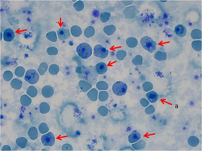
Rizzuto et al.                                                                                      NGS for Dominant Beta-Thalassemia Diagnosis

INTRODUCTION                                                             asthenia, jaundice, and short stature. No family history of
                                                                         hemolytic anemia. Examination of blood smear revealed
Beta-thalassemia major (BTHAL) is a well-known                           polychromasia,      anisopoikilocytosis,   basophil     stippling,
life-threatening condition characterized by severe transfusion-          Cabot rings, schistocytes, and spherocytes. Separation and
dependent anemia. BTHAL is an autosomal recessive disorder               quantification of Hb fractions did not reveal any extraordinary
presenting with high frequencies in populations from the                 peak and showed normal values for HbA2 and HbF. At 5-
Mediterranean area. Currently, up to 257 genetic variants in             year-old he underwent splenectomy. After the surgery, Heinz
the beta-globin gene (HBB) have been identified as BTHAL                 bodies were present (Figures 1, 2) and isopropanol stability
disease-causing, leading to a total or partial reduction of              test, performed according to standard methodology, appeared
beta-globin chain synthesis. The clinical severity of BTHAL              positive (Figure 3). Family studies in both parents were strictly
is related to the extent of imbalance between the alpha and              normal, including evaluation of Hb fractions. Enzyme activity
non-alpha-globin chains, while clinical management consists              assays, EMA-binding test, and osmotic gradient ectacytometry
of regular life-long red blood cell (RBC) transfusions and iron          (LoRRca MaxSis) were performed to rule out hemolytic anemia
chelation therapy. At present, the only definitive cure is bone          due to RBC defects other than hemoglobinopathy. Results,
marrow transplant (Efremov, 2007; Galanello and Origa, 2010).            although not strictly normal, did not reveal any RBC defect.
Both BTHAL patients and carriers are usually easily diagnosed            However, they should be taken with caution since the patient was
through routine laboratory tests. However, there is an ultra-rare        intensively transfused. Genetic analysis was performed on PKLR
condition overlapping BTHAL clinical manifestations known                and G6PD genes failing to reveal any disease-causing mutation.
as dominant beta-thalassemia (dBTHAL), which is caused by                   The second case is a female adult patient with mild chronic
the presence of certain unstable (UH) or hyper unstable (HUH)            compensated hemolysis referred for diagnosis when she was
hemoglobinopathies.                                                      20 years old. The father also presented with mild compensated
    UHs are a group of congenital disorders caused by mutations          hemolysis. No further examinations were performed before
in globin genes leading to destabilization of hemoglobin (Hb)            referral. Although the presence of extravascular hemolysis, the
molecules as a consequence of (a) amino acid substitutions               examination of blood smear was not informative. Separation and
within the heme pocket, (b) disruption of secondary structure,           quantification of Hb fractions did not reveal any extra peaks and
(c) substitution in the hydrophobic interior of the subunit, (d)         Heinz body and stability tests were normal. Further laboratory
amino acid deletions, and (e) elongation of the subunit. Thus,           tests were performed to rule out hemolytic anemia due to RBC
altering any of the steps in globin processing, including subunit        enzyme and membrane defects, including enzyme activity assays,
folding, heme interaction, dimerization, or tetramerization              EMA-binding test, and osmotic gradient ectacytometry (LoRRca
(Bunn and Forget, 1986). These abnormal Hb variants undergo              MaxSis). All of them showed normal values.
rapid denaturation followed by precipitation, leading to the                The third case is a male adult patient. He presented with
formation of Heinz bodies, which cause hemolysis of RBCs.                several episodes of hemolytic crises during childhood requiring
Clinical manifestations may vary from asymptomatic to severely           blood transfusion on two occasions. He underwent splenectomy
affected forms. Treatment is mainly symptomatic and based on             at the age of 25-year-old. The patient was diagnosed with
transfusion requirements as for BTHAL (Steinberg et al., 2009;           hereditary spherocytosis (HS) following a previous HS diagnosis
Thom et al., 2013).                                                      of his mother and the absence of abnormal Hb peaks by
    UHs are dominantly inherited with a significant rate of de           conventional electrophoresis.
novo mutations. They generally do not separate from normal                  Patients who underwent splenectomy neither clinically
Hb using standard methods. Thus, diagnosis of dBTHAL can be              improved nor presented complications as pulmonary
challenging since it requires a high index of suspicion and the          hypertension, thrombosis or increased hemolysis during
diagnosis may be delayed for years hampering the access to timely        10-year follow-up.
treatment interventions.                                                    The last two cases are two unrelated children who presented
    The study we present herein confirms the relevance of                with macrocephaly and severe congenital anemia. The parents
including globin genes in next generation sequencing (NGS)               of both patients had no family history for abnormal Hb or
approaches for the diagnosis of rare anemia disorders (RADs),            thalassemia and had normal hematological features. Therefore,
especially for cases with no family history in which the anemia is       conventional testing for abnormal Hb was not performed. All
not easily explained.                                                    siblings were unaffected.
                                                                            The first of these two unrelated children is a male
                                                                         patient presenting with large head circumference and
PATIENTS AND METHODS                                                     hepatosplenomegaly. Congenital dyserythropoietic anemia
                                                                         was suspected. However, no genetic analysis was performed
Clinical Reports                                                         for confirmation. He underwent successfully bone marrow
Here we present five clinical cases diagnosed with UH                    transplant at the age of 4.
after NGS analysis. Clinical data and laboratory findings are               The second child is a female patient presenting with
shown in Table 1.                                                        frontal bossing, macrocephaly, and severe anemia at the age
   The first case is a male pediatric patient referred with severe       of 2. Congenital dyserythropoietic anemia was suspected.
chronic blood dependent anemia since he was 4-month-old,                 Therefore, genetic analysis of CDAN1 and SEC23B genes was

Frontiers in Physiology | www.frontiersin.org                        2                                  February 2021 | Volume 12 | Article 628236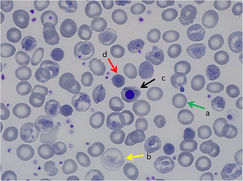
Rizzuto et al. NGS for Dominant Beta-Thalassemia Diagnosis
TABLE 1 | Overview on clinical and genetic data of the five reported clinical cases.
Parameters Case 1 Case 2 Case 3 Case 4 Case 5
Gender/Age Male/Pediatric Female/Adult Male/Adult Male/Pediatric Female/Pediatric
Hb (120–170 g/L) 70–80 119 141 82 79
MCV (80–100 fL) 110–115 97.8 102.3 83 Not done
MCHC (27–33.5 g/dL) 28 32.1 30.8 Not done Not done
Reticulocyte count (50–100 · 10ˆ9/L) 900 293 331 810 Not done
Reticulocyte count (%) 34 7.71 7.39 Not done Not done
Lactate dehydrogenase-LDH (U/L) 4,500–5,000 243 145 186 259
Hb Fractions Normal Normal Normal Not done Not done
Heinz bodies Positive Negative Positive Not done Not done
Stability test Positive Negative Positive Not done Not done
Age of onset (months) 4 Unknown Unknown Unknown 2
Family history No family history Father presents Mother diagnosed No family history No family history
mild compensated with hereditary
hemolysis spherocytosis
Transfusion need 8 U/Year No 2 times Multiple Multiple
Splenectomy Yes (5 y) No Yes (25 y) No No
Stem cell transplant (age years) No No No Yes (4 y) Yes (3 y)
Genotype HBBc.202G > A (p.Val67Met) HBA1c.187G > A HBBc.290T > C HBBc.442T > C HBBc.442T > A
(p.Val62Met) (p.Leu96Pro) (p.Ter147Glnext*21) (p.Ter147Lysext*21)
Hb variant name Hb Bristol-Alesha Hb Evans Hb Debrousse Hb Zunyi Hb Mokum
*Performed after the diagnosis of UH.
FIGURE 1 | Peripheral Blood Smear, May Grunwald Giemsa Stain. (a) Transfused red blood cells, (b) non-transfused red blood cells with hemoglobinization
abnormalities, (c) orthochromatic erythroblast, (d) erythrocitary inclusions that correspond to Heinz bodies.
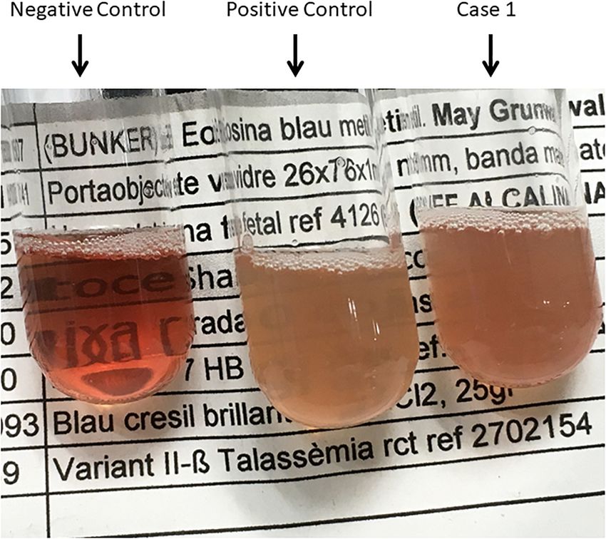
Frontiers in Physiology | www.frontiersin.org 3 February 2021 | Volume 12 | Article 628236Rizzuto et al. NGS for Dominant Beta-Thalassemia Diagnosis FIGURE 2 | Peripheral Blood Smear, Brilliant Cresyl Blue Stain. (a) Heinz bodies. FIGURE 3 | Isopropanol Test_01. Negative control (Hb AS), Positive control (Hb F), and Case 1. Frontiers in Physiology | www.frontiersin.org 4 February 2021 | Volume 12 | Article 628236
Rizzuto et al.                                                                                             NGS for Dominant Beta-Thalassemia Diagnosis

TABLE 2 | List of genes included in the t-NGS approach.

Symbol             Phenotype               Gene/Locus     Category                                         Description
                  MIM number               MIM number

ADA                   102700                    608958    Enzymopathy                                      Adenosine deaminase
AK1                   103000                    103000    Enzymopathy                                      Adenylate kinase 1
ALDOA                 611881                    103850    Enzymopathy                                      Aldolase, fructose-bisphosphate a
ANK1                  616089                    612641    Membranopathy                                    Ankyrin 1
ATRX                  301040                    300032    Alpha-thalassemia myelodysplasia syndrome,       Helicase 2, x-linked
                                                          somatic; Alpha-thalassemia/mental retardation
                                                          syndrome; Mental retardation-hypotonic facies
                                                          syndrome, X-linked
BPGM                  222800                    613896    Erythrocytosis and methemoglobinemia due to      Bisphosphoglycerate mutase
                                                          enzyme alteration
C15orf41              615631                    615626    Congenital dyserythropoietic anemia              Chromosome 15 open reading frame 41
CDAN1                 224120                    224120    Congenital dyserythropoietic anemia              Codanin 1
CYB5R3                250800                    613213    Methemoglobinemia, type I;                       Cytochrome b5 reductase 3
                                                          Methemoglobinemia, type II
EPB41                 611804                    130500    Membranopathy                                    Erythrocyte membrane protein band 4.1
EPB42                 612690                    177070    Membranopathy                                    Erythrocyte membrane protein band 4.2
EPO                   617907                    133170    Erythropoiesis modulator                         Erythropoietin
EPOR                  133100                    133171    Erythropoiesis modulator                         Erythropoietin receptor
G6PD                  300908                    305900    Enzymopathy                                      Glucose-6-phosphate dehydrogenase
GAPDH                    *                      138400    Enzymopathy                                      Glyceraldehyde-3-phosphate dehydrogenase
GATA1                 300835                    305371    Congenital dyserythropoietic anemia              Gata binding protein 1 (globin transcription
                                                                                                           factor 1)
GCLC                  230450                    606857    Enzymopathy                                      Glutamate-cysteine ligase, catalytic subunit
GPI                   613470                    172400    Enzymopathy                                      Glucose-6-phosphate isomerase
GSR                   618660                    138300    Enzymopathy                                      Glutathione reductase
GSS                   266130                    601002    Enzymopathy                                      Glutathione synthetase
GYPC                  616089                    110750    Membranopathy                                    Glycophorin c (gerbich blood group)
HBA1                  617981                    141800    Hemoglobinopathy                                 Hemoglobin–alpha locus 1
HBA2                  617981                    141850    Hemoglobinopathy                                 Hemoglobin–alpha locus 2
HBB                   617980                    141900    Hemoglobinopathy                                 Hemoglobin subunit beta
HBD                      *                      142000    Thalassemia due to Hb Lepore; Thalassemia,       Hemoglobin–delta locus
                                                          delta-
HBG1                  141900                    141749    Fetal hemoglobin quantitative trait locus 1      Hemoglobin, gamma a
HBG2                  613977                    142250    Cyanosis, transient neonatal; Fetal hemoglobin   Hemoglobin, gamma g
                                                          quantitative trait locus 1
HK1                   235700                    142600    Enzymopathy                                      Hexokinase 1
KCNN4                 616689                    602754    Membranopathy                                    Potassium channel, calcium activated
                                                                                                           intermediate/small conductance subfamily n
                                                                                                           alpha, member 4
KIF23                    *                      605064    Congenital dyserythropoietic anemia              Kinesin family member 23
KLF1                  613673                    600599    Congenital dyserythropoietic anemia              Kruppel-like factor 1 (erythroid)
NT5C3A                266120                    606224    Enzymopathy                                      50 -nucleotidase, cytosolic iiia
PFKL                     *                      171860    Hemolytic anemia due to phosphofructokinase      Phosphofructokinase, liver type
                                                          deficiency
PFKM                  232800                    610681    Enzymopathy                                      Phosphofructokinase, muscle
PGD                      *                      172200    Enzymopathy                                      6-phosphogluconate dehydrogenase,
                                                                                                           erythrocyte
PGK1                  300653                    311800    Enzymopathy                                      Phosphoglycerate kinase 1
PIEZO1                616089                    611184    Membranopathy                                    Piezo-type mechanosensitive ion channel
                                                                                                           component 1
PKLR                  266200                    609712    Enzymopathy                                      Pyruvate kinase, liver and rbc
RHAG                  185000                    180297    Membranopathy                                    rh-associated glycoprotein
SEC23B                224100                    610512    Congenital dyserythropoietic anemia              Sec23 homolog b, copii coat complex
                                                                                                           component

                                                                                                                                                 (Continued)

Frontiers in Physiology | www.frontiersin.org                                5                                 February 2021 | Volume 12 | Article 628236
Rizzuto et al. NGS for Dominant Beta-Thalassemia Diagnosis
TABLE 2 | Continued
Symbol Phenotype Gene/Locus Category Description
MIM number MIM number
SLC2A1 606777 138140 Membranopathy Solute carrier family 2 (facilitated glucose transporter), member 1
SLC4A1 612653 109270 Membranopathy Solute carrier family 4 (anion exchanger), member 1 (diego blood group)
SPTA1 130600 182860 Membranopathy Spectrin alpha, erythrocytic 1
SPTB 616649 182870 Membranopathy Spectrin beta, erythrocytic
TPI1 615512 190450 Enzymopathy Triosephosphate isomerase 1
UGT1A1 237900 191740 Gilbert syndrome udp glucuronosyltransferase 1 family, polypeptide a1
*Not available.
performed not revealing any disease-causing mutation. She performed using the SeqCap EZ Human Exome Library v3.0
underwent successfully bone marrow transplant when she was (Roche NimbleGen Madison, WI, United States). Sequencing
almost 3-year-old. was done on an Illumina HiSeq2500 HTv4 (Illumina, San
In all cases, RAD due to Hb variant was not suspected Diego, CA, United States) with paired-end 125-bp reads. Read
mainly due to the fact that parents did not present family alignment to hg19 and variant calling were done with a
history of RADs, except for case 3, RBC parameters were found pipeline based on BWA-MEM0.7 and GATK 3.3.0. The median
to be normal and abnormal Hb fractions were absent when coverage of the captured target region was at least 98×.
analyzed. Therefore, genetic testing was performed for genes Variant annotation and prioritizing were done using Cartagenia
associated with RADs other than globin genes, failing to show a Bench Lab NGS (Agilent Technologies). Variants located outside
conclusive diagnosis. the exons and intron/exon boundaries and variants with a
minor allele frequency (MAF) of >1% in control databases,
Genetics Analysis including dbSNP1374 , 1000 Genomes Project (phase 3)5 , and
Written informed consent was obtained from cases or legal Exome Variant Server (EVS), NHLBI Exome Sequencing Project
guardian. Targeted NGS (t-NGS) analysis was performed in cases National Heart, Lung, and Blood Institute GO Exome Sequencing
1, 2, and 3 while whole exome sequencing (WES) analysis was Project (ESP6500 release)6 and in-house exome controls were
performed in cases 4 and 5. For all the patients, genomic DNA excluded. Variants that fitted with a de novo or recessive mode
was extracted from peripheral blood. For patients who underwent of inheritance were further prioritized based on literature,
bone marrow transplant, DNA samples were previously stored. predicted (deleterious) effects on protein function by e.g.,
The designed t-NGS panel covered 46 genes described truncating the protein, affecting splicing, amino acid change, and
as disease causing for RADs, including genes responsible evolutionary conservation.
for membrane disorders, enzyme defects, congenital For case 5, WES was performed in a trio approach (patient
dyserytrhopoietic anemia and the HBA1/HBA2 and HBB and both parents). Exomes were captured using the Agilent
genes responsible for alpha and beta-globin chains, respectively. SureSelectXT Human All Exon v5 (Agilent, Santa Clara, CA,
The full list of genes included is shown in Table 2. Exon and United States) accompanied by Illumina paired-end sequencing
exon/intron boundaries were capture using a NimbleGen on the HiSeq2000 (Illumina, San Diego, CA, United States).
SeqCap EZ HyperCap (Roche) solution-based capture system The in-house sequence analysis pipeline Modular GATK-Based
followed by next generation sequencing on the MySeq (Illumina) Variant Calling Pipeline (MAGPIE) (LUMC Sequencing Analysis
with 150 bp paired-end reads. For the bioinformatics analysis, Support Core, LUMC) was used to call the SNVs/indels.
alignment to the hg38 genome was performed with BWA-MEM LOVDplus (Leiden Genome Technology Center, LUMC, Leiden)
(Li H. 203 arXIV:1303.3997v2) and detection of changes with was used for interpretation of variants.
GATK1 . Obtained variants were filtered and annotated based
on variant effect, coverage (>30) and MAF (>0.05). Resulting
variants were assessed for technique pitfalls through IGV. The RESULTS
nomenclature used was the recommended by HGVS2 . Finally,
disease-causing variants were prioritized based on inheritance Genetic variants in globin genes responsible for UH or HUH were
pattern and VarSome3 for previous evidence as disease causing found in all five cases as shown in Table 1. All variants were
mutations or predictions score information. Variants were confirmed by Sanger sequencing.
reported according to American College of Medical Genetics In case 1, variant HBB:c.202G > A (p.Val67Met) was found
(ACMG) guidelines. in exon 2 in the heterozygous state. This HBB variant is known
For case 4, WES was performed in a trio approach (patient as Hb Bristol-Alesha, a UH associated with moderate-severe
and both parents). Libraries were prepared using the Kapa HTP hemolytic anemia. The variant was not found in the parents,
kit (Illumina, San Diego, CA, United States) and capture was suggesting a de novo variant in the patient.
1 4
https://software.broadinstitute.org/gatk/ http://www.ncbi.nlm.nih.gov/projects/SNP
2 5
http://www.hgvs.org http://www.internationalgenome.org/
3 6
https://varsome.com http://evs.gs.washington.edu/EVS/
Frontiers in Physiology | www.frontiersin.org 6 February 2021 | Volume 12 | Article 628236Rizzuto et al. NGS for Dominant Beta-Thalassemia Diagnosis
In case 2, variant HBA1:c.187G > A (p.Val62Met) was found a 2-3–fold higher level of mRNA than HBA1 (Liebhaberts
in exon 2 in the heterozygous state. This HBA1 variant is known et al., 1986). Thus, the beta-globin gene is the first option
as Hb Evans and is associated wild with mild hemolytic anemia to investigate for disease-causing mutations leading to RADs
and classified as UH. Parents were not sequenced. due to UHs/HUHs especially in cases with moderate to
In Case 3, variant HBB:c.290T > C (p.Leu96Pro) was found severe phenotypes.
in in exon 2 in the heterozygous state. This HBB variant Interestingly, Hb Bristol-Alesha is classified as a UH variant,
is known as Hb Debrousse and is described as a moderate not as a HUH as we expected based on the severity of the patient’s
UH. Parents were not sequenced. Nevertheless, antecedents clinical picture. The change to methionine at position 67 of the
of hemolytic anemia are present in the mother, suggesting a beta-globin chain alters the hydrophobic heme pocket causing
dominant inheritance pattern. the instability of the protein (Kano et al., 2004). As described in
In cases 4 and 5, two missense stop-loss mutations at previous clinical reports, at physical examination, splenomegaly
position 422 of the HBB gene were found. The first variant and jaundice may be found. Iron overload and gallstones may
HBB:c.442T > C (p.Ter147Glnext∗ 21), found in case 4, is develop due to the rapid turnover of RBCs.
known as Hb Zunyi, while the second variant HBB:c.442T > A Hb Debrousse, reported twice in literature, is a UH
(p.Ter147Lysnext∗ 21), found in case 5, constitutes a novel variant characterized by well-compensated chronic hemolytic anemia
which was called Hb Mokum. Both variants cause the loss of a due to its high oxygen affinity. Hb Debrousse is caused by leucine
stop codon and elongation of the translated beta-globin chain of to proline substitution at position 96 involving the hydrophobic
21 amino acids due to a new stop codon in the 30 untranslated environment of the proximal side of the heme. In the previously
region (30 UTR) of the HBB gene. The variants were not found in reported cases, Hb Debrousse discovery was possible after a
the parents suggesting de novo variants in the patients. Parvovirus B19 infection that caused a hemolytic crisis (Lacan
According to the ACMG guidelines, all the variants were et al., 1996). Indeed, since affected patients show a chronic well-
classified as pathogenic (Richards et al., 2015). compensated hemolytic anemia, the diagnosis of such a variant
is unlikely until the globin genes are investigated. Such a study is
usually performed only when some complications occur.
DISCUSSION Hb Zunyi was recently reported for the first time as a de
novo mutation in a Chinese child with severe anemia requiring
We highlight the importance of including globin genes in the blood transfusion, malnutrition, growth delay, splenomegaly and
NGS analysis of RAD for enabling the diagnosis of UH. We hepatomegaly (Su et al., 2019). In the study herein, we identified
present five clinical cases affected with RAD due to UH variants, both Hb Zunyi and Hb Mokum as de novo mutations in the
four are related to the beta-globin gene (Hb Bristol—Alesha, Hb heterozygous state. Hb Zunyi and the novel Hb Mokum are stop-
Debrousse, Hb Zunyi, and the novel Hb Mokum), meanwhile, loss mutations at position 442 in HBB, resulting in an elongated
one is related to the alpha-globin gene (Hb Evans). The use of beta-globin chain leading to HUHs. The extra amino acids in
NGS has been crucial for the final conclusive diagnosis. the elongated beta-globin chain (169 a.a.) are probably affecting
The severity of RADs due to UHs depends on the mutation’s its helical sequence, interfering with its tertiary structure and
impact on protein stability and consequently on the degree of causing an unstable tetramer. Frameshift mutations in the HBB
hemolysis and inefficient erythropoiesis. Patients’ RBCs typically gene, resulting in the elongated beta-globin chain, have been
display abnormal but unspecific morphology with microcytosis, described before but resulted in shorter beta-chains (max. 157
hypochromia, moderate to severe anisopoikilocytosis, basophilic aa.) and milder phenotypes than the mutations described here
stippling, and inclusions that may become particularly prominent (Su et al., 2019).
following splenectomy (Steinberg et al., 2009; Kent et al., Finally, Hb Evans is classified as UH. It is consequence of a
2014). UHs are commonly inherited in a dominant way or valine to methionine substitution at position 62 of the alpha2-
presented as de novo, although there are some examples of globin chain encoding gene HBA2. Hb Evans has been reported in
recessive inheritance leading to mild phenotypes. According patients presenting with mild hemolytic anemia that was getting
to results obtained through the HbVar Query page (dated worse particularly in case of stress (Wilson et al., 1989).
14th January 2020), 1,534 Hb variants have been described The standard tests to detect abnormal anemias are High
so far due to mutations on either HBA1/HBA2 or the HBB Precision Liquid Chromatography (HPLC) or conventional
genes. Up to 251 variants (16.4%) are classified as UH or or capillary electrophoresis (CE). However, UHs/HUHs do
HUH based on heat or isopropanol stability tests and/or low not normally appear in the peak-patterns or appear as
Hb abundancy (Giardine et al., 2007, 2014). It is worthy small peaks that may be mistaken for degradation products.
to highlight that all the HUH variants involving the HBB In three of the five UH cases reported here, extra peaks
gene reported positive stability tests, meanwhile in most of were not detected. More confined methods are Heinz Bodies
the HUH involving the alpha-globin genes, hyper instability test or stability tests as isopropanol precipitation or heat
has been only deduced from low abundance. This must be stability tests, which are affordable screening techniques for
cautiously taken since mutations in alpha-globin genes are UHs/HUHs variants. In the patient with Hb Bristol-Alesha,
lower expressed (Rizzuto et al. NGS for Dominant Beta-Thalassemia Diagnosis
were not performed. Genetic analysis of globin genes should be the already established European Reference Networks for rare
performed for diagnosis confirmation. Inclusion of HBA1/HBA2 hematological disorders, ERN-EuroBloodNet.
and HBB in NGS approaches will facilitate timely conclusive
diagnosis. as a screening tool for hemolytic anemias will assist in
reaching a definitive diagnosis sooner. DATA AVAILABILITY STATEMENT
Furthermore, the occurrence of de novo mutations causing
UHs/HUHs should also be considered in the analysis of The datasets generated for this study can be found in the
genetic variants. online repositories. The names of the repository/repositories and
The usefulness of NGS in improving the diagnosis of RADs accession number(s) can be found below: www.ithanet.eu and
has already been demonstrated in several studies as well as https://ithanet.eu/db/ithagenes?ithaID=3697.
its relevance in new gene discovery (Shang et al., 2017; Duez
et al., 2018). In the case of overlapping phenotypes, which
frustrate proper diagnosis, the use of NGS may be beneficial ETHICS STATEMENT
for ultra-rare RADs. In a recent publication, 36% of patients
initially diagnosed with congenital dyserytrhopoietic anemia, Written informed consent was obtained from the cases/legal
received a final diagnosis of pyruvate kinase deficiency after guardian for the publication of any potentially identifiable images
NGS analysis (Russo et al., 2018). Nevertheless, in the majority or data included in this article.
of the t-NGS panels reported, globin genes are not included,
since globin genes are quite short and molecular diagnosis of
most common Hb disorders, such as sickle cell disease (SCD) AUTHOR CONTRIBUTIONS
and thalassemia syndromes, is well-established through Sanger
sequencing and GAP-PCR/MLPA. Therefore, dBTHAL disorders VR, MM-P, CLH, TK, and DB wrote the manuscript. All authors
due to UH/HUH may also benefit from NGS approaches for critically revised the manuscript.
RADs by including globin genes, as presented herein.
Current literature on dBTHAL and UH/HUH variants is
mainly composed of retrospective case reports, which makes FUNDING
evidenced-based management of this RAD unlikely. In addition,
to benefit from the most adequate management it is necessary to This study was supported by funding from the authors’
achieve a diagnosis as early as possible. In conclusion, this study institutions and the European Commission H2020-MSCA-ITN-
confirms the importance of NGS as a fundamental tool to early 2019, Grant Agreement N860436, “EVIDENCE.”
identify and treat UH/HUH in patients with RAD without an
established diagnosis after standard methodologies.
Future challenges include a better understanding of disease ACKNOWLEDGMENTS
characteristics and management, and consideration of bone
marrow transplant as a curative option. Therefore, we encourage This work was generated within the European Reference
that these patients are referred to expert Units in referral centers Network on Rare Hematological Diseases (ERN-
for enabling basic and clinical research taking advantage of EuroBloodNet, FPA 739541).
REFERENCES Kano, G., Morimoto, A., Hibi, S., Tokuda, C., Todo, S., and Sugimoto, T. (2004). Hb
Bristol-Alesha presenting Thalassemia-Type hyperunstable hemoglobinopathy.
Bunn, H. F., and Forget, B. G. (1986). Hemoglobin: Molecular, Genetic and Int. J. Hematol. 80, 410–415. doi: 10.1532/IJH97.04048
Clinical Aspects. Philadelphia, PA: W. B. Saunders Company, doi: 10.1016/0092- Kent, M. W., Oliveira, J. L., Hoyer, J. D., Swanson, K. C., Kluge, M. L.,
8674(87)90069-9 Dawson, D. B., et al. (2014). Hb grand junction (HBB: C.348-349delinsG;
Duez, J., Carucci, M., Garcia-Barbazan, I., Corral, M., Perez, O., Luis Presa, P.His117IlefsX42): a new hyperunstable hemoglobin variant. Hemoglobin 38,
J., et al. (2018). High-throughput microsphiltration to assess red blood cell 8–12. doi: 10.3109/03630269.2013.853672
deformability and screen for malaria transmission-blocking drugs. Nat. Protoc. Lacan, P., Kister, J., Francina, A., Souillet, G., Galactéros, F., Delaunay, J.,
13, 1362–1376. doi: 10.1038/nprot.2018.035 et al. (1996). Hemoglobin debrousse (B96[FG3]Leu → Pro): a new unstable
Efremov, G. D. (2007). Dominantly inherited β-Thalassemia. Hemoglobin 31, hemoglobin with twofold increased oxygen affinity. Am. J. Hematol. 51, 276–
193–207. doi: 10.1080/03630260701290092 281. doi: 10.1002/(SICI)1096-8652(199604)51:4Rizzuto et al. NGS for Dominant Beta-Thalassemia Diagnosis Russo, R., Manna, F., Gambale, A., Marra, R., Rosato, B. E., Caforio, P., et al. (2018). Wilson, J. B., Webber, B. B., Kutlar, A., Reese, A. L., Mckie, V. C., Lutcher, C. L., Multi-Gene panel testing improves diagnosis and management of patients with et al. (1989). Hb evans or A262(E11)Val→metβ2; an unstable hemoglobin Hereditary Anemias. Am. J. Hematol. 93, 672–682. doi: 10.1002/ajh.25058 causing a mild hemolytic anemia. Hemoglobin 13, 557–566. doi: 10.3109/ Shang, X., Peng, Z., Ye, Y., Asan, Zhang, X., Chen, Y., et al. (2017). Rapid targeted 03630268908993106 next-generation sequencing platform for molecular screening and clinical genotyping in subjects with hemoglobinopathies. EBioMedicine 23, 150–159. Conflict of Interest: The authors declare that the research was conducted in the doi: 10.1016/j.ebiom.2017.08.015 absence of any commercial or financial relationships that could be construed as a Steinberg, M. H., Forget, B. G., Higgs, D. R., and Weatherall, D. J. (2009). Disorders potential conflict of interest. of Hemoglobin: Genetics, Pathophysiology, and Clinical Management, Second Edition, Vol. 94. Cambridge: Cambridge University Press, i–iv. doi: 10.1017/ Copyright © 2021 Rizzuto, Koopmann, Blanco-Álvarez, Tazón-Vega, Idrizovic, Díaz CBO9780511596582 de Heredia, Del Orbe, Pampliega, Velasco, Beneitez, Santen, Waisfisz, Elting, Smiers, Su, Q., Chen, S., Wu, L., Tian, R., Yang, X., Huang, X., et al. (2019). Severe de Pagter, Kerkhoffs, Harteveld and Mañú-Pereira. This is an open-access article thalassemia caused by Hb Zunyi [B147(HC3)Stop→Gln; HBB: C.442T>C)] on distributed under the terms of the Creative Commons Attribution License (CC BY). the β-Globin gene. Hemoglobin 43, 7–11. doi: 10.1080/03630269.2019.1582430 The use, distribution or reproduction in other forums is permitted, provided the Thom, C. S., Dickson, C. F., Gell, D. A., and Weiss, M. J. (2013). Hemoglobin original author(s) and the copyright owner(s) are credited and that the original variants: biochemical properties and clinical correlates. Cold Spring Harb. publication in this journal is cited, in accordance with accepted academic practice. No Perspect. Med. 3, 1–22. doi: 10.1101/cshperspect.a011858 use, distribution or reproduction is permitted which does not comply with these terms. Frontiers in Physiology | www.frontiersin.org 9 February 2021 | Volume 12 | Article 628236
You can also read