PRIMARY EXTRANODAL NON-HODGKIN'S LYM-PHOMA INVOLVING RIGHT MAXILLA
←
→
Page content transcription
If your browser does not render page correctly, please read the page content below
https://doi.org/10.5272/jimab.2020262.3187
Journal of IMAB
                                               Journal of IMAB - Annual Proceeding (Scientific Papers). 2020 Apr-Jun;26(2)
ISSN: 1312-773X
https://www.journal-imab-bg.org
                                                 Case report

                                          PRIMARY EXTRANODAL NON-HODGKIN’S LYM-
                                          PHOMA INVOLVING RIGHT MAXILLA
                                          Elitsa Deliverska1, Evgenii Hadjiev2, Lutchezar Stefanov1, Gergana Tsvetkova2,
                                          Emel Bekirova2, Sadeta Parusheva1
                                          1) Department of Dental, Oral and Maxillofacial surgery, Faculty of Dental
                                          Medicine, Medical University - Sofia.
                                          2) Clinic of Clinical Hematology, UMHAT “Alexandrovska” - Sofia, Bulgaria.

        ABSTRACT                                                comprising the extranodal lymphomas arise from lymphoid
        Background: Lymphoma in a single bone without           tissue in other organs and from sites normally lacking or-
any nodal or visceral involvement is known as primary in-       ganized lymphoid tissue such as the stomach, skin, brain and
tra-osseous lymphoma, which is a very uncommon malig-           testis [1, 4]. The most common site of extranodal NHL in
nancy. It constitutes 3.1% of malignant bone tumours and        the head and neck region is Waldeyer’s ring [5].
5% of extranodal lymphomas, and the incidence is only                   Lymphoma arising from a single bone without any
0.6% in the jaws. These tumours can be primary or second-       nodal or visceral involvement is known as primary intra-
ary (in the disseminated forms). The primary intra-osseous      osseous lymphoma. It is a very uncommon malignancy and
lymphoma of the jaws is a challenge to diagnose, because        constitutes 3.1% of malignant bone tumours and 5% of
of their various clinical presentation, which can mimic an      extranodal lymphomas. [1] Extranodal non-Hodgkin lym-
odontogenic tumour, cyst or a fibro-osseous lesion.             phoma (NHL) represent 20% to 30% of all the NHL. [6]
        Purpose: This case report discusses a rare malig-       Among the NHL that occurs in the maxillofacial area, 15-
nancy of jaw bones, including clinical presentation, his-       45% arise in the oral cavity [7] engaging the upper jaw
topathologic features, immunologic profile, PET scan,           (11%), the lower jaw (8%), the palatal soft tissue (8%) and
management and prognosis.                                       gum (7%). The incidence in jaw bones is only 0.6%. [8]
        Material and methods: We present a case of a pri-               Extranodal lymphoma of the head and neck comprise
mary intraosseous lymphoma in a 45-year-old female who          a heterogeneous group of tumours with different histologi-
presented with a swelling in the right maxilla. It highlights   cal types, modes of presentation and prognosis. The unique
the importance of recognizing rare entities that may present    jaw bone and soft localization are very rare for the NHL,
in the jaws, the impact of the disease and its management.      and in some cases, differential diagnosis with the most
        Results: Jaw localization of Non-Hodgkin’s lym-         common dental lesions and other soft tissue pathologies
phoma is rare. Clinical symptomatology and radiological         and tumours may be difficult.
signs are poorly contributive. The diagnosis relies on the              One of the diagnostic problems with regard to NHL
histopathological analysis.                                     is the variable nature of clinical symptoms. Although the
        Conclusion: One of the diagnostic problems with re-     lymph nodes are the principal location for these prolifera-
gard to NHL is the variable nature of clinical symptoms. A      tions, all other organs, in particular, those containing nor-
biopsy allowed diagnosing an intra-oral bone lymphoma,          mal lymphoid tissues can also develop lymphoma [9].
and the patient was referred to the hematooncology unit         These tumours can be primary or secondary (in the dissemi-
for treatment.                                                  nated forms) [9, 10]
                                                                        Patients may be of any age group, but there is a ten-
        Keywords: non Hodgkin’s lymphoma, extranodal,           dency to involve adults, especially older adults. The Epstein
jaws,                                                           Barr virus (EBV) is specifically associated with Burkitt’s
                                                                lymphoma. Lymphomas can be associated with other viruses
       INTRODUCTION                                             such as that of hepatitis C (HCV) and herpes virus type 8
       The malignant lymphomas are neoplastic transforma-       (HHV-8). The stages of passage between infection by a vi-
tions of cells that reside predominantly in lymphoid tissues.   rus and the appearance of lymphoma are not known. Patients
The two main variants of malignant lymphoma are Hodg-           with immunodeficiencies, such as transplant patients or those
kin’s disease and Non-Hodgkin’s lymphoma (NHL) [1, 2].          with Human immunodeficiency virus (HIV), would be prone
Unlike Hodgkin’s disease, Non-Hodgkin’s lymphoma has a          to the development of NHL [10, 11]
firmly established cellular origin with morphologic subtypes            In 1994, the International Lymphoma Study Group
corresponding to various stages of lymphocyte differentia-      [11] proposed a classification of various uniform groups
tion. The appropriate treatment depends on the histology        of lymphoma, taking into consideration different morpho-
and extent of the disease [3]. Approximately 70% of the cases   logical, histopathological, phenotypic, anatomic and clini-
of NHL arise from lymph nodes while the remaining 30%,          cal data. Depending on the criteria of malignancy and the

J of IMAB. 2020 Apr-Jun;26(2)                  https://www.journal-imab-bg.org                                           3187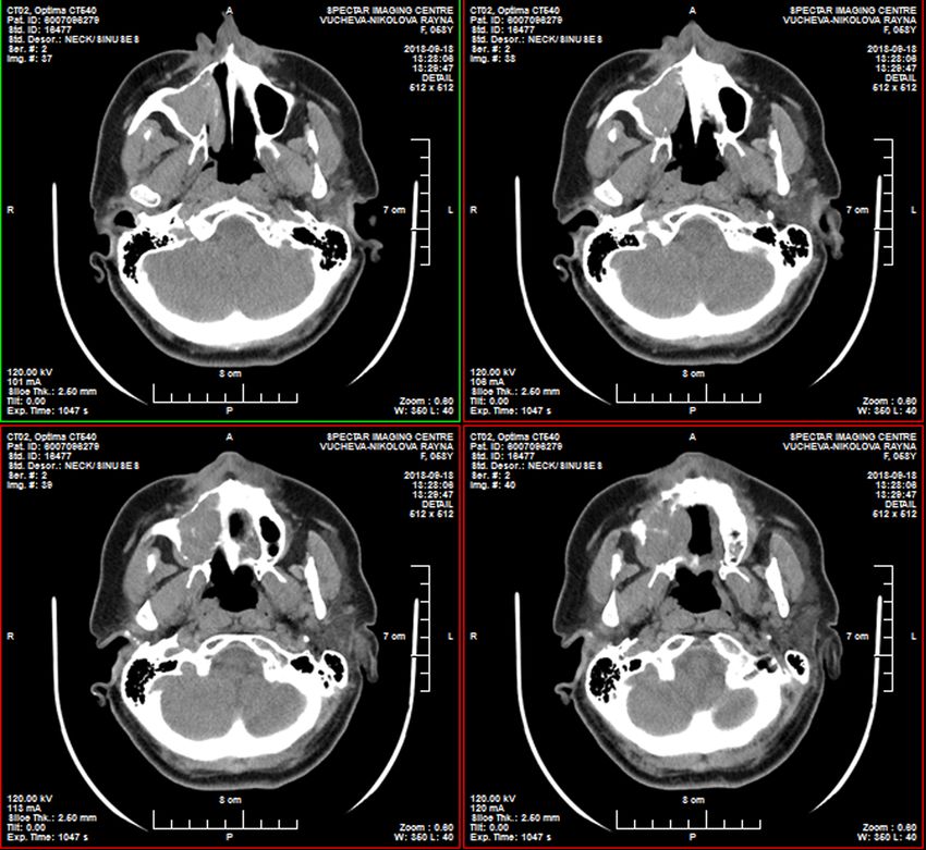
prognosis index, a treatment strategy is defined and then        the air filled cavities of the face (one third of extra-nodal
assessed [10].                                                   NHL), the salivary glands and exceptionally in the mouth
        Most of the lymphomas are of B cell lineage (85%         [13, 14] Primary NHL’s make up 8% of mandibular tumours
of NHL in adults). The following can be discerned:               and 0.6% of all NHL’s [15]
        - Follicular lymphoma: the proliferation phenotypes             The bone lesions of Burkitt’s disease are character-
are CD10+, CD5 and CD23. These lymphomas are rarely              ized by a high incidence of mandibular tumours (more than
localized and generally appear as deep peripheral adenopa-       50% of cases). This localization is also present in 15% of
thies, with damage to the spleen and invasion of bone mar-       non-epidemic Burkitt’s disease [9, 10]. Tumours of the jaw
row.                                                             are generally seen in adults (between the ages of 40 and
        - Mantle cell lymphomas: phenotypes CD5+, CD10,          50) with a male to female ratio - 0.5 [10, 15].
CD23 and CD43+.
        - Extranodalmarginalzonelymphomasoflymphoid                     CASE REPORT
tissue associated with mucous membranes (MALT): pheno-                  A 58- years- old female presented with an intraoral
types CD5, CD10 and CD23.                                        swelling in the right molar and palatal area noticed around
        - Other small-cell lymphomas.                            three weeks ago. There was not any extraoral swelling caus-
        - Diffuse, large-B-cell lymphoma: phenotypes             ing facial asymmetry, nor any history of trauma to the site
CD20+, ( CD79a+. They account for 40% of NHL and are             or toothache in the region. She gave no history of nasal
considered to be “aggressive.”                                   congestion or watering of the eyes or any changes in the
        - Burkitt’s lymphoma: initially described in Africa,     surface sensations. The intraoral swelling was not associ-
this is associated with the EBV and observed particularly        ated with pain. At the time of presentation, the patient did
in children and patients with HIV. [10]                          not report any systemic symptoms and had no fever or his-
         (Phenotype T lymphoma, counts only for 15% of           tory of weight loss or sweating. She was not on any medi-
NHL in adults. Diagnosis of NHL is based principally on          cation, was a non-smoker and non-alcoholic. The patient’s
morphological, histological and cytological investigations,      previous medical, family and past dental histories were non-
and is confirmed immunohistochemically. The biopsy               contributory. The patient had a history of Lime disease and
specimen should be large enough to enable standard mor-          allergy to doxycycline and sulfonamides. On local exami-
phological and pathological examinations, culture and his-       nation, a diffuse intraoral swelling involving the upper mo-
tochemical analyses (as well as, sometimes, the freezing of      lar and palatal area was detected, which several days after
tissue for future additional tests). It needs to be emphasized   the incisional biopsy reduced its size (fig.1). No palpable
that a superficial biopsy may lead to an error in diagnosis.     lymph nodes were present. None of the teeth in the quad-
Once the diagnosis is confirmed, the staging is clinical and     rant were decayed, no periodontal pockets were present in
radiological [10]                                                relation to teeth # 13, 14, 15 but the grade I mobility was
        - Estimation of the tumour mass.                         noted and upper molar teeth were tender on percussion. No
        - Examination of lymph node regions.                     other abnormalities were detected on hard or soft tissue ex-
        - Standard X-rays (panoramic jaw, chest) and ab-         amination. The clinical differential diagnosis included the
dominal and chest scans.                                         plasmocytoma, fibro-osseous lesions such as ossifying fi-
        - Bone marrow sampling.                                  broma, fibrous dysplasia, nonodontogenic and odontogenic
        Chemotherapy must be adapted to the staging and          bone tumours involving the maxilla. The panoramic radio-
histological classification of the lymphoma associated with      graph showed obliteration of the right maxillary sinus with
local radiation treatment of the region (35-40 Gy). This         loss of continuity in the floor of the maxillary sinus and
combination is recommended in particular for high grade          the alveolar bone in the region of teeth # 14, 15 appeared
localized lymphomas [11, 12]. As a general rule, this means      dense and granular. Obliteration of maxillary sinus with the
3–4 courses of CHOP chemotherapy (Cyclophosphamide,              destruction of the posterior and anterior walls was noted
Adriamycin, Vincristine and Prednisolone); or other proto-       on axial sections. (fig. 2, 3) On computed tomography (CT),
col such as ACVBP chemotherapy (Adriamycin, Cyclophos-           the coronal section of paranasal sinus region revealed a soft
phamide, Vindesine, Bleomycin and Prednisolone) [11, 12].        tissue lesion causing local destruction of the floor of the
Once remission has been obtained, the aim of clinical moni-      right maxillary antrum and encroaching onto the alveolar
toring is to detect early signs of recurrence. The follow up     process. (fig. 4) An incisional biopsy of the swelling was
must be at least 10 years, an examination every 3 months         performed, and the histopathology showed sheets of round
is appropriate during the first 5 years. Patient information     and spindle shaped cells with vesiculated nuclei in a fi-
and the role of the general practitioner are important. [10]     brous connective tissue stroma. Poorly differentiated tu-
        The prognosis of NHL does not depend on the mul-         mour cells with increased mitotic figures and vascularity
tiplicity of clinical aspects, but mainly on the histology       with multinucleated giant cells were seen. The features were
and the stage of the tumour (whether it is localized or not,     suggestive of malignant tumour of mesenchymal origin.
stage of evolution, number of extra-nodal localizations,         Immunohistochemistry played an important role in distin-
etc.). Age is also an important factor [10, 12]                  guishing the cell type and differential diagnosis (# 181478/
        Tumour localization (e.g.in jaws) is not found to be     18.09. 2018). In the present case the tumour cells were posi-
a significant prognosis factor. Although rare in the jaws        tive for Leucocyte common antigen (LCA/CD45), CD20+
[12], it is most often found in Waldeyer’s ring and then in      a marker that recognizes the surface antigen which is ex-

3188                                            https://www.journal-imab-bg.org               J of IMAB. 2020 Apr-Jun;26(2)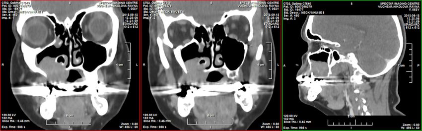
pressed on B-cells); CD5+; CD10+; BCL6+;MUM1+;                        Fig. 1. Intraoral photograph showing clinical features
BCL2+, TdT-; C- MYC+ in15% of all cells, Ki67+ in 80%          of a patient: the appearance of a mass and buccal expansion
of cells. PET scan reveals tumour mass in right maxillary      in the right maxillary premolar and molar sector with low
sinus 22\26\29mm. (fig. 3.) Cytokeratin AE1-AE3- nega-         mobility of related teeth 7 days after the incisional biopsy.
tive. The above findings of the histochemical study indi-
cate that the tumour cells were of large B-cell origin. Thus
based on the immunologic profile, a diagnosis of diffuse
large B-cell lymphoma was given IV stage, NCCN-IPI-2,
R-IPI 1, CNS-IPI 1. The patient was referred to medical on-
cology for chemotherapy and was managed with cyclophos-
phamide, doxorubicin (hydro doxorubicin), vincristine
(oncovin) and prednisone (CHOP) regimen and 3 courses
R-CHOP. The patient was on periodic follow-up; PET scan
(fig. 5) on 20.02.2019 reveal fully regression of the dis-
ease and after restaging again CHOP and R- CHOP(8 courses
from the beginning). The decision was two more courses
with the same scheme and two more courses after that with
R- CHOP. 05.2019 dated CT scan after 6 courses of chemo-
therapy revealed no tumour lesion or any changes of the
maxillary sinus, lymph nodes and liver.

        Fig. 2. Axial CT scan of the patient: tumour mass in the right maxillary sinus and osteolytic image concerning
lateral wall of the maxillary sinus

J of IMAB. 2020 Apr-Jun;26(2)                 https://www.journal-imab-bg.org                                         3189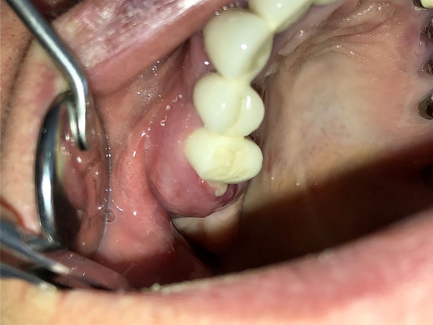
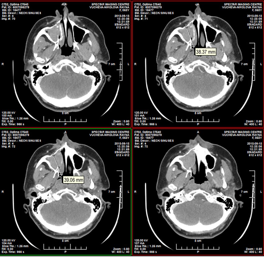
Fig. 3. Axial CT scan of the patient: tumour mass in the right maxillary sinus invading jaw and nasal cavity.

       Fig. 4. Coronal CT image(soft tissue window)showing space occupying lesion in the right maxillary sinus with
destructive margins

3190                                          https://www.journal-imab-bg.org             J of IMAB. 2020 Apr-Jun;26(2)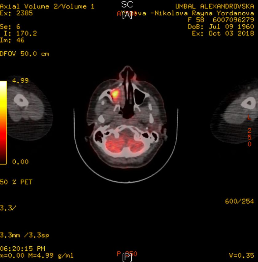
Fig. 5. PET scan of the NHL                             along with immunohistochemical evaluation is instrumen-
                                                              tal in arriving at the diagnosis of lymphoma. The immu-
                                                              nologic markers will help in detecting the cell lineage and
                                                              various subtypes of lymphomas. Diffuse large B cell lym-
                                                              phoma is one of the most common lymphoid malignan-
                                                              cies in adults, representing about 30–40% of adult NHL
                                                              diagnosed de novo on the basis of morphology and
                                                              immunophenotype. Diffuse large B-cells typically express
                                                              the B-cell markers CD19, CD20 and CD22 and the sur-
                                                              face immunoglobulin. The tumour cells are larger and more
                                                              irregular than immunoblasts, and the cytoplasm is less ba-
                                                              sophilic [19].
                                                                     When tissue diagnosis of NHL is given, whole body
                                                              survey using CT or MRI to evaluate the extent of the dis-
                                                              ease involvement is necessary for accurate staging and ap-
                                                              propriate management of the disease. Positron emission
                                                              tomography (PET) scanning is reported to be comparable
                                                              to CT and provides no additional information than the
                                                              typical work-up [18]
                                                                     Bone involvement in itself, however, does not ap-
                                                              pear to be a poor prognostic sign; rather, the extensive
                                                              disease is a more important determinant of treatment out-
                                                              come [10]
                                                                     The patient presented here was treated with chemo-
       DISCUSSION                                             therapy - on CHOP (cyclophosphamide, doxorubicin (hy-
       Non-Hodgkin’s lymphomas (NHL) are a heteroge-          dro doxorubicin), vincristine (oncovin) and prednisone
neous group of lymphoproliferative disorders originating      regimen and is on regular follow-up.
in B-lymphocytes, T-lymphocytes or natural killer (NK)               The international prognostic index (IPI) was devel-
lymphocytes. Primary non- Hodgkin’s lymphoma (NHL)            oped to predict outcome in patients with aggressive NHL,
is found outside nodal tissues in 24–45% of cases. Com-       based on patients’ clinical characteristics before treatment.
mon extranodal sites include the GI tract, skin, and less     The predictors which signify worst prognosis include ad-
often, bones. In the head and neck region, Waldeyer’s         vancing age, advanced disease, more extranodal sites of
ring, oral mucosa, salivary glands, paranasal sinuses, la-    disease, elevated serum lactate dehydrogenase levels, and
ryngeal tissue, and osseous structures have been found to     delayed response to chemotherapy. Based on these pre-
exhibit primary NHL. The incidence of NHL has increased       dictors patients are grouped under low risk; low-interme-
dramatically in the last 3 decades, which has been attrib-    diate risk; high-intermediate risk; and high risk with pre-
uted partly to the human immunodeficiency virus (HIV)         dicted 5-year survivals of 75%, 51%, 43%, and 26%, re-
epidemic and the development of AIDS-related NHL [1]          spectively [20]. The patients with NHL involving the Wal-
       We present a case of primary intraosseous lym-         deyer’s ring had a low-risk international prognostic index
phoma in a 58-year-old female who presented with a            (IPI) [21]. The 5-year survival rate for stage I NHL of
swelling in the right maxilla. It highlights the importance   maxillo-mandibular region is reported to be approxi-
of recognizing rare entities that may present in the jaws,    mately 50%. The patients with primary intra bony-
the impact of the disease and its management.                 lymphoma have an excellent prognosis, with a 5-year sur-
       The most common presentation of the intrabony          vival rate of 95% with combined chemo radiation therapy
lymphomas would be a painless local mass that gradually       [22].
increases in size. This can be associated with tooth mo-             In the cohort described by Pazoki et al. [13], the
bility and alveolar bone loss. The other clinical features    mean age of patients was around 40 years at time of bi-
include pain, paraesthesia of the lip, cervical lymphad-      opsy. The diagnosis may be difficult because there is fre-
enopathy [1]. Due to the varied clinical presentation of      quently a low index of clinical suspicion. The following
lymphoma, this has often led to misdiagnosis and a de-        unexplained symptoms should urge the referring physi-
lay in the treatment until proven by biopsy. [1]              cian or dentist to request a Computerized Axial Tomog-
       The radiological features of intraosseous lympho-      raphy (CAT) scan, as well as a bone and/or gum biopsy
mas reveal the lowering of the alveolar margin and dif-       [10, 13]:
fuse bone destruction or solitary bone defect, and these             - Persistence of pain.
findings resemble those of periodontal inflammation, os-             - Persistent ulceration of mouth mucosa.
teomyelitis, and other malignant tumours. Almost all                 - Neurological disorders.
NHLs involving the jaw bone tend to have ill-defined mar-            - Tumour mass of gums.
gins [5]                                                             - Unexplained mobility of a tooth (or teeth).
       The histopathological study of the tumour cells,              - A mass in an extraction socket.

J of IMAB. 2020 Apr-Jun;26(2)                https://www.journal-imab-bg.org                                         3191
- Ill defined osteolytic changes. studies can help for early diagnosis. Early and accurate
According to Edeiken-Monroe et al. [16], the ra- diagnosis followed by aggressive chemotherapy is crucial,
diological aspect of these lesions showed lyses in 80% and any delay in management warrants a poor prognosis.
of cases. The process is poorly delimited with a wide area This case report also emphasizes the importance of includ-
of transition to normal bone. Differential diagnosis in- ing a rare entity like primary intraosseous lymphoma in
cludes odontogenic inflammatory or periodontal disease the differential diagnosis of unilateral jaw swellings. Our
and squamous cell carcinoma. case is a rare presentation of Non-Hodgkin’s lymphoma
There was approximately 10 weeks delay between confined to the maxilla, seen in a 58-year-old immuno-
initial presentation and the diagnosis [15], and in our case, competent individual with impeccable medical history
it was at about 5 weeks. Surgical contribution is limited and yet immunohistochemically very aggressive. Very few
to obtaining a specimen representative of the lesion and such cases are reported in the literature. It has broadened
sufficient for complete histological examination [15]. our horizon of differential diagnosis and persuade us not
Where lymphoma is suspected the specimen is placed in to accept any swellings involving the orofacial structures
saline not formalin solution to facilitate flow cytometry. at its face value.
A long-term retrospective study showed a 5-year survival
rate around 50%. [10, 13, 23, 24, 25] CONCLUSION
We have reported a rare case of primary Jaw localization of non Hodgkin’s lymphoma is
intraosseous lymphoma in a 58-year-old female patient rare. Clinical symptoms and radiological signs are poorly
who presented to us with a swelling in the right maxilla. contributive. The diagnosis relies on a histopathological
Primary intra-osseous lymphomas are relatively rare and analysis.
are often difficult to diagnose as the clinical features may One of the diagnostic problems with regard to NHL
mimic other pathological entities like odontogenic cyst, is the variable nature of clinical symptoms. A biopsy al-
odontogenic tumour, fibro-osseous lesion or other malig- lowed diagnosing an intra-oral bone lymphoma, and the
nancies. A careful clinical evaluation that is supported by patient should be referred to the hematooncology unit for
radiologic, histopathologic and immunohistochemical treatment.
REFERENCES:
1. Ramprakash CH, Padmashree S, Therapeutic Progress in Diffuse Large mas.] [in French] Rev Prat. 2002 Oct
Rema J. Primary intraosseous lym- B Cell Lymphoma. Ann Hematol. 2014 1;52(15):1711-8. [PubMed]
phoma of the maxilla – A case report Apr;93(4):541-56. [PubMed] 12. Ugar DA, Turker M, Memis L.
and review of the literature? J Oral [Crossref] Primary lymphoma of the mandible:
Maxillofac Surg Med Pathol. 2015 7. Weber AL, Rahemtullah A, Ferry report of a case. J Oral Maxillofac
Sep; 27(5):712-21. [Crossref] JA. Hodgkin and non-Hodgkin lym- Surg. 1995 Jul;53(7):827-9. [PubMed]
2. Nadler ML. The malignant lym- phoma of the head and neck: clinical, [Crossref]
phomas. In: Wilson JD, Braunwald E, pathologic, and imaging evaluation. 13. Pazoki A, Jansisyanont P, Ord
Iselbacher KJ, Martin JB, Fauci AS, Neuroimaging Clin N Am. 2003 RA. Primary non-Hodgkin’s lym-
Root RK, editors. Harrison’s principles Aug;13(3):371-92. [PubMed] phoma of the jaws: Report of 4 cases
of internal medicine, vol. 2, 12th edi- [Crossref] and review of the literature. J Oral
tion New York: McGraw-Hill, Inc; 8. Kemp S, Gallagher G, Kabani S, Maxillofac Surg. 2003 Jan;61(1):112-
1991. p. 1599–612. Noonan V, O’Hara C. Oral non-Hodg- 7. [PubMed] [Crossref]
3. Eric Scholar, Non-Hodgkin’s kin’s lymphoma: review of the litera- 14. Eisenbud L, Sciubba J, Mir R,
lymphoma. Pharm: The Comprehen- ture and World Health Organization Sachs SA. Oral presentations in non-
sive Pharmacology Reference. 2007:1- classification with reference to 40 Hodgkin’s lymphoma: a review of
5. cases. Oral Surg Oral Med Oral Pathol thirty-one cases. Part I. Data analysis.
4. Issacson PG. Non-Hodgkin’s Oral Radiol Endod. 2008 Feb;105(2): Oral Surg Oral Med Oral Pathol. 1983
lymphoma. Encyclopedia of genetics; 194-201. [PubMed] [Crossref] Aug; 56(2):151-6. [PubMed] [Crossref]
2001. p.1347-9. 9. Gabarre J, Bikandou B, Binet 15. Longo F, De Maria G, Esposito
5. Matsuzaki H, Katase N, Hara M, JL,Localisations des lymphomes. La P, Califano L. Primary non Hodgkin’s
Asaumi J, Yanagi Y, Unetsubo T, et al. Revue du praticien, 1993,43, 1633- lymphoma of the mandible. Report of
Primary extranodal lymphoma of the 1639. a case. Int J Oral Maxillofac Surg.
maxilla: a case report with imaging 10. Djavanmardi L, Oprean N, 2004 Dec;33(8):801-3. [PubMed]
features and dynamic data analysis of Alantar A, Bousetta K, Princ G. Malig- [Crossref]
magnetic resonance imaging. Oral nant non-Hodgkin’s Lymphoma (NHL) 16. Edeiken-Monroe B, Edeiken J,
Surg Oral Med Oral Pathol Oral of the Jaws: A Review of 16 Cases. J Kim EE. Radiologic concepts of lym-
Radiol Endod. 2011 Sep;112(3):e59- Craniomaxillofac Surg. 2008 Oct; phoma of bone. Radiol Clin North Am.
69. [PubMed] [Crossref] 36(7):410-4. [PubMed] [Crossref] 1990 Jul;28(4):841-64. [PubMed]
6. Cai Q, Westin J, Fu K, Desai M, 11. Ferme C. [Malignant Lympho- 17. Javier G, Ferres R. Large B cell
Zhang L, Huang H, et al. Accelerated
3192 https://www.journal-imab-bg.org J of IMAB. 2020 Apr-Jun;26(2)diffuse lymphoma. Orphanet Encylo- of Non- Hodgkin’s lymphoma (NHL) tients: A 10 years retrospective clin-
pedia. March 2004. [Internet] in head and neck: analysis of 138 icopathological study. J Oral
18. Kostakoglu L, Leonard JP, Kuji cases.] [in Chinese]. Shanghai Kou Maxillofac Pathol. 2018 May-
I, Coleman M, Vallabhajosula S, Gold- Qiang Yi Xue. 2011 Apr;20(2):179-82. Aug;22(2):280-281. [PubMed]
smith SJ. Comparison of fluorine-18 [PubMed] [Crossref]
fluorodeoxyglucose positron emission 21. Urquhart A, Berg R. Hodgkin’s 24. Dubal PM, Dutta R, Vazquez A,
tomogra- phy and Ga-67 scintigraphy and non-Hodgkin’s lymphoma of the Patel TD, Baredes S, Eloy JA. A com-
in evaluation of lymphoma. Cancer. head and neck. Laryngoscope. 2001 parative population-based analysis of
2002 Feb 15;94(4):879-88. [PubMed] Sep;111(9):1565-9. [PubMed] sinonasal diffuse large B-cell and
[Crossref] [Crossref] extranodal NK/T-cell lymphomas.
19. Kawasaki G, Nakai M, Mizuno 22. Dinakar J, Lakshmi Priya, Laryngoscope. 2015 May;125(5):
A, Nakamura T, Okabe H. Malignant Samyukta Reddy. Primary non-Hodg- 1077-83. [PubMed] [Crossref]
lymphoma of the mandible: report of kin’s lymphomaof the mandible - a 25. Hsueh CY, Yang CF, Gau JP,
a case. Oral Surg Oral Med Oral case report. JOMFP. 2010; 14(2):73-6. Kuan EC, Ho CY, Chiou TJ, et al. Na-
Pathol Oral Radiol Endodr. 1997 [Crossref] sopharyngeal Lymphoma: A 22-Year
Mar;83(3): 345-9. [PubMed] [Crossref] 23. Shirazi N, Bist SS, Puri N, Review of 35 Cases. J Clin Med. 2019
20. Liu HW, Wen WS, Yang G. Harsh M, Ahmad S. Primary sinonasal Oct 3;8(10). pii: E1604. [PubMed]
[Clinical manifestation and diagnosis lymphoma in immunocompetent pa- [Crossref]
Please cite this article as: Deliverska E, Hadjiev E, Stefanov L, Tsvetkova G, Bekirova E, Parusheva S. Primary Extranodal
Non-Hodgkin’s Lymphoma Involving Right Maxilla. J of IMAB. 2020 Apr-Jun;26(2):3187-3193.
DOI: https://doi.org/10.5272/jimab.2020262.3187
Received: 01/07/2019; Published online: 11/06/2020
Address for correspondence:
Elitsa Georgieva Deliverska
Department of Dental, Oral and Maxillofacial surgery, Faculty of Dental Medi-
cine, Medical University- Sofia.
1, Georgi Sofiiski Blvd., Sofia 1431, Bulgaria.
E-mail: elitsadeliverska@yahoo.com,
J of IMAB. 2020 Apr-Jun;26(2) https://www.journal-imab-bg.org 3193You can also read