PITUITARY TUMOR, MENINGIOMA AND GLIOBLASTOMA - CURRENT RESEARCH AND INNOVATIVE TREATMENTS
←
→
Page content transcription
If your browser does not render page correctly, please read the page content below

VICKIE AND JACK FARBER INSTITUTE FOR NEUROSCIENCE
               OUTCOMES & RESEARCH: VOLUME 6

PITUITARY TUMOR, MENINGIOMA AND GLIOBLASTOMA
    CURRENT RESEARCH AND INNOVATIVE TREATMENTS
A MESSAGE FROM THE PRESIDENT/CEO, VICKIE AND JACK FARBER INSTITUTE FOR NEUROSCIENCE

Dear Colleagues,

Thomas Jefferson University Hospital’s Departments of Neurology and Neurosurgery and the
Vickie and Jack Farber Institute for Neuroscience – Jefferson Health are constantly engaged in
research that will help discover innovative treatments for various neurologic conditions, thus
improving patient care.

I am pleased to be sending you another volume of our series that describes research and superior
patient care in which our physicians and departments engage. In previous brochures, we have
described our research in spinal cord injury; cognitive functioning in temporal lobe epilepsy; ALS;
ongoing clinical trials and treatment for both aneurysm and stroke; and Jefferson’s advancements
in the treatment of Parkinson’s disease.

In this current volume, we highlight the work Jefferson Health is doing in the area of brain
tumor research and patient care, under the direction of Division Chief James J. Evans, MD, FACS,
FAANS in collaboration with David W. Andrews, MD; Nina Martinez, MD; Iyad Alnahhas, MD, MSc;
and others who are involved in various innovative projects, including but not limited to,
glioblastoma immunotherapy.

At Jefferson Health, we value our clinicians and researchers who contribute to the well-being of
patients with neurological impairment. We continue to improve treatments for patients with various
neurological conditions. Substantive research guides all of our treatment modalities.

Now, I invite you to explore current research and treatment options for patients with brain tumors.
I hope you will find it informative to read about the ways we are working to enhance the quality of
life for our patients and those at other hospitals.

Sincerely,

                       Robert H.Rosenwasser MD, MBA, FACS, FAHA
                       Jewell L. Osterholm, MD, Professor and Chair, Department of Neurological Surgery
                       Professor of Radiology, Neurovascular Surgery, Interventional Neuroradiology
                       President/CEO: Vickie and Jack Farber Institute for Neuroscience
                       Medical Director, Jefferson Health Neuroscience Network
                       Senior Vice President, Jefferson Enterprise Neuroscience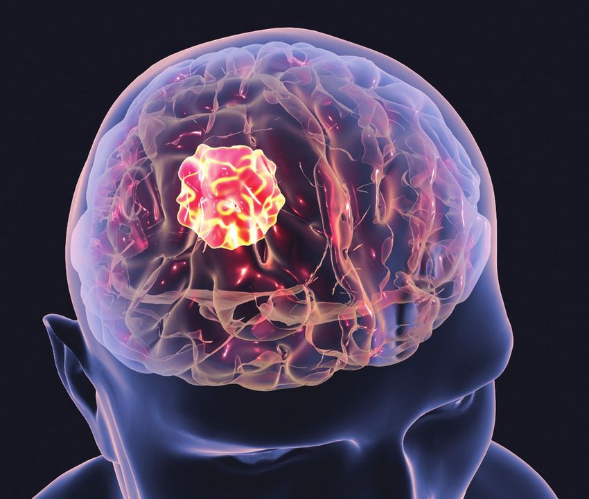
RESEARCH AND TREATMENT IN PATIENTS WITH BRAIN TUMORS

Brain Tumor Statistics

A    diagnosis of brain tumor is a life-altering event. Over 780,000 people in the United States are
currently living with a brain tumor, and nearly 84,000 will be diagnosed with a primary brain tumor
in 2021. Overall, the chance that a person will develop a malignant tumor of the brain or spinal cord
in his or her lifetime is less than 2%, but brain tumors account for the fourth-highest cancer for
numbers of years lost, averaging 20 years lost per patient. Despite advances in research and clinical
treatment, survival for malignant brain tumors remains poor overall. Up to 30% of patients with
metastatic cancer will have disease to the brain, which is even a more common finding in the era
of improved lifespan with cancer due to targeted molecular therapy and immunotherapy.

Brain Tumor Care at Jefferson Health

Jefferson Health performs a high volume of surgical and stereotactic radiation treatments annually,
in addition to clinical trial enrollment and non-operative management, making it one of the top-ranked
neuroscience and cancer care centers for brain tumors. Jefferson Health is one of the National
Cancer Institute’s 70 designated cancer centers in the nation and continues to have the highest
volume of treated brain tumors in the Greater Philadelphia region.

At Jefferson Health, our neurosurgeons and neuro-oncologists provide innovative treatment options
for tumors that are both complex and require a combination of treatments for the best outcomes.
Strong interdisciplinary patient management between neurosurgery, neuro-oncology, radiation
oncology, otolaryngology, neuro-ophthalmology, and critical care offer patients the best possible
treatment options. Clinicians also engage with community providers to allow patients the choice of
treatment closer to home if they desire. Jefferson Health serves as a major teaching facility with
professors and fellows visiting from around the world to learn leading-edge surgical techniques
as well as neuro-oncology and radiation oncology treatments.

The following pages describe some of the work currently taking place within the Vickie and Jack
Farber Institute of Neuroscience and Sidney Kimmel Cancer Center including Jefferson Health’s
treatment modalities and research activities in the area of brain tumor care. Our team of neurosurgeons,
neuro-oncologists, medical and radiation oncologists, dedicated nurses, and other allied healthcare
specialists combine their expertise to provide an aggressive treatment plan for brain tumor
patients and provide support for patients’ loved ones.

ONCOLOGY NURSE
                                                    NAVIGATORS

                           CASE
                         MANAGERS                                    SURGEONS

                                                                                    MEDICAL
   PATHOLOGISTS                                                                   ONCOLOGISTS

                                                   PATIENT

   RADIOLOGISTS                                                                    RADIATION
                                                                                  ONCOLOGISTS

                          PHYSICAL                                     GENETIC
                         THERAISTS                                   COUNSELORS

                                                      NURSING
                                                       TEAM

Figure 1: Multidisciplinary patient care Source: Jefferson Health.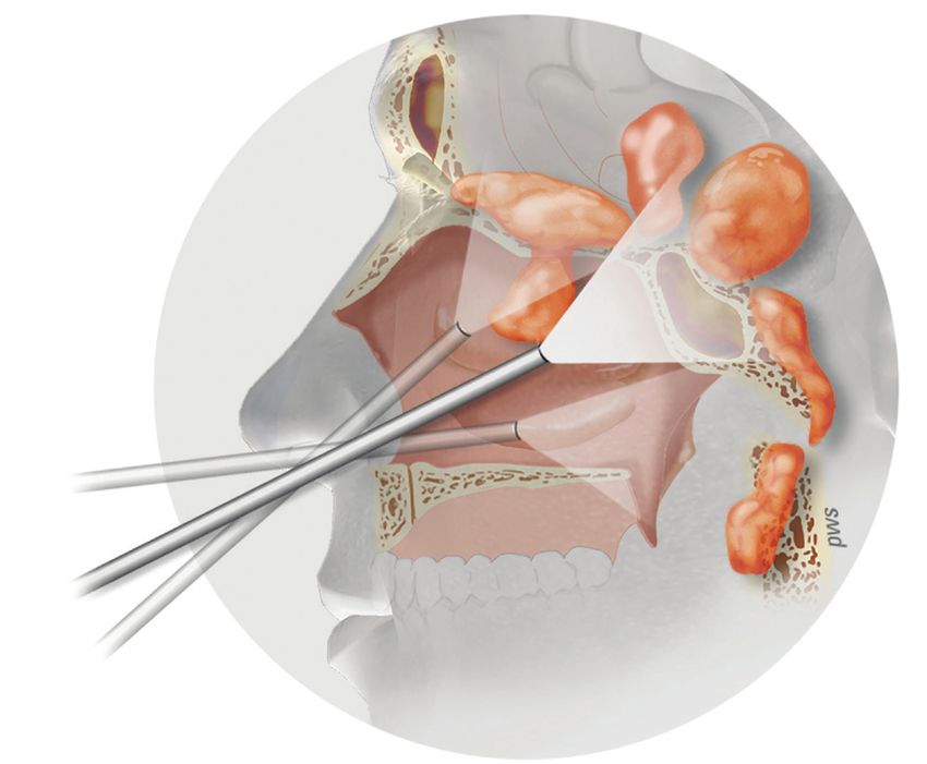
RESEARCH AT JEFFERSON ON BRAIN TUMORS

Dr. James J. Evans, Chief of the Brain Tumor Division, and Past President of the North American
Skull Base Society, along with his team of neurosurgeons, performs complex cranial, skull base, and
endoscopic minimally invasive neurosurgery to treat a wide range of disorders and tumors of the
brain and skull base. Below are several of his team’s studies.

Pituitary Adenoma

Giant pituitary adenomas, defined as >4 cm in maximal tumor diameter, compose ≈5% to 14% of
adenomas and present a particular surgical challenge given their size and frequent involvement of
critical neurovascular structures. Dr. Evans and colleagues reviewed the cases of 55 patients (mean age
55.2 years) with giant pituitary adenomas who underwent surgery with an endoscopic endonasal
approach (EEA) between 2008 and 2016. Factors affecting the extent of tumor removal were evaluated.

Gross total resection was achieved in 24 patients (44%), and near-total resection (>90%) in 26 patients
(47%) (Table 1). A multilobular configuration (P=0.002) and cavernous sinus invasion (P=0.044)

Figure 2. Outcomes after endonasal resection of giant pituitary adenomas. Source: Jefferson Health.

              Extent of Resection                                Outcomes in Patient with
                                                                    Visual Imparment

        Clinical Improvement of Presenting Symptoms                         Pituitary Outcomes
negatively affected the extent of resection, whereas tumor size, intraventricular, and anterior or
posterior fossa extension did not. Ten patients underwent adjuvant radiotherapy.

With regard to patient outcomes, 66% of patients showed visual improvements or even normalization,
whereas 31% exhibited stable vision. Visual deterioration occurred in only 1 patient. A new hormonal
deficit occurred in 8 patients (17%), whereas recovery of an existing hormonal deficit occurred in 6
patients (20%). Functional tumors were only seen in 4 patients and all patients with hormone-secreting
adenomas required adjuvant medical and/or radiotherapy to achieve biochemical remission. After
a mean follow-up of 41 months, tumor recurrence/progression occurred in 6 patients (11%).
Complications included apoplexy of residual tumor resulting in ischemic stroke in 1 patient,
postoperative cerebrospinal fluid leak in 1 patient, permanent diabetes insipidus in 4 patients (7%),
and medical complications in 3 patients (5%).

Table 1. Clinical Outcomes after Endonasal Resection Giant Pituitary Adenomas

     OUTCOME                                                                                         VALUE (%)

     Extent of resection (n=55)
       • Gross total resection                                                                       • 24 (44)
       • Near total resection (>90%)                                                                 • 26 (47)
       • Subtotal resection (1 pituitary axis)                                                             • 8/45 (17)
       • Unchanged function                                                                          • 40 (73)
       • Chemical remission (in secretory adenomas)                                                  • 4/4 (100)

     Clinical improvement of presenting symptoms
       • Headache                                                                                    • 7/9 (78)
       • Cranial neuropathy                                                                          • 3/3 (100)
       • Altered mental status                                                                       • 3/3 (100)
       • Hydrocephalus                                                                               • 22/22 (100)

Phasellus dictum dictum sagittis. Nam velit erat, elementum vitae semper nec, efficitur a nibh. Nam congue ullamcorper quam
eget auctor. Praesent scelerisque augue eros, vitae congue nunc semper eu. Etiam venenatis eleifend nibh semper laoreet. Sed ex
lorem, mollis ut commodo sed, semper quis tortor. Etiam congue hendrerit purus, eu venenatis felis bibendum vel.
Source: Jefferson Health
A                                            B                                           C

 D                                            E                                           F

 G                                            H

Figure 3: Examples of tumor resection in our series.
(A and B) Case 1. (A) Preoperative contrast-enhanced sagittal T1-weighted magnetic resonance imaging (MRI) showing a giant
adenoma with invasion of the sphenoid sinus, clivus, and anterior skull base and extension into the anterior cranial fossa and the
third ventricle. (B) Postoperative MRI showing gross total resection (GTR) after 2 staged endonasal surgeries. (C and D) Case 2. (C)
Preoperative contrast-enhanced coronal MRI demonstrating a giant adenoma with extension into the third ventricle and invasion
of the right cavernous sinus. (D) Postoperative MRI showing near- total resection (NTR) with small residual inside the cavernous
sinus (arrow). (E and F) Case 3. (E) Preoperative contrast-enhanced sagittal MRI showing a giant adenoma with suprasellar extension
and bilobular superior pole of the tumor, with 1 lobe extending into the anterior cranial fossa and the other lobe extending into
the third ventricle (arrows). (F) Postoperative MRI showing GTR of the tumor after an expanded endonasal approach. (G and H)
Case 4. (G) Preoperative contrast-enhanced coronal MRI showing extension of the tumor into the third ventricle causing localized
hydrocephalus within the right lateral ventricle (white arrow). (H) Postoperative MRI showing NTR of the lesion after a standard
transsellar approach with resolution of the hydrocephalus and very small residual attached to the anterior cerebral artery (arrow).
Republished with permission from Elshazly et al. Clinical Outcomes after Endoscopic Endonasal Resection of Giant Pituitary
Adenomas. World Neurosurg. (2018) 114:e447-e456.
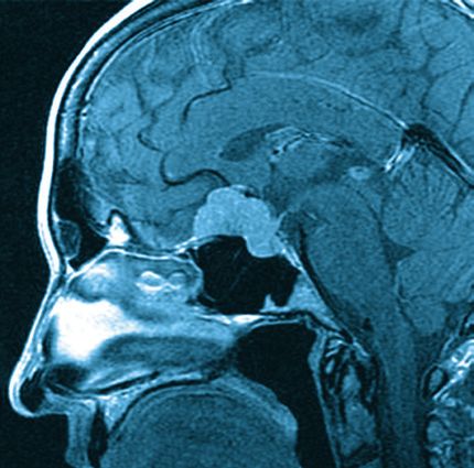
Figure 4: Potential treatment sites accessible with endonasal approaches. Source: Jefferson Health

This study supported the use of EEA as an excellent option for managing giant pituitary adenomas
and demonstrated at least comparable efficacy to traditional microscopic transsphenoidal and
transcranial approaches. The EEA for giant pituitary adenomas achieved high rates of near or
complete resection, and visual improvement as well as low rates of complications. Adjuvant radiation
treatment was useful for unresectable tumors and functional adenomas. Some tumors may require
staged approaches with either staged endonasal or staged craniotomy/endonasal surgeries, but one
must be aware of the risk of postoperative ischemia and apoplexy within giant pituitary adenomas
that have undergone subtotal resection.

REFERENCES

Khaled Elshazly, Varun R. Kshettry, Christopher J. Farrell, Gurston Nyquist, Marc Rosen, James J. Evans. Clinical
Outcomes after Endoscopic Endonasal Resection of Giant Pituitary Adenomas. World Neurosurg. (2018)
114:e447-e456.

Tuberculum Sella Meningiomas

Minimally invasive treatment of anterior skull base meningiomas is another area of focus for Jefferson
Health physicians. Led by Dr. Evans, Jefferson Health’s team is among the most experienced in the
world at treating these common benign brain tumors, using the most current endoscopic, microsurgical,
and stereotactic radiosurgery techniques.

Transcranial microsurgical resection via various approaches (e.g., pterional, orbitozygomatic) has
historically been the standard treatment for symptomatic tuberculum sella meningiomas (TSM). The
development of endoscopic endonasal approaches (EEA) has generated interest in treating these
tumors by less invasive procedures. Surgical goals are the same regardless of approach: preservation
or restoration of visual and neurological function with maximal safe tumor removal, including any
areas of dural and bony involvement.

In a recent study, Dr. Evans and his colleagues reviewed an institutional database of over 1000 EEA
cranial base cases for histologically confirmed meningiomas originating from the tuberculum sella
treated by an endonasal approach, with 25 consecutive cases identified from the time period of
March 2008 to January 2016.

Figure 5. Outcomes after endonasal resection of tuberculum sella meningiomas. Source: Jefferson Health.

             Extent of Resection                           Visual Outcomes After Surgery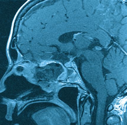
Table 2: Clinical Outcomes after Endonasal Resection of Tuberculum Sellae Meningiomas

     OUTCOME                                                                                         VALUE (%)

     Extent of resection after surgery
       • GTR overall                                                                                 • 19 (76)
       • GTR for patients with planned GTR                                                           • 19 (95)
       • NTR                                                                                         • 3 (12)
       • STR                                                                                         • 3 (12)

     Length of stay, days                                                                            • 6.6

     Visual impaired before surgery
       • Improved                                                                                    • 15/17 (88)
       • Stable                                                                                      • 2/17 (12)
       • Worsened                                                                                    • 0 (0)

     Headache improvement                                                                            • 4/4 (100)

     Complications
      • CSF leak                                                                                     • 2 (8)
      • Transient visual deterioration                                                               • 4 (16)
      • Transient diabetes insipidus                                                                 • 3 (12)
      • Permanent diabetes insipidus                                                                 • 0 (0)
      • New pituitary dysfunction                                                                    • 0 (0)
      • Hemorrhage                                                                                   • 1 (4)
      • Medical complications                                                                        • 1 (4)

Phasellus dictum dictum sagittis. Nam velit erat, elementum vitae semper nec, efficitur a nibh. Nam congue ullamcorper quam
eget auctor. Praesent scelerisque augue eros, vitae congue nunc semper eu. Etiam venenatis eleifend nibh semper laoreet. Sed ex
lorem, mollis ut commodo sed, semper quis tortor. Etiam congue hendrerit purus, eu venenatis felis bibendum vel.
Source: Jefferson Health

The mean patient age was 53.9 years, with female predominance (84%). Preoperatively, 84% of patients
had vision impairment and 68% had optic canal tumor invasion. Tumor was abutting or partially encasing
the anterior cerebral artery in 14 (56%) and 3 (12%) patients, respectively. The supraclinoid internal
carotid artery (ICA) was partially or completely encased in 4 (16%) and 4 (16%) patients, respectively.
Gross total resection (GTR) was achieved in 19/25 (76%) cases. Among patients without complete
ICA encasement, GTR was achieved in 19/20 (95%) patients. Optic canal invasion, tumor volume,
intratumoral calcifications, and partial vascular encasement were not limiting factors for GTR. A total
of 88% of patients with preoperative visual impairment had improvement or normalization of vision.
No patient experienced permanent visual deterioration or new permanent pituitary dysfunction.
Complete ICA encasement was the most common reason for subtotal resection. Complications
included cerebrospinal fluid leak in 2 (8%) cases, transient diabetes insipidus in 3 (12%) cases,
hemorrhage in 1 (4%) case, and medical complications in 1 (4%) case.
PREOPERATIVE POSTOPERATIVE Figure 6: EPreoperative and postoperative images demonstrate the endonasal resection of a tuberculum sellae meningioma. Source: Jefferson Health. These results strengthen the growing body of literature suggesting that EEA can provide high rates of complete resection and visual improvement with low rates of complications for TSM. Nonetheless, successful removal of these tumors requires significant experience in endoscopic endonasal cranial base surgery and an understanding of the indications and limitations of the EEA. In our experience, the primary limitation precluding GTR was complete ICA encasement. Conversely, optic canal invasion, tumor volume, partial ACA or ICA encasement, and significant tumor calcification were not found to be limiting factors.
Table 3: Anatomical and Pathological Factors Determining Optimal Surgical Approaches for Tuberculum
Sellae Meningiomas

     TRANSCRANIAL                                              TRANSNASAL

     Absolute indication                     Absolute indication
       • Extension lateral to optic nerve	  • Sellar extension with prefixed optic chiasm
       • Anterior cerebral artery encasement
       • “Kissing” carotids

     Relative indication                               Relative indication
       • Abuts anterior cerebral artery with history 	  • Tumor inferior to ipsilateral optic nerve
         of radiation
       • Ventral extension to cribiform

REFERENCES

Khaled Elshazly, Varun R. Kshettry, Christopher J. Farrell, Gurston Nyquist, Marc Rosen, James J. Evans. Clinical
Outcome after Endoscopic Endonasal Resection of tuberculum Sella Meningiomas. Oper Neurosurg. 2018
14(5):494-502.
Jefferson Health Researchers Challenge “Straw” Dogma
Evans and colleagues recently published results challenging the dogma of banning straws after
endonasal surgery, often an important tool in allowing patients to stay hydrated after surgery. The
intent was to study the common belief that the use of straws postoperatively in this patient population
is contraindicated due to the negative pressure created and the risk of exacerbating bleeding or
CSF leakage. This dogma has led to the restriction of straw use after endonasal procedures, such
as pituitary surgery, at most medical centers.
Pressure catheters were placed in the nasal cavity of healthy volunteers (N=20), and pressure
measurements were recorded while participants drank liquids of different viscosities from a cup
versus through a straw. Measurements were recorded with and without subjects occluding their nose
to simulate postoperative nasal obstruction. (See Figure 5). There were no statistically significant
differences in pressure when comparing drinking from a cup, using a straw, or occluding the nose
and using a straw (P>0.05).
10
9
Mean Negative Pressure (cm H2O)
8
7
6
5
4
3
2
1
0
Water (cm H2O) Milkshake (cm H2O)
n Cup n Straw n Occluding Nose and Using Straw
Figure 7. Pressure during Drinking Water vs Milkshake. Source: Jefferson Health
Conclusion: Straw use is not associated with the generation of significant negative pressure in the
nasal cavity. The pressure generated when drinking from a straw is not significantly different from that
of drinking from a cup. This supports that straw use may be safe for patients following endoscopic
skull base surgery.
REFERENCES
Erin K Reilly, et al. Straws Don’t Suck: Are Straws Dangerous after Endoscopic Skull Base Surgery? Journal of
Neurological Surgery, Part B: Skull Bas; August 2020, DOI: 10.1055/s-0040-1714095David W. Andrews, MD: Imvax and a Glioblastoma Immunotherapy Trial The Search for a Cure for Glioblastomas David W. Andrews, MD, is the Anthony Alfred Chiurco, MD Professor of Neurological Surgery, Vice Chair of Clinical Services in the Department of Neurological Surgery at Thomas Jefferson University Hospital. Dr. Andrews has spent the past two decades leading a team of researchers and clinicians at Jefferson Health working on immunotherapy treatments for rare and deadly types of brain cancers, such as glioblastoma multiforme. It was Dr. Andrews who established the Brain Tumor Division at Jefferson in 1995. In addition to his many duties and responsibilities at Jefferson Health, Dr. Andrews is also the Chief Medical Officer and Founder of Imvax, Inc. His work, at Jefferson Health and Imvax, is also dedicated to finding a vaccine to alleviate or eradicate glioblastoma, the most aggressive type of primary brain tumor with a prognosis of little more than a year with standard treatment. He is joined in this endeavor by immunologist, D. Craig Hooper, PhD, a professor of cancer biology at the Sidney Kimmel Cancer Center—Jefferson Health. Glioblastomas are deadly tumors – the same that killed Senators John McCain and Edward Kennedy, as well as Beau Biden. Imvax, an immunotherapy startup, is advancing a new treatment option for glioblastomas, using autologous tumor cell vaccine. Recently, interim results from a phase 1b clinical trial were released. This new experimental glioblastoma vaccine – developed by Jefferson Health and Imvax – showed the treatment was tolerated well by patients, slowed tumor recurrence, and prolonged patient survival. Recently, interim results from a phase 1b clinical trial were released and presented at the American Association for Cancer Research’s annual meeting in early 2019 and published in the journal, Clinical Cancer Research. Despite standard of care (SOC) established by Stupp (2005), glioblastoma remains a uniformly poor prognosis. The study evaluated IGV-001, which combines autologous glioblastoma tumor cells and an antisense oligonucleotide against insulin-like growth factor type 1 receptor (IMV-001), in newly diagnosed glioblastoma. Results were published by Dr. Andrews, Dr. Hooper and their team at Jefferson Health. This open-label protocol, approved by the IRB at Jefferson, collected tumor cells during resection. They were treated ex vivo with IMV-001, encapsulated in biodiffusion chambers with additional IMV-001, irradiated, then implanted in abdominal acceptor sites. Patients were randomized to 4 exposure levels and SOC was initiated 4-6 weeks later. Based on clinical improvements, randomization was halted after patient 23 and subsequent patients received only the highest exposure. Safety and tumor progression were primary and secondary objectives, respectively. Time-to-event outcomes were compared to the SOC arms of published studies.
The trial enrolled 33 patients with newly diagnosed glioblastoma who were treated with the novel
cancer vaccine (IGV-001) in the prospective phase 1b study. Median follow-up was 3.1 years. Six
patients had adverse events (grade =3), possibly related to IGV-001. Median progression-free survival
(PFS) was 9.8 months in the intent-to-treat population (vs. SOC, 6.5 months; P=.0003). In IGV-001-
treated patients who met Stupp-eligible criteria, PFS was 11.6 months overall (n=22; P=.001) and
17.1 months at the highest exposure (n=10; P=.0025). The greatest overall survival was observed in
Stupp-eligible patients receiving the highest exposure (median, 38.2 months; P=.044). Stupp-eligible
patients with methylated O6-methylguanine–DNA methyltransferase promoter (n=10) demonstrated
median PFS of 38.4 months (P=.0008). Evidence of immune activation was noted. IGV-001 was
well-tolerated, PFS compared favorably to SOC, and evidence suggested an immune-mediated
mechanism. (Clinicaltrials.gov NCT02507583). Imvax now plans a phase 2b trial for newly diagnosed
glioblastoma patients. In addition to Jefferson Health, several other medical centers on the East
Coast (Boston, Cleveland, and Washington, DC), will be involved in this trial.

Imvax has been named by Fierce Biotech as one of 2020’s Fierce 15 biotechnology companies,
designating the company as one of the most promising private biotechnology companies in the
industry. For patients diagnosed with the type of cancer that the Imvax vaccine work is focused on
there is great enthusiasm that the development of novel patient-specific vaccines and immunotherapy
strategies will provide the possibility of hope and someday, a cure.

REFERENCES

Stupp R, Mason WP, van den Bent MJ, Weller M, Fisher B, Taphoorn MJ, et al. Radiotherapy plus 410concomitant
and adjuvant temozolomide for glioblastoma. N Engl J Med. 2005;352(10):987-96 411doi 10.1056/NEJMoa043330.

David W. Andrews, Kevin D. Judy, Charles B. Scott, Samantha Garcia, Larry A. Harshyne, Lawrence Kenyon, Kiran
Talekar, Adam Flanders, Kofi-Buaku Atsina, Lyndon Kim, Nina Martinez, Wenyin Shi, Maria Werner-Wasik, Haisong
Liu, Mikhail Prosniak, Mark Curtis, Rhonda Kean, Donald Y. Ye, Emily Bongiorno, Sami Sauma, Mark A. Exley, Kara
Pigott and D. Craig Hooper. Phase 1b Clinical trial of IFV-001 for Patients with Newly Diagnosed Glioblastoma.
Cancer Res February 10 2021 DOI:10.1158/1078-0432.CCR-20-3805.
NEURO-ONCOLOGY RESEARCH                                       or Optune is an FDA-approved treatment for patients
                                                              with glioblastoma in the adjuvant phase of treatment
Iyad Alnahhas, MD, MSc, is a board-certified neuro-
                                                              after completion of radiation therapy. It delivers low
oncologist actively involved in translational and clinical
                                                              intensity alternating electric fields, which interfere
research projects in the Brain Tumor Division at
                                                              with cell division, using transducer arrays, and has
Jefferson. His primary focus of research is in cancer
                                                              shown positive results in a phase III clinical trial
genomics for gliomas and CNS lymphoma. One
                                                              (EF-14). In this investigator-initiated pilot trial, we
notable current translational research project addresses
                                                              are combining TTF upfront with radiation aiming to
the role of 5-Methylthioadenosine phosphorylase               increase the efficacy of this treatment against the
(MTAP) in isocitrate dehydrogenase (IDH) mutant               deadly disease. In addition, this effort is now being
gliomas, and the interaction between the epidermal            expanded to a large national phase III trial (the
growth factor receptor (EGFR) and platelet-derived            TRIDENT trial).
growth factor receptor (PDGFR) in IDH-wild-type
glioblastoma. These projects are in the process of being      Scalp-Sparing Intensity-Modulated Stereotactic
translated to investigator-initiated clinical trials. Other   Radiation Therapy in Treating Patients with Grade
projects include evaluating serum markers for radiation       II-IV Glioma
necrosis as well as advanced radiomics research.              Radiation Therapy (RT) is one of the mainstay treatments
                                                              for patients with gliomas. The goal of this phase II
INVESTIGATOR-INITIATED CLINICAL TRIALS                        clinical trial is to make RT better tolerated for patients.
                                                              The main objective is to determine the effect of
SPARE Scalp Preservation and Radiation Plus
                                                              scalp-sparing intensity-modulated (IM)-stereotactic
Alternating Electric Tumor Treatment Field (novoTTF,
                                                              radiotherapy (SRT) on the incidence of wound
Optune) for Patients with Glioblastoma: A Pilot Study
                                                              infection and/or wound dehiscence as well as scalp
Glioblastoma (GBM) is the most common malignant               thickness for patients with grade II-IV gliomas. The
brain cancer. Unfortunately, few advancements have            study also looks at the incidence of hair loss and
been made in the management of GBM despite                    recovery rate as well as quality of life of patients
extensive research efforts. Tumor-treating fields (TTF)       living with this disease.
Table 4. Current Open Central Nervous System Trials
Disease Title Note
Newly SPARE Scalp Preservation and Radiation Plus Alternating Electric Optune Spare
Diagnosed Tumor Treatment Field (novoTTF, Optune) for Patients with
GBM Glioblastoma: A Pilot Study
A Single Institution Pilot Study using head and neck Maskless MID
Immobilization Device (MID) for patients with intracranial tumors PI- Bar-Ad
Laser Ablation of Abnormal Neurological Tissue using Robotic LAANTERN
NeuroBlate System (LAANTERN) Prospective Registry
Various
Phase II Randomized Trial Of Stereotactic Radiotherapy (SRT) Aflibercept
Tumors
Followed by intravitreal Aflibercept Injection for patients with ocular
melanoma.
Intensity-modulated Stereotactic Radiotherapy as an Upfront Scalp- Scalp-Sparing
sparing Intervention for the Treatment of Newly Diagnosed Grade
II-IV Gliomas
Alliance Codel N0577 Phase III Intergroup Study of Radiotherapy
Anaplastic
with concomitant and adjuvant Temozolomide vs Radiotherapy
or Low N0577
Adjuvant PCV chemotherapy in Patients with 1p/19q deleted
Grade Glioma
Anaplastic Glioma
STELLAR Phase 3 Randomized, Open Label Study to evaluate the Stellar
efficacy and safety of Eflornithine with Lomustine compared to
Lomustine alone in patients with AA that progress/recur after
irradiation and adjuvant TMZ chemotherapy
Recurrent A Phase 1, Open-Label, Multicenter, Dose Escalation and Expansion PRT-811
Glioma Study of PRT811 in Subjects with Relapsed/Refractory Myelofibrosis,
Advanced Solid Tumors and Recurrent High-Grade Gliomas
Standard Chemotherapy versus Chemotherapy Chosen by Cancer Chemo ID
Stem Cell Chemosensitivity Testing in the Management of Patients
with Recurrent Glioblastoma Multiforme (GBM)
Neurocognition in Patients with Multiple Brain Metastases treated Neurocog
with Neurosurgery: A Phase II Study
Brain Phase III Trial of Post-Surgical Single Fraction Stereotactic SRS
Metastasis Radiosurgery (SRS) Compared with Fractionated SRS (FSRS) PI Dr. Wenyin
for Resected Metastatic Brain Disease Shi
Genomically-Guided Treatment Trial in Brain Metastases Genom
Surgery Ethicon: EVICEL Dual Sealing Clinical Trial EVICELVICKIE AND JACK FARBER INSTITUTE FOR NEUROSCIENCE
Department of Neurology
Department of Neurological Surgery
Department of Neuroscience
Department of Psychiatry
Patient Appointments: 1-800-JEFF-NOW
Patient Transfers: 1-800-JEFF-121
Physician Referrals: 215-503-5700
JeffersonHealth.org/Farber
PHILADELPHIA • MONTGOMERY COUNTY • BUCKS COUNTY • SOUTH JERSEY
CS 21-0427You can also read