"True" Versus "Bay" Apical Cysts: Clinical, Radiographic, Histopathologic, and Histobacteriologic Features
←
→
Page content transcription
If your browser does not render page correctly, please read the page content below
CLINICAL RESEARCH

“True” Versus “Bay” Apical                                                                                          Domenico Ricucci, MD, DDS,*
                                                                                                                                ^ças, DDS, MSc,
                                                                                                                    Isabela N. Ro
                                                                                                                    PhD,†‡ Sandra Herna ndez, DDS,
Cysts: Clinical, Radiographic,                                                                                      MSc,†x and Jose F. Siqueira, Jr.,
                                                                                                                    DDS, MSc, PhD†‡
Histopathologic, and
Histobacteriologic Features

ABSTRACT
                                                                                                                    SIGNIFICANCE
Introduction: This study compared the main clinical, radiographic, and histologic features of
true and bay apical cysts. Methods: The study material comprised 95 biopsy specimens of                             This study found no
apical periodontitis lesions obtained attached to the root tip of both untreated and root canal–                    differences between true and
treated teeth. Clinical and radiographic data were recorded. Specimens were obtained by                             bay cysts concerning clinical
extraction or periradicular surgery and were meticulously processed for histopathologic and                         and histopathologic
histobacteriologic methods. All cases diagnosed as apical cysts (n 5 23) were divided into the                      manifestations. Both types
true and bay types, which were then compared for tooth location, patient’s sex, lesion size,                        always exhibited intraradicular
severity of clinical symptoms, presence of a sinus tract, previous abscess episodes, and                            and sometimes extraradicular
prevalence of bacteria in the main root canal lumen and ramifications, on the outer root                             infection. Findings do not
surface, and within the cyst cavity. Results: Eleven specimens were classified as true (48%)                         support the assumption that
and 12 (52%) as bay cysts. Bacteria were found in all specimens, regardless of the                                  true cysts are self-sustainable
histopathologic diagnosis. Planktonic bacteria were observed in the main root canal in all true                     entities not associated with
cysts and in 11 of 12 (92%) bay cyst cases. Biofilms were detected in the main canal in 10                           infection.
cases from each diagnostic group and were frequently observed in ramifications.
Extraradicular biofilms occurred in a few specimens only. Bacteria were visualized within the
cavity of both true (4/11, 36%) and bay (6/12, 50%) cyst specimens. The severity of histologic
inflammation was always high. There were no significant differences between true and bay
cysts for all the clinical, radiographic, histopathologic, and histobacteriologic parameters
assessed. Conclusions: Except for the morphologic relationship of the cyst cavity with the
root canal space, true and bay cysts exhibited no other significant differences in the various
parameters evaluated. The 2 cyst types were always associated with an intraradicular
infection and sometimes with an extraradicular infection. Findings question the need to
differentiate true and bay cysts and do not support the assumption that true cysts are self-
sustainable entities not maintained by infection. (J Endod 2020;46:1217–1227.)

KEY WORDS
Apical periodontitis; bay apical cyst; biofilm; endodontic infection; true apical cyst
                                                                                                                    From the *Private Practice, Cetraro, Italy;
                                                                                                                    †
                                                                                                                      Department of Endodontics, Faculty of
In response to root canal infection, the periradicular tissues mount an immune reaction that may give rise          Dentistry, Grande Rio University, Rio de
to bone resorption and granuloma formation1. With the passage of time, the lesion may become                        Janeiro; ‡Department of Endodontics and
                                                                                                                    Dental Research, Iguaçu University, Nova
epithelialized as the epithelial cell rests of Malassez start to proliferate in the granuloma, and, ultimately, a
                                                                                                                    Iguaçu, Rio de Janeiro, Brazil; and
cavity lined by an epithelium is formed, which characterizes the apical cyst. The lumen of the apical cyst          x
                                                                                                                     Department of Endodontics, Francisco
cavity is usually lined by a stratified squamous epithelium, although in about 8% of the apical cysts the            Marroquín University, Guatemala City,
cavity may be partially or predominantly lined by ciliated columnar cells of respiratory origin2,3. Four            Guatemala
theories have tried to explain the genesis of the apical cyst cavity, including the breakdown theory4, the          Address requests for reprints to Dr
abscess theory5,6, the immunologic theory7, and the trapped connective tissue theory8, but none of them             Domenico Ricucci, Piazza Calvario, 7,
has been clearly demonstrated to be true.                                                                           87022, Cetraro (CS), Italy.
                                                                                                                    E-mail address: dricucci@libero.it
       Numerous studies have evaluated the prevalence of apical cysts among periradicular lesions.                  0099-2399/$ - see front matter
Apical granuloma is the most common histopathologic form of apical periodontitis in the large majority of
                                                                                                                    Copyright © 2020 American Association
studies, and the prevalence of cysts ranges from 6%–55%9–16. Cysts and granulomas cannot be                         of Endodontists.
distinguished by radiographic examination alone9,13,14, although large lesions are more likely to be                https://doi.org/10.1016/
cysts17,18. Although some studies have suggested that cysts can be differentiated from granulomas by                j.joen.2020.05.025

JOE  Volume 46, Number 9, September 2020                                                                                   True Versus Bay Cysts        1217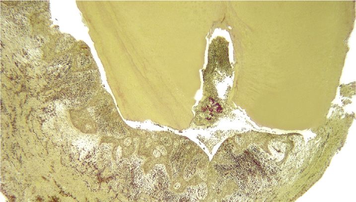
other approaches, including polyacrylamide           the bay cyst may be more prone to become             2. mild, when the patient reported no
gel electrophoresis of the periapical fluid,          infected by bacteria advancing directly from            episodes of spontaneous pain and no self-
tomography, and ultrasound real-time                 the canal into the cyst cavity, which might             medication with analgesics, but the tooth
imaging18–21, a definitive diagnostic                 impair healing even after proper intracanal             was slightly tender to chewing and
differentiation can only be attained by              antimicrobial treatment. Indeed, an infected            pressure;
histopathology, especially using a serial            bay cyst has been reported as the cause of           3. moderate, when the patient declared
sectioning approach to include the entire            nonsurgical treatment failure27.                        episodes of spontaneous pain and self-
lesion. Most of the studies that evaluated the               Accurate histopathologic diagnosis of           medication with analgesics, which
cyst prevalence did not perform serial               apical periodontitis lesions is reliant on serial       succeeded in resolving pain, and exhibited
sectioning, and this may have compromised            sectioning evaluation of a biopsy specimen              tenderness to percussion/palpation; and
the results and help explain the wide range          that represents the entire lesion.                   4. severe, when there was excruciating pain,
reported. The few studies that used serial           Histopathology of a limited number of sections          not resolved with analgesics, associated
sectioning or serial step sectioning showed a        may be confusing and may make the operator              with a painful response to percussion/
cyst prevalence of 15%–32% of the apical             erroneously classify an epithelialized                  palpation.
periodontitis lesions3,10,22.                        granuloma as a cyst or a bay cyst as a true
                                                                                                                  Information on the occurrence of acute
        Depending on the relationship of the         cyst. In addition, a proper differentiation
                                                                                                          abscess episodes related to the affected tooth
cyst cavity with the root canal via the apical       between true and bay cysts can only be made
                                                                                                          any time before surgery/extraction was also
foramen, the apical cyst has been classified as       if the lesion specimen remains attached to the
                                                                                                          available. These cases had been diagnosed by
a “true” or “bay” (also known as a “pocket”)         root apex obtained by periradicular surgery or
                                                                                                          the clinician based on the development of
cyst22,23. The lumen of the bay cyst cavity          extraction because the continuity of the cyst
                                                                                                          swelling and redness of the skin associated
communicates directly with the root canal            cavity with the root canal is essential
                                                                                                          with pain that prompted the patient to seek
system through the apical foramen with the           information. Comparisons of the
                                                                                                          professional aid. Some patients with
root apex protruding into the cavity, whereas        histopathologic features of the 2 conditions are
                                                                                                          abscesses had been treated with antibiotics.
the true cyst has a completely independent           limited in the literature, and no study has so far
                                                                                                                  The radiographic lesion size was
cavity with no continuity or connection to the       evaluated the histobacteriology of true and bay
                                                                                                          determined as the mean diameter of the
root canal. Simon22 used the serial sectioning       cysts.
                                                                                                          periapical radiolucency and categorized as
approach to evaluate apical periodontitis                    The present study used meticulous
                                                                                                          small if they were 5 mm and large if they were
lesions in untreated teeth that remained             serial sectioning and histopathologic/
                                                                                                          .5 mm. Teeth with periodontal pockets
attached to the root apices after extraction and     histobacteriologic evaluations to compare the
                                                                                                          communicating with the periapical lesion or
reported that true and bay cysts occurred in         main features of true and bay cysts that might
                                                                                                          teeth with vertical fractures were excluded
similar prevalences (ie, 9% of the lesions).         justify the alleged different biological behavior
                                                                                                          from the study.
Therefore, true and bay cysts each                   between them after root canal treatment.
                                                                                                                  To be included in the study, the
corresponded to 50% of the apical cysts.
                                                                                                          specimen obtained by extraction or surgery
Another study using serial sections or serial
                                                                                                          should have consisted of the entire apical
step sections of lesions adhered to the apices       MATERIALS AND METHODS
                                                                                                          periodontitis lesion still adhered to the root
reported that 9% were true cysts and 6% were
                                                     Clinical Specimens                                   apex. Periradicular surgery was performed as
bay cysts10. Of the apical cysts, 61.5% were
                                                     The study material was composed of 95                follows. After elevation of a full-thickness
true, and 38.5% were bay cysts. The study by
                                                     human biopsies of apical periodontitis lesions       periosteal flap, the buccal bone covering the
Ricucci et al3 was another one that evaluated
                                                     obtained attached to the root tip of both            lesion was carefully removed until the
serial sections from whole lesion specimens
                                                     untreated and root canal–treated teeth. The          pathologic tissue and the root tip were
attached to the apices of untreated extracted
                                                     specimens were obtained by endodontic                exposed. The root tip was first resected
teeth. Although 42% of the lesions showed an
                                                     specialists or oral surgeons through                 approximately 3 mm short of the apex with a
epithelium, the frequency of cysts was 32%.
                                                     periradicular surgery or extraction in private       fissure bur. Subsequently the soft tissue was
True cysts occurred in 16% of the lesions,
                                                     dental practices and dental schools and sent         carefully enucleated from the bone crypt with
whereas bay cysts corresponded to 18%. Of
                                                     consecutively over a period of 12 years to a         smooth microelevators, in an attempt to obtain
the apical cysts, 50% were true, and 56% were
                                                     single histologic laboratory. At the time of         the resected root tip and the surrounding
bay cysts (1 lesion contained the 2 types).
                                                     treatment, the patients were presented with          pathologic soft tissue in one piece.
        It was proposed in a study of untreated
                                                     risks, benefits, treatments, and options and                  The teeth were processed for light
teeth22 and was reinforced by only 1 case
                                                     had given consent for examination of their           microscopy. From this pool, only specimens in
report24 that the true cyst, assumed to be a
                                                     teeth. The patients’ mean age was 37.3 years         which a cyst cavity was detected in the
self-sustaining lesion, cannot heal after
                                                     (range, 15–82 years). The protocol for this          histologic sections (23 cases) were selected
nonsurgical root canal treatment, whereas bay
                                                     retrospective study was approved by the              for the present study.
cysts, especially the smaller lesions, can10,22.
                                                     institutional review board.
The rationale provided was that the bay cyst is
                                                             Clinical and radiographic data were
open to the root canal and then amenable to                                                               Histopathologic and
                                                     available for all teeth. Symptoms were
intracanal infection control, whereas the true                                                            Histobacteriologic Analyses
                                                     categorized as follows:
cyst represents a disease entity no more                                                                  Specimens were fixed in 10% buffered
dependent on the root canal infection and then       1. absent (asymptomatic), when the patient           formalin for at least 48 hours. Demineralization
not responsive to root canal treatment22,23.            reported no pain episodes and the tooth           was performed in a solution of 22.5% (vol/vol)
This theory has been strongly questioned                was comfortable with normal response to           formic acid and 10% (wt/vol) sodium citrate for
because it does not exhibit biological                  vertical/lateral percussion and periapical        a period of 3–4 weeks. The end point was
plausibility8,25. Siqueira26 raised the point that      palpation;                                        determined radiographically. For the

1218    Ricucci et al.                                                                                       JOE  Volume 46, Number 9, September 2020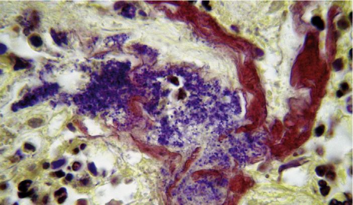
specimens obtained by extraction, the apical           presenting patterns of calcification                     Eleven specimens were identified as
4-to 5-mm segment of the root was separated            (calculuslike structures)                        true cysts (48%) and 12 (52%) as bay cysts.
with a sharp razor blade. At the end of the         3. The presence of bacteria/bacterial               The mean age for patients with true cysts
demineralization process, specimens were               aggregations within the cyst lumen. If           was 37 years (men 5 34 years and women 5
washed in running water for 24 hours and               present, occasional bacterial cells or           43 years), and for bay cysts, it was 43 years
dehydrated in ascending grades of ethanol              aggregations observed on the peripheral          (men 5 39 years and women 5 46 years).
(50%–100%). After clearing in xylene, they             collagenous surface of the periapical lesion     Treated teeth with true cysts were followed
were infiltrated and embedded in paraffin.               and not surrounded by an inflammatory             up for 5–10 years. One treated tooth with a
Next, the biopsies were oriented parallel to the       infiltrate were regarded as contaminants,         bay cyst was followed up for 4 years,
long axis of the main root canal in the apical         possibly resulting from the passage of the       whereas another tooth with a bay cyst had
third in order to obtain sections with the main        specimen through the socket during               showed persistent exudation and symptoms
canal and periapical tissue in direct continuity.      extraction; as such, they were excluded          that could not be resolved after several
Serial sections were taken with the microtome          from evaluation.                                 sessions of nonsurgical treatment and
set at 4–5 mm. Every fifth slide was stained with    4. The intensity of histologic inflammation was      required periradicular surgery.
hematoxylin-eosin for screening purposes in            ranked as none; mild, when limited                      The frequency of true cysts according to
order to locate the areas with the most severe         aggregations of exclusively chronic              the tooth type was as follows: maxillary
reactions. Additional slides were stained as           inflammatory cells were seen in the cyst          premolars (5), maxillary molars (3), maxillary
needed. Selected slides were stained with              lumen and/or a few chronic inflammatory           incisors (1), mandibular premolars (1) and
Masson trichrome to identify collagen and with         cells infiltrated the subepithelial connective    mandibular molars (1). For bay cyst, frequency
the Taylor modification of the Brown-Brenn              tissue; moderate, when discrete                  was as follows: maxillary molars (6), maxillary
stain for the presence of bacteria28.                  accumulations of chronic inflammatory             incisors (3), mandibular premolars (2) and
        Apical periodontitis lesions were              cells occurred in the cyst lumen and             maxillary premolars (1).
classified according to agreed                          epithelial walls with some occasional                   Bacteria were found in all cases
histomorphologic criteria into granulomas,             polymorphonuclear leukocytes (PMNs); or          examined, regardless of the histopathologic
abscesses, and cysts29,30. The diagnosis of a          severe, when large accumulations of acute        diagnosis (Figs. 1–5). Planktonic bacteria were
cyst was made when a distinct cavity lined by          and chronic inflammatory cells with a             observed in the main root canal in all true cyst
epithelium and filled with semisolid material           prevalence of PMNs were evident in the           cases and in 11 of 12 (92%) bay cyst cases.
was observed. Depending on the relationship            cyst lumen intermixed with necrotic debris       Biofilms were detected in the main canal in 10
between the epithelial lining of the cyst cavity       or cholesterol crystals or in the cyst walls,    cases from each of the diagnostic groups.
and the root and between the cyst cavity and           heavily infiltrating the epithelium.              These appeared to be very thick, often filling
the root canal space, cystic lesions were                                                               the entire canal lumen in the apical portion
differentiated into “true” or “bay” cysts22. The                                                        (Figs. 1A, C, and D and 2A, C, and D) and
lesions classified as true cysts were                                                                    faced with a severe concentration of PMNs
                                                    Statistical Analysis
characterized by the presence of a cavity                                                               (Fig. 1C and D). Biofilms were frequently
                                                    Statistical analysis was performed to evaluate if
bordered by an epithelial wall that was not                                                             observed in ramifications from both treated
                                                    there were differences between true and bay
continuous with the canal lumen in any of the                                                           and untreated teeth (Figs. 1B, 4B and D).
                                                    cysts regarding tooth location (maxillary or
serial histologic sections. The lesions classified                                                       Ramifications were sometimes clogged by
                                                    mandibular); patient’s sex; lesion size (small or
as bay cysts showed a cystic space                                                                      dense biofilms exhibiting different bacterial
                                                    large); severity of clinical symptoms (severe or
surrounded by an epithelial wall that joined the                                                        morphotypes, with the bacterial cells prevailing
                                                    not); presence of a sinus tract; history of a
external root surface forming a “sac,” isolating                                                        over the extracellular matrix component
                                                    previous acute abscess; and prevalence of
the foramen from the rest of the lesion. The bay                                                        (Fig. 4D).
                                                    planktonic bacteria or bacterial biofilms in the
cyst cavity had a direct opening into the canal                                                                Extraradicular biofilms formed on the
                                                    main canal lumen, biofilms in ramifications,
lumen.                                                                                                  external apical surface were detected only in a
                                                    extraradicular biofilm, and bacteria in the cyst
        In the 23 cases diagnosed as apical                                                             few specimens from both types of cysts. None
                                                    lumen. The Fisher exact test was used for all
cysts (true or bay), the following aspects were                                                         of them showed signs of calcification. These
                                                    analyses with the level of significance set at 5%
specifically looked for in the histopathologic                                                           biofilms exhibited varying proportions of cells
                                                    (P , .05).
and histobacteriologic analyses (Table 1):                                                              and extracellular substance, and in 1 instance
                                                                                                        the biofilm was observed between the layers of
1. The presence, location, and arrangement
                                                                                                        cementum that were detached from the
   of bacteria (planktonic or biofilm structures)    RESULTS                                             radicular surface (Fig. 4E).
   in the apical portion of the root canal
                                                    Twenty-three cystic lesions were obtained                  Bacterial cells occurred in the lumen of
   system, including the main canal lumen and
                                                    from 21 patients (11 men and 10 women)              true cysts in 4 of 11 (36%) specimens (Figs. 2A
   walls and apical ramifications (intraradicular
                                                    without contributory medical conditions. These      and B and 3A and B) and in 6 of 12 (50%) bay
   infection). The parameter used for biofilm
                                                    patients ranged in age from 20–70 years             cyst specimens (Figs. 4B, C, and F and 5B and
   classification was as defined elsewhere as
                                                    (mean age 5 40 years). Cyst specimens were          C). These bacteria were arranged in
   follows: “populations of microorganisms
                                                    collected from maxillary and mandibular             aggregations of varying sizes intermixed with
   that are concentrated at an interface and
                                                    treated and untreated teeth. Of the 23              necrotic debris and inflammatory cells,
   typically surrounded by an extracellular
                                                    specimens, 2 true cysts from different teeth        apparently free in the cyst lumen (Figs. 3A and
   polymeric substance matrix”31.
                                                    belonged to the same patient, and 2 bay cysts       B; 4B, C, and F; and 5B and C). In 2 cases (1
2. The presence of bacterial aggregations
                                                    from different teeth were from another patient.     bay and 1 true cyst), they were present in the
   adhering to the outer apical root surface
                                                    Twenty specimens were obtained from tooth           cyst lumen in the form of typical actinomycotic
   (extraradicular biofilm) eventually
                                                    extraction and 3 from apical surgery.               colonies, with intertwining branching

JOE  Volume 46, Number 9, September 2020                                                                                True Versus Bay Cysts     1219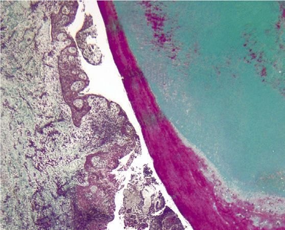
TABLE 1 - Clinical, Radiographic, Histopathologic, and Histobacteriologic Findings Associated with True and Bay Apical Cysts
True cyst Bay cyst
Total, N (%) Untreated, n (%) Treated, n (%) Total, n (%) Untreated, n (%) Treated, n (%) P value*
Specimens 11 7 4 12 10 2
Sex† .2
Male 7 (70) 4 (67) 3 (75) 4 (36) 3 (33) 1 (50)
Female 3 (30) 2 (33) 1 (25) 7 (64) 6 (67) 1 (50)
Tooth location 1.0
Maxillary 9 (82) 5 (71) 4 (100) 10 (83) 8 (80) 2 (100)
Mandibular 2 (18) 2 (29) 0 (0) 2 (17) 2 (20) 0 (0)
Lesion size .68
Small (5 mm) 5 (45) 4 (57) 1 (25) 7 (58) 7 (70) 0 (0)
Large (.5 mm) 6 (55) 3 (43) 3 (75) 5 (42) 3 (30) 2 (100)
Clinical symptoms .4
None 2 (18) 1 (14) 1 (25) 2 (17) 2 (20) 0 (0)
Mild 0 (0) 0 (0) 0 (0) 3 (25) 2 (20) 1 (50)
Moderate 6 (55) 4 (57) 2 (50) 1 (8) 1 (10) 0 (0)
Severe 3 (27) 2 (29) 1 (25) 6 (50) 5 (50) 1 (50)
Previous acute abscess .4
Yes 6 (55) 4 (57) 2 (50) 9 (75) 8 (80) 1 (50)
No 5 (45) 3 (43) 2 (50) 3 (25) 2 (20) 1 (50)
Sinus tract .6
Yes 1 (9) 1 (14) 0 (0) 3 (25) 2 (20) 1 (50)
No 10 (91) 6 (86) 4 (100) 9 (75) 8 (80) 1 (50)
Planktonic bacteria 1.0
Main canal 11 (100) 7 (100) 4 (100) 11 (92) 10 (100) 1 (50)
Bacterial biofilm
Main canal 10 (91) 6 (86) 4 (100) 10 (83) 10 (100) 0 (0) 1.0
Ramifications 10 (91) 7 (100) 3 (75) 9 (75) 7 (70) 2 (100) .59
Extraradicular 1 (9) 1 (14) 0 (0) 3 (25) 2 (20) 1 (50) .59
Bacteria in the cyst cavity 4 (36) 3 (43) 1 (25) 6 (50) 5 (50) 1 (50) .68
*Fisher’s exact test.
†
One patient contributed 2 true cysts and another patient contributed 2 bay cysts.
filamentous bacteria surrounded by a severe histobacteriologic parameters evaluated (P . between the cyst cavity and the root canal
accumulation of PMNs (Fig. 2A and B). .05). Data are shown in Table 1. space, there were no other significant
The severity of inflammation was high in differences between true and bay cysts in
all specimens, so it was not subjected to terms of clinical, radiographic, histopathologic,
statistical analysis. All kinds of inflammatory DISCUSSION and histobacteriologic features. These findings
cells were observed in the cyst lumen question the need to subdivide the
This study compared the prevalence and
together with varying amounts of amorphous several features of true and bay cysts classification of apical cysts.
necrotic debris. The epithelial lining of all but 4 diagnosed histopathologically using a The frequency of cysts observed in this
radicular cysts was constituted by a typical study was 24% (23/95) of the apical
meticulous research protocol. All apical
stratified squamous epithelium. One true cyst periodontitis lesions included in this study were periodontitis lesions examined. When
and 3 bay cysts in 4 maxillary (one third, one obtained attached to the teeth and therefore compared with other studies that also
second, and two first) molars showed the maintaining their natural morphologic evaluated serial or serial step sections of
characteristics of a respiratory epithelium, with lesions attached to the apex, this figure is
relationship with the root apex. This strict
ciliated epithelial cells and goblet cells in criterion was not used in the large majority of higher than the 15% and 17% reported by Nair
limited portions of the cyst walls. The epithelial studies evaluating the prevalence of the et al10 and Simon22, respectively, but is lower
lining was in general thick and irregular, with than the 32% reported by Ricucci et al3
different types of apical periodontitis lesions
extensive proliferation and the tendency to because it is more difficult to meet. This (Table 2). As for the prevalence of the 2 types
form arcading structures (Figs. 1A; 2A; 3A; justifies the smaller number of cases evaluated of apical cysts, this study is in agreement with
4A; and 5A, B, and D) with entrapped islands two others that shown that each type occurs in
herein. Serial sectioning from 1 side to the
of highly vascularized connective tissue other of the apical periodontitis lesion was also approximately half of the cases with a cyst
(Fig. 5D and E). The strands of epithelium and performed in this study to report on the diagnosis3,22. The true cyst corresponded to
the connective tissue islands were heavily 12% of all lesions, which is within the range
relationship of the lesion to the apical foramina.
infiltrated by acute and chronic inflammatory These 2 approaches are essential for an previously reported of 9% to 16% of all lesions.
cells (Fig. 5E and F). accurate histopathologic diagnosis of cysts Corresponding figures for the bay cyst were
There were no significant differences and their classification as true or bay types. 13% in this study, in a previously reported
between true and bay cysts for all the clinical, range of 6% to 18% of all lesions3,10,22
Findings from the present study showed that,
radiographic, histopathologic, and apart from the morphologic relationship (Table 2).
1220 Ricucci et al. JOE Volume 46, Number 9, September 2020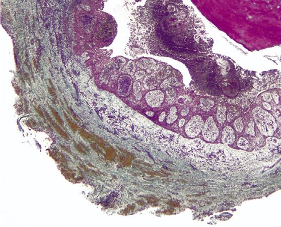
A B
C D
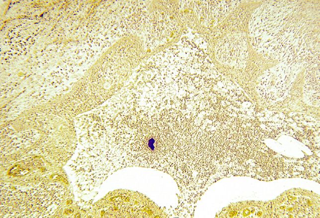
FIGURE 1 – A bay cyst. (A ) A section cut through the main canal and foramen. The cyst lumen is continuous with the root canal space (Taylor modified Brown and Brenn, original
magnification !16). (B ) An area of the canal located approximately 5 mm coronal to the foramen (not shown in A ). Biofilms with low bacterial density in the main canal and
ramifications (original magnification !50). (C ) Detail of the area of the apical canal indicated by the arrow in A. A thick bacterial biofilm fills the entire lumen and is faced with an
accumulation of inflammatory cells. Note the “floc” displaced apically in the inflammatory tissue (arrow ) (original magnification !100). (D ) A high-power view of the biofilm showing a
dense aggregation of filamentous forms (original magnification !400).
No previous study had compared the assumption that true cysts can be an cavity and 1 as a biofilm adhered to the outer
histobacteriology of true and bay cysts. No independent entity in the absence of root surface. Of these 5 cases, 4 had a history
distinct pattern of infection was observed concomitant bacterial infection because of previous acute abscess, and 1 had a sinus
between the 2 types. Intraradicular bacteria infection occurred in all cases. tract. These may have been the most possible
were found in all cases, regardless of the Extraradicular bacteria were found in explanations for the bacteria detected in the
associated cyst type and the root canal status many cases, either inside the cyst lumen or in extraradicular space, particularly in the cyst
(untreated or treated). Planktonic bacteria were fewer cases as a biofilm adhered to the outer lumen. After resolution of the abscess,
observed in the main canal in all teeth, except root surface near the exits of apical foramina. bacteria may have persisted in the
for 1 bay cyst specimen, in which bacteria The histobacteriologic method permits better periradicular tissues and maintained a chronic
occurred as a biofilm in ramifications and in the distinction of bacterial contaminants when infectious process. Even though the case with
extraradicular space. Bacteria organized in compared with other methods of bacterial a sinus tract had not had a previous report of
biofilm structures were observed in the main detection, such as culture and molecular acute abscess, this might have occurred with
apical canal in the large majority of specimens, methods, because it provides information on subclinical symptoms to justify the fistula.
except for 1 true cyst and 2 bay cyst cases. In bacterial spatial location in the lesion and the Cases with a sinus tract have been shown to
response to bacteria present in the root canal association with inflammation. Specimens harbor an extraradicular infection in about
and sometimes inside the lesion, severe with contaminants were excluded from the 83% of the teeth32. Bacteria present within
inflammation was observed in both cyst types. study. Extraradicular bacteria were found in 5 the cyst cavity are beyond the reach of
The present findings do not give support to the specimens of true cysts, 4 within the cyst nonsurgical root canal procedures and are
JOE Volume 46, Number 9, September 2020 True Versus Bay Cysts 1221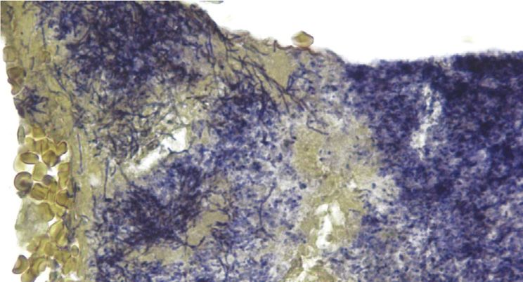
A                                                               B

                           C                                                                                                                             D

FIGURE 2 – A true cyst. (A ) A section cut through 2 ramifications filled with bacterial biofilms. The cyst lumen is separated from the root canal space (this anatomic feature is
maintained in all sections of the series) (Taylor modified Brown and Brenn, original magnification !16). (B ) Detail of the cyst cavity whose lumen is filled with cells and debris. A high-
power view of the blue spot at the center of the cyst lumen reveals an actinomycoticlike colony surrounded by a severe concentration of PMNs (original magnification !50, inset !
630). (C ) A section cut approximately 150 sections from that in A disclosing another large ramification clogged with a thick bacterial biofilm (original magnification !16). (D ) Middle
magnification from C (original magnification !100).

located in an area that makes it difficult for the              However, the fate of bacteria eventually                        There are no reports demonstrating that
host defenses to eliminate, given the type and                 persisting in a cyst cavity after elimination of                bacterial colonies located inside the cyst
consistency of the cyst lumen content.                         the intraradicular component is not known.                      cavity may cause root canal treatment failure

                           A                                                                   B

FIGURE 3 – A true cyst. (A ) A large cyst cavity containing some debris (Taylor modified Brown and Brenn, original magnification !16). (B ) Magnification of the rectangular area in A.
Large numbers of bacteria colonize the necrotic debris in the lumen (original magnification ! 100, inset ! 400).

1222      Ricucci et al.                                                                                                           JOE  Volume 46, Number 9, September 2020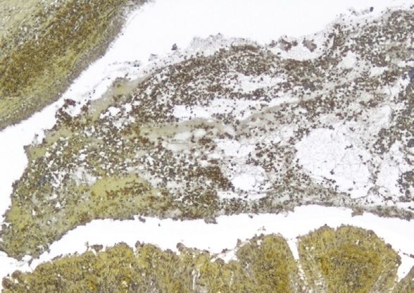
A                                                                   B                                                   C

                                                                               E

       D                                                                               E                                             F

FIGURE 4 – A bay cyst. (A ) A section encompassing the root canal, foramen, and cyst cavity. Overview (hematoxylin-eosin, original magnification !8). (B ) A section taken 80
sections away passing through the canal but not including the main foramen. Several ramifications can be seen in the thickness of the left canal wall (Taylor modified Brown and Brenn,
original magnification !16). (C ) Magnification of the area demarcated by the rectangle in B (original magnification !100). (D ) A high-power view of the exit of the lateral canal
indicated by the arrow in B. Its lumen is occupied by a dense biofilm showing varying bacterial morphotypes (original magnification !400). (E ) A high-power view of the area indicated
by the arrow in C. Filaments prevail in a biofilm structure adhering to the radicular surface and showing varying bacterial concentrations (original magnification !400). (F ) A high-
power view of the elongated free colony in C (original magnification !400).

in the absence of a concomitant intraradicular                cutting approach is used in that stained                      including the lesions examined in the present
infection.                                                    sections are examined under the light                         investigation.
        The only case report that suggested                   microscope to locate sites for further                                The assumption that the true cyst
that true cysts could be a self-sustaining entity             sectioning and evaluation in a transmission                   becomes a pathologic entity independent of
independent from canal infection found no                     electron microscope. Miniature pyramids are                   the root canal system and is not affected by
bacteria in the canal by culture and a                        prepared at the selected sites that show                      nonsurgical root canal treatment is only
correlative light and electron microscopic                    bacteria in light microscopy or that are likely to            speculative and has neither scientific evidence
approach24. However, bacteria may have                        harbor bacteria34. The low sensitivity for                    support nor biological plausibility8,25. Although
passed unnoticed when using methods with                      bacterial detection is recognized by the                      this cyst type has no apparent communication
low sensitivity for bacterial detection. A                    authors themselves—“the extremely limited                     with the root canal, this by no means can be
negative culture does not guarantee that the                  area that still can be covered by this means of               interpreted as being a separate disease entity.
root canal is free of bacteria, especially                    investigation makes it rather easy for bacteria               The etiology is the same as the bay cyst (ie,
because of the limitations of the culture                     to go undetected”34. This is because of the                   infection of the root canal system that causes
technologies in detecting difficult-to-grow and                loss of biopsy material due to preparation for                periradicular inflammation), and some locally
as-yet-uncultivated bacteria and the inability                transmission electron microscopy and the                      released inflammatory mediators serve as
to detect bacteria at low levels33. In addition,              very small area that can be examined under a                  growth factors for epithelium proliferation and
the correlative light and electron microscopic                transmission electron microscope. In turn, the                cyst formation. There is no reason to believe
approach used in the previous study can                       histobacteriology approach used in this study                 that the epithelium lining of the true cyst can
provide highly detailed information from some                 has been very successful in revealing bacteria                become self-sustainable, like neoplastic
selected areas but has a very low sensitivity to              in the vast majority of cases with                            lesions8,25. The present study lends
detect bacteria. In that method, a serial step-               posttreatment apical periodontitis27,35,36,                   substantial additional information to debunk

JOE  Volume 46, Number 9, September 2020                                                                                                        True Versus Bay Cysts         1223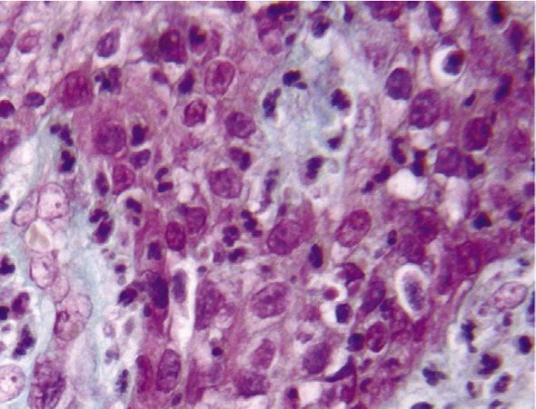
A                                                                 B                                          C

         D                                                                 E

         F

FIGURE 5 – A bay cyst. (A ) A section cut through the apical canal and foramen (Taylor modified Brown and Brenn, original magnification !25). (B ) A section taken approximately 80
sections away from that in A, not encompassing the apical foramen (original magnification !25). (C ) A high-power view of the area of the cyst lumen indicated by the arrow in B.
Bacterial aggregation intermixed with amorphous fuchsin-stained material and inflammatory cells (original magnification !630). (D ) Another section of the series taken 40 sections
after that in B. The cyst lumen is lined by a thick wall of arcading epithelium (Masson trichrome, original magnification !25). (E ) A high-power view of epithelial strands indicated by the
arrow in D. The epithelium surrounds cores of connective tissue infiltrated with inflammatory cells (original magnification !400). (F ) The strands appear infiltrated by polymor-
phonuclear neutrophils (original magnification !1000).

1224      Ricucci et al.                                                                                                             JOE  Volume 46, Number 9, September 2020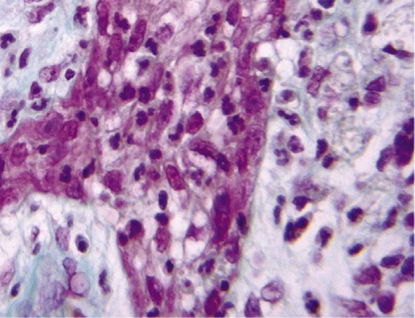
TABLE 2 - The Prevalence of Apical Cysts and Their 2 Types in Studies That Used Histopathologic Serial or Serial Step Sectioning to Evaluate Lesions Attached to the Root Apices

                                   Cyst among                 True cyst among                Bay cyst among               True cyst among                Bay cyst among
 Study                          all lesions, n (%)            all lesions, n (%)             all lesions, n (%)            all cysts, n (%)               all cysts, n (%)
 Simon, 198022                        6/35 (17)                     3/35 (9)                      3/35 (9)                      3/6 (50)                      3/6 (50)
 Nair et al, 199610                 39/256 (15)                   24/256 (9)                    15/256 (6)                    24/39 (61.5)                  15/39 (38.5)
 Ricucci et al, 20063                16/50 (32)                     8/50 (16)                     9/50 (18)                    8/16 (50)                     9/16 (56)
 This study                          23/95 (24)                    11/95 (12)                    12/95 (13)                   11/23 (48)                    12/23 (52)

this theory because true and bay cysts                      complete healing in 74% and incomplete                       true and bay cysts as to their prevalence,
showed no significant differences for all                    healing in 9.5% of cases40. Therefore, it seems              clinical, radiographic, histopathologic, and
variables evaluated. In addition, the cysts that            reasonable to assume that apical cysts,                      histobacteriologic manifestations. The 2 types
represented posttreatment apical periodontitis              regardless of their types, can heal provided the             of cysts were always associated with an
were of both types, and concurrent bacterial                source of epithelial proliferation (ie, the root             intraradicular infection and sometimes with an
infection was always present inside the canal               canal infection) is eliminated by treatment. If              extraradicular infection as well. The fact that
and sometimes outside. Therefore, it seems                  this is true, differentiation between granulomas             the 2 types of cysts only apparently differ from
evident that failure of the endodontic                      and cysts or true and bay cysts would be of no               the morphologic relationship of the cavity to
treatment, including cases of true cysts, is                clinical relevance.                                          the root canal puts into question the real need
primarily caused by persistent or secondary                         A curious finding from this study was                 to differentiate them and subdivide the apical
intraradicular bacterial infection with, in some            that from the 2 patients who contributed 2                   cyst into categories. Finally, the fact that all
cases, concurrent extraradicular bacterial                  specimens each, 1 had 2 true cysts associated                cases of true cysts from both untreated and
infection.                                                  with 2 different teeth and the other had 2 bay               treated teeth had an infectious component
         A definitive answer as to whether cysts             cysts from different teeth as well. This might               does not support the assumption that true
(all or only the “true” ones) can heal or not after         suggest an individual predisposition to develop              cysts are self-sustainable entities not
nonsurgical root canal treatment would only be              a true or a bay cyst, but the limited number of              associated with infection.
provided if cysts and other lesions could be                patients providing more than 1 specimen was
distinguished without biopsy. A differentiation             too small to draw such a conclusion.
                                                                                                                         CREDIT AUTHORSHIP
cannot be achieved by radiographs alone, and                        As typically found in apical cysts, a
the effectiveness of the other methods has not              stratified squamous epithelium was found
                                                                                                                         CONTRIBUTION STATEMENT
been confirmed; only an old study using                      composing the lining of the cyst cavity in the               Domenico Ricucci: Investigation,
polyacrylamide gel electrophoresis of the root              large majority of specimens. However, 4 of 23                Methodology, Writing - review & editing,
canal aspirates to differentiate granulomas and             cysts (17%), all in the maxillary region, showed             Visualization. Isabela N. Ro^ ças: Data
cysts suggested that many cysts healed after                a ciliated columnar epithelium partially or                  curation, Formal analysis, Validation. Sandra
root canal treatment37. Indirect evidence that              completely lining the cyst wall. All involved                Herna  ndez: Data curation, Formal analysis.
cysts heal can be inferred from the fact that the           teeth were maxillary molars, and the                         Jose F. Siqueira: Conceptualization, Formal
success rate of endodontic treatment is higher              occurrence of a ciliated columnar epithelium in              analysis, Writing - review & editing.
than the cyst prevalence9,15,38,39. Moreover,               the lesions may be related to the proximity of
suggestive evidence is given by a study in                  the root apexes and lesions to the maxillary
which the nonsurgical root canal treatment of               sinus floor2.
                                                                                                                         ACKNOWLEDGMENTS
large cysticlike lesions with a fluid content                        In conclusion, the present findings                   The authors deny any conflicts of interest
containing cholesterol crystals resulted in                 revealed no significant differences between                   related to this study.

                                                            REFERENCES
                                                             1.    Sasaki H, Stashenko P. Interrelationship of the pulp and apical periodontitis. In: Hargreaves KM,
                                                                   Goodis HE, Tay FR, editors. Seltzer and Bender’s Dental Pulp. 2nd ed. Chicago: Quintessence
                                                                   Publishing; 2012. p. 277–99.
                                                             2.    Ricucci D, Loghin S, Siqueira JF Jr, Abdelsayed RA. Prevalence of ciliated epithelium in apical
                                                                   periodontitis lesions. J Endod 2014;40:476–83.
                                                             3.    Ricucci D, Pascon EA, Pitt Ford TR, Langeland K. Epithelium and bacteria in periapical lesions.
                                                                   Oral Surg Oral Med Oral Pathol Oral Radiol Endod 2006;101:239–49.

                                                             4.    Ten Cate AR. The epithelial cell rests of Malassez and the genesis of the dental cyst. Oral Surg Oral
                                                                   Med Oral Pathol 1972;34:956–64.

                                                             5.    Summers L. The incidence of epithelium in periapical granulomas and the mechanism of
                                                                   cavitation in apical dental cysts in man. Arch Oral Biol 1974;19:1177–80.
                                                             6.                            €gren U. Experimental evidence supports the abscess theory of
                                                                   Nair PN, Sundqvist G, Sjo
                                                                   development of radicular cysts. Oral Surg Oral Med Oral Pathol Oral Radiol Endod
                                                                   2008;106:294–303.

JOE  Volume 46, Number 9, September 2020                                                                                                    True Versus Bay Cysts        1225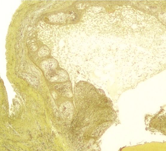
7.    Torabinejad M. The role of immunological reactions in apical cyst formation and the fate of
       epithelial cells after root canal therapy: a theory. Int J Oral Surg 1983;12:14–22.

 8.    Lin LM, Huang GT, Rosenberg PA. Proliferation of epithelial cell rests, formation of apical cysts,
       and regression of apical cysts after periapical wound healing. J Endod 2007;33:908–16.
 9.    Bhaskar SN. Periapical lesion: types, incidence, and clinical features. Oral Surg Oral Med Oral
       Pathol 1966;21:657–71.
10.    Nair PN, Pajarola G, Schroeder HE. Types and incidence of human periapical lesions obtained
       with extracted teeth. Oral Surg Oral Med Oral Pathol Oral Radiol Endod 1996;81:93–102.

11.    Block RM, Bushell A, Rodrigues H, Langeland K. A histopathologic, histobacteriologic, and
       radiographic study of periapical endodontic surgical specimens. Oral Surg Oral Med Oral Pathol
       1976;42:656–78.

12.    Sommer RF, Ostrander FD, Crowley MC. Clinical Endodontics: A Manual of Scientific
       Endodontics. Philadelphia: WB Saunders Co; 1966.
13.    Mortensen H, Winther JE, Birn H. Periapical granulomas and cysts. An investigation of 1,600
       cases. Scand J Dent Res 1970;78:241–50.

14.    Priebe WA, Lazansky JP, Wuehrmann AH. The value of the roentgenographic film in the
       differential diagnosis of periapical lesions. Oral Surg Oral Med Oral Pathol 1954;7:979–83.

15.    Lalonde ER, Luebke RG. The frequency and distribution of periapical cysts and granulomas. An
       evaluation of 800 specimens. Oral Surg Oral Med Oral Pathol 1968;25:861–8.
16.    Lin LM, Pascon EA, Skribner J, et al. Clinical, radiographic, and histologic study of endodontic
       treatment failures. Oral Surg Oral Med Oral Pathol 1991;71:603–11.

17.    Lalonde ER. A new rationale for the management of periapical granulomas and cysts: an
       evaluation of histopathological and radiographic findings. J Am Dent Assoc 1970;80:1056–9.

18.    Morse DR, Patnik JW, Schacterle GR. Electrophoretic differentiation of radicular cysts and
       granulomas. Oral Surg Oral Med Oral Pathol 1973;35:249–64.
19.    Trope M, Pettigrew J, Petras J, et al. Differentiation of radicular cyst and granulomas using
       computerized tomography. Endod Dent Traumatol 1989;5:69–72.
20.    Simon JH, Enciso R, Malfaz JM, et al. Differential diagnosis of large periapical lesions using cone-
       beam computed tomography measurements and biopsy. J Endod 2006;32:833–7.

21.    Cotti E, Campisi G, Ambu R, Dettori C. Ultrasound real-time imaging in the differential diagnosis
       of periapical lesions. Int Endod J 2003;36:556–63.
22.    Simon JH. Incidence of periapical cysts in relation to the root canal. J Endod 1980;6:845–8.

23.    Nair PN. Non-microbial etiology: periapical cysts sustain post-treatment apical periodontitis.
       Endod Topics 2003;6:96–113.

24.    Nair PN, Sjo€gren U, Schumacher E, Sundqvist G. Radicular cyst affecting a root-filled human
       tooth: a long-term post-treatment follow-up. Int Endod J 1993;26:225–33.
25.    Lin LM, Ricucci D, Lin J, Rosenberg PA. Nonsurgical root canal therapy of large cyst-like
       inflammatory periapical lesions and inflammatory apical cysts. J Endod 2009;35:607–15.

26.    Siqueira JF Jr. Aetiology of root canal treatment failure: why well-treated teeth can fail. Int Endod J
       2001;34:1–10.

27.    Ricucci D, Siqueira JF Jr, Lopes WS, et al. Extraradicular infection as the cause of persistent
       symptoms: a case series. J Endod 2015;41:265–73.
28.    Taylor RD. Modification of the Brown and Brenn Gram stain for the differential staining of gram-
       positive and gram-negative bacteria in tissue sections. Am J Clin Pathol 1966;46:472–6.

29.    Ricucci D, Siqueira JF Jr. Endodontology. An Integrated Biological and Clinical View. London,
       UK: Quintessence Publishing; 2013.

30.    Ricucci D, Mannocci F, Pitt Ford TR. A study of periapical lesions correlating the presence of a
       radiopaque lamina with histological findings. Oral Surg Oral Med Oral Pathol Oral Radiol Endod
       2006;101:389–94.

31.    Hall-Stoodley L, Costerton JW, Stoodley P. Bacterial biofilms: from the natural environment to
       infectious diseases. Nat Rev Microbiol 2004;2:95–108.
32.    Ricucci D, Loghin S, Gonçalves LS, et al. Histobacteriologic conditions of the apical root canal
       system and periapical tissues in teeth associated with sinus tracts. J Endod 2018;44:405–13.

33.                     ^ças IN. Exploiting molecular methods to explore endodontic infections: Part 1-
       Siqueira JF Jr, Ro
       current molecular technologies for microbiological diagnosis. J Endod 2005;31:411–23.

1226     Ricucci et al.                                                                                          JOE  Volume 46, Number 9, September 2020
34. €gren U, Krey G, et al. Intraradicular bacteria and fungi in root-filled, asymptomatic
Nair PN, Sjo
human teeth with therapy-resistant periapical lesions: a long-term light and electron microscopic
follow-up study. J Endod 1990;16:580–8.
35. Ricucci D, Siqueira JF Jr. Biofilms and apical periodontitis: study of prevalence and association
with clinical and histopathologic findings. J Endod 2010;36:1277–88.
36. Ricucci D, Siqueira JF Jr, Bate AL, Pitt Ford TR. Histologic investigation of root canal-treated teeth
with apical periodontitis: a retrospective study from twenty-four patients. J Endod 2009;35:493–
502.
37. Morse DR, Wolfson E, Schacterle GR. Nonsurgical repair of electrophoretically diagnosed
radicular cysts. J Endod 1975;1:158–63.
38. Ricucci D, Russo J, Rutberg M, et al. A prospective cohort study of endodontic treatments of
1,369 root canals: results after 5 years. Oral Surg Oral Med Oral Pathol Oral Radiol Endod
2011;112:825–42.
39. Ørstavik D. Time-course and risk analyses of the development and healing of chronic apical
periodontitis in man. Int Endod J 1996;29:150–5.
40. Çalısxkan MK. Prognosis of large cyst-like periapical lesions following nonsurgical root canal
treatment: a clinical review. Int Endod J 2004;37:408–16.
JOE Volume 46, Number 9, September 2020 True Versus Bay Cysts 1227You can also read