Relationships of Overt and Silent Brain Lesions With Cognitive Function in Patients With Atrial Fibrillation
←
→
Page content transcription
If your browser does not render page correctly, please read the page content below
JOURNAL OF THE AMERICAN COLLEGE OF CARDIOLOGY VOL. 73, NO. 9, 2019
ª 2019 BY THE AMERICAN COLLEGE OF CARDIOLOGY FOUNDATION
PUBLISHED BY ELSEVIER
ORIGINAL INVESTIGATIONS
Relationships of Overt and Silent Brain
Lesions With Cognitive Function in
Patients With Atrial Fibrillation
David Conen, MD, MPH,a,b,c Nicolas Rodondi, MD, MAS,d,e Andreas Müller, MD,f Juerg H. Beer, MD,g
Peter Ammann, MD,h Giorgio Moschovitis, MD,i Angelo Auricchio, MD, PHD,j Daniel Hayoz, MD,k Richard Kobza, MD,l
Dipen Shah, MD,m Jan Novak, MD,n Jürg Schläpfer, MD,o Marcello Di Valentino, MD,p Stefanie Aeschbacher, PHD,a,b
Steffen Blum, MD,a,b Pascal Meyre, MD,a,b Christian Sticherling, MD,a,b Leo H. Bonati, MD,q Georg Ehret, MD,m
Elisavet Moutzouri, MD,d,e Urs Fischer, MD, MS,r Andreas U. Monsch, PHD,s Christoph Stippich, MD,t
Jens Wuerfel, MD,u Tim Sinnecker, MD,q,u Michael Coslovsky, PHD,b Matthias Schwenkglenks, PHD, MPH,v
Michael Kühne, MD,a,b,* Stefan Osswald, MD,a,b,* for the Swiss-AF Study Investigatorsy
ABSTRACT
BACKGROUND Patients with atrial fibrillation (AF) have an increased risk of cognitive decline, potentially resulting
from clinically unrecognized vascular brain lesions.
OBJECTIVES This study sought to assess the relationships between cognitive function and vascular brain lesions in
patients with AF.
METHODS Patients with known AF were enrolled in a multicenter study in Switzerland. Brain magnetic resonance im-
aging (MRI) and cognitive testing using the Montreal Cognitive Assessment (MoCA) were performed in all participants.
Large noncortical or cortical infarcts (LNCCIs), small noncortical infarcts (SNCIs), microbleeds, and white matter lesions
were quantified by a central core laboratory. Clinically silent infarcts were defined as infarcts on brain MRI in patients
without a clinical history of stroke or transient ischemic attack.
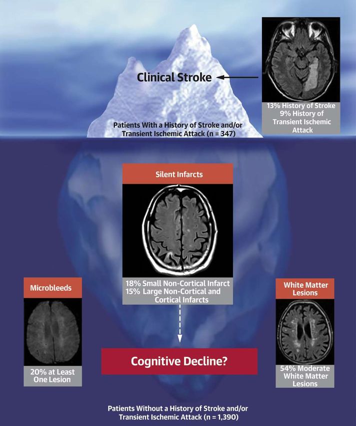
RESULTS The study included 1,737 patients with a mean age of 73 8 years (28% women, 90% taking oral antico-
agulant agents). On MRI, LNCCIs were found in 387 patients (22%), SNCIs in 368 (21%), microbleeds in 372 (22%), and
white matter lesions in 1715 (99%). Clinically silent infarcts among the 1,390 patients without a history of stroke or
transient ischemic attack were found in 201 patients with LNCCIs (15%) and 245 patients with SNCIs (18%). The MoCA
score was 24.7 3.3 in patients with and 25.8 2.9 in those without LNCCIs on brain MRI (p < 0.001). The difference in
MoCA score remained similar when only clinically silent LNCCIs were considered (24.9 3.1 vs. 25.8 2.9; p < 0.001). In
a multivariable regression model including all vascular brain lesion parameters, LNCCI volume was the strongest predictor
of a reduced MoCA (b ¼ 0.26; 95% confidence interval: 0.40 to 0.13; p < 0.001).
CONCLUSIONS Patients with AF have a high burden of LNCCIs and other brain lesions on systematic brain MRI
screening, and most of these lesions are clinically silent. LNCCIs were associated with worse cognitive function,
even among patients with clinically silent infarcts. Our findings raise the question of MRI screening in patients with AF.
Listen to this manuscript’s
(J Am Coll Cardiol 2019;73:989–99) © 2019 by the American College of Cardiology Foundation.
audio summary by
Editor-in-Chief
Dr. Valentin Fuster on
JACC.org.
From the aCardiology Division, Department of Medicine, University Hospital Basel, Basel, Switzerland; bCardiovascular Research
Institute Basel, Basel, Switzerland; cPopulation Health Research Institute, McMaster University, Hamilton, Canada; dInstitute of
Primary Health Care (BIHAM), University of Bern, Bern, Switzerland; eDepartment of General Internal Medicine, Inselspital, Bern
University Hospital, University of Bern, Bern, Switzerland; fDepartment of Cardiology, Triemli Hospital Zurich, Zurich,
Switzerland; gDepartement of Medicine, Cantonal Hospital of Baden and Molecular Cardiology, University Hospital of Zurich,
Zurich, Switzerland; hDepartment of Cardiology, Kantonsspital St. Gallen, St. Gallen, Switzerland; iDepartment of Cardiology,
ISSN 0735-1097/$36.00 https://doi.org/10.1016/j.jacc.2018.12.039990 Conen et al. JACC VOL. 73, NO. 9, 2019
Swiss-AF Brain Lesions and Cognition in AF Patients MARCH 12, 2019:989–99
T he prevalence of atrial fibrillation
ABBREVIATIONS association, but systematic investigations in patients
AND ACRONYMS (AF) in the general population is with AF are currently lacking. Microbleeds are of
increasing rapidly (1). Patients with particular interest because patients with AF usually
AF = atrial fibrillation
AF are at high risk of adverse events. need lifelong oral anticoagulation for stroke preven-
FLAIR = fluid-attenuated
Although the relationships of AF with death, tion (7). Although prior studies did not show a
inversion recovery
stroke, and congestive heart failure have consistent trend of more microbleeds among patients
LNCCI = large noncortical or
cortical infarct been known for many years (2,3), more using oral anticoagulation, the use of this therapy
MoCA = Montreal Cognitive
recent evidence suggests that patients with among patients with a significant burden of micro-
Assessment AF also face an increased risk of cognitive bleeds remains controversial (8–10).
MRI = magnetic resonance dysfunction and dementia (4,5). This The aim of the current study was to assess the re-
imaging growing awareness is reflected by a recent lationships of clinically known and unknown (silent)
SNCI = small noncortical publication of an international expert vascular brain lesions detected on brain magnetic
infarct
consensus paper on this topic (6). resonance imaging (MRI) with cognitive function in a
TIA = transient ischemic attack Meta-analyses suggest that part of the as- large sample of patients with AF. We focused on large
sociation between AF and dementia is explained by infarcts and infarcts involving the brain cortex, which
the higher stroke risk among patients with AF, but the may originate from embolic mechanisms and as such
risk of dementia was also increased in patients with represent AF-related sequelae. We also considered
AF but without a clinical history of stroke (5). Clini- imaging markers of cerebral small vessel disease,
cally unrecognized (silent) cerebral infarcts, micro- which share vascular risk factors with AF, including
bleeds, or other brain lesions may explain this white matter disease, small noncortical infarcts
Ospedale Regionale di Lugano, Lugano, Switzerland; jDivision of Cardiology, Fondazione Cardiocentro Ticino, Lugano,
Switzerland; kDepartment of Internal Medicine, HFR-Hôpital Cantonal Fribourg, Fribourg, Switzerland; lDepartment of Car-
diology, Luzerner Kantonsspital, Lucerne, Switzerland; mDivision of Cardiology, Department of Medical Specialties, University
Hospital Geneva, Geneva, Switzerland; nDepartment of Cardiology, Bürgerspital Solothurn, Solothurn, Switzerland; oDepart-
ment of Cardiology, Centre Hospitalier Universitaire Vaudois, Lausanne, Switzerland; pDepartment of Cardiology, Ospedale
San Giovanni, Bellinzona, Switzerland; qDepartment of Neurology and Stroke Center, University Hospital Basel, University of
Basel, Basel, Switzerland; rDepartment of Neurology, Inselspital, Bern University Hospital, University of Bern, Bern, Switzerland;
s
Memory Clinic, Universitäre Altersmedizin, Felix Platter Spital Basel, University of Basel, Basel, Switzerland; tDepartment of
Neuroradiology, University Hospital Zurich, Zurich, Switzerland; uMedical Image Analysis Center (MIAC AG) and Department of
Biomedical Engineering, University of Basel, Basel, Switzerland; and the vEpidemiology, Biostatistics, and Prevention Institute,
University of Zurich, Zurich, Switzerland. *Drs. Kühne and Osswald are joint senior authors and contributed equally to this work. yA
list of all Swiss-AF investigators is provided in the Online Appendix. The Swiss-AF cohort study is supported by grants of the Swiss
National Science Foundation (grants 33CS30_1148474 and 33CS30_177520), the Foundation for Cardiovascular Research Basel, and
the University of Basel. The Department of Radiology, University Hospital Basel, Basel holds a general research agreement with
Siemens and receives support from Guerbet, Bracco, and Bayer all unrelated to this work. Dr. Conen has a McMaster University
Department of Medicine Mid-Career Research Award; his work is supported by the Hamilton Health Sciences RFA Strategic Initiative
Program; and has received consulting fees from Servier, Canada. Dr. Rodondi has received a grant from the Swiss Heart Foundation.
Dr. Müller has received consulting fees from Biosense Webster, Switzerland. Dr. Beer has received grants from the Swiss National
Science Foundation, the Swiss Heart Foundation, and Bayer; and has received consultancy honoraria from Bayer and Daiichi-Sankyo.
Dr. Auricchio has received speaker fees from Boston Scientific and Microport; and is a consultant to Boston Scientific, Microport,
Daiichi-Sankyo, and Biosense Webster. Dr. Kobza has received grants from Biotronik, Biosense Webster, Boston Scientific, Medtronic,
and Abbott. Dr. Shah has received speaker fees from Biosense Webster, Daiichi-Sankyo, Boehringer Ingelheim, Bristol-Myers Squibb,
and Bayer; and has received consultancy honoraria from Biosense Webster. Dr. Sticherling has received speaker honoraria from
Biosense Webster and Medtronic; and has received research grants from Biosense Webster, Daiichi-Sankyo, and Medtronic. Dr.
Bonati has received grants from the Swiss National Science Foundation, the University of Basel, the Swiss Heart Foundation, The
Stroke Association, and AstraZeneca; and has received consulting and advisory board fees from Amgen, Bayer, Bristol-Myers Squibb,
and Claret Medical. Dr. Monsch has received honoraria or grant support from AC Immune, AbbVie, Roche, Takeda, and Vifor Pharma.
Dr. Stippich has received grants from the Swiss National Science Foundation, Siemens, Bracco, Guerbert, Schering, Bayer, Amgen,
Merck Sharp and Dohme, Novartis, Pfizer, and The Medicines Company. Dr. Wuerfel is CEO of the Medical Image Analysis Center,
Basel; has served on advisory boards for Actelion, Biogen, Genzyme-Sanofi, Novartis, Roche, and the Guthy Jackson Charitable
Foundation; has received research grants from Novartis; has received speaker honoraria from Bayer, Biogen, Genzyme, Novartis, and
Teva; and has received support by the European Union (Horizon2020), the German Research Association, the German Ministry of
Education and Research (BMBF/KKNMS), and the German Ministry of Economy (BMWi). Dr. Schwenkglenks has received grants
unrelated to the submitted work from Amgen, Merck Sharp and Dohme, Novartis, Pfizer, and The Medicines Company; has received
fees unrelated to the submitted work from Amgen; and has received a grant from the Swiss National Science Foundation. Dr. Kühne
has received consultant fees from Bayer, Boehringer Ingelheim, Pfizer-BMS, Daiichi-Sankyo, Medtronic, Biotronik, Boston Scientific,
Biosense Webster, AstraZeneca, and Novartis. All other authors have reported that they have no relationships relevant to the contents
of this paper to disclose.
Manuscript received September 13, 2018; revised manuscript received November 14, 2018, accepted December 3, 2018.JACC VOL. 73, NO. 9, 2019 Conen et al. 991
MARCH 12, 2019:989–99 Swiss-AF Brain Lesions and Cognition in AF Patients
(SNCIs), and microbleeds (11). Both cerebral small (spatial resolution 1.0 1.0 3.0 mm3 ) sequence
vessel disease and embolic stroke are key mecha- with whole brain coverage and without interpolation.
nisms underlying the development of vascular de- In addition, either a 2-dimensional axial
mentia (12). susceptibility-weighted imaging (spatial resolution
1.0 1.0 3.0 mm 3) or a 2-dimensional axial T2*-
METHODS weighted (spatial resolution of 1.0 1.0 3.0 mm 3)
sequence was applied.
STUDY DESIGN AND PARTICIPANTS. The Swiss-AF All brain MRI scans were analyzed centrally in a
(Swiss Atrial Fibrillation) study is an ongoing pro- specialized imaging core laboratory (Medical Image
spective cohort study that enrolled 2,415 patients Analysis Centre, Basel, Switzerland). MRI scans were
between 2014 and 2017 across 14 centers in analyzed by blinded expert raters unaware of personal
Switzerland. The detailed methodology has been characteristics or cognitive function. They marked
described previously (13). Patients were eligible for and segmented lesions in a standardized fashion using
Swiss-AF if they had a history of documented AF and an in-house procedure approved for international
if they were 65 years old or older. In addition, we clinical studies. Board-certified neuroradiologists
aimed to enroll 10% to 15% of patients between 45 confirmed all ratings. SNCIs were defined as hyperin-
and 65 years of age to assess the effects of AF on in- tense lesions on FLAIR #20 mm in diameter on axial
dividuals in the active workforce. We excluded pa- sections and not involving the cortex, consistent
tients with secondary forms of AF and patients who with ischemic infarction in the territory of a
were unable to provide informed consent. Patients perforating arteriole (located in the white matter,
with an acute illness within the last 4 weeks could be internal or external capsule, deep brain nuclei,
enrolled only once the acute episode had resolved. thalamus, or brainstem) (11). We did not further
differentiate between SNCIs and lacunes on the basis
SEE PAGE 1000
of the presence or absence of a central fluid-filled
Of the 2,415 patients enrolled in Swiss-AF, 667 did cavity. Large noncortical infarcts were noncortical
not have a brain MRI at baseline. The main reason for infarcts with a diameter >20 mm. Cortical infarcts
a missing brain MRI was the presence of a cardiac were defined as hyperintense lesions on FLAIR
device (n ¼ 461; 69%). Other reasons included further involving the cortex irrespective of their size and
contraindications to perform an MRI or claustro- whether or not they also involved subcortical areas.
phobia of the patient. An additional 11 patients did For the present analysis we differentiated between
not undergo cognitive testing, thus leaving 1,737 SNCIs and large noncortical infarcts or cortical in-
participants for the present analysis. The local ethics farcts (LNCCIs). Hyperintense white matter lesions
committees approved the study protocol, and written were graded using the Fazekas scale, and at least
informed consent was obtained from all participants. moderate disease was defined as a score $2 in either
CLINICAL MEASURES. Information on personal the periventricular or the deep white matter region
characteristics, risk factors, comorbidities, antith- (14). Perivascular spaces were identified, differenti-
rombotic treatment, and other factors was obtained ated by their tubular morphology, and subsequently
through standardized case report forms. Weight and excluded. FLAIR-hyperintense lesions not meeting
height were directly measured, and body mass index the criteria mentioned earlier were identified as white
was calculated as weight in kilograms divided by matter lesions. Microbleeds were identified and
height in meters squared. At baseline, 3 consecutive counted as nodular, strongly hypointense lesions on
blood pressure measurements were obtained, and the either T 2*-weighted or susceptibility-weighted imag-
mean of them was used in all analyses. We classified ing. T 2-weighted volumes of noncortical and cortical
AF into paroxysmal, persistent, and permanent AF infarcts as well as white matter lesions were
according to recommended definitions (7). segmented and quantified semiautomatically using
Amira (Mercury Computer Systems Inc., Chelmsford,
BRAIN MAGNETIC RESONANCE IMAGING. Brain MRI
Massachusetts). Lesions with a central FLAIR-
was acquired on a 1.5-T or 3.0-T scanner. The stan-
hypointense core were segmented in total without
dardized protocol included a 3-dimensional T 1 -
differentiating between hyperintense and hypo-
weighted magnetization-prepared rapid gradient echo
3 intense lesion areas.
(MPRAGE) (spatial resolution 1.0 1.0 1.0 mm ), a
2-dimensional axial fluid-attenuated inversion recov- COGNITIVE TESTING. All study personnel were cen-
ery (FLAIR) (spatial resolution 1.0 1.0 3.0 mm3 ), trally trained to perform a standardized neuro-
and 2-dimensional axial diffusion-weighted imaging cognitive assessment. The Montreal Cognitive992 Conen et al. JACC VOL. 73, NO. 9, 2019
Swiss-AF Brain Lesions and Cognition in AF Patients MARCH 12, 2019:989–99
Assessment (MoCA) evaluates visuospatial and exec- diagnostics were performed, and no major violations
utive functions, confrontation naming, memory, of model assumptions of homogeneity and normally
attention, language, and abstraction (15). Patients can distributed residuals were detected. The microbleed
obtain a maximum of 30 points, with higher scores count was truncated at 20, and 3 outliers were given a
indicating better cognitive function. One point was count of 20 to minimize their influence on the asso-
added to the total test score if the patient had 12 years ciations. All analyses were performed on an available
or less of formal education. data basis and conducted using R version 3.5.1 (R Core
Team, 2018, R Foundation, Vienna, Austria); mixed
STATISTICAL ANALYSIS. Baseline characteristics
effects models were constructed using the nlme
were stratified by the presence or absence of a clinical
package (16).
history of stroke or transient ischemic attack (TIA)
and presented as mean SD for continuous variables RESULTS
or as counts (percentages) for nominal variables. We
compared differences across groups with Wilcoxon Baseline characteristics are shown in Table 1. Mean
rank sum tests or chi-square tests, as appropriate. age was 73 8 years, 28% of participants were
Silent cerebral infarcts were defined as cerebral in- women, 90% were anticoagulated at the time of study
farcts (LNCCIs or SNCIs) on brain MRI in patients enrollment (54% direct oral anticoagulant agents,
without a history of stroke or TIA. Lesion volumes 36% vitamin K antagonists), and 18% were receiving
were indicated as median (interquartile range) given antiplatelet therapy. Patients with a history of stroke
their skewed distribution. or TIA were older, had a higher prevalence of hyper-
Given that history of stroke or TIA is a key pre- tension and diabetes, and were more often taking oral
dictor of lower cognitive function, we repeated all anticoagulant agents.
main analyses in patients without a history of stroke Prevalence and size of vascular brain lesions
or TIA. To assess the associations of vascular brain detected on brain MRI are shown in Table 2. At least 1
lesion parameters with MoCA score values and to LNCCI was detected in 22% of participants, with a
adjust for potential confounders, we constructed median volume of 1,623 mm 3. SNCIs were observed in
linear mixed effects regression models in which study 21% (median volume 63 mm3). The overlap between
center was included as a random intercept to account LNCCI and SNCI was small; 68% of patients with SNCIs
for potential differences across study centers. We first had no LNCCIs, and 75% of patients with LNCCIs had
fitted univariable models using log-transformed and no SNCIs, such that 30% of the study population had
centered vascular brain lesion parameters (or counts either LNCCIs or SNCIs. Microbleeds were observed in
for microbleeds) as MoCA score predictors. Separate 22% (median count 1) and white matter lesions in 99%
models were constructed for each brain lesion (median volume 3,918 mm3 ). The extent of white
parameter. Univariable models were adjusted for a matter lesions was at least moderate in 54% of par-
pre-defined set of covariates, including age, sex, body ticipants. When patients with a history of stroke or TIA
mass index, education level, smoking status, history were excluded, 201 of 1,390 (15%) participants had
of hypertension, history of diabetes, AF type, and use evidence of a silent LNCCI (median volume 525 mm 3),
of oral anticoagulation. Finally, we constructed a and 245 (18%) had evidence of a silent SNCI (median
combined multivariable model including all struc- volume 57 mm 3) (Table 2).
tural brain lesion parameters in a single model. We Comparisons of MoCA scores between patients
performed 2 sensitivity analyses, 1 that additionally with and without a specific vascular brain lesion are
adjusted the combined model for a history of car- shown in Figures 1A and 1B and in Online Table 1. The
dioversion or left atrial ablation and 1 adjusting for least square mean MoCA was 24.9 (95% confidence
time since first AF diagnosis. Finally, to examine the interval [CI]: 24.3 to 25.5) and 25.9 (95% CI: 25.3 to
independence of SNCI and LNCCI, we performed 2 26.5) among patients with and without an LNCCI on
additional analyses in which we excluded patients brain MRI (p < 0.001). The MoCA was 25.0 (95% CI:
who had both SNCIs and LNCIs on brain MRI. 24.4 to 25.6) versus 25.9 (95% CI: 25.3 to 26.4) in pa-
MoCA scores were compared across different strata tients with and without an SNCI (p < 0.001) and 25.4
and subgroups by using likelihood ratio tests between (95% CI: 24.7 to 26.0) versus 25.8 (95% CI: 25.2 to 26.4)
mixed effects models with and without the stratum as in patients with and without microbleeds (p ¼ 0.07).
single predictor. In all models, we included dummy When patients with a clinical history of stroke or TIA
indicators representing the presence or absence of were excluded, the MoCA score difference between
each vascular brain lesion type, in addition to the patients with or without silent LNCCI (25.1; 95% CI:
actual volume or count measurement. Visual model 24.4 to 25.7 vs. 26.0; 95% CI: 25.4 to 26.5; p < 0.001),JACC VOL. 73, NO. 9, 2019 Conen et al. 993
MARCH 12, 2019:989–99 Swiss-AF Brain Lesions and Cognition in AF Patients
T A B L E 1 Baseline Characteristics
All Patients No History of Stroke/TIA History of Stroke/TIA
(N ¼ 1,737) (n ¼ 1,390) (n ¼ 347) p Value*
Age, yrs 73 8 72 9 75 7994 Conen et al. JACC VOL. 73, NO. 9, 2019
Swiss-AF Brain Lesions and Cognition in AF Patients MARCH 12, 2019:989–99
score (Table 3, Online Table 3). When we excluded
F I G U R E 1 Least Square Mean MoCA Score According to the Presence or Absence of a
Specific Vascular Brain Abnormality
patients with a history of stroke or TIA, presence of
LNCCI ( b ¼ 0.53; 95% Cl: 0.94 to 0.12; p ¼ 0.012),
LNCCI volume ( b ¼ 0.18; 95% Cl: 0.39 to 0.02;
A
p ¼ 0.072), and presence of at least moderate white
30 matter lesions ( b ¼ 0.46; 95% CI: 0.77 to 0.16;
p < 0.001 p < 0.001 p < 0.001 p = 0.066 p ¼ 0.003) remained associated with the MoCA score
(Table 3, Online Tables 4 and 5). Sensitivity analyses
28
with models including covariates for a baseline his-
tory of cardioversion and left atrial ablation, or time
Predicted MoCA
26 since first AF diagnosis provided very similar results
(Online Tables 6 to 9). Excluding patients with both
SNCIs and LNCCIs on brain MRI also provided similar
24 results (Online Tables 10 and 11).
Stratified MoCA score results are shown in
Figure 2. The overall mean MoCA score was 25.5
22
3.1. It was 24.7 3 .7 and 25.6 2.9 among patients
with and without a clinical history of stroke (p <
20 0.001), respectively. MoCA scores also differed
across strata of age, AF type, hypertension, diabetes,
SNCI LNCCI Faz. ≥2 Microbleeds
oral anticoagulation, and CHA 2DS 2-VASc (congestive
heart failure, hypertension, age $75 years [2 points],
B diabetes, prior stroke or TIA or thromboembolism
[2 points], vascular disease, age 65 to 74 years, fe-
30
male sex) score, but not sex. Patients with perma-
p < 0.001 p < 0.001 p < 0.001 p = 0.15 nent AF had lower MoCA scores than patients with
28 paroxysmal or persistent AF, a finding that persisted
in multivariable models (Online Table 3).
The same subgroups were assessed for differences
Predicted MoCA
26 in brain lesion parameters (Online Table 12). Larger
LNCCI volumes were observed in patients with per-
manent AF, higher CHA2DS 2-VASc scores, and those
24
with a history of stroke, hypertension, or diabetes.
Oral anticoagulation with either direct oral anticoag-
22
ulant agents or vitamin K antagonists was not asso-
ciated with a higher microbleed count compared with
no anticoagulation. Higher CHA2 DS2-VASc scores
20 were associated with significantly higher infarct and
white matter lesion volumes, but lower microbleed
SNCI LNCCI Faz. ≥2 Microbleeds
counts (Online Table 12).
Absent Present
DISCUSSION
(A) All patients. (B) Patients without a history of stroke or transient ischemic attack.
Least square means were obtained from linear mixed effects models that included
Several important findings emerged from this
covariates for the presence versus absence of a specific damage and damage volume
comprehensive analysis of vascular brain lesions and
(count for microbleeds), and study center as random intercept. Faz ¼ Fazekas scale;
LNCCI ¼ large noncortical or cortical infarcts; MoCA ¼ Montreal Cognitive Assessment; cognitive function in patients with AF (Central
SNCI ¼ small noncortical infarcts. Illustration). First, participants had a substantial
burden of vascular brain lesions detected on sys-
tematic brain MRI: 22% and 21% had evidence of a
(Table 3). Coefficients of all covariates included in the previous LNCCI and SNCI, respectively. Second,
multivariable models are presented in Online Table 2. among patients without a history of stroke or TIA,
In a combined multivariable model including all 15% and 18% still had evidence of a previous clinically
vascular brain lesion parameters, LNCCI count and silent LNCCI and SNCI, respectively. Thus, most of
volume were the strongest predictors of the MoCA the observed lesions were clinically silent. Third,JACC VOL. 73, NO. 9, 2019 Conen et al. 995
MARCH 12, 2019:989–99 Swiss-AF Brain Lesions and Cognition in AF Patients
T A B L E 3 Linear Mixed Effect Models Assessing the Relationships of MoCA Score With Vascular Brain Lesion Parameters
Presence of Damage Type Volume/Count
Predictor of Interest b Coefficient 95% CI p Value b Coefficient 95% CI p Value
All patients
Small noncortical infarcts
Univariable 0.87 (1.22 to 0.53)996 Conen et al. JACC VOL. 73, NO. 9, 2019
Swiss-AF Brain Lesions and Cognition in AF Patients MARCH 12, 2019:989–99
F I G U R E 2 MoCA Score Across Predefined Strata
p Value for
Subgroup Mean (SD) Difference
Atrial fibrillation type
Paroxysmal 25.7 (3.1)
Persistent 25.7 (3.0)JACC VOL. 73, NO. 9, 2019 Conen et al. 997 MARCH 12, 2019:989–99 Swiss-AF Brain Lesions and Cognition in AF Patients C ENTR AL I LL U STRA T I O N Brain Lesions and Cognition in Patients With Atrial Fibrillation Conen, D. et al. J Am Coll Cardiol. 2019;73(9):989–99. Potential relationships of overt and silent brain lesions with cognitive function in patients with atrial fibrillation.
998   Conen et al.                                                                                                      JACC VOL. 73, NO. 9, 2019

      Swiss-AF Brain Lesions and Cognition in AF Patients                                                                 MARCH 12, 2019:989–99

                     cognitive impairment in the combined multivariable                22% of participants had microbleeds. This prevalence
                     model. Therefore, silent LNCCIs but not small vessel              is similar to that in elderly individuals from the
                     disease may explain the association between AF and                general population (25), or patients with ischemic
                     dementia in the absence of clinically overt strokes               stroke (26). Anticoagulation use was not associated
                     (Central Illustration) (5).                                       with a higher microbleed count, and microbleed
                        Although a 1-point MoCA score difference may                   count was not associated with cognitive function.
                     seem small on an absolute scale, it is similar to a 10-           This finding is in agreement with data showing that
                     year age difference or the presence of hypertension or            aspirin and apixaban had a similar impact on the
                     diabetes (Figure 2). We therefore expect such a dif-              incidence of microbleeds (27). Currently available
                     ference to be relevant from a clinical and societal               data therefore suggest that anticoagulation is safe in
                     perspective. Because the majority of brain infarcts               most patients with microbleeds, although it remains
                     were clinically silent and observed in patients                   controversial whether there is a subgroup of patients
                     without a history of stroke or TIA, our data raise the            with a high microbleed burden who should not be
                     issue of brain MRI screening in patients with AF.                 started on anticoagulation despite their high stroke
                     Future studies should develop risk scores for AF pa-              risk (28,29).
                     tients that identify subgroups of AF patients who may
                                                                                       STUDY STRENGTHS AND LIMITATIONS. Strengths of
                     benefit from brain imaging to better guide antith-
                                                                                       this study include the large sample size of well-
                     rombotic treatment in a cost-efficient manner. It is
                                                                                       characterized patients with AF, including the avail-
                     intriguing that permanent AF was associated with a
                                                                                       ability of brain MRI and cognitive testing. A potential
                     lower MoCA score independent of covariates and
                                                                                       limitation is the cross-sectional design of this anal-
                     brain lesion volumes (Online Table 3), a finding sug-
                                                                                       ysis, precluding assessment of causality or direc-
                     gesting that the arrhythmia itself or associated
                                                                                       tionality of effect. Participants in our study were
                     treatments may have a direct effect on cognition. This
                                                                                       mostly white, and all were enrolled in the compulsory
                     supports findings from a prior study showing worse
                                                                                       Swiss health insurance system. Whether our data are
                     cognition in patients with AF but without evidence of
                                                                                       applicable to other population groups or settings re-
                     brain infarcts on MRI (19).
                                                                                       mains to be determined. Finally, our study included
                        Oral anticoagulation effectively prevents stroke in
                                                                                       only patients with AF, and it is unclear how the
                     patients with AF (20). Although our cross-sectional
                                                                                       prevalence of vascular brain lesions compares with
                     analysis cannot address the question whether the
                                                                                       that of other sample groups.
                     cerebral infarcts occurred before or after initiation of
                     oral anticoagulation, it nevertheless raises the issue
                     that anticoagulation may not be sufficient to prevent
                                                                                       CONCLUSIONS
                     a significant number of silent infarcts, especially
                                                                                       In this large study of well-treated patients with AF we
                     those caused by mechanisms other than cardiac em-
                                                                                       found a high burden of vascular brain lesions on
                     bolism. A combination of aspirin and low-dose rivar-
                                                                                       systematic brain MRI screening. Most of these lesions
                     oxaban was significantly better than aspirin alone for
                                                                                       were previously unrecognized. Our analyses show
                     stroke    prevention       among       patients   with   stable
                                                                                       that the presence of overt or silent LNCCIs on MRI
                     vascular disease but without AF (21). Whether such a
                                                                                       have a similar impact on cognitive function as overt
                     treatment strategy may also benefit patients with AF
                                                                                       strokes, a finding suggesting that these lesions may
                     is currently unknown. Finally, both a history of hy-
                                                                                       explain at least part of the increased risk of cognitive
                     pertension and diabetes were significantly associated
                                                                                       dysfunction in these patients. Conversely, micro-
                     with lower MoCA scores in multivariable analyses, a
                                                                                       bleeds were not significantly associated with cogni-
                     finding suggesting that the high prevalence of car-
                                                                                       tive function. Finally, the value of routine MRI
                     diovascular risk factors in patients with AF may also
                                                                                       scanning and cognitive function testing for better risk
                     contribute to the occurrence of overt and silent ce-
                                                                                       stratification of patients with AF should be assessed
                     rebral lesions.
                                                                                       in further studies.
                        Although its detailed histopathological correlates
                     still   need   to   be    investigated,     microbleeds    are
                     considered to be small hemorrhages in the brain (22).             ADDRESS         FOR   CORRESPONDENCE:        Dr.   David
                     Patients with microbleeds have an increased risk of               Conen, Population Health Research Institute, 237
                     stroke and intracranial hemorrhage (23,24). In our                Barton Street East, Hamilton, Ontario, Canada.
                     study with a high prevalence of anticoagulation use,              E-mail: conend@mcmaster.ca. Twitter: @CRIBasel.
JACC VOL. 73, NO. 9, 2019 Conen et al. 999
MARCH 12, 2019:989–99 Swiss-AF Brain Lesions and Cognition in AF Patients
PERSPECTIVES
COMPETENCY IN MEDICAL KNOWLEDGE: Our TRANSLATIONAL OUTLOOK: Future studies should
study shows that most brain infarcts observed in patients evaluate whether and which patients with AF may benefit
with AF are clinically silent. Large numbers of silent and from brain MRI screening. These studies should also
overt lesions were observed even though 90% of the address the question of optimal antithrombotic treatment
patients were taking oral anticoagulant agents at the time to prevent silent infarcts in patients with AF.
of the brain MRI.
REFERENCES
1. Krijthe BP, Kunst A, Benjamin EJ, et al. Pro- neurodegeneration. Lancet Neurol 2013;12: 22. van Veluw SJ, Charidimou A, van der
jections on the number of individuals with atrial 822–38. Kouwe AJ, et al. Microbleed and microinfarct
fibrillation in the European Union, from 2000 to detection in amyloid angiopathy: a high-resolution
12. O’Brien JT, Thomas A. Vascular dementia.
2060. Eur Heart J 2013;34:2746–51. MRI-histopathology study. Brain 2016;139:
Lancet 2015;386:1698–706.
3151–62.
2. Conen D, Chae CU, Glynn RJ, et al. Risk of death
13. Conen D, Rodondi N, Mueller A, et al. Design of
and cardiovascular events in initially healthy 23. Charidimou A, Karayiannis C, Song TJ, et al.
the Swiss Atrial Fibrillation Cohort Study (Swiss-
women with new-onset atrial fibrillation. JAMA Brain microbleeds, anticoagulation, and hemor-
AF): structural brain damage and cognitive decline
2011;305:2080–7. rhage risk: meta-analysis in stroke patients with
among patients with atrial fibrillation. Swiss Med
AF. Neurology 2017;89:2317–26.
3. Chatterjee NA, Chae CU, Kim E, et al. Modifiable Wkly 2017;147:w14467.
risk factors for incident heart failure in atrial 24. Wilson D, Ambler G, Shakeshaft C, et al. Ce-
fibrillation. J Am Coll Cardiol HF 2017;5:552–60. 14. Fazekas F, Chawluk JB, Alavi A, Hurtig HI, rebral microbleeds and intracranial haemorrhage
Zimmerman RA. MR signal abnormalities at 1.5 T in risk in patients anticoagulated for atrial fibrillation
4. Thacker EL, McKnight B, Psaty BM, et al. Atrial Alzheimer’s dementia and normal aging. AJR Am J after acute ischaemic stroke or transient ischaemic
fibrillation and cognitive decline: a longitudinal Roentgenol 1987;149:351–6. attack (CROMIS-2): a multicentre observational
cohort study. Neurology 2013;81:119–25.
15. Nasreddine ZS, Phillips NA, Bedirian V, et al. cohort study. Lancet Neurol 2018;17:539–47.
5. Kalantarian S, Stern TA, Mansour M, Ruskin JN.
The Montreal Cognitive Assessment, MoCA: a brief 25. Poels MM, Vernooij MW, Ikram MA, et al.
Cognitive impairment associated with atrial fibril-
screening tool for mild cognitive impairment. J Am Prevalence and risk factors of cerebral micro-
lation: a meta-analysis. Ann Intern Med 2013;158:
Geriatr Soc 2005;53:695–9. bleeds: an update of the Rotterdam scan study.
338–46.
16. Pinheiro J, Bates D, DebRoy S, Sarkar D, R Stroke 2010;41:S103–6.
6. Dagres N, Chao TF, Fenelon G, et al. European
Core Team. nlme: Linear and Nonlinear Mixed 26. Charidimou A, Shakeshaft C, Werring DJ. Ce-
Heart Rhythm Association (EHRA)/Heart Rhythm
Effects Models. R package version 3.1-137 2018. rebral microbleeds on magnetic resonance imag-
Society (HRS)/Asia Pacific Heart Rhythm Society
Vienna, Austria: R Foundation, 2018. Available ing and anticoagulant-associated intracerebral
(APHRS)/Latin American Heart Rhythm Society
at: https://CRAN.R-project.org/package¼nlme. hemorrhage risk. Front Neurol 2012;3:133.
(LAHRS) expert consensus on arrhythmias and
Accessed January 16, 2019.
cognitive function: what is the best practice? 27. O’Donnell MJ, Eikelboom JW, Yusuf S, et al.
Heart Rhythm 2018;15:e37–60. 17. Price TR, Manolio TA, Kronmal RA, et al. Silent Effect of apixaban on brain infarction and micro-
brain infarction on magnetic resonance imaging bleeds: AVERROES-MRI assessment study. Am
7. Camm AJ, Kirchhof P, Lip GY, et al. Guidelines
and neurological abnormalities in community- Heart J 2016;178:145–50.
for the management of atrial fibrillation: The Task
dwelling older adults. the Cardiovascular Health
Force for the Management of Atrial Fibrillation of 28. Diener HC, Selim MH, Molina CA,
Study. CHS Collaborative Research Group. Stroke
the European Society of Cardiology (ESC). Eur Greenberg SM. Embolic stroke, atrial fibrillation,
1997;28:1158–64.
Heart J 2010;31:2369–429. and microbleeds: is there a role for anti-
18. Vermeer SE, Prins ND, den Heijer T, Hofman A, coagulation? Stroke 2016;47:904–7.
8. Horstmann S, Mohlenbruch M, Wegele C, et al.
Prevalence of atrial fibrillation and association of Koudstaal PJ, Breteler MM. Silent brain infarcts 29. Charidimou A, Shoamanesh A, Al-Shahi
previous antithrombotic treatment in patients and the risk of dementia and cognitive decline. Salman R, et al. Cerebral amyloid angiopathy, ce-
with cerebral microbleeds. Eur J Neurol 2015;22: N Engl J Med 2003;348:1215–22. rebral microbleeds and implications for anti-
1355–62. 19. Knecht S, Oelschlager C, Duning T, et al. Atrial coagulation decisions: the need for a balanced
fibrillation in stroke-free patients is associated approach. Int J Stroke 2018;13:117–20.
9. Saito T, Kawamura Y, Sato N, et al. Non-vitamin
k antagonist oral anticoagulants do not increase with memory impairment and hippocampal atro-
cerebral microbleeds. J Stroke Cerebrovasc Dis phy. Eur Heart J 2008;29:2125–32.
2015;24:1373–7. KEY WORDS atrial fibrillation, cognitive
20. Hart RG, Pearce LA, Aguilar MI. Meta-analysis:
dysfunction, microbleeds, silent cerebral
10. Akoudad S, Darweesh SK, Leening MJ, et al. antithrombotic therapy to prevent stroke in pa-
infarcts, white matter lesions
Use of coumarin anticoagulants and cerebral tients who have nonvalvular atrial fibrillation. Ann
microbleeds in the general population. Stroke Intern Med 2007;146:857–67.
2014;45:3436–9.
21. Eikelboom JW, Connolly SJ, Bosch J, et al. A PP END IX For a complete list of Swiss-AF
11. Wardlaw JM, Smith EE, Biessels GJ, et al. Rivaroxaban with or without aspirin in stable car- study investigators as well as supplemental
Neuroimaging standards for research into small diovascular disease. N Engl J Med 2017;377: tables, please see the online version of this
vessel disease and its contribution to ageing and 1319–30. paper.You can also read