Clinical application of thioredoxin reductase as a novel biomarker in liver cancer
←
→
Page content transcription
If your browser does not render page correctly, please read the page content below
www.nature.com/scientificreports
OPEN Clinical application of thioredoxin
reductase as a novel biomarker
in liver cancer
Xuping Wu1, Qi Wang2, Yousheng Lu3, Jinye Zhang4, Hanwei Yin2 & Yongxiang Yi1*
Hepatic cancer is often amenable to surgery, including percutaneous ablation, trans-arterial
chemoembolization. However, in metastatic cases, surgery is often not an effective option.
Chemotherapy as a conventional clinical method for treatment of malignant diseases may be useful in
such cases, but it is likewise not always able to slow or halt progression, therefore novel approaches
for treatment of hepatic cancer are needed. Current research suggests that molecular tumor markers
(TM) can play a crucial role for diagnosis and prognostic evaluation of malignancies, and TM such as
AFP, CEA, CA19-9 have been reported in many malignant diseases. Thioredoxin reductase (TrxR),
a type of anti-oxidant biomarker, has become a TM of significant interest. However, little is known
about the above TM and TrxR activity in liver cancer. Therefore, this paper aimed to assess these TM
with regards to diagnosis and and monitoring treatment efficacy in both primary and metastatic liver
cancer. Our results showed TrxR had superior performance for discriminating between liver cancer
patients and healthy controls than AFP, CEA, and CA19-9. TrxR also exhibited superior performance
for assessing benefits of chemotherapy regardless if patients had PLC or MLC. Meanwhile, due to
diagnostic efficiency of unresponsive chemotherapy patients, TrxR also showed a higher activity levels
than other general markers in liver metastasis patients. Our results suggest that application of TrxR
in combination with other tumor markers may maximize the efficiency of diagnosis and assessment
of therapeutic efficiency, and provide new insights for the clinical application of TrxR as a candidate
biomarker for liver cancer.
Primary liver cancer (PLC) is a common life-threatening t umor1,2 with a current mortality rate close to that of
lung cancer3. Due to the long latency period of PLC, a large number of patients reach advanced stages before
diagnosis and clinical intervention4–7. Many patients are therefore also characterized by high recurrence and
high metastasis r ates8–11. Numerous studies have shown that several risk factors, such as excessive alcohol con-
sumption, smoking, cirrhosis, and type 2 diabetes, predispose to PLC progression12–15, and these risk factors
may also contribute to disease initiation. However, a main cause of PLC is viral hepatitis, which accounts for a
large percentage of cases in C hina16,17. Approximately 85% of cirrhosis and liver cancer patients are diagnosed
with viral hepatitis, which indicates that viral hepatitis constitutes a significant risk factor for liver cancer18,19.
Hepatic metastasis as an advanced stage of secondary growth carcinoma is etiologically different from PLC20,21.
In these patients, tumors originate from distant liver metastasis through epithelial-mesenchymal transformation
and circulating tumor cells, such as from esophageal and gastric cancers22. These tumor cells can circulate to
the liver through the bloodstream, even in patients who underwent s urgery23,24, and a large number of patients
experience recurrence. Application of transcatheder arterial chemoembolization (TACE) might alleviate the
progression of liver metastases to some extent25,26, but the treatment of invasive secondary cancers is often inef-
fective. Therefore, it would be helpful to improve the early detection of (metastatic) liver cancers and to evaluate
the efficacy of chemotherapy with regards to survival of patients.
Timely control of tumor progression is beneficial to improving the quality of life of patients. Tumor markers
have been used for almost 160 years and constitute an important clinical auxiliary tool. Specific antigens, includ-
ing carbohydrate antigen (CEA) and carbohydrate antigen 19-9 (CA19-9), exhibit a partial diagnostic accuracy
for some cancers27–29, and alpha-fetoprotein (AFP) levels are abnormally elevated in liver disease30. Unfortunately,
1
The Second Hospital of Nanjing, Nanjing University of Chinese Medicine, Nanjing 210000, Jiangsu,
China. 2Keaise Center for Clinical Laboratory, Wuhan, China. 3Jiangsu Provincial Cancer Hospital and Jiangsu
Provincial Cancer Institute, Medical Department of Cancer Hospital Affiliated to Nanjing Medical University,
Nanjing, China. 4Nantong Tumor Hospital, Nantong, China. *email: ian0126@126.com
Scientific Reports | (2021) 11:6069 | https://doi.org/10.1038/s41598-021-85688-3 1
Vol.:(0123456789)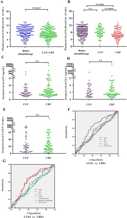
www.nature.com/scientificreports/
the aforementioned tumor markers lack significant diagnostic value after chemotherapy, which make it difficult
to evaluate disease progression (PD)29. Novel circulating biomarkers are therefore being explored.
TrxR is an enzyme of the triphosphopyridine nucleotide (NADPH) oxidative pathway and plays a key role in
several physiological activities, such as redox pathways and DNA s ynthesis31,32. Previous studies suggested that
TrxR indicates higher levels of abnormally proliferating cells, and may have a superior diagnostic efficacy than
other conventional tumor markers. In addition, TrxR activity is rapidly downregulated after chemotherapy in
non-small cell lung cancer (NSCLC), gastric cancer, and breast c ancer33–35. Previous studies consistently dem-
onstrated that TrxR has potentially high value for clinical diagnosis and assessment of therapeutic efficacy. Here,
we assess the role of TrxR and other TM in primary and metastatic liver cancer (PLC and MLC, respectively).
To the best of our knowledge, it is the first study to investigate the role of TrxR in the evaluation of therapeutic
efficiency in the primary liver cancer (PLCs). Furthermore, this study included the comparison of TrxR activity
between PLCs and MLCs, providing a new insight to the clinical appliance of TrxR in the diagnosis and monitor-
ing therapeutic efficiency in liver cancer.
Methods
Patients. Cancer patients were eligible for enrollment based on histologically confirmed liver cancer, as
described in Supplementary materials (Supplemental Table S1), and were consecutively recruited from The
Second Hospital of Nanjing (Nanjing, China), Jiangsu Cancer Hospital (Jiangsu, China), and Nantong Tumor
Hospital (Jiangsu, China). Enrollment occurred from 2017 to 2020. Sex- and age-matched health controls and
patients diagnosed into other diseases (such as hepatitis) based on hematological, histopathological and com-
puted tomography (CT) analyses36, were also enrolled.
Specimen properties. Sample collection was conducted as described in the literature34. Extra-cellular
blood samples were obtained in tubes containing EDTA or no anticoagulant for 2 h preoperatively, after which
the samples followed by centrifugation at 3500 rpm for 5 min at room temperature. The upper serum plasma was
collected and stored in EP tubes at 4 °C.
The tumor marker analysis. Levels of CEA, CA19-9, and AFP as tumor markers associated with liver can-
cer were obtained at the indicated times during patient visits. Following previous literature29,34, CEA, AFP and
CA19-9 were analyzed by electrochemiluminescence-based immunoassay (ECLIA) with Cobas analyzer (Roche
Diagnostics, Mannheim, Germany), following the manufacturer’s instructions. Reference values for all tumor
markers were selected based on the Chinese Society of Clinical Oncology, and three clinical thresholds were set
at 39 U/mL for CA19-9, 7.0 ng/mL for AFP, and 3.5 ng/mL for CEA37.
Assay for TrxR activity analysis. The activity of thioredoxin reductase (TrxR) in plasma was measured
by UV spectrophotometry as previously described in the literature34,38–40. The kits used were commercial kits
approved for marketing and purchased from Clairvoyance Health Technology Co., Ltd, Wuhan, China. All
operations were performed according to the manufacturer’s instructions34,38,39. A single-blinded experimental
protocol was designed.
Statistical analysis. The diagnostic efficacy of the biomarker was assessed by the receiver operator curve
(ROC), assessing the value of the area under the curve (AUC), and the operating characteristics of the 95%
confidence interval (CI). All statistical analyzes were performed in GraphPad Prism 7 (Graphprism, USA) and
SPSS19.0 (SPSS Inc, USA). The correlation between TrxR and other tumor markers was analyzed in R by regres-
sion correlation. The non-parametric Mann–Whitney U test was applied to evaluate the difference for two paral-
lel groups. Statistical significance was considered as P < 0.0541.
Ethics statement. This current study, including all experimental protocols, was approved by the ethics
committees of The Second Hospital of Nanjing (Nanjing, China), Jiangsu Cancer Hospital (Jiangsu, China) and
Nantong Tumor Hospital (Nantong, China). The methods were carried out in accordance with the approved
guidelines and regulations. Informed consent was obtained from all patients.
Results
This retrospective analysis was carried out on 1286 specimens enrolled from Jiangsu Province Cancer Hospi-
tal, Nanjing Second People’s Hospital, and Nantong Cancer Hospital between 2017 and 2020, including 327
patients with primary liver cancer before (n = 183) or after (n = 144) clinical intervention, 809 patients with liver
metastases (n = 161 for therapeutic evaluation), and 150 healthy controls, respectively. Besides, 510 patients
with other liver diseases [hepatitis (n = 327), liver injury (n = 61), liver dysfunction (n = 26), cirrhosis (n = 85),
and fatty liver (n = 11)], were also enrolled for the comparison of TrxR levels between liver cancer and other
common liver diseases.
Pre‑intervention plasma TrxR activity and AFP, CEA, and CA19‑9 levels in primary liver cancer
patients and healthy controls. To assess potential differences in TrxR activity in healthy individuals
and patients with primary liver cancers (PLCs), 183 PLCs and 150 of healthy individuals were enrolled in this
study. TrxR activity and levels of AFP, CEA, and CA19-9 [median (IQR)] were measured in PLCs and healthy
controls before the clinical intervention. TrxR activity was significantly elevated in PLCs [8.63 (6.38, 10.05) U/
mL] relative to healthy controls [2.80 (1.7, 3.6) U/mL] (Fig. 1A). Likewise, serum AFP, CEA, and CA19-9 levels
Scientific Reports | (2021) 11:6069 | https://doi.org/10.1038/s41598-021-85688-3 2
Vol:.(1234567890)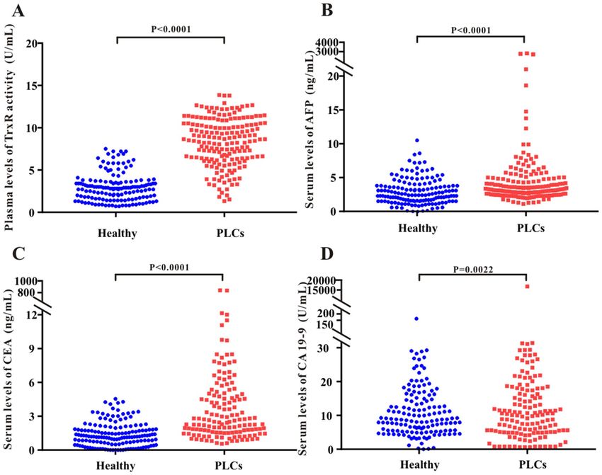
www.nature.com/scientificreports/

                                 Figure 1.  Scatter plot of the distribution of plasma TrxR (A), serum AFP (B), CEA (C), and CA19-9 (D)
                                 levels between healthy people and PLC groups before clinical interventions. P values were calculated by the
                                 nonparametric Mann–Whitney U test. Statistical significance was considered as P < 0.05.

                                 were significantly increased in PLCs relative to healthy controls, indicating that TrxR activity and AFP, CEA, and
                                 CA19- are potentially sensitive biomarkers for liver cancer before clinical intervention (Fig. 1B–D).

                                 Potential suitability of TrxR activity as a diagnostic biomarker for primary liver cancer. Analy-
                                 sis of ROC curves was performed to evaluate the suitability of TrxR plasma activity as a biomarker for the
                                 diagnosis of primary liver cancers (PLCs)42. The optimal cutoff for TrxR activity was calculated using the maxi-
                                 mum Youden index (sensitivity + specificity-1) to distinguish PLC patients from healthy controls. As presented
                                 in Fig. 2A, the diagnotic cut-off value of TrxR activity in PLC patients was calculated to be 3.85 U/mL for a
                                 sensitivity of 92.31% and a specificity of 81.33% based on the ROC curve (AUC 0.939; 95% CI 0.915–0.964). In
                                 comparison, CEA showed the second highest AUC for distinguishing liver cancer patients from healthy controls
                                 (Table 1; 0.838; 95% CI 0.797–0.879). Simultaneously, we showed that CA19-9 and AFP have a moderate ability
                                 to discriminate between PLC patients and healthy controls, with AUCs of 0.677 (95% CI 0.618–0.735) and 0.598
                                 (95% CI 0.537–0.658), respectively. The sensitivity of CA19-9 was less than 50%, which suggested that the use of
                                 CA19-9 and AFP for PLC diagnosis had a high risk for false negatives. It was therefore evident that TrxR activity
                                 was superior to other tumor markers for clinical diagnosis in PLC patients.
                                     Furthermore, combinations of CA19-9, CEA and AFP displayed an increased efficacy for detecting PLC
                                 patients (AUC 0.887; 95% CI 0.853–0.920) compared with the three individual levels (P < 0.5). Remarkably, by
                                 adding TrxR to this combination group, there was further improvement in the diagnostic efficiency for PLC (AUC
                                 0.984; 95% CI 0.887–0.984). These results provide a promising diagnostic combination of four biomarkers for
                                 PLC diagnosis, which could be used for future clinical applications (Fig. 2B and Table 1).
                                     Additionally, we also investigated the level of TrxR activity in other liver diseases including hepatitis, liver
                                 injury, liver dysfunction, cirrhosis, and fatty liver. As shown in Supplemental Fig. S1, other liver diseases also
                                 showed lower levels of TrxR activity compared to PLCs, suggesting TrxR level was specifically elevated in PLCs
                                 instead of other liver diseases.

Scientific Reports |   (2021) 11:6069 |               https://doi.org/10.1038/s41598-021-85688-3                                                   3

                                                                                                                                              Vol.:(0123456789)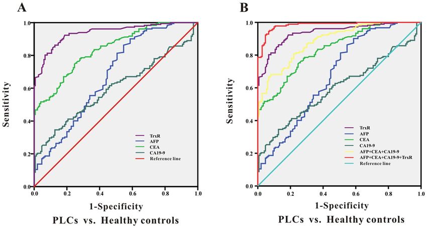
www.nature.com/scientificreports/

                                           Figure 2.  ROC curve analyses of TrxR, AFP, CEA, CA19-9 (A), and the combinations (B) for the differentiation
                                           of PLCs and healthy controls.

                                                                           PLC patients before clinical intervention
                                            Tumor markers                  AUC (95%CI)           SEN%     SPE%     PPV%    NPV%    PLR     NLR
                                            PLC patients vs. healthy controls
                                            TrxR                           0.939 (0.915–0.964)   92.31    81.33    83.18   91.36    4.95   10.57
                                            AFP                            0.677 (0.618–0.735)   86.26    45.33    61.21   76.75    1.58    3.30
                                            CEA                            0.838 (0.797–0.879)   74.73    75.33    75.18   74.88    3.03    2.98
                                            CA19-9                         0.598 (0.537–0.658)   34.62    87.33    73.21   57.19    2.73    1.34
                                            CEA + CA19-9 + AFP             0.887 (0.853–0.920)   68.13    92.00    89.49   74.27    8.52    2.89
                                            CEA + CA19-9 + AFP + TrxR      0.984 (0.974–0.994)   94.51    93.33    93.41   94.44   14.18   16.99

                                           Table 1.  The diagnostic efficiency of TrxR, CA19-9, CEA , AFP and their combinations in distinguishing PLC
                                           patients from healthy controls. SPE: specificity; SEN: sensitivity; NPV: negative predictive value; PPV: positive
                                           predictive value;NLR: negative likelihood ratio. PLR: positive likelihood ratio; the diagnosic threshold of TrxR
                                           activity was 3.85 U/mL.

                                           Assessment of therapeutic efficacy by monitoring TrxR activity after chemotherapy in PLC
                                           patients. To further investigate TrxR activity with regards to response to chemotherapy in patients with
                                           PLC, two groups of 144 PLC patients were divided based on clinical results. These patients were classified as
                                           Clinical Unresponsive Patients (CUP, 49 patients) or Clinical Responsive Patients (CRP, 95 patients) according
                                           to CT results. Patients with complete response (CR), partial response (PR) or stable disease (SD) mostly benefted
                                           from the chemotherapy and were included into CRP group. On the contrary, patients with progressive disease
                                           (PD) or uncontrolled condition afer chemotherapy were included into CUP group.Further statistical analysis
                                           was performed by measuring plasma TrxR levels in the CUP and CRP groups.
                                               Firstly, we investigated if TrxR activity is an independent indicator for the diagnosis and therapeutic evalua-
                                           tion in liver cancer. As shown in Supplemental Fig. S2, correlation analysis indicated no significant correlation
                                           between TrxR activity and CEA, CA19-9, or AFP in either healthy group or liver patients. Thus, TrxR activity
                                           can be considered as an independent indicator for the diagnosis and therapeutic evaluation in liver cancer, and
                                           TrxR level was not affected by other TMs. Meanwhile, a detailed analysis has been performed to compare the
                                           TrxR activity among different histological types of PLCs. As shown in Supplemental Fig. S3, TrxR activity was
                                           not significantly different among hepatocellular carcinoma (HCCs), intrahepatic cholangiocarcinoma (ICCs),
                                           and combined HCCs/ICCs in PLCs either before clinical interventions or after chemotherapy, suggesting that
                                           TrxR levels in PLCs were not affected by the histological types of primary liver cancer.

          Scientific Reports |   (2021) 11:6069 |                   https://doi.org/10.1038/s41598-021-85688-3                                              4

Vol:.(1234567890)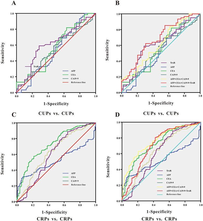
www.nature.com/scientificreports/

                                 Figure 3.  (A) Scatter plot of the distribution of plasma TrxR activity levels between liver cancer patients before clinical
                                 interventions and PLC patients after chemotherapy. (B–E) Scatter plot of the distribution of plasma TrxR (B), serum AFP
                                 (C), CEA (D), and CA19-9 (E) among PLC patients with different clinical outcome after chemotherapy (CUP vs. CRP). CUP:
                                 clinical unresponsive patient; CRP: clinical responsive patients. P values were determined by the Mann–Whitney U test.
                                 N.S: no statistical significance. (F–G) ROC curve analyses of TrxR, AFP, CEA, CA19-9 (F), and the combinations (G) for the
                                 differentiation of PLCs with different clinical outcome after chemotherapy (CUP vs. CRP).

Scientific Reports |   (2021) 11:6069 |                 https://doi.org/10.1038/s41598-021-85688-3                                                          5

                                                                                                                                                       Vol.:(0123456789)
www.nature.com/scientificreports/
PLCs after chemotherapy
Tumor markers AUC (95%CI) SEN% SPE% PPV% NPV% PLR NLR
Primary carcinoma of liver: CUPs vs.CRPs
TrxR 0.776 (0.687–0.865) 73.47 80.00 78.60 75.10 3.67 3.02
AFP 0.624 (0.529–0.718) 75.51 47.37 58.93 65.92 1.43 1.93
CEA 0.588 (0.485–0.691) 36.73 87.37 74.41 58.00 2.91 1.38
CA19-9 0.433 (0.333–0.534) 22.45 82.11 55.64 51.43 1.25 1.06
CEA + CA19-9 + AFP 0.574 (0.466–0.681) 44.90 80.00 69.18 59.21 2.24 1.45
CEA + CA19-9 + AFP + TrxR 0.781 (0.693–0.869) 75.51 78.95 78.20 76.32 3.59 3.22
Table 2. The assessment of therapeutic efficiency by TrxR, AFP, CA19-9, and CEA and their combinations
after chemotherapy in PLC patients. SPE: specificity; SEN: sensitivity; NPV: negative predictive value; PPV:
positive predictive value;NLR: negative likelihood ratio. PLR: positive likelihood ratio; the diagnosic threshold
of TrxR activity level was 7.45 U/mL in PLC patients.
Figure 3A shows overall post-chemotherapy TrxR levels in the PLC group [7.09 (5.90, 8.87) U/mL], which
were lower than in pre-intervention patients [8.63 (6.38, 10.50) U/mL]. Notably, TrxR activity levels in the CRP
group [6.60 (5.70, 8.20) U/mL] were significantly decreased compared with the CUP group [8.80 (7.53, 10.23) U/
mL], indicating that TrxR levels decrease in PLC patients benefitting of chemotherapy (Fig. 3B). The TrxR levels
in the CUP group remained unchanged compared with patients before the clinical intervention. Consistent with
TrxR activity, AFP levels were also reduced in CRP patients compared with CUP patients (Fig. 3C). Downregula-
tion of other tumor markers, including CEA and CA19-9 levels was not significantly different between the CRP
and CUP groups (Fig. 3D,E and Supplemental Fig. S4).
To further confirm that TrxR offers effective therapeutic evaluation in PLC patients after chemotherapy, we
analysed the therapeutic value by ROC curve. As shown in Fig. 3F and Table 2, the threshold for TrxR activity to
discriminate between CUPs and CRPs was calculated at 7.45 U/mL, with a sensitivity of 73.74% and a specific-
ity of 80.00% (AUC 0.776; 95% CI 0.687–0.865). Meanwhile, AFP showed the second highest AUC level (AUC
0.624; 95% CI 0.529–0.718) in distinguishing CUPs from CRPs. CA19-9 and CEA had very limited ability to
discriminate between CUPs and CRPs, with corresponding AUCs of 0.433 (95% CI 0.333–0.534) and 0.588 (95%
CI 0.485–0.691). The sensitivity of CA19-9 and CEA was less than 50%, which indicates that CA19-9 and CEA
for therapeutic evaluation had a high risk for false negatives. When adding TrxR to a combination panel of AFP,
CA19-9 and CEA, the value of therapeutic evaluation in patients with PLC was further strengthened relative to
TrxR alone or to the other three biomarker combination panel (AUC 0.781; 95% CI 0.693–0.869) (Fig. 3G and
Table 2). In summary, the ability to monitor therapeutic evaluation by plasma TrxR activity was superior to CEA,
CA19-9 and AFP, but combination of all four markers exhibited the highest AUC.
Assessment of therapeutic efficacy by monitoring TrxR activity after chemotherapy in MLC
patients. Above studies have investigated the relevance between TrxR and PLCs, however, so far it was not
known whether TrxR is associated with the prognosis of metastatic liver cancer (MLCs). As shown in Supple-
mental Fig. S5, TrxR activity was not significantly different among MLCs originated from different tumor enti-
ties, which includes intestinal cancer, gastric cancer, breast cancer, lung cancer, pancreatic cancer, esophageal
cancer and nasopharyngeal cancer, suggesting that TrxR levels in MLCs were not affected by the primary organs
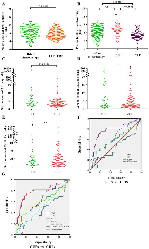
of the tumor. Consistent with the observation in PLCs, analysis of TrxR activity revealed a significant decrease
after chemotherapy [7.35(5.62, 9.79) U/mL] in MLCs compared with those before clinical intervention [8.63
(6.38, 10.50) U/mL] (Fig. 4A).
Image-based methods such as CT were often used to observe the efficacy of chemotherapy for metastatic
liver cancer42,43. Here in MLC patients, the decrease in TrxR activity was greater in the CRP group [7.70 (5.78,
10.21) U/mL] than in the CUP group [9.40 (7.55, 11.27) U/mL] (Fig. 4B). However, other TMs such as CEA,
CA19-9 and AFP, were not significantly different between CRP and CUP groups in MLCs (Fig. 4C–E and Sup-
plemental Fig. S6), suggesting TrxR exerted a significant advantage over other TMs in the therapeutic evaluation
of chemotherapy in MLCs. Similar to the observations in PLC patients, plasma TrxR activity exhibited higher
sensitivity and specificity (AUC 0.630; 95% CI 0.542–0.718) than CEA, CA19-9 and AFP levels in discrimination
between CRPs and CUPs in MLCs based on ROC analysis (Fig. 4F and Table 3). Application of TrxR in combina-
tion with other TMs enhanced the sensitivity and specificity of assessment of therapeutic efficacy (AUC 0.643;
95% CI 0.556–0.729) compared with the other tumor markers alone (Fig. 4G and Table 3). The above results
provide an insight for evaluating the efficiency of chemotherapy in MLC patients using biomarkers. TrxR appears
to play an important role as a novel serum biomarker and was able to effectively assess therapeutic efficacy in
hepatic metastasis patients.
TrxR, AFP, CA19‑9 and CEA were elevated in MLC patients compared to PLC patients in both
CUP and CRP group. Within the CUP or CRP group, it was crucial to further compare biomarkers and
combination panels between the MLC and PLC groups to explore if the therapeutic efficiency of TrxR and other
TMs were affected by the type of liver cancer.
Scientific Reports | (2021) 11:6069 | https://doi.org/10.1038/s41598-021-85688-3 6
Vol:.(1234567890)www.nature.com/scientificreports/
Figure 4. (A) Scatter plot of the distribution of plasma TrxR activity levels between liver cancer patients before clinical
interventions and MLC patients after chemotherapy. (B-E) Scatter plot of the distribution of plasma TrxR (B), serum AFP
(C), CEA (D), and CA19-9 (E) among MLC patients with different clinical outcome after chemotherapy (CUP vs. CRP).
CUP: clinical unresponsive patient; CRP: clinical responsive patients. P values were determined by the Mann–Whitney U test.
N.S: no statistical significance. (F-G) ROC curve analyses of TrxR, AFP, CEA, CA19-9 (F), and the combinations (G) for the
differentiation of MLCs with different clinical outcome after chemotherapy (CUP vs. CRP).
Scientific Reports | (2021) 11:6069 | https://doi.org/10.1038/s41598-021-85688-3 7
Vol.:(0123456789)www.nature.com/scientificreports/
MLCs after chemotherapy
Tumor markers AUC (95%CI) SEN% SPE% PPV% NPV% PLR NLR
Metastatic carcinoma of liver: CUPs vs. CRPs
TrxR 0.630 (0.542–0.718) 63.93 63.00 63.34 63.59 1.73 1.75
AFP 0.544 (0.453–0.636) 57.38 58.00 57.74 57.64 1.37 1.36
CEA 0.511 (0.417–0.605) 13.11 97.00 81.38 52.75 4.37 1.12
CA19-9 0.519 (0.427–0.611) 24.59 84.00 60.58 52.69 1.54 1.11
CEA + CA19-9 + AFP 0.570 (0.476–0.663) 63.93 53.00 57.63 59.51 1.36 1.47
CEA + CA19-9 + AFP + TrxR 0.643 (0.556–0.729) 68.85 63.00 65.05 66.92 1.86 2.02
Table 3. Assessment of therapeutic efficiency using TrxR, AFP, CA19-9, CEA and their combinations after
chemotherapy in MLC patients. SPE: specificity; SEN: sensitivity; NPV: negative predictive value; PPV: positive
predictive value;NLR: negative likelihood ratio. PLR: positive likelihood ratio; the diagnosic threshold of TrxR
activity level was 8.285 U/mL in MLC patients.
Among all CUP patients, as shown in Fig. 5A,B and Table 4, TrxR activities and other TMs in MLC patients
were significantly higher than those in PLC patients. The diagnostic cut-off value of TrxR activity to distinguish
between PLC and MLC patients was calculated at 8.48 U/mL, with a sensitivity of 59.02% and a specificity of
71.43% based on ROC results (AUC 0.615; 95% CI 0.508–0.721). The combination of TrxR with other TMs
could significantly improve the diagnostic sensitivity in the CUP group (AUC 0.683; 95% CI 0.583–0.783),
suggesting that levels of TrxR and other TMs were significantly higher in MLCs compared with PLCs in CUP
group. Consistently, in CRP group, TrxR activities in MLC patients were also remarkably higher than those in
PLC patients, with a sensitivity of 88.82% and a specificity of 35.56% based on ROC results (AUC 0.605; 95% CI
0.538–0.718) (Fig. 5C,D and Table 4). Collectively, these data suggested that in both CRP and CUP groups, all
these TM markers (TrxR, CEA, CA19-9, AFP) were consistently elevated in MLCs in comparison with PLCs.
Discussion
Primary liver cancer (PLC) is a common clinical malignant tumor44,45 with high mortality rates especially in
western developed c ountries16,46. The causes of PLC are so far not completely elucidated. It is generally considered
that PLC is closely associated with viral hepatitis, liver cirrhosis, excessive drinking, and other risk f actors47,48.
Most of the patients are diagnosed at low-differentiated stages, which seriously impacts the prognosis of these
patients49. Screening tests and timely treatment could significantly improve the quality of life and survival in PLC
patients. Different from the pathogenesis of PLC, patients with liver metastasis as secondary liver tumors develop
these tumors as distal metastasis of other cancers, such as colorectal c ancer50,51. In these cases, surgical resection
alone may offer little benefit, and chemotherapy is the main therapeutic strategy for advanced p atients52. However,
the clinical benefits of liver metastasis chemotherapy are not always obvious and multiple chemotherapy leads to
an additional burden of patients due to adverse effects52,53. Therefore, the therapeutic response of patients with
liver metastasis and timely control of the progression of liver metastases are of great significance to improve the
survival and clinical benefits of patients with liver metastasis.
Tumor markers have become an important auxiliary tool for clinical d iagnosis54. It has been reported that
CA19-9 has high accuracy and detection efficiency as a tumor marker for the diagnosis of pancreatic cancer
and liver c ancer55,56. CEA also offers diagnostic value for liver cancer, breast cancer, intestinal cancer, and other
malignant tumors27. The expression of AFP is elevated in liver cirrhosis and liver cancer57. However, although
these tumor markers offer certain diagnostic value for liver cancer, they offer limited value for assessing thera-
peutic efficacy following chemotherapy and thus new strategies are being developed.
Thioredoxin reductase (TrxR) is a NADPH-dependent dimeric selenide containing FAD domains. Numer-
ous studies have shown that levels of TrxR are significantly higher than those of CEA and CA19-9 in malignant
diseases such as breast, lung, and colorectal c ancers34,58. In addition, TrxR has shown great therapeutic value in
malignant diseases such as renal and lung cancer, and the combined sensitivity of TrxR compared with conven-
tional tumor markers alone seems to be superior34. Therefore, the combination of TrxR with other conventional
tumor markers has important clinical implications for the diagnosis and monitoring of therapeutic efficacy.
To further study the diagnostic value of TrxR and conventional tumor markers for liver cancer, this study
assessed TrxR and CEA, CA19-9, AFP in a primary liver cancer group and a healthy control group. The results
show that TR activity was significantly enhanced in PLC patients compared with healthy controls (Fig. 1A and
Table 1). Other tumor markers, including CEA and AFP, also had a high diagnostic sensitivity, which could
therefore also be used to distinguish the liver cancer patients and healthy controls. The diagnostic value of CA19-9
was not as high, which may be due to the fact the control group included people with chronic basic diseases,
such as chronic pharyngitis and diabetes. In addition, we also used TrxR combined with conventional tumor
markers to evaluate the diagnostic efficacy of liver cancers (Fig. 2B), which showed the highest performance of
markers assessed in this study.
Analysis of the levels of TMs which indicate clinical response may also be of great value to monitor the thera-
peutic efficacy after chemotherapy. The role of TrxR levels in liver cancer following chemotherapy was so far
unknown. Here, we showed that TrxR levels decreased following chemotherapy in both PLC and MLC (Figs. 3A,
4A). The levels of TrxR and AFP were lower in the CRP group compared with those in the CUP group, while
other tumor markers showed no significant difference before and after chemotherapy (Figs. 3B–E, 4B–E). These
Scientific Reports | (2021) 11:6069 | https://doi.org/10.1038/s41598-021-85688-3 8
Vol:.(1234567890)www.nature.com/scientificreports/
Figure 5. (A, B) ROC curve analyses of TrxR, AFP, CEA, CA19-9 (A), and the combinations (B) for the
differentiation of CUPs with different cancer types (PLCs vs. MLCs). (C, D) ROC curve analyses of TrxR, AFP,
CEA, CA19-9 (C), and the combinations (D) for the differentiation of CRPs with different cancer types (PLCs
vs. MLCs).
results revealed a suitability of TrxR for monitoring chemotherapy response. Furthermore, plasma TrxR activity
and combination panels had an obvious effect on improving assessment of therapeutic efficiency over individual
TM levels (Tables 2, 3). These results suggested that AFP and other conventional tumor markers alone may
not be sufficient to distinguish CRP from CUP patients. Combination of TrxR and the other TMs significantly
improved diagnostic efficiency in both MLC and PLC patients. Regardless of clinical outcome (CUP or CRP),
the TrxR levels were significantly elevated in MLCs compared with PLCs.
In summary, this is the first study to identify the role of TrxR activity in the diagnosis and therapeutic evalua-
tion of liver cancer. According to our results, TrxR activity was generally more effective than other routine tumor
markers including AFP, CEA, and CA19-9 in the diagnosis and monitoring the therapeutic efficiencies in both
Scientific Reports | (2021) 11:6069 | https://doi.org/10.1038/s41598-021-85688-3 9
Vol.:(0123456789)www.nature.com/scientificreports/
Liver cancer patients after chemotherapy
Tumor markers AUC (95%CI) SEN% SPE% PPV% NPV% PLR NLR
CUPs for differences between PLCs and MLCs
TrxR 0.615 (0.508–0.721) 59.02 71.43 67.38 63.54 2.07 1.74
AFP 0.529 (0.417–0.642) 100.00 14.29 53.85 100.00 1.17 —
CEA 0.568 (0.459–0.678) 81.97 36.73 56.44 67.07 1.30 2.04
CA19-9 0.540 (0.431–0.648) 59.02 53.06 55.70 56.42 1.26 1.29
CEA + CA19-9 + AFP 0.618(0.513–0.724) 83.61 38.78 57.73 70.29 1.37 2.37
CEA + CA19-9 + AFP + TrxR 0.683 (0.583–0.783) 62.30 69.39 67.05 64.79 2.03 1.84
CRPs for differences between PLCs and MLCs
TrxR 0.605 (0.538–0.718) 88.82 35.56 57.95 76.08 1.38 3.18
AFP 0.522 (0.456–0.587) 31.18 92.59 80.80 57.36 4.21 1.35
CEA 0.728 (0.672–0.785) 57.65 78.52 72.85 64.96 2.68 1.85
CA19-9 0.631 (0.567–0.695) 64.12 60.00 61.58 62.58 1.60 1.67
CEA + CA19-9 + AFP 0.763 (0.771–0.816) 62.94 79.26 75.21 68.14 3.03 2.14
CEA + CA19-9 + AFP + TrxR 0.705 (0.645–0.766) 80.59 57.78 65.62 74.85 1.91 2.98
Table 4. Assessment of therapuetic efficiency by TrxR, CEA, AFP, and CA19-9 and their combinations
in distinguishing MLC from PLC patients in the CUP and CRP group. SPE: specificity; SEN: sensitivity;
NPV: negative predictive value; PPV: positive predictive value;NLR: negative likelihood ratio. PLR: positive
likelihood ratio.
PLCs and MLCs. Combination of TrxR with other TMs may significantly enhance the clinical diagnostic value
and assessment of therapeutic efficacy of liver cancer patients. Furthermore, this study included the compari-
son of TrxR activity between PLCs and MLCs, providing a new insight to the clinical appliance of TrxR in the
diagnosis and monitoring therapeutic efficiency in liver cancer. Among common liver diseases, TrxR level was
specifically elevated in primary liver cancer compared with other liver diseases or healthy controls, suggesting
the upregulation of TrxR is only sensitive to the carcinogenesis in liver, and can be considered as a promising
auxiliary tool in the clinical diagnosis of liver cancer. Taken together, this study has revealed TrxR as a novel
biomarker in liver cancer with strong clinical relevance, suggesting TrxR as a promising tool in future clinical
application of liver cancer diagnosis and therapeutic evaluation.
Received: 12 November 2020; Accepted: 26 February 2021
References
1. Li, C. et al. Primary liver cancer presenting as pyogenic liver abscess: Characteristics, diagnosis, and management. J. Surg. Oncol.
105, 687–691. https://doi.org/10.1002/jso.22103 (2012).
2. Frangov, T., Tasev, V., Bulanov, D., Gaĭdarski, R. & Dimitrova, V. Postoperative liver failure after hepatic resections for hepatocel-
lular carcinoma. Khirurgiia 59, 14–16 (2003).
3. Bray, F. et al. Global cancer statistics 2018: GLOBOCAN estimates of incidence and mortality worldwide for 36 cancers in 185
countries. CA 68, 394–424. https://doi.org/10.3322/caac.21492 (2018).
4. De Castro, J. et al. Long-term survival in advanced non-squamous NSCLC patients treated with first-line bevacizumab-based
therapy. Clin. Transl. Oncol. 19, 219–226. https://doi.org/10.1007/s12094-016-1527-8 (2017).
5. Arends, J. et al. ESPEN guidelines on nutrition in cancer patients. Clin. Nutr. 36, 11–48. https://doi.org/10.1016/j.clnu.2016.07.015
(2017).
6. Zimmermann, C. et al. Early palliative care for patients with advanced cancer: a cluster-randomised controlled trial. Lancet 383,
1721–1730. https://doi.org/10.1016/s0140-6736(13)62416-2 (2014).
7. El Jabbour, T. et al. Update on hepatocellular carcinoma: Pathologists’ review. World J. Gastroentero. https://doi.org/10.3748/wjg.
v25.i14.1653 (2019).
8. Lee, Y. J. et al. Risk stratification system for groups with a low, intermediate. Cancer Res. Treat. 179, 315–324. https://doi.
org/10.1007/s10549-019-05469-5 (2019).
9. Serrano, P. E. et al. Risk factors for survival following recurrence after first liver resection for colorectal cancer liver metastases. J.
Surg. Oncol. 120, 1420–1426. https://doi.org/10.1002/jso.25735 (2019).
10. Wakizaka, K. et al. Clinical and pathological features of combined hepatocellular–cholangiocarcinoma compared with other liver
cancers. J. Gastroen Hepatol. 34, 1074–1080. https://doi.org/10.1111/jgh.14547 (2019).
11. Llovet, J. M. et al. Hepatocellular carcinoma. Nat. Rev. Dis. Primers 2, 16018. https://doi.org/10.1038/nrdp.2016.18 (2016).
12. Mehta, G. et al. Short-term abstinence from alcohol and changes in cardiovascular risk factors, liver function tests and cancer-
related growth factors: a prospective observational study. BMJ Open 8, e020673. https://doi.org/10.1136/bmjopen-2017-020673
(2018).
13. Yu, H., Harris, R. E., Kabat, G. C. & Wynder, E. L. Cigarette smoking, alcohol consumption and primary liver cancer: A case-control
study in the USA. Int. J. Cancer 42, 325–328. https://doi.org/10.1002/ijc.2910420304 (1988).
14. Zhang, X.-X. et al. Primary biliary cirrhosis-associated hepatocellular carcinoma in Chinese patients: incidence and risk factors.
World J. Gastroentero. 21, 3554–3563. https://doi.org/10.3748/wjg.v21.i12.3554 (2015).
15. Su, Q., Sun, F., Li, J., Zhang, H. & Qiao, L. The correlation analysis of primary liver cancer with Type 2 diabetes. Indian J. Cancer
52, 148. https://doi.org/10.4103/0019-509X.186557 (2015).
Scientific Reports | (2021) 11:6069 | https://doi.org/10.1038/s41598-021-85688-3 10
Vol:.(1234567890)www.nature.com/scientificreports/
16. Collaboration GBoDLC. The Burden of Primary Liver Cancer and Underlying Etiologies From 1990 to 2015 at the Global, Regional,
and National Level: Results From the Global Burden of Disease Study 2015. JAMA Oncol. 3, 1683–1691. https://doi.org/https://
doi.org/10.1001/jamaoncol.2017.3055 (2017)
17. Cui, Y. A. et al. Efficacy of a self-management program in patients with chronic viral hepatitis in China. BMC Nurs. 18, 44. https
://doi.org/10.1186/s12912-019-0366-7 (2019).
18. van Gemert, C. et al. Improving the identification of priority populations to increase hepatitis B testing rates, 2012. BMC Public
Health 16, 95. https://doi.org/10.1186/s12889-016-2716-7 (2016).
19. Batyrbekova, N., Aleman, S., Lybeck, C., Montgomery, S. & Duberg, A.-S. Hepatitis C virus infection and the temporal trends
in the risk of liver cancer: a national register-based cohort study in Sweden. Cancer Epidem. Biomar. 29, 63–70. https://doi.
org/10.1158/1055-9965.EPI-19-0769 (2020).
20. Zhou, X. D. Recurrence and metastasis of hepatocellular carcinoma: progress and prospects. Hepatob. Pancreat Dis. 1, 35 (2002).
21. Liu, Q., Zhang, A., Xu, W. & Dong, J. A new view of the roles of blood flow dynamics and Kupffer cell in intra-hepatic metastasis
of hepatocellular carcinoma. Med. Hypotheses. 77, 87–90. https://doi.org/10.1016/j.mehy.2011.03.033 (2011).
22. Hayes, T., Smyth, E., Riddell, A. & Allum, W. Staging in esophageal and gastric cancers. Hematol. Oncol. Clin. North Am. 31, 427.
https://doi.org/10.1016/j.hoc.2017.02.002 (2017).
23. Kamel, S. I. et al. The role of liver-directed surgery in patients with hepatic metastasis from a gynecologic primary carcinoma.
World J. Surg. 35, 1345–1354. https://doi.org/10.1007/s00268-011-1074-y (2011).
24. Shin, H. et al. Solitary colorectal liver metastasis after curative intent surgery: prognostic factors affecting outcomes and survival.
ANZ J. Surg. 89, 61–67. https://doi.org/10.1111/ans.14933 (2019).
25. Kodama, K. et al. Comparison of clinical outcome of hepatic arterial infusion chemotherapy and sorafenib for advanced hepatocel-
lular carcinoma according to macrovascular invasion and transcatheter arterial chemoembolization refractory status. J. Gastroen.
Astroen. Hepatol. 33, 1780–1786. https://doi.org/10.1111/jgh.14152 (2018).
26. Hiraoka, A. et al. Hepatic function during repeated TACE procedures and prognosis after introducing sorafenib in patients with
unresectable hepatocellular carcinoma: Multicenter analysis. Digest Dis. 35, 602–610. https://doi.org/10.1159/000480256 (2017).
27. Hao, C., Zhang, G. & Zhang, L. Serum CEA levels in 49 different types of cancer and noncancer diseases. Prog. Mol. Biol. Transl.
162, 213–227. https://doi.org/10.1016/bs.pmbts.2018.12.011 (2019).
28. Wang, W. et al. The diagnostic value of serum tumor markers CEA, CA19-9, CA125, CA15-3, and TPS in metastatic breast cancer.
Clin. Chim. Acta 470, 51–55. https://doi.org/10.1016/j.cca.2017.04.023 (2017).
29. Feng, F. et al. Diagnostic and prognostic value of CEA, CA19-9, AFP and CA125 for early gastric cancer. BMC Cancer 17, 737.
https://doi.org/10.1186/s12885-017-3738-y (2017).
30. Galle, P. R. et al. Biology and significance of alpha-fetoprotein in hepatocellular carcinoma. Liver Int. 39, 2214–2229. https://doi.
org/10.1111/liv.14223(2019).
31. Arner, E. S. & Holmgren, A. The thioredoxin system in cancer. Semin. Cancer Biol. 16, 420–426. https://doi.org/10.1016/j.semca
ncer.2006.10.009 (2006).
32. Ouyang, Y. et al. Modulation of thiol-dependent redox system by metal ions via thioredoxin and glutaredoxin systems. Metallomics
10, 218–228. https://doi.org/10.1039/c7mt00327g (2018).
33. Peng, W. et al. Plasma activity of thioredoxin reductase as a novel biomarker in gastric cancer. Sci. Rep. 9, 19084. https://doi.
org/10.1038/s21598-019-55641-6 (2019).
34. Ye, S. et al. Thioredoxin reductase as a novel and efficient plasma biomarker for the detection of non-small cell lung cancer: a
large-scale, multicenter study. Sci. Rep. 9, 2652. https://doi.org/10.1038/s41598-018-38153-7 (2019).
35. Bhatia, M. et al. The thioredoxin system in breast cancer cell invasion and migration. Redox Biol. 8, 68–78. https: //doi.org/10.1016/j.
redox.2015.12.004 (2016).
36. Flejou, J. F. WHO: Classification of digestive tumors: the fourth edition. Ann. Pathol. 31, S27-31. https://doi.org/10.1016/j.annpa
t.2011.08.001 (2011).
37. Cong, W. M., Bu, H., Chen, J., Dong, H. & Committee, G. Practice guidelines for the pathological diagnosis of primary liver cancer:
2015 update. World J. Gastroentero. 22, 9279–9287. https://doi.org/10.3748/wjg.v22.i42.9279 (2016).
38. Chen, G. et al. The serum activity of thioredoxin reductases 1 (TrxR1) is correlated with the poor prognosis in EGFR wild-type
and ALK negative non-small cell lung cancer. Oncotarget 8, 115270. https://doi.org/10.18632/oncotarget.23252 (2017).
39. Dong, C. et al. Role of thioredoxin reductase 1 in dysplastic transformation of human breast epithelial cells triggered by chronic
oxidative stress. Sci. Rep. 6, 36860–36860. https://doi.org/10.1038/srep36860 (2016).
40. Zhang, W., Zheng, X. & Wang, X. Oxidative stress measured by thioredoxin reductase level as potential biomarker for prostate
cancer. Am. J. Cancer Res. 5, 2788 (2015).
41. Bai, W., Gao, J., Qian, C. & Zhang, X. A bioinformatics analysis of differentially expressed genes associated with liver cancer. Chin.
J. Hepatol. 25, 435. https://doi.org/10.3760/cma.j.issn.1007-3418.2017.06.009 (2017).
42. Gao, H. et al. Value of spectral CT-based quantitative analysis in differential diagnosis of liver cancer and liver abscess. Chin. J.
Hepatol. 24, 676. https://doi.org/10.3760/cma.j.issn.1007-3418.2016.09.008 (2016).
43. Sivesgaard, K. et al. Diagnostic accuracy of CE-CT, MRI and FDG PET/CT for detecting colorectal cancer liver metastases in
patients considered eligible for hepatic resection and/or local ablation. Eur. Radiol. 28, 4735–4747. https://doi.org/10.1007/s0033
0-018-5469-0 (2018).
44. Sui, G.-D., Zhang, G.-Y., Niu, Z.-J. & Hu, S.-Y. Expression of farnesyltransferase in primary liver cancer. Chin. Med. J.-Peking 125,
2427–2431 (2012).
45. Cong, W. & Wu, M. New insights into molecular diagnostic pathology of primary liver cancer: Advances and challenges. Cancer
Lett. 368, 14–19. https://doi.org/10.1016/j.canlet.2015.07.043 (2015).
46. Islami, F. et al. Disparities in liver cancer occurrence in the United States by race/ethnicity and state. CA 67, 273–289. https://doi.
org/10.3322/caac.21402(2017).
47. Akinyemiju, T. et al. The burden of primary liver cancer and underlying etiologies from 1990 to 2015 at the global, regional, and
national level: results from the global burden of disease study 2015. JAMA Oncol. 3, 1683–1691. https://doi.org/10.1001/jamao
ncol.2017.3055 (2017).
48. Simpson, R. F. et al. Alcohol drinking patterns and liver cirrhosis risk: analysis of the prospective UK Million Women Study. Lancet
Public Health 4, e41–e48. https://doi.org/10.1016/s2468-2667(18)30230-5 (2019).
49. Ogasawara, N. et al. Poorly differentiated hepatocellular carcinoma in a low-risk patient with an otherwise normal liver. Int. Med.
59, 365–372. https://doi.org/10.2169/internalmedicine.3577-19 (2020).
50. Kim, E. K., Song, M. J., Jung, Y., Lee, W. S. & Jang, H. H. Proteomic analysis of primary colon cancer and synchronous solitary
liver metastasis. Cancer Genom. Proteom. 16, 583–592. https://doi.org/10.21873/cgp.20161 (2019).
51. Engstrand, J. et al. Colorectal cancer liver metastases—a population-based study on incidence, management and survival. BMC
Cancer 18, 78. https://doi.org/10.1186/s12885-017-3925-x (2018).
52. Schirren, M. et al. Liver and lung metastases of colorectal cancer. Long-term survival and prognostic factors. Chirurg 87, 151–156.
https://doi.org/10.1007/s00104-015-0024-x (2016).
53. Inoue, I. et al. Preoperative chemotherapy may not influence the remnant liver regenerations and outcomes after hepatectomy for
colorectal liver metastasis. World J. Surg. 42, 3316–3330. https://doi.org/10.1007/s00268-018-4590-1 (2018).
Scientific Reports | (2021) 11:6069 | https://doi.org/10.1038/s41598-021-85688-3 11
Vol.:(0123456789)www.nature.com/scientificreports/
54. Zhang, S., Zhao, Y., Zhang, M. & Wu, X. The diagnostic value of tumor markers in bronchoalveolar lavage fluid for the peripheral
pulmonary carcinoma. Clin. Respir. J. 11, 481–488. https://doi.org/10.1111/crj.12362 (2017).
55. Zeng, P., Li, H., Chen, Y., Pei, H. & Zhang, L. Serum CA199 levels are significantly increased in patients suffering from liver, lung,
and other diseases. Prog. Mol. Biol. Transl. 162, 253–264. https://doi.org/10.1016/bs.pmbts.2018.12.010 (2019).
56. Honda, K. et al. CA19-9 and apolipoprotein-A2 isoforms as detection markers for pancreatic cancer: a prospective evaluation. Int.
J. Cancer 144, 1877–1887. https://doi.org/10.1002/ijc.31900 (2019).
57. Liu, X. et al. Association of serum level of growth differentiation factor 15 with liver cirrhosis and hepatocellular carcinoma. PLoS
ONE https://doi.org/10.1371/journal.pone.0127518 (2015).
58. Peng, W., Zhou, Z., Zhong, Y., Sun, Y. & Lu, J. Plasma activity of thioredoxin reductase as a novel biomarker in gastric cancer. Sci.
Rep. 9, 19084. https://doi.org/10.1038/s41598-019-55641-6 (2019).
Author contributions
X.W. and Y.Y. conceived the research plan. X.W., Q.W. and H.Y. devised experiments. X.W., Q.W. and Y.L. con-
ducted experiments. Q.W., Y.L., and J.Z. analyzed data. X.W., Q.W. and H.Y. wrote the manuscript text. X.W.
and Y.Y. edited the manuscript.
Funding
This work was supported by grants from Jiangsu Provincial Special Program of Medical Science (BL2014005).
Competing
The authors declare no competing interests.
Additional information
Supplementary Information The online version contains supplementary material available at https://doi.
org/10.1038/s41598-021-85688-3.
Correspondence and requests for materials should be addressed to Y.Y.
Reprints and permissions information is available at www.nature.com/reprints.
Publisher’s note Springer Nature remains neutral with regard to jurisdictional claims in published maps and
institutional affiliations.
Open Access This article is licensed under a Creative Commons Attribution 4.0 International
License, which permits use, sharing, adaptation, distribution and reproduction in any medium or
format, as long as you give appropriate credit to the original author(s) and the source, provide a link to the
Creative Commons licence, and indicate if changes were made. The images or other third party material in this
article are included in the article’s Creative Commons licence, unless indicated otherwise in a credit line to the
material. If material is not included in the article’s Creative Commons licence and your intended use is not
permitted by statutory regulation or exceeds the permitted use, you will need to obtain permission directly from
the copyright holder. To view a copy of this licence, visit http://creativecommons.org/licenses/by/4.0/.
© The Author(s) 2021
Scientific Reports | (2021) 11:6069 | https://doi.org/10.1038/s41598-021-85688-3 12
Vol:.(1234567890)You can also read