Cancer Association of South Africa (CANSA) - The Cancer ...
←
→
Page content transcription
If your browser does not render page correctly, please read the page content below
Cancer Association of South Africa (CANSA)

                                                                               Fact Sheet
                                                                                   on
                                                                           Childhood Gliomas

Introduction
Brain and central nervous system (CNS) tumours
are of the most common group of cancers in
children, and said to account for a quarter of all
childhood cancers. Paediatric brain tumours are
very different biologically than adult brain tumours.
Having a tumour in the brain is always a very
serious matter. Brain tumours generally have a
better outcome in children than in adults, although
children with brain tumours are frequently unwell
for months prior to diagnosis and the prolonged
period between symptom onset and diagnosis is
often associated with increased morbidity (the rate
of incidence of a disease).

                                                                                                 [Picture Credit: Glioma]

Jones, D.T.W., Kieran, M.W., Bouffet, E., Alexandrescu, S., Bandopadhayay, P., Bornhorst, M. Ellison,
D., Fangusaro, J., Fisher, M.J., Foreman, N., Fouladi, M., Hargrave, D., Hawkins, C., Jabado,
N.,Massimino, M., Mueller, S., Perilongo, G., Schouten van Meeteren, A.Y.N., Tabori, U. Warren, K.,
Waanders, A.J., Walker, D., Weiss, W., Witt, O., Wright, K., Shu, Y., Bowers, D.C., Pfister, S.M. &
Packer, R.J. 2018.
“Despite the fact that they are not typically life-threatening, low-grade gliomas (LGGs) remain a
significant clinical challenge in pediatric neuro-oncology due to comorbidities associated with these
tumors and/or their treatments, and their propensity to multiply recurs. LGGs, in total the most
common brain tumors arising in childhood, can often become a chronic problem requiring decades of
management. The Second International Consensus Conference on Pediatric Low-Grade Gliomas held
in Padua, Italy in 2016 was convened in an attempt to advance the pace of translating biological
discoveries on LGGs into meaningful clinical benefit. Topics discussed included: the implications of our
growing biological understanding of the genomics underlying these tumors; the assessment of the
model systems available; the implications of the molecular and histopathologic differences between
adult and pediatric diffuse gliomas; and steps needed to expedite targeted therapy into late-stage
clinical trials for newly diagnosed cases. Methods for the diagnostic assessment of alterations in the
Ras/mitogen-activated protein kinase pathway, typical for these tumors, were also considered. While

Researched and Authored by Prof Michael C Herbst
[D Litt et Phil (Health Studies); D N Ed; M Art et Scien; B A Cur; Dip Occupational Health; Dip Genetic Counselling; Dip
Audiometry and Noise Measurement; Diagnostic Radiographer; Medical Ethicist]
Approved by Ms Elize Joubert, Chief Executive Officer [BA Social Work (cum laude); MA Social Work]
February 2021                                                                                                    Page 1
the overall tone was positive, with a consensus that progress is being and will continue to be made,
the scale of the challenge presented by this complex group of tumors was also acknowledged. The
conclusions and recommendations of the meeting panel are provided here as an outline of current
thinking and a basis for further discussion.”

Childhood Gliomas
A glioma is a primary brain tumour that originates from the supportive cells of the brain, called glial
cells. Glial cells are the most common cellular component of
the brain. There are five to 10 times more glial cells than
neurons. There are four principle types of glial cells:

•   Astrocyte cells
•   Oligodendroglial cells
•   Ependymal cells
•   Microglial cells
                                              [Picture Credit: Glial Cells]

Unlike neurons, glial cells have the ability to divide and
multiply. If this process occurs too rapidly and without
control, a glioma forms.

Different gliomas form from different glial cells:
• Tumours with characteristics of abnormal astrocytes are
    called astrocytomas. More than three quarters of
    all gliomas are astrocytomas.
• Other types of gliomas are the oligodendroglioma and ependymoma.
• In some cases, tumours can have mixed features and                                      therefore      be    named
    mixed glioma (oligoastrocytoma, for example).

Subramanian, S. & Ahmed, T. 2020.
“Pediatric brain tumors are the most common type of solid childhood cancer and only second to
leukemia as a cause of pediatric malignancies. They are classified into supra and infratentorial tumors.
They could also be classified according to the age of diagnosis into congenital brain tumors (CBT)
(diagnosed antenatally in the first 60 days of life), tumors of the infancy (younger than 1 year of age),
and older children. The prognosis of pediatric brain tumors depends on the age at presentation,
histological type, and extent of resection. CBT behaves in different ways than pathologically similar
tumors in older children. For example, high-grade gliomas exhibit sightly better prognosis in very
young children compared to their older counterparts. With the advances in imaging techniques,
molecular biology, and genetics, pediatric brain tumors are increasingly being diagnosed early in the
disease course, sub-grouped, and treated with more targeted strategies. Among all childhood cancers,
brain tumors are the leading cause of death.”

Incidence of Childhood Gliomas in South Africa
The National Cancer Registry (2017) does not provide information regarding the incidence of Gliomas
in South Africa.
Researched and Authored by Prof Michael C Herbst
[D Litt et Phil (Health Studies); D N Ed; M Art et Scien; B A Cur; Dip Occupational Health; Dip Genetic Counselling; Dip
Audiometry and Noise Measurement; Diagnostic Radiographer; Medical Ethicist]
Approved by Ms Elize Joubert, Chief Executive Officer [BA Social Work (cum laude); MA Social Work]
February 2021                                                                                                    Page 2
Stages of Childhood Gliomas
Staging is the process used to find out how much cancer there is and if the cancer has metastasised
(spread). It is important to know the stage in order to plan treatment.

There is no standard staging system for childhood malignant glioma. Instead the grade of the tumour
is used to plant treatment. The grade of the tumour describes how abnormal the cancer cells look
under a microscope and how quickly the tumour is likely to grow and spread.

The following grades are used:

•   Low-grade cerebral glioma is a tumour that is very slow-growing and rarely spreads
•   High-grade or malignant cerebral glioma is a tumour that is fast-growing and which may spread
    throughout the brain

Childhood cerebral glioma may form in more than one place in the brain and usually does not spread
to other parts of the body.

Signs and Symptoms of Low Grade Childhood Gliomas
Low grade gliomas are benign (non-cancerous) tumours (grade I or II) that develop from brain cells
called astrocytes.

Astrocytomas are the most common type of glioma. One often hears the terms astrocytoma and
glioma used interchangeably. Different types include; pilocytic, pilomyxoid and fibrillary astrocytomas.
In children, more than 80% of astrocytomas are low grade.

Low grade astrocytomas usually develop only where they originated and grow slowly over a period of
time. These tumours can arise anywhere in the brain or spinal cord. The most common areas for them
to occur are the cerebral hemispheres (frontal, parietal, temporal lobes) and the cerebellum.

They can spread (disseminate) to other areas of the brain or spinal cord in the cerebrospinal fluid,
although this is rare. Tumours can be solid or part cystic (fluid filled).

The symptoms depend on where the tumour is located and also whether there is increased pressure
in the head (raised intracranial pressure).

Symptoms include:
• nausea and vomiting (most common)
• lethargy and irritability
• headaches
• clumsiness
• seizures (fits)
• difficulty with tasks like handwriting
• gradual decline in school work (where applicable)
• changes in personality and behaviour

Researched and Authored by Prof Michael C Herbst
[D Litt et Phil (Health Studies); D N Ed; M Art et Scien; B A Cur; Dip Occupational Health; Dip Genetic Counselling; Dip
Audiometry and Noise Measurement; Diagnostic Radiographer; Medical Ethicist]
Approved by Ms Elize Joubert, Chief Executive Officer [BA Social Work (cum laude); MA Social Work]
February 2021                                                                                                    Page 3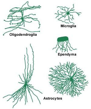
•   abnormal gait (the way the child walks).

If the tumour spreads or is located in the spinal cord, the signs and symptoms may include:
• back pain
• difficulty walking
• problems with bowel and bladder control.

Signs and Symptoms of High Grade Childhood Gliomas
High grade gliomas are malignant (cancerous) tumours that develop from brain cells called astrocytes.
Astrocytomas are the most common type of glioma. One often hears the terms astrocytoma and
glioma used interchangeably.

In children, only 20% of astrocytomas are high grade. High grade gliomas are classified according to
the grade of aggressiveness (how quickly they grow) as either anaplastic astrocytomas (grade III) or
glioblastoma multiforme (GBM).

These tumours often spread into the
healthy tissue that surrounds the
tumour. This makes them difficult to
remove surgically. They most commonly
arise in the cerebral hemispheres
(frontal, parietal and temporal lobes) or
centre of the brain (thalamus). The
tumour can spread to other parts of the
brain and spinal cord.

The symptoms depend on where in the
brain the tumour is and also whether
there is increased pressure in the head
(raised intracranial pressure).

                   [Picture Credit: Glioma Sites]

Symptoms include:
• nausea and vomiting (most common)
• lethargy and irritability
• headaches
• clumsiness
• seizures (fits)
• difficulty with tasks like handwriting
• gradual decline in school work (where applicable)
• changes in personality and behaviour
• abnormal gait (the way the child walks).

If the tumour spreads or is located in the spinal cord, the signs and symptoms may include:
Researched and Authored by Prof Michael C Herbst
[D Litt et Phil (Health Studies); D N Ed; M Art et Scien; B A Cur; Dip Occupational Health; Dip Genetic Counselling; Dip
Audiometry and Noise Measurement; Diagnostic Radiographer; Medical Ethicist]
Approved by Ms Elize Joubert, Chief Executive Officer [BA Social Work (cum laude); MA Social Work]
February 2021                                                                                                    Page 4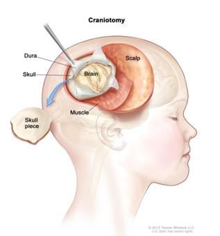
•   back pain
•   difficulty walking
•   problems with bowel and bladder control.

Risk Factors for Childhood Brain Tumours
Primary brain tumours account for only a small percentage of cancer deaths.

Ostrom, Q.T., Adel Fahmideh, M., Cote, D.J., Muskens, I.S., Schraw, J.M., Scheurer, M.E. & Bondy,
M.L. 2020.
“Primary brain tumors account for ~1% of new cancer cases and ~2% of cancer deaths in the United
States; however, they are the most commonly occurring solid tumors in children. These tumors are
very heterogeneous and can be broadly classified into malignant and benign (or non-malignant), and
specific histologies vary in frequency by age, sex, and race/ethnicity. Epidemiological studies have
explored numerous potential risk factors, and thus far the only validated associations for brain tumors
are ionizing radiation (which increases risk in both adults and children) and history of allergies (which
decreases risk in adults). Studies of genetic risk factors have identified 32 germline variants associated
with increased risk for these tumors in adults (25 in glioma, 2 in meningioma, 3 in pituitary adenoma,
and 2 in primary CNS lymphoma), and further studies are currently under way for other histologic
subtypes, as well as for various childhood brain tumors. While identifying risk factors for these tumors
is difficult due to their rarity, many existing datasets can be leveraged for future discoveries in multi-
institutional collaborations. Many institutions are continuing to develop large clinical databases
including pre-diagnostic risk factor data, and developments in molecular characterization of tumor
subtypes continue to allow for investigation of more refined phenotypes. Key Point 1. Brain tumors
are a heterogeneous group of tumors that vary significantly in incidence by age, sex, and
race/ethnicity.2. The only well-validated risk factors for brain tumors are ionizing radiation (which
increases risk in adults and children) and history of allergies (which decreases risk).3. Genome-wide
association studies have identified 32 histology-specific inherited genetic variants associated with
increased risk of these tumors.”

Diagnosis of Childhood Gliomas
The following tests and procedures may be used:
CT Scan (CAT scan) – a procedure that makes a series of detailed pictures of areas inside the body,
taken from different angles. The pictures are made by a computer linked to an X-ray machine. A dye
may be injected into a vein or swallowed by the patient to help the organs or tissues show up more
clearly. This procedure is also called computed tomography, computerised tomography, or
computerised axial tomography

MR Scan (magnetic resonance imaging) with gadolinium – a procedure that uses a magnet, radio
waves, and a computer to make a series of detailed pictures of areas inside the brain and spinal cord.
A substance called gadolinium is injected through a vein. The gadolinium collects around the cancer
cells so they show up brighter in the picture. This procedure is also called nuclear magnetic resonance
imaging (NMRI).

A brain biopsy - if a brain tumour is suspected, a brain biopsy is done by removing part of the skull and
using a needle to remove a sample of the tumour tissue. A pathologist views the tissue under a
Researched and Authored by Prof Michael C Herbst
[D Litt et Phil (Health Studies); D N Ed; M Art et Scien; B A Cur; Dip Occupational Health; Dip Genetic Counselling; Dip
Audiometry and Noise Measurement; Diagnostic Radiographer; Medical Ethicist]
Approved by Ms Elize Joubert, Chief Executive Officer [BA Social Work (cum laude); MA Social Work]
February 2021                                                                                                    Page 5
microscope to look for cancer cells. If cancer cells are found, the doctor will remove as much tumour
as safely possible during the same surgery.

Johansen, M.L., Vincent, J., Gittleman, H., Craig, S.E.L., Couce, M., Sloan, A.E., Barnholtz-Sloan, J.S.
& Brady-Kalnay, S.M. 2019.
“An integrated approach has been adopted by the World Health Organization (WHO) for diagnosing
brain tumors. This approach relies on the molecular characterization of biopsied tissue in conjunction
with standard histology. Diffuse gliomas (grade II to grade IV malignant brain tumors) have a wide
range in overall survival, from months for the worst cases of glioblastoma (GBM) to years for lower
grade astrocytic and oligodendroglial tumors. We previously identified a change in the cell adhesion
molecule PTPmu in brain tumors that results in the generation of proteolytic fragments. We developed
agents to detect this cell surface-associated biomarker of the tumor microenvironment. In the current
study, we evaluated the PTPmu biomarker in tissue microarrays and individual tumor samples of
adolescent and young adult (n = 25) and adult (n = 69) glioma populations using a fluorescent
histochemical reagent, SBK4-TR, that recognizes the PTPmu biomarker. We correlated staining with
clinical data and found that high levels of the PTPmu biomarker correlate with increased survival
of glioma patients, including those with GBM. Patients with high PTPmu live for 48 months on average,
whereas PTPmu low patients live only 22 months. PTPmu high staining indicates a doubling of patient
survival. Use of the agent to detect the PTPmu biomarker would allow differentiation
of glioma patients with distinct survival outcomes and would complement current molecular
approaches used in glioma prognosis.”

Where in the Brain Childhood Gliomas Grow
Gliomas may grow in different parts of the brain.

Gliomas in the cerebellum - the cerebellum is the back part of the brain. It is also called the posterior
fossa or hindbrain. Gliomas in the cerebellum are nearly all low grade. The main treatment is surgery
and about 9 out of 10 can be completely removed. If the tumour is not completely removed, the
specialist may decide to monitor the tumour with MRI scans and delay treatment until there are signs
that it is growing again. The child's specialist may also suggest radiotherapy or chemotherapy straight
away. Sometimes it is possible to carry out more surgery.

Gliomas in the brain stem - these tumours can be very difficult to treat. The brain stem is a very
delicate area that controls many vital body functions. It includes an area called the midbrain. As the
brain stem controls many vital body functions it is not always possible to operate in this area.

Gliomas in the brain stem can be slow growing and just in one area (these are called focal tumours).
Other tumours are faster growing and tend to grow into the surrounding brain tissue (called diffuse
tumours). There are various sub-types of brain stem glioma.

If possible, surgeons remove focal type gliomas. The child's specialist will then keep a very close eye
on the child to see if any remaining tumour starts to grow again. Sometimes, the specialist may advise
delaying surgery. Some tumours in this area grow so slowly that they do not really need treatment if
they are not causing symptoms.

Researched and Authored by Prof Michael C Herbst
[D Litt et Phil (Health Studies); D N Ed; M Art et Scien; B A Cur; Dip Occupational Health; Dip Genetic Counselling; Dip
Audiometry and Noise Measurement; Diagnostic Radiographer; Medical Ethicist]
Approved by Ms Elize Joubert, Chief Executive Officer [BA Social Work (cum laude); MA Social Work]
February 2021                                                                                                    Page 6
Some tumours are very small, but cause fluid to collect around the brain (hydrocephalus). If this
happens, the child's doctor may suggest that they put in a tube to drain the fluid, but they may give
no other treatment until the tumour grows.

Depending on their age, the child may have radiotherapy for a diffuse type glioma in the brain stem.
For very young children, specialists tend to give chemotherapy because this can work well for these
tumours in young children. They delay giving radiotherapy until the child is older. Fortunately brain
stem gliomas are very rare in young children.

Gliomas of the eyesight nerves - the eyesight nerves (optical nerves) are also called the visual nerves
or the optic pathway. Gliomas in this area are most often quite slow growing (low grade). In some
situations the child's specialist may suggest that the child does not have treatment straight away.
Some of these tumours grow so slowly that they may not cause any symptoms or problems for years.
This is most often the case with tumours in children who have the genetic condition
neurofibromatosis.

If there is any risk that the child may lose his/her eyesight they need to have treatment. Their specialist
will try to remove as much of the tumour as possible.

Surgery can be difficult in this area of the brain. If the tumour cannot be completely removed, the
child's specialist may suggest:

•   Radiotherapy or chemotherapy straight away if the child is having problems with sight
•   Chemotherapy to shrink the tumour, with radiotherapy delayed until the child is older

Gliomas that come back - unfortunately childhood gliomas can come back some time after they have
first been treated. The treatment for a brain tumour that has come back depends on the treatment
that the child had first time round.

The child's surgeon may be able to operate again. If the child did not have radiotherapy before, then
the specialist may suggest it now. If the child has had radiotherapy, they may still be able to have it
again. Or the doctor may suggest chemotherapy instead.

Treatment of Childhood Glioma
Paediatric high-grade gliomas (HGGs)—including glioblastoma multiforme, anaplastic astrocytoma,
and diffuse intrinsic pontine glioma—are difficult to treat and are associated with an extremely poor
prognosis. There are no effective chemotherapeutic regimens for the treatment of paediatric HGG,
but many new treatment options are in active investigation. There are crucial molecular differences
between adult and paediatric HGG such that results from adult clinical trials cannot simply be
extrapolated to children. Molecular markers overexpressed in paediatric HGG include
PDGFRα and P53. Amplification of EGFR is observed, but to a lesser degree than in adult HGG
(MacDonald, et al., 2011).

Researched and Authored by Prof Michael C Herbst
[D Litt et Phil (Health Studies); D N Ed; M Art et Scien; B A Cur; Dip Occupational Health; Dip Genetic Counselling; Dip
Audiometry and Noise Measurement; Diagnostic Radiographer; Medical Ethicist]
Approved by Ms Elize Joubert, Chief Executive Officer [BA Social Work (cum laude); MA Social Work]
February 2021                                                                                                    Page 7
There are very few randomised controlled trials looking at the
effectiveness of surgical resection, radiotherapy and
chemotherapy. Management also includes treatment of
complications (e.g. raised intracranial pressure, hydrocephalus,
seizures, pituitary hormone deficiencies), support for the child
and their family and addressing any associated psychological and
educational difficulties.

                                                 [Picture Credit: Craniotomy]

PDQ Pediatric Treatment Editorial Board. 2020.
There are different types of treatment for children with brain
stem glioma.
Children with brain stem glioma should have their treatment planned by a team of health care
providers who are experts in treating childhood brain tumours.
Five types of standard treatment are used:
• Surgery
• Radiation therapy
• Chemotherapy
• Cerebrospinal fluid diversion
• Observation
New types of treatment are being tested in clinical trials.

Surgical resection
Surgical resection is very important and recent data suggest that complete total resection, especially
of gliomas, should always be the aim and is associated with improved survival in children.

•   However, complete resection of the tumour by means of craniotomy is very rarely achievable as
    the margins of most tumours are indistinct. This means that during surgical resection it becomes
    difficult to determine whether abnormal or normal tissue is being resected. Resection also allows
    for a biopsy to be taken which in some types of brain tumour alters therapy.

•   Biopsy may be performed beforehand and usually direct open biopsy is preferred at the time of
    surgery although, for basal ganglia and brain stem lesions, stereotactic biopsies are taken.
    Preoperatively, children may be given phenytoin to prevent seizures and corticosteroids to reduce
    brain oedema.
•   Hydrocephalus is common postoperatively and
    therefore at the time of surgery an external ventricular
    drain or ventriculoperitoneal shunt is inserted which
    will be removed a few days later once the CSF clears.
•   Very young children (under the age of 2 years) require
    radical resection as radiotherapy is delayed until they
    are older, as it will damage local normal tissue which is
    still developing. This is usually followed by
    chemotherapy.

                                        [Picture Credit: Gamma Knife]

Researched and Authored by Prof Michael C Herbst
[D Litt et Phil (Health Studies); D N Ed; M Art et Scien; B A Cur; Dip Occupational Health; Dip Genetic Counselling; Dip
Audiometry and Noise Measurement; Diagnostic Radiographer; Medical Ethicist]
Approved by Ms Elize Joubert, Chief Executive Officer [BA Social Work (cum laude); MA Social Work]
February 2021                                                                                                    Page 8
Radiotherapy • This is provided in low doses and to very localised areas to avoid damage to surrounding normal brain tissue. There are various techniques that can be employed, e.g. gamma knife (used for slow- growing lesions) and interstitial seeds which are implanted during surgery. Ajithkumar, T., Taylor, R. & Kortmann, R.D. 2019. “Paediatric low-grade (World Health Organization grade I-II) gliomas (LGGs) represent a spectrum of primary central nervous system tumours. Local tumour control is the cornerstone in the general management of childhood gliomas. Surgery is the primary treatment of choice in the majority. Non- surgical treatments are recommended for progressive or symptomatic inoperable disease. Although chemotherapy is increasingly used as first non-surgical treatment, radiotherapy remains standard as salvage treatment or as primary treatment in selected cases in which surrounding normal tissue can be optimally preserved. The role of targeted therapies is currently under investigation in clinical trials. Modern high-precision radiotherapy techniques, including proton therapy, have the potential to improve long-term toxicities. There is therefore an urgent need for prospective studies to compare the efficacy and safety of modern radiotherapy with systemic treatment in children with LGGs. New information on molecular genetic patterns in LGGs may also have an impact on the selection and sequencing of radiotherapy.” Chemotherapy There are various chemotherapy regimens in use and they usually involve vincristine. In the rare primary CNS lymphoma, chemotherapy alone has been used with good outcomes. In low-grade gliomas, if residual disease remains after excision then chemotherapy has been used. Newer chemotherapeutic regimens are being used including vincristine, etoposide, cyclophsphamide and 5-fluorouracil. Follow-up after treatment Children have MRI scans every six months for the first two years and then annually (although this varies according to the centre and may become less frequent after the first few years). Complications • Intellectual decline - a recent study of 120 young patients with primary brain tumours showed a decline in sustained attention span and reaction times. This appeared to be caused by multiple factors including local tumour effects, surgery and radiotherapy. More recently, guidance on detecting and monitoring cognitive decline have been proposed. • Growth hormone deficiency is common (thyroid hormone deficiency is less common). • Neurological handicap may occur and be permanent. • Increased risk of a second brain tumour 10-20 years down the line due to irradiation (e.g. developing meningioma or sarcoma) - risk is increased if the brain is irradiated at a very young age. • Reduced bone mineral density of multifactorial origin. • Cavernomas presenting as haemorrhagic lesions are increasingly being associated with CNS irradiation. • Prognosis • Resection of the tumour may resolve seizures and headaches. The surgical mortality is 1% for a paediatric craniotomy. The morbidity is higher and depends on the child's condition Researched and Authored by Prof Michael C Herbst [D Litt et Phil (Health Studies); D N Ed; M Art et Scien; B A Cur; Dip Occupational Health; Dip Genetic Counselling; Dip Audiometry and Noise Measurement; Diagnostic Radiographer; Medical Ethicist] Approved by Ms Elize Joubert, Chief Executive Officer [BA Social Work (cum laude); MA Social Work] February 2021 Page 9
preoperatively. Figures such as, 5-10% for gliomas and 20-30% for basal ganglia gliomas have been
    reported.
•   Tumours of the brain and central nervous system are the most common cause of deaths from
    cancer in childhood, accounting for around a third of all cancer deaths in children.
•   A recent meta-analysis suggests that prenatal supplementation with multivitamins, including folic
    acid, is associated with a reduced incidence of brain tumours and leukaemia. However, this
    evidence still needs further validation.

About Clinical Trials
Clinical trials are research studies that involve people. They are conducted under controlled
conditions. Only about 10% of all drugs started in human clinical trials become an approved drug.

Clinical trials include:
     • Trials to test effectiveness of new treatments
     • Trials to test new ways of using current treatments
     • Tests new interventions that may lower the risk of developing certain types of cancers
     • Tests to find new ways of screening for cancer

The South African National Clinical Trials Register provides the public with updated information on
clinical trials on human participants being conducted in South Africa. The Register provides
information on the purpose of the clinical trial; who can participate, where the trial is located, and
contact details.

For additional information, please visit: www.sanctr.gov.za/

Medical Disclaimer
This Fact Sheet is intended to provide general information only and, as such, should not be considered
as a substitute for advice, medically or otherwise, covering any specific situation. Users should seek
appropriate advice before taking or refraining from taking any action in reliance on any information
contained in this Fact Sheet. So far as permissible by law, the Cancer Association of South Africa
(CANSA) does not accept any liability to any person (or his/her dependants/estate/heirs) relating to
the use of any information contained in this Fact Sheet.

 Whilst CANSA has taken every precaution in compiling this Fact Sheet, neither it, nor any
contributor(s) to this Fact Sheet can be held responsible for any action (or the lack thereof) taken by
any person or organisation wherever they shall be based, as a result, direct or otherwise, of
information contained in, or accessed through, this Fact Sheet.

Researched and Authored by Prof Michael C Herbst
[D Litt et Phil (Health Studies); D N Ed; M Art et Scien; B A Cur; Dip Occupational Health; Dip Genetic Counselling; Dip
Audiometry and Noise Measurement; Diagnostic Radiographer; Medical Ethicist]
Approved by Ms Elize Joubert, Chief Executive Officer [BA Social Work (cum laude); MA Social Work]
February 2021                                                                                                  Page 10
Sources and References Consulted or Utilised Ajithkumar, T., Taylor, R. & Kortmann, R.D. 2019. Radiotherapy in the management of paeditraic low-grade gliomas. Clin Oncol (R Coll Radiol). 2019 Mar;31(3):151-161. doi: 10.1016/j.clon.2018.11.032. Epub 2018 Dec 7. Cancer Research UK http://www.cancerresearchuk.org/cancer-help/type/brain-tumour/treatment/types/treatment-for-gliomas-in-children Childhood Cerebral Astrocytoma http://cancer.osu.edu/patientsandvisitors/cancerinfo/cancertypes/brain/about/cerebral/pages/index.aspx Gamma Knife http://www.monmouthgammaknife.com/physician-info/gamma-knife-system/ Glial Cells http://www.google.co.za/imgres?imgurl=&imgrefurl=http%3A%2F%2Fneuroscience.uth.tmc.edu%2Fs1%2Fchapter08.html &h=0&w=0&tbnid=DBh06mJCD14U7M&zoom=1&tbnh=247&tbnw=204&docid=b5k- vVhugkqKAM&tbm=isch&ei=rgp_U_LbEoX80QWs3IHYBw&ved=0CAgQsCUoAg Glioma https://www.google.co.za/search?q=signs+and+symptoms+childhood+gliomas&source=lnms&tbm=isch&sa=X&ei=Vh5_U7 D5DtGO7QbW_4CADA&ved=0CAYQ_AUoAQ&biw=1517&bih=714&dpr=0.9#q=gliomas+brain+tumor&tbm=isch&facrc=_&i mgdii=_&imgrc=Z9jlOLQ4JqQRfM%253A%3BnoZ1UpozLopebM%3Bhttps%253A%252F%252Fbaptisthealth.net%252Fen%2 52Fhealth-services%252Fgamma- knife%252FPublishingImages%252FGlioma.jpg%3Bhttps%253A%252F%252Fbaptisthealth.net%252Fen%252Fhealth- services%252Fgamma-knife%252Fpages%252Fglioma.aspx%3B1000%3B883 Glioma Sites https://www.google.co.za/search?q=signs+and+symptoms+childhood+gliomas&source=lnms&tbm=isch&sa=X&ei=Vh5_U7 D5DtGO7QbW_4CADA&ved=0CAYQ_AUoAQ&biw=1517&bih=714&dpr=0.9#q=gliomas+brain+tumor&tbm=isch&facrc=_&i mgdii=bbC-CNxn-rf0aM%3A%3BHFAB0AxbAxITyM%3BbbC-CNxn-rf0aM%3A&imgrc=bbC-CNxn- rf0aM%253A%3BcMgrNLtK7gsPvM%3Bhttp%253A%252F%252Fhealthcare.utah.edu%252Fhealthlibrary%252Fhealth-lib- image.php%253Fimageid%253D279257%3Bhttp%253A%252F%252Fhealthcare.utah.edu%252Fhealthlibrary%252Frelated %252Fdoc.php%253Ftype%253D34%2526id%253D17824-1%3B530%3B500 Johansen, M.L., Vincent, J., Gittleman, H., Craig, S.E.L., Couce, M., Sloan, A.E., Barnholtz-Sloan, J.S. & Brady-Kalnay, S.M. 2019. A PTPmu Biomarker is associated with increased survival in gliomas. In J Mol Sci. Int J Mol Sci. 2019 May 14;20(10). pii: E2372. doi: 10.3390/ijms20102372. Jones, D.T.W., Kieran, M.W., Bouffet, E., Alexandrescu, S., Bandopadhayay, P., Bornhorst, M. Ellison, D., Fangusaro, J., Fisher, M.J., Foreman, N., Fouladi, M., Hargrave, D., Hawkins, C., Jabado, N.,Massimino, M., Mueller, S., Perilongo, G., Schouten van Meeteren, A.Y.N., Tabori, U. Warren, K., Waanders, A.J., Walker, D., Weiss, W., Witt, O., Wright, K., Shu, Y., Bowers, D.C., Pfister, S.M. & Packer, R.J. 2018. Pediatric low-grade gliomas: next biologically driven steps. Neru Oncol. 2018 Jan 22;20(2):160-173. doi: 10.1093/neuonc/nox141. MacDonald, T.J., Aguilera, D. & Kramm, C.M. 2011. Treatment of high-grade glioma in children and adolescents. Neuro- Oncology, doi: 10.1093/neuonc/nor092. National Cancer Institute http://www.cancer.gov/about-cancer/treatment/clinical-trials/what-are-trials Ostrom, Q.T., Adel Fahmideh, M., Cote, D.J., Muskens, I.S., Schraw, J.M., Scheurer, M.E. & Bondy, M.L. 2020. Risk factors for childhood and adult primary brain tumors. Neuro Oncol. 2019 Nov 4;21(11):1357-1375. PDQ Pediatric Treatment Editorial Board. 2020. Childhood brain stem glioma treatment. In: PDQ Cancer Information Summaries [Internet]. Bethesda (MD): National Cancer Institute (US); 2002–2020 Nov 12. Subramanian, S. & Ahmed, T. 2020. Childhood brain tumors. In: StatPearls [Internet]. Treasure Island (FL): StatPearls Publishing; 2020 Jan. 2020 Aug 10. Researched and Authored by Prof Michael C Herbst [D Litt et Phil (Health Studies); D N Ed; M Art et Scien; B A Cur; Dip Occupational Health; Dip Genetic Counselling; Dip Audiometry and Noise Measurement; Diagnostic Radiographer; Medical Ethicist] Approved by Ms Elize Joubert, Chief Executive Officer [BA Social Work (cum laude); MA Social Work] February 2021 Page 11
The Royal Marsden
http://www.royalmarsden.nhs.uk/cancer-information/children/pages/high-grade-glioma.aspx
http://www.royalmarsden.nhs.uk/cancer-information/children/pages/low-grade-glioma.aspx

UCLA Neurosurgery
http://neurosurgery.ucla.edu/body.cfm?id=159

Researched and Authored by Prof Michael C Herbst
[D Litt et Phil (Health Studies); D N Ed; M Art et Scien; B A Cur; Dip Occupational Health; Dip Genetic Counselling; Dip
Audiometry and Noise Measurement; Diagnostic Radiographer; Medical Ethicist]
Approved by Ms Elize Joubert, Chief Executive Officer [BA Social Work (cum laude); MA Social Work]
February 2021                                                                                                  Page 12
You can also read