Ki-67 Proliferation Index in Neuroendocrine Tumors: Can Augmented Reality Microscopy With Image Analysis Improve Scoring? - Augmentiqs
←
→
Page content transcription
If your browser does not render page correctly, please read the page content below
Original Article
Ki-67 Proliferation Index in Neuroendocrine Tumors:
Can Augmented Reality Microscopy With Image Analysis
Improve Scoring?
1
Swati P. Satturwar, MD ; Joshua L. Pantanowitz, BS2; Christopher D. Manko, BS2; Lindsey Seigh, BS1;
1 1
Sara E. Monaco, MD ; and Liron X. Pantanowitz, MD
BACKGROUND: The Ki-67 index is important for grading neuroendocrine tumors (NETs) in cytology. However, different
counting methods exist. Recently, augmented reality microscopy (ARM) has enabled real-time image analysis using glass
slides. The objective of the current study was to compare different traditional Ki-67 scoring methods in cell block material
with newer methods such as ARM. METHODS: Ki-67 immunostained slides from 50 NETs of varying grades were retrieved
(39 from the pancreas and 11 metastases). Methods with which to quantify the Ki-67 index in up to 3 hot spots included:
1) “eyeball” estimation (EE); 2) printed image manual counting (PIMC); 3) ARM with live image analysis; and 4) image analysis
using whole-slide images (WSI) (field of view [FOV] and the entire slide). RESULTS: The Ki-67 index obtained using the dif-
ferent methods varied. The pairwise kappa results varied from no agreement for image analysis using digital image analysis
WSI (FOV) and histology to near-perfect agreement for ARM and PIMC. Using surgical pathology as the gold standard, the
EE method was found to have the highest concordance rate (84.2%), followed by WSI analysis of the entire slide (73.7%) and
then both the ARM and PIMC methods (63.2% for both). The PIMC method was the most time-consuming whereas image
analysis using WSI (FOV) was the fastest method followed by ARM. CONCLUSIONS: The Ki-67 index for NETs in cell block
material varied by the method used for scoring, which may affect grade. PIMC was the most time-consuming method, and EE
had the highest concordance rate. Although real-time automated counting using image analysis demonstrated inaccuracies,
ARM streamlined and hastened the task of Ki-67 quantification in NETs. Cancer Cytopathol 2020;0:1-10. © 2020 American
Cancer Society.
KEY WORDS: augmented reality microscope; cell block; digital image analysis; Ki-67 quantification; manual count;
neuroendocrine tumors.
INTRODUCTION
Grading of gastrointestinal neuroendocrine tumors (NETs) is performed by quantifying their mitotic activity
and/or Ki-67 index.1 The grade is critical in NET prognostication and treatment decisions, along with other
parameters such as tumor size, lymphovascular invasion, and stage of disease. The World Health Organization
(WHO) classifies neuroendocrine neoplasms as well-differentiated NETs, poorly differentiated neuroendo-
crine carcinoma (NEC), and mixed neuroendocrine-nonneuroendocrine neoplasms. WHO categorizes NETs
as grade 1 (those with a Ki-67 index 20
per 10 HPF]); NECs are grade 3 by definition.1-3 The Ki-67 index is determined by counting the percentage of
Corresponding Author: Liron X. Pantanowitz, MD, Department of Pathology, University of Pittsburgh Medical Center Cancer Pavilion, Ste 201, 5150 Centre
Ave, Pittsburgh, PA 15232 (pantanowitzl@upmc.edu).
1
Department of Pathology, University of Pittsburgh Medical Center, Pittsburgh, Pennsylvania; 2 Department of Biology, University of Pittsburgh, Pittsburgh,
Pennsylvania
We thank Ahmed Ishtiaque for his technical support and Colleen Vrbin from Analytical Insights for help with the statistical analysis.
Received: January 13, 2020; Revised: February 18, 2020; Accepted: March 11, 2020
Published online Month 00, 2020 in Wiley Online Library (wileyonlinelibrary.com)
DOI: 10.1002/cncy.22272, wileyonlinelibrary.com
Cancer Cytopathology Month 2020 1
Original Article

positively stained tumor cells in an area of the tumor with     acquired by this unit occur in real time and can be dis-
the highest nuclear labeling (ie, a hot spot) based on 500      played on an attached computer monitor. In addition, if
to 2000 tumor cells.                                            the end user looks through the eyepieces of the microscope,
      NETs involving the pancreas and their metastases          they can see computer-generated output, such as annota-
to visceral sites (eg, the liver) frequently are diagnosed      tions, being projected as an overlay directly on the glass
using fine-needle aspiration (FNA).4-7 Often, these FNA         slide. ARM thus permits real-time image analysis to be
specimens represent the primary diagnostic material for         performed on glass slides with the output of the algorithm
patients. Hence, the preoperative grading of NETs on            superimposed on the slide, thereby generating a composite
such cytology material typically is required to guide           field of view (FOV) that can be used to supervise data col-
treatment decisions.8-11 Currently, to our knowledge, it        lection. This can be used to perform simple measurements
remains questionable whether a Ki-67 index obtained on          (eg, depth of tumor invasion) and, when coupled with
cytology material, such as a cell block, is truly represen-     image analysis software, to quantify immunohistochemical
tative of the entire tumor compared with the surgical re-       stains (eg, Ki-67 proliferation index) or it can be combined
section specimen.12 Several studies also have attempted         with more sophisticated deep learning algorithms (eg, to
to establish the best counting method for determining           rapidly detect metastases in lymph nodes).23 As opposed
the Ki-67 index.13-18 However, to the best of our knowl-        to DIA applied to whole-slide imaging (WSI), ARM is
edge, there is no accepted standard as yet and the best         quicker to use, provides for the real-time and seamless
scoring method still is debatable. Different methods for        integration of algorithms without the need to first acquire
Ki-67 index quantification include “eyeball” estimation         digital images, may be cheaper than buying a whole-slide
(EE) performed manually by a pathologist counting in            scanner, and does not require special technical skills to
real time through their microscope eyepiece, manual             operate. Based on our calculation of direct equipment
counting using a printed image that was captured using a        costs, the ARM device is approximately 20 times less
camera mounted to a microscope at ×20 magnification,19          expensive than a high-capacity whole-slide scanner.
and computer-assisted quantification using digital image              To the best of our knowledge, no studies to date
analysis (DIA).15,20,21 Manual methods suffer from low          have applied ARM to cytology. We anticipate that
reproducibility and high interreader variability among          ARM may overcome many of the aforementioned issues
pathologists, especially for low-grade NETs. Moreover,          related to Ki-67 quantification. Therefore, the objective
the manual methods, especially if they involve the print-       of the current study was to compare ARM with different
ing of images, are impractical in certain clinical prac-        (manual and DIA with WSI) scoring methods used for
tice settings and are labor-intensive. To overcome the          determining the Ki-67 proliferation index of NETs in cell
interobserver variabilities inherent in human counting,         block material.
researchers accordingly have explored automated count-
ing methods using digital images and a variety of image         MATERIALS AND METHODS
analysis algorithms. Although faster and more precise,
                                                                Case Collection
DIA systems can be costly because they may require an
expensive whole-slide scanner to digitize slides and infor-     After institutional review board approval, a total of
mation technology support.                                      50 cytology NETs from 45 patients were retrieved (39
      Recently, augmented reality microscopy (ARM) has          pancreatic tumors and 11 metastases to the liver) from
become available that enables real-time image analysis to       January 2014 to July 2019. Data including patient age,
be conducted using a traditional light microscope and glass     sex, subsequent surgical resection, and American Joint
slides,22 which avoids the need to first photograph or dig-     Committee on Cancer clinical stage of disease were
itize slides. An augmented reality microscope is a modi-        recorded. A total of 19 of the 39 pancreatic tumors
fied (“smart”) microscope that includes a small computer        (48.71%) underwent a subsequent surgical resection.
unit. This computer unit can be attached to the side of         The inclusion criteria for cases to be enrolled in this
any microscope or it can be inserted between the micro-         study were specimens for which a satisfactory cell block
scope’s objective lenses and eyepiece unit. It incorporates a   was available (ie, tumor cells were present), a NET
built-in camera to capture high-quality images. The images      proven by immunohistochemistry, the Ki-67 index was

2                                                                             Cancer Cytopathology  Month 2020
Ki-67 Proliferation Index in NETs/Satturwar et al

available in the cytology report, and the original Ki-67
immunostained slide was available for further analy-
sis. Exclusion criteria included cases with a cell block
for which the tumor cellularity was
Original Article
magnification and 1 Z-plane. DIA then was performed
using the Aperio immunohistochemistry color convo-
luted, nuclearV9 quantitative image analysis algorithm
(Leica Biosystems). This allowed for the percentage of
positive over total tumor nuclei (Ki-67 index) to be cal-
culated. DIA-WSI was performed for each hot-spot FOV
(FOV analysis) and also for the entire cell block (slide-
level analysis).
Histology analysis
These data were obtained from the 19 surgical resection
pathology reports. The Ki-67 index had been calculated
by 7 different surgical pathologists with expertise in gas-
trointestinal pathology, using manual counting of printed
images of at least 500 cells.
Statistical Analysis
Statistical analysis was performed using IBM SPSS
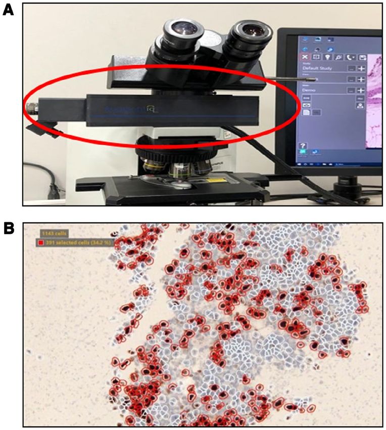
FIGURE 2. (Top) Light microscope with the Augmentiqs Statistics 22 (IBM Corporation, Armonk, New York),
device (red circle) fitted between the objective lenses and the
eyepiece. (Bottom) Screenshot showing a field of view using and statistical significance was assumed at P ≤ .05.
the augmented reality microscopy counting method in which
the image analysis segments individual nuclei (white circles)
The Cohen kappa was used to determine interrater
and the end user manually selects positive nuclei (red circles). reliability for tumor grade assessment (grades 1-3) for
pairwise comparison of Ki-67 quantification methods.
The normality of the distribution of continuous vari-
composite images within the microscope eyepiece, and ables (number of minutes and percentage of Ki-67) was
simultaneously displayed this composite image on the examined using the Shapiro-Wilk normality test.
attached computer monitor. For quantitative analyt- Because the data were not normally distributed, non-
ics, this ARM setup was coupled with image analysis parametric statistical tests were used. The Kendall tau-b
software modified from QuPath software (Queen’s correlation coefficient was used to determine the rela-
University Belfast, Belfast, Northern Ireland; https:// tionship between the amount of time spent in assessing
qupath.github.io) for the detection, segmentation, and the presence of Ki-67 and the amount of Ki-67 iden-
scoring of nuclear stains (Fig. 2 Bottom). For all NET tified. The interclass correlation coefficient (ICC) was
cases, the same hot spots at ×20 magnification were calculated to determine interrater reliability among all
analyzed. After image analysis results were superim- cytological methods, and when available with and with-
posed on glass slides, 2 of the authors (J.P. and C.M.) out subsequent surgical resection procedures.
manually selected all stained nuclei. These selected
stained cells then were divided by the total number of RESULTS
cells that were automatically detected by the software The clinicopathologic characteristics of the NETs
to calculate the Ki-67 index for each FOV. Hence, the included in the current study are summarized in Table 1.
actual quantitative assessment component still required The 50 cases were obtained from 45 patients with an
human involvement and was not automated. average age of 62 years and demonstrated a male-to-
female ratio of 1:1. The most common location for pancre-
Digital image analysis using whole- atic tumors was the body, followed by the tail. Pancreatic
slide images NETs measured on average 3.12 cm in size. Of the cases
Ki-67–stained slides were scanned using an Aperio AT2 that had subsequent surgical resection specimens, >50%
scanner (Leica Biosystems, Wetzlar, Germany) using ×40 of these patients had lymph node metastases and 10%
4 Cancer Cytopathology Month 2020Ki-67 Proliferation Index in NETs/Satturwar et al
TABLE 1. Clinicopathologic Characteristics of NETs very faint staining nuclei) not being counted individually.
Studied (50 by Cytology and 19 by Histology For the subset of cases with subsequent pancreatic resec-
Characteristic Value tions (19 cases), a Wilcoxon signed rank test indicated
Average patient age (range), y 62 (35-79) that the Ki-67 index of these NETs in the surgical spec-
Sex imen procedure (median, 3.5%) was significantly higher
Male 22 (49%)
Female 23 (51%) than the index obtained using the traditional EE method
Average tumor size (range), cm 3.12 (1.5-8.5)
Location of tumor, no. (%)
(median, 1.5%) when applied to corresponding preoper-
Pancreatic head 9 (23%) ative cytology samples (Z [Z-statistic] = −2.136; r [effect
Pancreatic body 14 (36%)
Pancreatic tail 13 (33.33%)
size] = .35 [P = .033]). This equates to a difference of
Pancreatic body-tail 3 (7.69%) tumor grade 1 as determined by the traditional EE man-
Liver (metastasis) 11 (22%)
Histologic grade (n = 19) ual method in cell blocks versus grade 2 in the follow-up
1 9 (47%) surgical specimens. For NETs with surgical follow-up in
2 9 (47%)
3 1 (5%) which the Ki-67 index in cell blocks was assessed using
T classification, no. (%) PIMC, ARM, and DIA-WSI (entire slide), these scores
T1 3 (15.7%)
T2 10 (52.63%) similarly resulted in a median equivalent of grade 1, but
T3 6 (31.57%)
T4 0
the difference in the median Ki-67 index for these com-
N classification, no. (%) puter-assisted methods was not statistically significant.
N0 7 (36.84%)
N1 10 (52.63%)
Tumor grade assessment using WHO guidelines
NX 2 (10.52%) and shown by different methods is shown in Table 2.
M classification, no. (%)
M0 17 (89.47%) The percentage of cases scored using DIA-WSI (FOV)
M1 2 (10.52%) with grade 2 was higher at 50% compared with approx-
AJCC stage, no. (%)
I 4 (21%) imately 30% for the other methods being used to score
II 6 (31.57%) cell blocks. The main reason for the difference in the
III 7 (36.84%)
IV 2 (10.52%) number of grade 1 NETs versus grade 2 NETs using the
Abbreviation: AJCC, American Joint Committee on Cancer eighth edition; different methods was the narrow cutoff value of the
NETs, neuroendocrine tumors.
Ki-67 index. The DIA-WSI (FOV) method had inaccu-
racies (typically causing upgrading of the tumor grade)
had other distant metastases. Of the 11 cases of liver due to miscounting of nontumoral material (debris in
metastases, 6 (54.55%) originated from the pancreas and cell blocks, tissue folds, and proliferating inflammatory
5 (45.45%) were metastases from the lung or intestinal cells). Of the grade 3 tumors, 8 of 11 tumors were grade
NETs. 3 NECs and all were scored as grade 3 using different
The Ki-67 proliferation index based off the origi- cytology methods.The interrater reliability for tumor
nal cytopathology reports indicated that the average grade by paired Ki-67 assessment method is shown in
Ki-67 score for the EE method of grading cell blocks was Table 3. The pairwise kappa results varied from no agree-
18.17% (range, 0.5%-95.0%) and that for the surgical ment (0.04 for DIA-WSI [FOV] and histology compar-
resection specimens (19 cases) was 8.91% (range, 0.7%- isons) to near-perfect agreement (0.81 for the ARM and
95.0%). When averaging the 3 hot spots, the Ki-67 index PIMC methods) based on a scale of 0.81 indicating
12.35% (range, 0.1%-85.83%) for the ARM method near-perfect agreement. Using the surgical resection spec-
and was 19.01% (range, 0.57%-94.0%) when using imens (19 cases) as the gold standard, Table 4 compares
DIA-WSI (FOVs) and 11.48% (range, 0.31%-87.98%) the concordance rate between cytology and histology
when using DIA-WSI (entire slide). When using the cases. Compared with this histopathology gold standard,
ARM method, some inaccuracies in cell segmentation the concordance rate was highest for the EE method at
were noted with the image analysis algorithm, resulting 84.2% (kappa, 0.71) followed by the DIA-WSI (entire
in certain cells (eg, overlapping cells present in clusters, slide) method at 73.7% (kappa, 0.52). ARM and PIMC
Cancer Cytopathology Month 2020 5Original Article
TABLE 2. Tumor Grade Assessment by Different Ki-67 Quantification Methods
DIA-WSI (Entire
EE PIMC ARM DIA-WSI (FOV) Slide) Histology
Tumor Grade Ki-67 Index No. % No. % No. % No. % No. % No. %
1 20% 11 22% 11 22% 10 20% 12 24% 8 16% 1 5%
Total 50 100% 50 100% 50 100% 50 100% 50 100% 19 100%
Abbreviations: ARM, augmented reality microscopy; DIA, digital image analysis; EE, eyeball estimation; FOV, field of view; PIMC, printed image manual counting;
WSI, whole-slide imaging.
TABLE 3. Pairwise Kappa Calculations to Determine Interrater Reliability for Tumor Grade Assessment Among
Ki-67 Calculation Methods
Scoring Method EE PIMC ARM DIA-WSI (FOV) DIA-WSI (Entire Slide) Histology
EE — 0.74 0.68 0.46 0.61 0.71
PIMC 0.74 — 0.81 0.50 0.43 0.33
ARM 0.68 0.81 — 0.44 0.45 0.33
DIA-WSI (FOV) 0.46 0.50 0.44 — 0.37 0.04
DIA-WSI (entire slide) 0.61 0.43 0.45 0.37 — 0.52
Histology 0.71 0.33 0.33 0.04 0.52 —
Abbreviations: ARM, augmented reality microscopy; DIA, digital image analysis; EE, eyeball estimation; FOV, field of view; PIMC, printed image manual counting;
WSI, whole-slide imaging.
Based on a scale of 0.81 indicating near-perfect agreement.
TABLE 4. Tumor WHO Grading Based on Ki-67 Quantification of Cell Block Material by Different Methods and
Concurrent Surgical Resection Specimens
Histology (Surgical Resection) Total
Scoring Method Cytology Grades Grade 1 Grade 2 Grade 3 All Grades Percent Agreementa Kappa
EE 1 9 3 0 12 84.2% 0.71
2 0 6 0 6
3 0 0 1 1
PIMC 1 6 4 0 10 63.2% 0.33
2 3 5 0 8
3 0 0 1 1
ARM 1 7 5 0 12 63.2% 0.33
2 2 4 0 6
3 0 0 1 1
DIA-WSI (FOV) 1 2 3 0 5 47.4% 0.04
2 7 6 0 13
3 0 0 1 1
DIA-WSI (entire 1 8 4 0 12 73.7% 0.52
slide) 2 1 5 0 6
3 0 0 1 1
Abbreviations: ARM, augmented reality microscopy; DIA, digital image analysis; EE, eyeball estimation; FOV, field of view; PIMC, printed image manual counting;
WHO, World Health Organization; WSI, whole-slide imaging.
a
The percent agreement shows the correlation of the Ki-67 index between cytology cell block and subsequent surgical resection specimen assessments.
each had a concordance rate of 63.2% (kappa, 0.33). the different methods for Ki-67 quantification varied
There was excellent reliability among all scoring methods markedly (Table 6). The traditional EE and manual
when 3 hot spots (FOV) were evaluated (Table 5) com- histology methods were excluded for statistical analy-
pared with just counting 1 or 2 hot spots (ICC of 0.912 sis because the time spent by the pathologists was not
[P < .001] for 3 hot spots vs ICC of 0.706 [P = .13] for recorded in the original reports. Generally, cases with
1 hot spot and ICC of 0.606 [P = .015] for 2 hot spots). higher percentages of Ki-67 immunostained cells were
The time spent, including the time required for associated with taking longer times to score (Fig. 3).
the annotation of slides, as measured in minutes for DIA-WSI scoring of only the hot spots was the fastest
6 Cancer Cytopathology Month 2020Ki-67 Proliferation Index in NETs/Satturwar et al
TABLE 5. Interclass Correlation Coefficient Results of surgically resected tumors (19 tumors). Moreover, even
for Tumor Grade Assessment Based on the Number when we used the PIMC or computer-assisted (ARM or
of Hot Spot FOVs
DIA-WSI) methods for the grading of cell blocks, they
Interclass still led to undergrading. Similarly, Tang et al reported
Different methods (PIMC, ARM, Correlation
DIA-WSI [FOV]) Coefficient 95% CI that EE, manual counting, and even DIA all can provide
1 FOV Average measures 0.706 0.144-0.927
incorrect Ki-67 assessments and therefore false grading of
2 FOV 0.606 0.095-0.876 NETs.15 This suggests that FNA does not lend itself to the
3 FOV 0.912 0.833-0.956
accurate grading of NETs, which may be due to limited
Abbreviations: ARM, augmented reality microscopy; DIA, digital image analy-
sis; FOV, field of view; PIMC, printed image manual counting; WSI, whole- sampling and tumor heterogeneity.26 For FNA samples
slide imaging.
that contain ≥1000 cells, one group of researchers dem-
onstrated that counting hot spots instead of the complete
method of calculating the Ki-67 proliferation index cell block provided better correlation with surgical speci-
(average, 2.6 minutes per case), whereas the PIMC mens.17 We did analyze the same 3 hot spots using differ-
method was the most time-consuming (average, 15.3 ent methods, and found excellent reliability among these
minutes per case). Assessments using ARM took an av- methods based on these hot spots versus analyzing just 1
erage of 7.6 minutes per case, which was approximately or 2 of the FOVs. One limitation of the current study was
one-half the time of the PIMC method. Using the the relatively small number of follow-up surgical resection
Kendall tau-b correlation (τ), there was a strong, posi- specimens (19 specimens), which may have influenced
tive correlation noted between time spent scoring and effective correlation with cytology specimens.
the Ki-67 score for the ARM (τ = .536; P < .001) and As noted earlier, several methods have been pro-
DIA-WSI (FOV) (τ = .408; P < .001) methods. There posed for determining the Ki-67 proliferation index in
also was a positive relationship observed between time NETs. These include the manual EE method using a light
to quantify and the Ki-67 index for manual assessments microscope, the PIMC method that requires pathologists
(τ = .221; P = .024). However, there was no statis- to print out photographs to count positive cells in hot
tically significant correlation noted between the time spots, and DIA techniques. The data from the current
involved and the Ki-67 score for the DIA-WSI (entire study indicated that the level of reliability for these dif-
slide) assessment method (τ = .069; P = .447). ferent methods when tested on cell block material was
good to excellent for Ki-67 quantification. However, the
DISCUSSION
average Ki-67 scores did differ with these various meth-
The Ki-67 index clearly plays a critical role in the patho- ods. It is interesting to note that the pairwise kappa result
logic assessment of and clinical decisions related to the demonstrated near-perfect agreement for the PIMC and
management of gastroenteropancreatic NETs.24,25 The ARM methods. When using the Ki-67 score obtained
variabilities in different scoring methods, as well as inter- from the 19 surgical pathology cases as the “gold stan-
laboratory and interobserver reproducibility, have been ad- dard,” we found that the EE method of scoring cell blocks
dressed in part through the use of standardized guidelines had the highest concordance rate. This result is surpris-
from the WHO and European Neuroendocrine Tumor ing because EE is subjective and therefore prone to high
Society (ENETS) for calculating the Ki-67 proliferation interobserver variability. Indeed, prior studies have re-
index in histopathology material.1-4,24 These guidelines ported that EEs of pancreatic NETs were inaccurate.27,28
have been extrapolated to grading NET samples (eg, cell To overcome this problem, Reid et al recommended the
blocks) procured by FNA. However, several studies have PIMC method.19 These authors found in their experi-
shown that the grading of cell blocks using the Ki-67 ence that the manual counting of camera-captured and/
proliferation index frequently results in undergrading or printed images was more reliable and had higher repro-
of these tumors when compared with follow-up surgical ducibility than DIA, but took longer than EE. We also
resection specimens.12 This fact was corroborated in the found the PIMC method to be the most time-consuming
current study, in which the majority of NETs that were in the current study. However, Dogukan et al mastered
graded as 1 on cell blocks as per the original cytology re- this laborious task by using a monitor image instead of
ports subsequently were found to be grade 2 in the subset the printout image method for Ki-67 scoring.29 These
Cancer Cytopathology Month 2020 7Original Article
TABLE 6. Amount of Time Spent to Quantify Ki-67 According to Different Scoring Methods
Scoring Method Average Time, Minutes Median Time, Minutes Minimum Time, Minutes Maximum Time, Minutes
PIMC 15.3 11.5 2.5 62.0
ARM 7.6 4.0 0.5 60.0
DIA-WSI (FOV) 2.6 3.2 0.3 3.5
DIA-WSI (entire slide) 14.2 9.4 0.7 46.6
Abbreviations: ARM, augmented reality microscopy; DIA, digital image analysis; FOV, field of view; PIMC, printed image manual counting; WSI, whole-slide imaging.
FIGURE 3. Scatterplots of time spent versus the Ki-67 index for different assessment methods. ARM indicates augmented reality
microscopy; DIA, digital image analysis; FOV, field of view; PIMC, printed image manual counting; WSI, whole-slide imaging.
authors demonstrated that although monitor image and patients with breast cancer.31 A limited number of publi-
printout image methods for Ki-67 scoring of gastroen- cations to date have reported outcomes when using com-
teropancreatic NETs resulted in comparable scores, they mercial and open-source (eg, ImmunoRatio http://wsise
were able to save time performing this task by using a grid rver.jilab.fi/old-jvsmicroscope-software/) image analysis
and a monitor. software to calculate the Ki-67 index in NETs.16,19,32,33
For assessing biomarkers in breast cancer (eg, ER, However, for this task, the majority of these authors
PR, or HER2), DIA has been shown to outperform man- conveyed that manual counting was more accurate than
ual scoring.30 Similar supportive findings using DIA have DIA. Some of the reasons for this discordance with DIA
been reported for determining the Ki-67 labeling index in included nontumor cell contamination (eg, proliferating
8 Cancer Cytopathology Month 2020Ki-67 Proliferation Index in NETs/Satturwar et al
tumor–infiltrating lymphocytes) and background nonspe- in real time during this undertaking, we overcame any
cific staining. In the current study, we also noted inaccura- focus issues that typically plague the digital imaging of
cies in some cases when individual cells were not counted cytology material. In summary, we found that ARM with
using the image analysis algorithm applied via ARM. DIA when directly supervised by a human streamlined
The ARM vendor (Augmentiqs) in the current study has and hastened the task of assessing the Ki-67 proliferation
since modified various parameters of the image analysis index in NETs. Given the versatility and cost benefit of
algorithm for quantifying nuclear staining, and there- microscope-based AR technology, we anticipate that there
fore improved results are expected in future studies. The will be other innovative studies using ARM in the near
majority of the prior studies using DIA to determine the future.
Ki-67 index in NETs used static images of representative
tumor regions. Very few researchers used WSI. Hasegawa FUNDING SUPPORT
et al did use WSI with DIA to evaluate the Ki-67 index No specific funding was disclosed.
in 58 patients and reported high concordance (90%) with
surgically resected specimens, but only when their FNA CONFLICT OF INTEREST DISCLOSURES
samples had adequate cellularity (>2000 tumor cells).9 The authors made no disclosures.
WSI most likely aided their findings by allowing a greater
percentage of the tumor to be analyzed, instead of just AUTHOR CONTRIBUTIONS
limiting their analyses to smaller FOVs (ie, snapshots). Swati P. Satturwar: Data collection, methodology (performed
This also may explain why in the current study DIA-WSI manual Ki-67 quantification), and writing–original draft. Joshua
of the entire slide demonstrated a better concordance with L. Pantanowitz: Methodology (performed Ki-67 quantification
using an augmented reality microscope). Christopher D. Manko:
surgical resection specimens than the PIMC method. Methodology (performed Ki-67 quantification using an augmented
To the best of our knowledge, the current study reality microscope). Lindsey Seigh: Digital analysis. Sara E.
is the first to evaluate the use of ARM in cytology. Monaco: Conceptualization, writing–original draft, and writing–
Augmented reality (AR) refers to technology that com- review and editing. Liron X. Pantanowitz: Conceptualization,
supervision, validation, and writing–review and editing.
bines reality and digital information.34 This is created by
superimposing a computer-generated digital image onto
an object or user’s view of the “real world.” This differs REFERENCES
1. Fang JM, Shi J. A clinicopathologic and molecular update of
from virtual reality, in which a complete digital or com- pancreatic neuroendocrine neoplasms with a focus on the new
puter-generated environment is created. AR technol- World Health Organization classification. Arch Pathol Lab Med.
ogy (eg, HoloLens; Microsoft Corporation, Redmond, 2019;143:1317-1326.
2. Guilmette JM, Nose V. Neoplasms of the neuroendocrine pancreas:
Washington) has been applied successfully to anatomic an update in the classification, definition and molecular genetic
pathology for unique applications such as 3-dimensional advances. Adv Anat Pathol. 2019;26:13-30.
3. Klimstra DA, Kloppel G, La Rosa S, Rindi G. The WHO Classification
image viewing and real-time pathology-radiology correla- of Tumors: Digestive System Tumors. 5th ed. IARC Press; 2019.
tion.35 Recently, investigators introduced the novel AR 4. Sigel CS. Advances in the cytologic diagnosis of gastroenteropancreatic
microscope.22 By attaching an AR unit to a conventional neuroendocrine neoplasms. Cancer Cytopathol. 2018;126:980-991.
5. Atiq M, Bhutani MS, Bektas M, et al. EUS-FNA for pancreatic neu-
microscope, this accessory device converts the microscope roendocrine tumors: a tertiary cancer center experience. Dig Dis Sci.
into a digital pathology solution that now can be used 2012;57:791-800.
6. Unno J, Kanno A, Masamune A, et al. The usefulness of endoscopic
to perform real-time telepathology and computer-assisted ultrasound–guided fine-needle aspiration for the diagnosis of pancre-
diagnostics (eg, image analysis and the application of atic neuroendocrine tumors based on the World Health Organization
artificial intelligence algorithms) in addition to AR.22,23,36 classification. Scand J Gastroenterol. 2014;49:1367-1374.
7. Fujimori N, Osoegawa T, Lee L, et al. Efficacy of endoscopic ultra-
In the current study, the AR microscope permitted us to sonography and endoscopic ultrasonography–guided fine-needle
rapidly execute image analysis to quantify Ki-67 directly aspiration for the diagnosis and grading of pancreatic neuroendocrine
tumors. Scand J Gastroenterol. 2016;51:245-252.
from cytology glass slides while they were present on the 8. Grosse C, Noack P, Silye R. Accuracy of grading pancreatic neuro-
microscope’s stage. This allowed us to avoid having to first endocrine neoplasms with Ki-67 index in fine-needle aspiration cell-
photograph these slides or digitize the entire slide with block material. Cytopathology. 2019;30:187-193.
9. Hasegawa T, Yamao K, Hijioka S, et al. Evaluation of Ki-67 index in
a whole-slide scanner. Moreover, because we were able EUS-FNA specimens for the assessment of malignancy risk in pancre-
to adjust the microscope’s fine and coarse magnification atic neuroendocrine tumors. Endoscopy. 2014;46:32-38.
Cancer Cytopathology Month 2020 9Original Article
10. Weynand B, Borbath I, Bernard V, et al. Pancreatic neuroendocrine 22. Chen PC, Gadepalli K, MacDonald R. An augmented reality micro-
tumour grading on endoscopic ultrasound–guided fine needle aspira- scope with real-time artificial intelligence integration for cancer diag-
tion: high reproducibility and inter-observer agreement of the Ki-67 nosis. Nat Med. 2019;25:1453-1457.
labelling index. Cytopathology. 2014;25:389-395. 23. Razavian N. Augmented reality microscopes for cancer histopathol-
11. Weiss VL, Kiernan C, Wright J, Merchant NB, Coogan AC, Shi C. ogy. Nat Med. 2019;25:1334-1336.
Fine-needle aspiration–based grading of pancreatic neuroendocrine 24. O’Toole D, Kianmanesh R, Caplin M. ENETS 2016 Consensus
neoplasms using Ki-67: is accurate WHO grading possible on cyto- Guidelines for the Management of Patients with Digestive
logic material? J Am Soc Cytopathol. 2018;7:154-459. Neuroendocrine Tumors: an update. Neuroendocrinology. 2016;103:
12. Farrell JM, Pang JC, Kim GE, Tabatabai ZL. Pancreatic neuroen- 117-118.
docrine tumors: accurate grading with Ki-67 index on fine-needle 25. Sadot E, Reidy-Lagunes DL, Tang LH, et al. Observation versus
aspiration specimens using the WHO 2010/ENETS criteria. Cancer resection for small asymptomatic pancreatic neuroendocrine tumors:
Cytopathol. 2014;122:770-778. a matched case-control study. Ann Surg Oncol. 2016;23:1361-1370.
13. Boutsen L, Jouret-Mourin A, Borbath I, van Maanen A, Weynand 26. Grillo F, Valle L, Ferone D, et al. KI-67 heterogeneity in well dif-
B. Accuracy of pancreatic neuroendocrine tumour grading by en- ferentiated gastro-entero-pancreatic neuroendocrine tumors: when is
doscopic ultrasound–guided fine needle aspiration: analysis of a biopsy reliable for grade assessment? Endocrine. 2017;57:494-502.
large cohort and perspectives for improvement. Neuroendocrinology. 27. Young HT, Carr NJ, Green B, Tilley C, Bhargava V, Pearce N.
2018;106:158-166. Accuracy of visual assessments of proliferation indices in gastroentero-
14. Goodell PP, Krasinskas AM, Davison JM, Hartman DJ. Comparison pancreatic neuroendocrine tumours. J Clin Pathol. 2013;66:700-704.
of methods for proliferative index analysis for grading pancre- 28. Cottenden J, Filter ER, Cottreau J. Validation of a cytotechnolo-
atic well-differentiated neuroendocrine tumors. Am J Clin Pathol. gist manual counting service for the Ki67 index in neuroendocrine
2012;137:576-582. tumors of the pancreas and gastrointestinal tract. Arch Pathol Lab
15. Tang LH, Gonen M, Hedvat C, Modlin IM, Klimstra DS. Objective Med. 2018;142:402-407.
quantification of the Ki67 proliferative index in neuroendocrine 29. Dogukan FM, Yilmaz Ozguven B, Dogukan R, Kabukcuoglu F.
tumors of the gastroenteropancreatic system: a comparison of Comparison of monitor-image and printout-image methods in Ki-67
digital image analysis with manual methods. Am J Surg Pathol. scoring of gastroenteropancreatic neuroendocrine tumors. Endocr
2012;36:1761-1770. Pathol. 2019;30:17-23.
16. Jin M, Roth R, Gayetsky V, Niederberger N, Lehman A, Wakely PE 30. Stalhammar G, Fuentes Martinez N, Lippert M, et al. Digital image
Jr. Grading pancreatic neuroendocrine neoplasms by Ki-67 staining analysis outperforms manual biomarker assessment in breast cancer.
on cytologic cell blocks: manual count and digital image analysis of Mod Pathol. 2016;29:318-329.
58 cases. J Am Soc Cytopathol. 2016;5:286-295. 31. Maeda I, Abe K, Koizumi H, et al. Comparison between Ki67 label-
17. Abi-Raad R, Lavik JP, Barbieri AL, Zhang X, Adeniran AJ, Cai G. ing index determined using image analysis software with virtual slide
Grading pancreatic neuroendocrine tumors by Ki-67 index evalu- system and that determined visually in breast cancer. Breast Cancer.
ated on fine-needle aspiration cell block material. Am J Clin Pathol. 2016;23:745-751.
2020;153:74-81. 32. Hacking SM, Sajjan S, Lee L, et al. Potential pitfalls in diagnostic
18. Niazi MKK, Tavolara TE, Arole V, Hartman DJ, Pantanowitz L, digital image analysis: experience with Ki-67 and PHH3 in gastroin-
Gurcan MN. Identifying tumor in pancreatic neuroendocrine testinal neuroendocrine tumors. Pathol Res Pract. 2020;216:152753.
neoplasms from Ki67 images using transfer learning. PLoS One. 33. Hwang HS, Kim Y, An S, et al. Grading by the Ki-67 labeling index
2018;13:e0195621. of endoscopic ultrasound–guided fine needle aspiration biopsy spec-
19. Reid MD, Bagci P, Ohike N, et al. Calculation of the Ki67 index imens of pancreatic neuroendocrine tumors can be underestimated.
in pancreatic neuroendocrine tumors: a comparative analysis of four Pancreas. 2018;47:1296-1303.
counting methodologies. Mod Pathol. 2015;28:686-694. 34. Berryman DR. Augmented reality: a review. Med Ref Serv Q.
20. Van Velthuysen ML, Groen EJ, Sanders J, Prins FA, van der Noort 2012;31:212-218.
V, Korse CM. Reliability of proliferation assessment by Ki-67 ex- 35. Hanna MG, Ahmed I, Nine J, Prajapati S, Pantanowitz L. Augmented
pression in neuroendocrine neoplasms: eyeballing or image analysis? reality technology using Microsoft HoloLens in anatomic pathology.
Neuroendocrinology. 2014;100:288-292. Arch Pathol Lab Med. 2018;142:638-644.
21. Volynskaya Z, Mete O, Pakbaz S, Al-Ghamdi D, Asa SL. Ki67 quan- 36. Siegel G, Regelman D, Maronpot R, Rosenstock M, Hayashi SM,
titative interpretation: insights using image analysis. J Pathol Inform. Nyska A. Utilizing novel telepathology system in preclinical studies
2019;10:8. and peer review. J Toxicol Pathol. 2018;31:315-319.
10 Cancer Cytopathology Month 2020You can also read