A Review of Self-supervised Learning Methods in the Field of Medical Image Analysis
←
→
Page content transcription
If your browser does not render page correctly, please read the page content below
I.J. Image, Graphics and Signal Processing, 2021, 4, 33-46
Published Online August 2021 in MECS (http://www.mecs-press.org/)
DOI: 10.5815/ijigsp.2021.04.03

A Review of Self-supervised Learning Methods
in the Field of Medical Image Analysis
Jiashu Xu
National Technical University of Ukraine “Igor Sikorsky Kyiv Polytechnic Institute”, Kyiv, 03056, Ukraine
Email: jiashu.xu.1@gmail.com

Received: 06 April 2021; Accepted: 20 May 2021; Published: 08 August 2021

Abstract: In the field of medical image analysis, supervised deep learning strategies have achieved significant
development, while these methods rely on large labeled datasets. Self-Supervised learning (SSL) provides a new
strategy to pre-train a neural network with unlabeled data. This is a new unsupervised learning paradigm that has
achieved significant breakthroughs in recent years. So, more and more researchers are trying to utilize SSL methods for
medical image analysis, to meet the challenge of assembling large medical datasets. To our knowledge, so far there still
a shortage of reviews of self-supervised learning methods in the field of medical image analysis, our work of this article
aims to fill this gap and comprehensively review the application of self-supervised learning in the medical field. This
article provides the latest and most detailed overview of self-supervised learning in the medical field and promotes the
development of unsupervised learning in the field of medical imaging. These methods are divided into three categories:
context-based, generation-based, and contrast-based, and then show the pros and cons of each category and evaluates
their performance in downstream tasks. Finally, we conclude with the limitations of the current methods and discussed
the future direction.

Index Terms: Medical image analysis, Self-Supervised learning, Unsupervised learning, Visual feature learning,
Contrastive Learning.

1. Introduction

      Self-supervised learning was first introduced in robotics, in which the training data was automatically labeled by
utilizing the relationship between different sensor signals. Then, Deep learning borrowed this idea, Unsupervised
Natural Language Processing tasks make significant development also benefited from this idea. Recent years, in
Computer Vision tasks, new SSL frameworks have seen a blossoming phenomenon. The paradigm of SSL algorithms is
conducive to medical image analysis tasks, there is a well-known phenomenon, labeled medical datasets are difficult to
assemble, and the time cost and labor cost of manual labeling are enormous, while unlabeled medical data is numerous.
SSL builds proxy tasks to perform representation learning from large-scale unlabeled data, through this process can
improve downstream task performance. Therefore, the application of self-supervised learning methods in the field of
medical image analysis is of great significance.
      However, there is a big difference between medical images and natural images. How to reasonably use the existing
SSL framework to solve the task of medical image analysis is the main research problem. This article reviews the self-
supervised learning methods applied in the field of medical image analysis, and aims to evaluate existing methods, find
new research directions, and provide help for subsequent researchers. Through the systematic analysis of the most
cutting-edge articles, found that the application of self-supervised learning in the medical field still has great application
potential. According to the characteristics of the proxy task of these self-supervised learning methods, we mainly divide
them into three categories: context-based, generation-based, and contrast-based. These methods will be specifically
introduced in Sections 2, 3, and 4. In Section 5, Integrate the experimental results of downstream tasks performed on
medical image datasets. Comprehensive evaluation of these existing SSL methods.

2. Context-based Self-supervised Learning

     In the self-supervised learning framework, the proxy tasks based on contextual semantics are usually geometric
transformation prediction, flipping, rotation angle prediction [1], Jigsaw Puzzles [2], etc. Some related research
demonstrated that these methods are more effective than transfer learning (from nature images domain) in specific
medical image analysis tasks [30].

Copyright © 2021 MECS                                          I.J. Image, Graphics and Signal Processing, 2021, 4, 33-46
34                        A Review of Self-supervised Learning Methods in the Field of Medical Image Analysis

2.1. Predicting Rotation
The pretext-task of predicting the rotation angle is to construct a pre-training dataset by randomly rotating the image of
the raw data, through this way pre-train the network. This process enables the network to understand the relationship
between spatial features within the image. A. Hatamizadeh.et al. used this method as a pretext-task to pre-train the
network architecture based on V-net [5] and Inception-ResNet V2 [4]. Then the pre-trained model is used for Lung lobe
segmentation tasks and DR classification tasks [3]. Their experimental results suggest that their method is improved
compared to the pre-trained model based on the ImageNet dataset. Imran, A. A. Z et al. proposed an SSL-based multi-
task learning model (S^4 MTL) [6], use geometric transformation function t(x) as a proxy task in the framework to
generate pseudo-label as supervision signals. In general, the predicting rotation is a simple pretext-task, compared with
the model trained from scratch, this pre-training model converges faster, but the model performance improvement is
limited.

Fig.1. Multi-task learning model (S^4 MTL) [6].

     Therefore, some researchers will combine multiple pretest-tasks to improve SSL framework performance. For
instance, a new SSL framework consists of the prediction of geometric transformations and the reconstruction of CT
scans [7]. Through reconstruction, the network can identify abnormal information at the pixel level. By predicting the
geometric transformation of the CT scans, the network can capture the semantic information of the global context. This
new framework has made a significant improvement for anomaly detection in brain CT scans. Since the rotation
prediction is performed on 2D images, some researchers try the 3D rotation prediction [9]. In the 3D rotation prediction
pretext-task, the input 3D image is randomly rotated by different degree {0°, 90°, 180°, 270°} in the 3D coordinate
system            . In Fig.2. Each axis has four rotation angles, so there are a total of 12 possible rotations. Whatever
which coordinate axis is rotated, when the rotation angle is zero, it is the same as the original image, so this is a 10-way
classification problem.

Fig.2. Predict the 3D image rotation degree [9].

2.2. Jigsaw Puzzles
     The jigsaw puzzle is another pretext-task based on context spatial semantic. Usually, this method to recognize the
order of the shuffled sequence of patches from the same image. Inspired by Jigsaw puzzles Y. Li, et al. [8] increased the
complexity of the pretext-task, on the basis of shuffled the order of the patches, also randomly rotated each patch by {0°,
90°, 180°, 270°}, as shown in Fig.3. These shuffled patches as the input of the network then trained to recognize the
right order, through the process the network can capture high-level semantic information.

Copyright © 2021 MECS                                               I.J. Image, Graphics and Signal Processing, 2021, 4, 33-46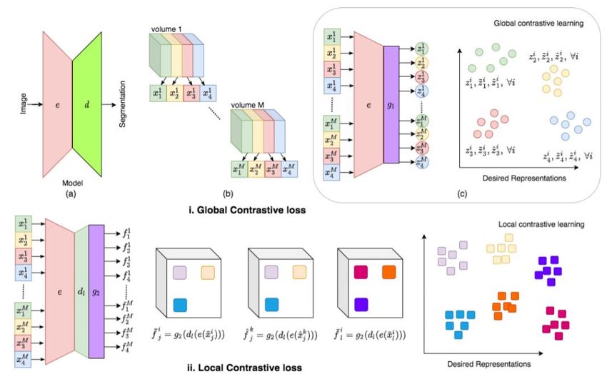
A Review of Self-supervised Learning Methods in the Field of Medical Image Analysis              35

Fig.3. Jigsaw puzzles for histopathological images

    In inspired by the 2D Jigsaw puzzle, Aiham et al. proposed 3D Jigsaw puzzle method as SSL proxy task [9].
    Similar to the 2D method, it is an n-way classification task, aims to find the right sequence index of the shuffled
3D patches.

Fig.4. 3D Jigsaw puzzles [9]

2.3. Rubik's Cube
      Rubik's Cube can be regarded as a derivative of the 3D Jigsaw puzzle. X. Zhuang et al. first proposed playing
Rubik’s cube as a proxy task for volumetric medical data [10]. Divide the 3D medical image into a set (2×2×2) of cubes,
and then randomly rotate cubes, the proxy task aims to recover the original 3D image, cubes rearrangement, and cubes
rotation. Compared with 3D Jigsaw puzzles, this method adds Cube’s rotation operations. Basis on this idea, Rubik's
Cube + [12] and Rubik's Cube ++ [11] methods have been developed. The pipeline of the Rubik's Cube is illustrated in
Fig.5. This method is more conducive to the contextual feature extraction of 3D unlabeled medical images.

Fig. 5. 3D Rubik's Cube [10]

2.4. Specific Pretext-tasks based on the Context of the Medical Image Domain
     Different from the previous context-based methods, Li Sun et al. [13] focuses on the context information of the
anatomical region of medical images and proposed a two-level feature learning method, first learning local textural
features from the regional anatomical level, and then learning global contextual features from the patient level.
Furthermore, in Bai’s work use the characteristics of different Cardiac MR view planes to build anatomical position
prediction pretext-task [14]. These ideas got rid of generic SSL methods and developed proxy tasks utilize the specific
context information of medical images.

3. Generated-based Self-supervised Learning

     In this section, we will introduce generation-based SSL methods in the medical image domain, including
autoencoder models [19] and GANs [20].

Copyright © 2021 MECS                                              I.J. Image, Graphics and Signal Processing, 2021, 4, 33-46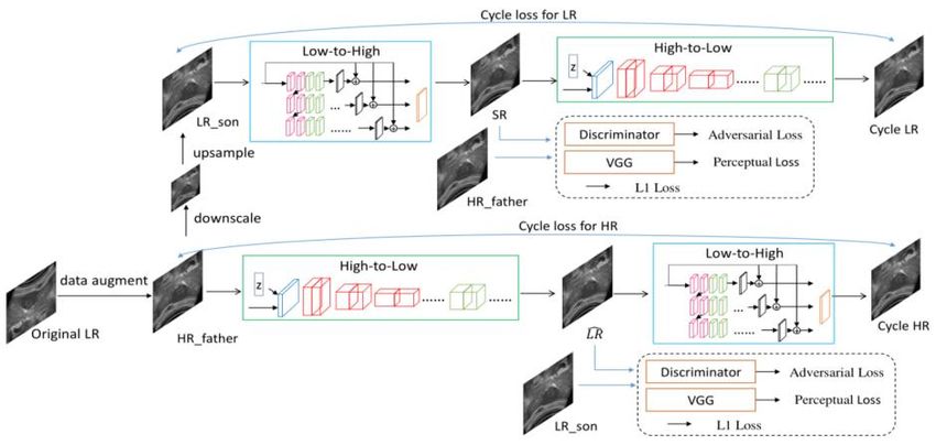
36 A Review of Self-supervised Learning Methods in the Field of Medical Image Analysis
3.1. Restore Based on the Autoencoder Model
The purpose of this SSL pretext-task is to recover the original sub-volume after its transformation (sub-volume,
cropped from unlabeled CT images by random size and random location). The original sub-volume through the
transformation function obtain ̃ . Take ̃ as the input of the autoencoder to restore the original sub-
volume. The restoration pretext-tasks optimizer function is expressed as:
min Lres G E xi xi 2 (1)
E G
xi D
Where G and E are the encoder and decoder. are the parameters of the autoencoder. Here the
transformations can be a non-linear transformation, local-shuffling, out-painting, in-painting. In the Models Genesis
[15][18] SSL framework integrates a variety of transformations and shared the same autoencoder to restore the . This
allows the model to learn latent representations from multiple perspectives and then transfer them to specific
downstream tasks. Inspired by Models Genesis, subsequent researchers proposed Semantic Genesis [16] and Universal
Model [17]. Semantic Genesis further extends the framework of Models Genesis to add self-classification branch, using
less computational overhead, actually a classification head is added at the end of the encoder in the autoencoder.
Semantic Genesis framework extracts semantics representation from the consistent and recurrent anatomical patterns.
The pipeline of the Models Genesis and Semantic Genesis are illustrated in Fig.6, 7.
Fig.6. The pipeline of the Models Genesis[15].
Fig.7. The pipeline of the Semantic Genesis[16].
Universal Model adds Modality Invariant Representation Learning tasks and Multi-level Feature Learning
classification tasks compare to the pipeline of the Models Genesis. Therefore, this method is dedicated to solving the
generalization problem of multi-task and multi-modality in the medical image analysis domain. The pipeline of the
Copyright © 2021 MECS I.J. Image, Graphics and Signal Processing, 2021, 4, 33-46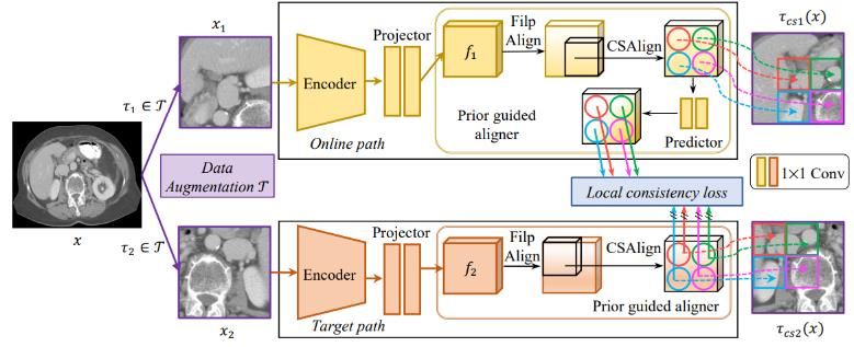
A Review of Self-supervised Learning Methods in the Field of Medical Image Analysis              37

Universal Model is illustrated in Fig.8.
3.2. Based on GANs
      As the most successful generative model in recent years, GAN is widely employed in computer vision tasks. Ross
et al. proposed a re-colorization proxy task that utilizes GAN to recolor medical images (endoscopic video data) [21].
First, the raw data is transformed into the CIELAB Color space, in CIELAB Color space the L-channel as input, train
the GAN to generate the a-channels and b-channels. Then transfer the generator network, which has the ability to
extract the low-level semantic information, to the target segmentation task. According to the variants of GAN, many
proxy tasks can be constructed, for instance the reconstruction of patches using Wasserstein GAN [22], use conditional
GAN [23] for image colorization [3], and self-supervised CycleGAN framework for ultrasound image super-resolution
[24]. Compared with previous pretext-tasks, the framework of GANs-based is more complicated, the self-supervised
CycleGAN framework is illustrated in Fig.9. it also means longer training time costs[58].

Fig.8. The pipeline of the Universal Model [17].

Fig.9. The self-supervised CycleGAN framework

4. Contrast-based Self-supervised Learning

     Contrast learning as a form of self-supervised learning has been the front-runner in the unsupervised natural image
domain, and these frameworks based on comparative learning constantly narrow the gap between unsupervised and
supervised learning, for instance, CPC [32], SimCLR [25], MoCo [26], BYOL [27], SimSiam [28], Twins [29]. The
basic idea of comparative learning is that different transformations of a sample image have similar representations and
these representations should be different from the different sample images, then, use unlabeled data to train the neural
network by minimizing the contrast loss [31]. However, its application in the medical imaging domain is still in infancy.
This section will introduce the application in the field of medical image analysis based on different contrastive learning
methods, here we divide those CL methods into 2 types: global-local contrast and context-context contrast.
4.1. Global-local Contrast
     Global-local Contrast is aims to modeling the relationship between local semantic representation and global
semantic representation of an instance. This method focuses on the prediction of the encoder-decoder architectures at
the pixel level and learns the global and local level representations while considering the local representation. For
medical images, the data of different patients have inherent consistency, because the structure of human organs is
consistent, such as MRI (magnetic resonance imaging) and CT (computed tomography). Therefore, encoding the same

Copyright © 2021 MECS                                              I.J. Image, Graphics and Signal Processing, 2021, 4, 33-46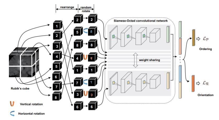
38 A Review of Self-supervised Learning Methods in the Field of Medical Image Analysis
anatomical region of the medical images will get the similar embedding, leverage this characteristic to construct the
global contrastive loss [33]. In contrast, Local representation aims to distinguishing pixel-level differences in
neighboring regions. In [33][35], by constructing InfoNCE loss [34], the network determines whether the output comes
from the similar distribution or the dissimilar distribution to define the global contrastive loss and the local contrastive
loss. InfoNCE loss is defined as:
LN Ex log
exp f x f x
T
(2)
exp f x f x J 1 exp f x f ( x j )
T N 1 T
Where is from a set { }, are transformed sample. Minimizing the loss narrows the distance
between the representations of and , while increasing the distance between the representation of x and other
dissimilar images.
The Global-local Contrast learning framework is illustrated in Fig.10. The input volumetric images divided into
four partitions, leverage the similarity of the slices in different volumetric images defining positive samples and
negative samples. Representations are extracted by encoder and projection head .
Fig.10. Global-local Contrast learning framework
Compared with [33], SAM [35] utilize global embedding and local embedding to construct InfoNCE loss in the
embedding space. This framework is more general, suitable for various tasks and has a relatively simple structure. The
pipeline of SAM is illustrated in Fig.11. First, the volumetric image and random transform sample as the input.
Then, generate global and local embedding information through the network. Finally, the global and the local InfoNCE
loss to encourage positive pairs to have similar embeddings, while pushing negative samples apart.
Fig.11. The pipeline of SAM
Copyright © 2021 MECS I.J. Image, Graphics and Signal Processing, 2021, 4, 33-46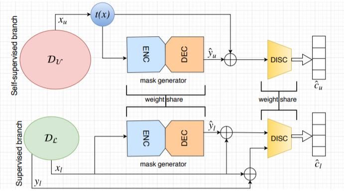
A Review of Self-supervised Learning Methods in the Field of Medical Image Analysis               39

4.2. Context-Context Contrast
     Context-context contrastive learning directly utilizes the relationships between the global representations of
different samples as what metric learning does, essentially, leveraging instance discrimination as a pretext task.
Recently, CPC[32], SimCLR[25], MoCo[26], BYOL[27] based on Context-context contrast methods achieved great
performance in the nature image domain,. These methods have inspired researchers in the medical domain, next we will
introduce the practical applications of these methods in the field of medical domain.
4.2.1 CPC
     Contrastive Predictive Coding (CPC) is a contrastive method that can be applied to any form of data such as text,
voice, video, image, etc. For image data, a sample can be seen as a sequence of pixels or image blocks, then CPC learns
the feature representation of spatial information. Inspired by CPC, TCPC [36] was proposed, which first uses Simple
Linear Iterative Clustering (SLIC) [37] to locate the potential lesion area, and then modified CPC to learn 3D feature
representation from the sub-volumes containing the lesion areas. TCPC framework as shown in the Fig.12.
     TCPC adopts a u-shaped path and there are fewer differences in the content of medical images compared with
natural images, so it uses a larger cube size than 2D CPC and 3D CPC.

Fig.12. TCPC framework [36]

4.2.2 MOCO
      In MoCo [26], the idea of using instance discrimination through momentum contrast was further developed, which
greatly increased the number of negative samples. Benefiting from the successful performance of MoCo, then
researchers proposed MoCo-CXR [38] framework on unlabeled chest X-ray’s dataset, and [39] further research was
conducted on the basis of MoCo-CXR.
      The medical image has the characteristics of small pixel area where the abnormality’s part is located, gray-scale
image, a large amount of unlabeled data [53], etc. So, some general data augmentations algorithms in MOCO are not
suitable for medical images. MoCo-CXR modified the data augmentation strategy used to generate views suitable for
the chest X-ray, such as horizontal flipping and random rotation. Experimental results show that MoCo-CXR-pretrained
model outperformance than ImageNet-pretrained model, further verify the availability of MoCo in chest X-rays.
following MoCo-CXR, MedAug [40] was proposed, which uses multiple images as a way to increase the number of
positive pair choices, in this process, patient metadata is introduced to assist in constructing positive pairs. The process
illustrated in Fig13. Their experiments proved that the performance of the model can be improved by using positive
pairs selected by patient metadata.

Copyright © 2021 MECS                                            I.J. Image, Graphics and Signal Processing, 2021, 4, 33-46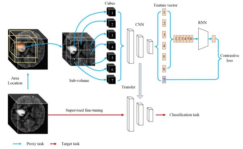
40                        A Review of Self-supervised Learning Methods in the Field of Medical Image Analysis

Fig.13. Self-Supervised Pretraining using Momentum Contrast Learning

      The medical image has the characteristics of small pixel area where the abnormality’s part is located, gray-scale
image, a large amount of unlabeled data, etc. So, some general data augmentations algorithms in MOCO are not
suitable for medical images. MoCo-CXR modified the data augmentation strategy used to generate views suitable for
the chest X-ray, such as horizontal flipping and random rotation. Experimental results show that MoCo-CXR-pretrained
model outperformance than ImageNet-pretrained model, further verify the availability of MoCo in chest X-rays.
following MoCo-CXR, MedAug [40] was proposed, which uses multiple images as a way to increase the number of
positive pair choices, in this process, patient metadata is introduced to assist in constructing positive pairs. The process
illustrated in Fig13. Their experiments proved that the performance of the model can be improved by using positive
pairs selected by patient metadata.
      In [39], a self-supervised learning algorithm for COVID-19 prediction is proposed, which actually borrows from
the overall framework of MoCo and adds some data augmentation strategy, such as, random Gaussian noise, random
cropping, interpolation. Then, this self-supervised pretraining achieved the highest AUC scores on Single Image
Prediction tasks of interest.

Fig. 14. The process selecting positive pairs used patient metadata for contrastive learning

4.2.3 SimCLR
      SimCLR follows the end-to-end framework and is different from MoCo, which uses momentum contrast, and in
order to deal with the large-scale negative sample problem, SimCLR chooses a larger batch. In [41] work, employ
SimCLR framework applies to histopathological image analysis. The author conducted different downstream tasks to
demonstrate that the feature extraction method based on comparative learning is significantly better than the baseline
based on ImageNet. Furthermore, Inspired by SimCLR, Multi-Instance Contrastive Learning (MICLe) [42] was
proposed. This method utilizes multiple images per patient to construct positive pairs, not only different augmentations
of the same image but also different images of the same medical pathology, the construct process are shown in the
Fig.15.
      In MICLe, only use N pair of minibatch and lightweight data augmentation. Experiments show that MICLe
significantly improves the accuracy and achieve the state-of-the-art result on the dermatology condition classification
task.

Copyright © 2021 MECS                                                        I.J. Image, Graphics and Signal Processing, 2021, 4, 33-46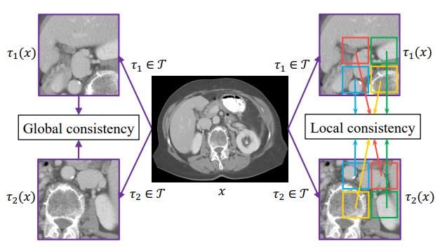
A Review of Self-supervised Learning Methods in the Field of Medical Image Analysis                      41

Fig.15. Multi-Instance Contrastive Learning (MICLe) framework.

4.2.4 BYOL
     BYOL is a more radical approach, first discards negative sampling in contrastive learning but achieves an even
better result, the model learns image representations using the online-target framework. The online network on an
augmented view of an image to predict the representation of another augmented view of the same image produced by
the target network. Based on the framework of BYOL, Prior-Guided Local (PGL) [43] focuses on Local consistency
loss and minimizes Local consistency loss based on the spatial and regional location relationship of the two augmented
views. Fig.16 illustrated the difference between global consistency loss and local consistency loss.

Fig.16. Global consistency loss (BYOL) and local consistency loss (PGL)

      PGL model framework including a data augmentation module and a prior dual-path module, as the Fig.17 shows.
First, the unlabeled training data is augmented by module to obtain , , and then fed to the prior dual-path for
feature extraction and alignment. The goal of the prior-guided aligner is to construct the spatial relation between and
   on the prior information of augmentation transformation and align the features.

Fig.17. PGL model framework.

Copyright © 2021 MECS                                                     I.J. Image, Graphics and Signal Processing, 2021, 4, 33-46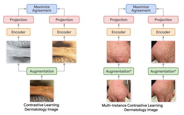
42 A Review of Self-supervised Learning Methods in the Field of Medical Image Analysis
5. Performance Comparison
For specific downstream tasks or target tasks in the field of medical imaging, they are mainly divided into two
categories: classification and segmentation. This section integrates the performance of SSL methods on different
datasets.
5.1. LUNA 2016
LUNA 2016 [46] dataset comes from the 2016 LUNA Nodule Analysis competition. Table 1. Shows the
classification task results of the self-supervised learning methods on the LUNA 2016 dataset, all of the results,
including the mean and standard deviation across ten trials. From the results, the self-supervised methods are conducive
to the improvement of the performance of the target task, even performance is better than supervised learning, as
illustrated in fig.18.
Fig.18. The result of SSL methods performed on the LUNA 2016 and BraTS-2018, the first line of result data is the supervised method.
Table 1. Performance on LUNA 2016
Method Supervised AUC%(mean±s.d.)
MedicalNet [45] 95.80±0.49
Autoencoder 88.43±10.25
In-painting [48] 91.46±2.97
Patch shuffling 91.93±2.32
De-noising [49] 95.92±1.83
Jigsaw 95.47±1.24
Rubik’s Cube 96.24±1.27
DeepCluster [50] 97.22±0.55
Self-restoration 98.07±0.59
3D Rotation 97.13±0.81
Semantic Genesis 3D 98.47±0.32
3D Jigsaw 96.12±0.63
3D CPC 92.88±1.56
TCPC 96.55±0.97
Model Genesis 98.34±0.44
Universal Model 99.04±0.23
5.2. BraTS-2018
BraTS 2018 [47] dataset comes from Multimodal Brain Tumor Segmentation Challenge. The results of these
experiments on the BraTS 2018 dataset are shown in Table 2.
Through these experimental results, it seems that whether segmentation tasks or classification tasks, the self-
supervised learning methods redesigned based on medical image characteristics has higher performance.
Copyright © 2021 MECS I.J. Image, Graphics and Signal Processing, 2021, 4, 33-46A Review of Self-supervised Learning Methods in the Field of Medical Image Analysis 43
Table 2. Performance on BraTS 2018
Method Supervised IoU%(mean±s.d.)
MedicalNet 66.09±1.35
Autoencoder 56.36±5.32
In-painting 61.38±3.84
Patch shuffling 52.95±6.92
De-noising 57.83±1.57
Jigsaw 63.33±1.11
Rubik’s Cube 62.75±1.93
DeepCluster 65.96±0.85
Self-restoration 67.96±1.29
3D Rotation 56.48±1.78
Semantic Genesis 3D 68.80±0.30
3D Jigsaw 59.65±0.81
Model Genesis 67.96±1.29
Universal Model 72.10±0.67
5.3. Performance of Contrastive Learning
Contrastive learning methods achieve The-state-of-the-art in self-supervised learning methods of natural images.
The experimental results in Table 3 show that in the medical image analysis domain, the contrast-based methods also
achieved The-state-of-the-art. The performance of the modified comparative learning method is more prominent and
those methods more suitable for medical image analysis.
Table 3. Performance of Contrastive Learning methods
Method Dataset Dice % Iou %
Random Init KiTS [51] 81.57 74.63
Models Genesis KiTS 82.32 75.51
BYOL KiTS 84.06 77.56
PGL KiTS 84.29 78.27
Random Init Liver dataset[52] 73.97 66.79
Models Genesis Liver dataset 74.74 67.68
BYOL Liver dataset 74.82 68.09
PGL Liver dataset 76.05 69.06
Random Init Spleen dataset [52] 93.23 88.08
Models Genesis Spleen dataset 94.20 89.44
BYOL Spleen dataset 94.56 89.83
PGL Spleen dataset 95.60 91.61
6. Discussions and Future Directions
The application of self-supervised learning methods in the medical field has achieved great success, and obtaining
good performance that exceeds the supervised pre-training model on certain medical image tasks. However, directly
applying existing self-supervised learning methods is not necessarily suitable for medical image tasks [57].
6.1. Selection of Data Augmentation and Pretext Tasks
In medical images, human organs are consistent, and only a small part of the lesions are different [55]. So, some
data augmentation methods and some pretext-tasks will destroy the main information of the raw data. Therefore, it is
necessary to construct suitable pretext-tasks and data augmentation strategies according to the characteristics of medical
image data. By designing effective pretexts, using the information other than the part to be predicted to predict a certain
subset of information, mining the potential laws of large-scale unsupervised data. Learn data representations and
inductive biases that help improve downstream task performance. These data representations cover good semantics or
structural meanings.
Copyright © 2021 MECS I.J. Image, Graphics and Signal Processing, 2021, 4, 33-4644 A Review of Self-supervised Learning Methods in the Field of Medical Image Analysis
6.2. Extract features with Multiple Pretext Tasks
Most existing self-supervised learning methods in the medical domain learn data features by training a pretext-task.
Different pretext-tasks mean different supervision signals, which can help the network learn more data features. The
existing methods construct pre-tasks from both local and global perspectives [33], so that the model not only learns
global features, but also focuses on fine-grained features, and has achieved good experimental results on medical image
segmentation tasks. Whether it is through the experimental results of the literature or the perspective of algorithm proof,
multiple Pretext Tasks are helpful to extract the potential law of data distribution.
6.3. Dataset Imbalance
The bias of the dataset is a normal phenomenon, but this phenomenon is more prominent in medical images. In a
large amount of unlabeled medical data, the data of the disease is actually much smaller than the data of the normal
person. Use self-supervision to overcome the inherent "data bias" and learn better initialization feature information that
is not related to labels from unbalanced datasets [44]. The problem of data imbalance needs to be considered in the
construction of the self-supervised framework in the medical domain.
6.4. Redesign Contrastive Learning
In the field of medical imaging, directly applying the existing Contrastive Learning framework has limited
improvement in experimental results. According to the characteristics of medical images, suitable negative samples can
be constructed, then extract more useful data features [[56],[58]]. In order to improve the performance of self-
supervised learning methods in the field of medical image analysis, we need to further explore how to construct
negative examples and how to better adapt self-supervised learning methods to downstream tasks.
7. Conclusions
This article has extensively reviewed the latest applications of self-supervised methods in the field of medical
image analysis. Self-supervised learning can use unlabeled data or even unbalanced data to extract latent features. This
unsupervised learning paradigm naturally adapts to the problems of medical image data. We have clearly categorized
recent methods and introduced the pipelines of these methods separately, introduces background knowledge and
important frameworks. Finally, discussed the future research directions, and puts forward the issues that need to be paid
attention to when designing new methods and paradigms. In short, our work fills the gap of review papers in the
medical image analysis based on self-supervised learning and researchers can easily grasp the cutting-edge ideas of this
domain.
Acknowledgment
This research has been partially supported by China Scholarship Council (CSC), and Special thanks should go to
my supervisor professor Sergii Stirenko, for his instructive advice and useful suggestions on my paper.
References
[1] S. Gidaris, P. Singh, N. Komodakis.” Unsupervised Representation Learning by Predicting Image Rotations,” Proceedings of
the International Conference on Learning Representations, 2018.
[2] M. Noroozi, P. Favaro. “Unsupervised Learning of Visual Representations by Solving Jigsaw Puzzles,” In European
conference on computer vision, 2016, pp: 69-84.
[3] T. Nima, Y. Hu, J. Cao, X. Yan, Y. Xiao, Y. Lu, J. Liang, et al. “Surrogate supervision for medical image analysis: Effective
deep learning from limited quantities of labeled data.” In 2019 IEEE 16th International Symposium on Biomedical
Imaging,2019, pp. 1251-1255.
[4] C. Szegedy, S. Ioffe, V. Vanhoucke, "Inception-v4, inception-resnet and the impact of residual connections on learning." In
Proceedings of the AAAI Conference on Artificial Intelligence, vol. 31, no. 1. 2017.
[5] A. Hatamizadeh, P. Shilpa, X. Ding, D. Terzopoulos, and N. Tajbakhsh. "Automatic segmentation of pulmonary lobes using a
progressive dense V-network." In Deep Learning in Medical Image Analysis and Multimodal Learning for Clinical Decision
Support, 2018, pp. 282-290.
[6] Imran, A. A. Z., Huang, C., Tang, H., Fan, W., Xiao, Y., Hao, D., et al. “Partly Supervised Multitask Learning.” arXiv preprint
arXiv:2005.02523,2020.
[7] A. R. Venkatakrishnan, S. T. Kim, R. Eisawy, F. Pfister, & Navab, N. “Self-Supervised Out-of-Distribution Detection in Brain
CT Scans.” arXiv e-prints, arXiv-2011,2020.
[8] Y. Li, J. Chen, X. Xie, K. Ma, & Y. Zheng. “Self-Loop Uncertainty: A Novel Pseudo-Label for Semi-supervised Medical
Image Segmentation. In International Conference on Medical Image Computing and Computer-Assisted Intervention.” pp. 614-
623, Oct. 2020
[9] A. Taleb,W. Loetzsch, N. Danz, J. Severin, T. Gaertner, B. Bergner, & C. Lippert. “3d self-supervised methods for medical
imaging.” arXiv preprint arXiv:2006.03829, 2020.
Copyright © 2021 MECS I.J. Image, Graphics and Signal Processing, 2021, 4, 33-46A Review of Self-supervised Learning Methods in the Field of Medical Image Analysis 45
[10] X. Zhuang, Y. Li, Y. Hu, K. Ma, Y. Yang, and Y. Zheng, “Self-supervised Feature Learning for 3D Medical Images by Playing
a Rubik’s Cube.” arXiv preprint arXiv:1910.02241, 2019.
[11] X. Tao, Y. Li, W. Zhou, K. Ma, & Y. Zheng, Revisiting Rubik’s cube: self-supervised learning with volume-wise
transformation for 3D medical image segmentation. In International Conference on Medical Image Computing and Computer-
Assisted Intervention, 2020, October. pp. 238-248.
[12] J. Zhu, Y. Li, Y. Hu, K. Ma, S. K. Zhou, & Y. Zheng. Rubik’s Cube+: A self-supervised feature learning framework for 3D
medical image analysis. Medical Image Analysis, 2020, 64, 101746.
[13] S. Li, K. Yu, and K. Batmanghelich. "Context Matters: Graph-based Self-supervised Representation Learning for Medical
Images." arXiv preprint arXiv:2012.06457 ,2020.
[14] Bai, W., Chen, C., Tarroni, G., Duan, J., Guitton, F., Petersen, S. E., ... & Rueckert, D. "Self-supervised learning for cardiac mr
image segmentation by anatomical position prediction." In International Conference on Medical Image Computing and
Computer-Assisted Intervention, pp. 541-549. 2019.
[15] Zhou, Z., Sodha, V., Pang, J., Gotway, M. B., & Liang, J. and Jianming Liang. "Model’s genesis." Medical image analysis 67:
101840, 2021.
[16] Haghighi, F., Taher, M. R. H., Zhou, Z., Gotway, M. B., & Liang, J. "Learning Semantics-enriched Representation via Self-
discovery, Self-classification, and Self-restoration." In International Conference on Medical Image Computing and Computer-
Assisted Intervention, pp. 137-147. 2020.
[17] Zhang, X., Zhang, Y., Zhang, X., & Wang, Y. Universal Model for 3D Medical Image Analysis. arXiv preprint
arXiv:2010.06107, 2020.
[18] Zhou, Z., Sodha, V., Siddiquee, M. M. R., Feng, R., Tajbakhsh, N., Gotway, M. B., & Liang, J. "Model’s genesis: Generic
autodidactic models for 3d medical image analysis." In International Conference on Medical Image Computing and Computer-
Assisted Intervention, pp. 384-393.2019.
[19] Chen, L., Bentley, P., Mori, K., Misawa, K., Fujiwara, M., & Rueckert, D. Self-supervised learning for medical image analysis
using image context restoration. Medical image analysis, 58, 101539. 2019.
[20] Yu, L., Zhang, W., Wang, J., & Yu, Y. Seqgan: Sequence generative adversarial nets with policy gradient. In Proceedings of
the AAAI conference on artificial intelligence. 2017, February,Vol. 31, No. 1.
[21] Ross, T., Zimmerer, D., Vemuri, A., Isensee, F., Wiesenfarth, M., Bodenstedt, S., ... & Maier-Hein, L. Exploiting the potential
of unlabeled endoscopic video data with self-supervised learning. International journal of computer assisted radiology and
surgery, 13(6), 925-933. 2018.
[22] M. Arjovsky, S. Chintala, L. Bottou, “Wasserstein gan,” arXiv:1701.07875,2017.
[23] Larsson, G., Maire, M., & Shakhnarovich, G. Colorization as a proxy task for visual understanding. In Proceedings of the IEEE
Conference on Computer Vision and Pattern Recognition. pp. 6874-6883. 2017.
[24] Liu, H., Liu, J., Hou, S., Tao, T., & Han, J. Perception consistency ultrasound image super-resolution via self-supervised
CycleGAN. Neural Computing and Applications, 1-11. 2021.
[25] Chen, T., Kornblith, S., Norouzi, M., & Hinton, G. A simple framework for contrastive learning of visual representations. In
International conference on machine learning. 2020, November, pp. 1597-1607.
[26] He, K., Fan, H., Wu, Y., Xie, S., & Girshick, R. Momentum contrast for unsupervised visual representation learning. In
Proceedings of the IEEE/CVF Conference on Computer Vision and Pattern Recognition. pp. 9729-9738. 2020.
[27] Grill, J. B., Strub, F., Altché, F., Tallec, C., Richemond, P. H., Buchatskaya, E., ... & Valko, M. Bootstrap your own latent: A
new approach to self-supervised learning. arXiv preprint arXiv:2006.07733. 2020.
[28] Chen, X., & He, K. Exploring Simple Siamese Representation Learning. arXiv preprint arXiv:2011.10566.2020.
[29] Zbontar, J., Jing, L., Misra, I., LeCun, Y., & Deny, S. Barlow Twins: Self-Supervised Learning via Redundancy Reduction.
arXiv preprint arXiv:2103.03230. 2021.
[30] Zhang, P., Wang, F., & Zheng, Y. Self-supervised deep representation learning for fine-grained body part recognition. In 2017
IEEE 14th International Symposium on Biomedical Imaging. April ,2017, pp. 578-582. 2017.
[31] Hadsell, R., Chopra, S., LeCun, Y.: Dimensionality reduction by learning an invariant mapping. In: 2006 IEEE Computer
Society Conference on Computer Vision and Pattern Recognition. vol. 2, pp. 1735–1742. 2006.
[32] Henaff, O.J. Data-efficient image recognition with contrastive predictive coding. In IEEE Conference on Computer Vision and
Pattern Recognition,pp. 4182-4192, 2019.
[33] Chaitanya, K., Erdil, E., Karani, N., & Konukoglu, E. Contrastive learning of global and local features for medical image
segmentation with limited annotations. arXiv preprint arXiv:2006.10511. 2020.
[34] Oord, A. V. D., Li, Y., & Vinyals, O. Representation learning with contrastive predictive coding. arXiv preprint
arXiv:1807.03748. 2018.
[35] Yan, K., Cai, J., Jin, D., Miao, S., Harrison, A. P., Guo, D., et al. Self-supervised Learning of Pixel-wise Anatomical
Embeddings in Radiological Images. arXiv preprint arXiv:2012.02383.2020.
[36] Zhu, J., Li, Y., Hu, Y., & Zhou, S. K. Embedding Task Knowledge into 3D Neural Networks via Self-supervised Learning.
arXiv preprint arXiv:2006.05798.2020
[37] Achanta, R., Shaji, A., Smith, K., Lucchi, A., Fua, P., Susstrunk, S.: SLIC superpixels compared to state-of-the-art superpixel
methods. vol. 34, pp. 2274–2282,2012.
[38] Sowrirajan, H., Yang, J., Ng, A. Y., & Rajpurkar, P. MoCo-CXR: MoCo Pretraining Improves Representation and
Transferability of Chest X-ray Models. E-print arXiv:2010.05352,2020
[39] Sriram, A., Muckley, M., Sinha, K., Shamout, F., Pineau, J., Geras, K. J., ... & Moore, W. (2021). COVID-19 Prognosis via
Self-Supervised Representation Learning and Multi-Image Prediction. E-print arXiv:2101.04909,2021.
[40] Vu, Y. N. T., Wang, R., Balachandar, N., Liu, C., Ng, A. Y., & Rajpurkar, P. MedAug: Contrastive learning leveraging patient
metadata improves representations for chest X-ray interpretation. arXiv preprint arXiv:2102.10663.2021.
[41] Ciga, O., Martel, A. L., & Xu, T. Self-supervised contrastive learning for digital histopathology. arXiv preprint
arXiv:2011.13971. 2020.
Copyright © 2021 MECS I.J. Image, Graphics and Signal Processing, 2021, 4, 33-4646 A Review of Self-supervised Learning Methods in the Field of Medical Image Analysis
[42] Azizi, S., Mustafa, B., Ryan, F., Beaver, Z., Freyberg, J., Deaton, J., et al. Big Self-Supervised Models Advance Medical Image
Classification. arXiv preprint arXiv:2101.05224. 2021.
[43] Xie, Y., Zhang, J., Liao, Z., Xia, Y., & Shen, C. (2020). PGL: Prior-Guided Local Self-Supervised Learning for 3D Medical
Image Segmentation. arXiv preprint arXiv:2011.12640. 2020.
[44] Yang, Y., & Xu, Z. Rethinking the value of labels for improving class-imbalanced learning. arXiv preprint arXiv:2006.07529.
2020.
[45] Chen, S., Ma, K., & Zheng, Y. Med3d: Transfer learning for 3d medical image analysis. arXiv preprint arXiv:1904.00625.
2019.
[46] Setio, A. A. A., Traverso, A., De Bel, T., Berens, M. S., van den Bogaard, C., Cerello, P., ... & Jacobs, C. Validation,
comparison, and combination of algorithms for automatic detection of pulmonary nodules in computed tomography images: the
LUNA16 challenge. Medical image analysis, 42, 1-13. 2017.
[47] Bakas, S., Reyes, M., Jakab, A., Bauer, S., Rempfler, M., Crimi, A., et al. Identifying the best machine learning algorithms for
brain tumor segmentation, progression assessment, and overall survival prediction in the BRATS challenge. arXiv preprint
arXiv:1811.02629. 2018.
[48] Pathak, D., Krahenbuhl, P., Donahue, J., Darrell, T., & Efros, A. A. Context encoders: Feature learning by inpainting. In
Proceedings of the IEEE conference on computer vision and pattern recognition. pp. 2536-2544. 2016.
[49] Vincent, P., Larochelle, H., Lajoie, I., Bengio, Y., Manzagol, P. A., & Bottou, L. Stacked denoising autoencoders: Learning
useful representations in a deep network with a local denoising criterion. Journal of machine learning research, 11(12). 2010.
[50] Caron, M., Bojanowski, P., Joulin, A., & Douze, M. Deep clustering for unsupervised learning of visual features. In
Proceedings of the European Conference on Computer Vision. pp.132-149. 2018.
[51] Heller, N., Isensee, F., Maier-Hein, K. H., Hou, X., Xie, C., Li, F., et al. The state of the art in kidney and kidney tumor
segmentation in contrast-enhanced ct imaging: Results of the kits19 challenge. Medical Image Analysis, 67, 101821. 2021.
[52] Simpson, A. L., Antonelli, M., Bakas, S., Bilello, M., Farahani, K., Van Ginneken, B., et al. A large annotated medical image
dataset for the development and evaluation of segmentation algorithms. arXiv preprint arXiv:1902.09063. 2019.
[53] Gordienko, Y., Gang, P., Hui, J., Zeng, W., Kochura, Y., Alienin, O., ... & Stirenko, S. Deep learning with lung segmentation
and bone shadow exclusion techniques for chest X-ray analysis of lung cancer. In International Conference on Computer
Science, Engineering and Education Applications. Springer, Cham, pp. 638-647, 2018.
[54] Yang, Y. H., Xu, J. S., Gordienko, Y., & Stirenko, S. Abnormal Interference Recognition Based on Rolling Prediction Average
Algorithm. In International Conference on Computer Science, Engineering and Education Applications. Springer, Cham, pp.
306-316. 2020.
[55] Sulema, Y., Kerre, E. and Shkurat, O. Vector Image Retrieval Methods Based on Fuzzy Patterns. International Journal of
Modern Education & Computer Science, 12(3), 2020.
[56] Md. Rahat Khan, A. S. M. Shafi, " Statistical Texture Features Based Automatic Detection and Classification of Diabetic
Retinopathy", International Journal of Image, Graphics and Signal Processing(IJIGSP), Vol.13, No.2, pp. 53-61, 2021.DOI:
10.5815/ijigsp.2021.02.05.
[57] Farzaneh Nikroorezaei, Somayeh Saraf Esmaili, " Application of Models based on Human Vision in Medical Image Processing:
A Review Article", International Journal of Image, Graphics and Signal Processing(IJIGSP), Vol.11, No.12, pp. 23-28,
2019.DOI: 10.5815/ijigsp.2019.12.03.
[58] Kama, R., Chinegaram, K., Tummala, R.B. and Ganta, R.R. Segmentation of Soft Tissues and Tumors from Biomedical
Images using Optimized K-Means Clustering via Level Set formulation. International Journal of Intelligent Systems and
Applications, 11(9), p.18, 2019.
Authors’ Profiles
Jiashu Xu, received a master's degree from the Department of computing engineering, National Technical
University of Ukraine “Igor Sikorsky Kyiv Polytechnic Institute”. Now is a Ph.D. student from the same
university. His research interests include self-supervised learning, unsupervised learning, computer vision,
GAN, and their applications in the medical image domain.
How to cite this paper: Jiashu Xu, " A Review of Self-supervised Learning Methods in the Field of Medical Image Analysis",
International Journal of Image, Graphics and Signal Processing(IJIGSP), Vol.13, No.4, pp. 33-46, 2021.DOI:
10.5815/ijigsp.2021.04.03
Copyright © 2021 MECS I.J. Image, Graphics and Signal Processing, 2021, 4, 33-46You can also read