Emerging and Emergency Animal Diseases of the Pig - VET 451 Lecture 12 Intensive Animal Industries 2014
←
→
Page content transcription
If your browser does not render page correctly, please read the page content below
Emerging and Emergency
 Animal Diseases of the Pig
           Lecture 12
            VET 451
Intensive Animal Industries 2014
            kim@portec.com.au
            kate@portec.com.au
           susan@portec.com.au
            www.portec.com.au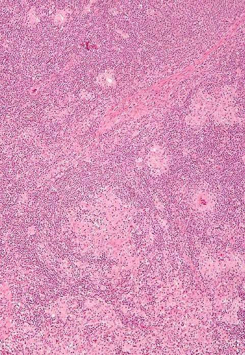
Learning Objectives

• What are the emergency and emerging
  diseases of significance in the pig
• How to diagnose these disorders
• How to prevent, and appropriately respond
  to, these conditions
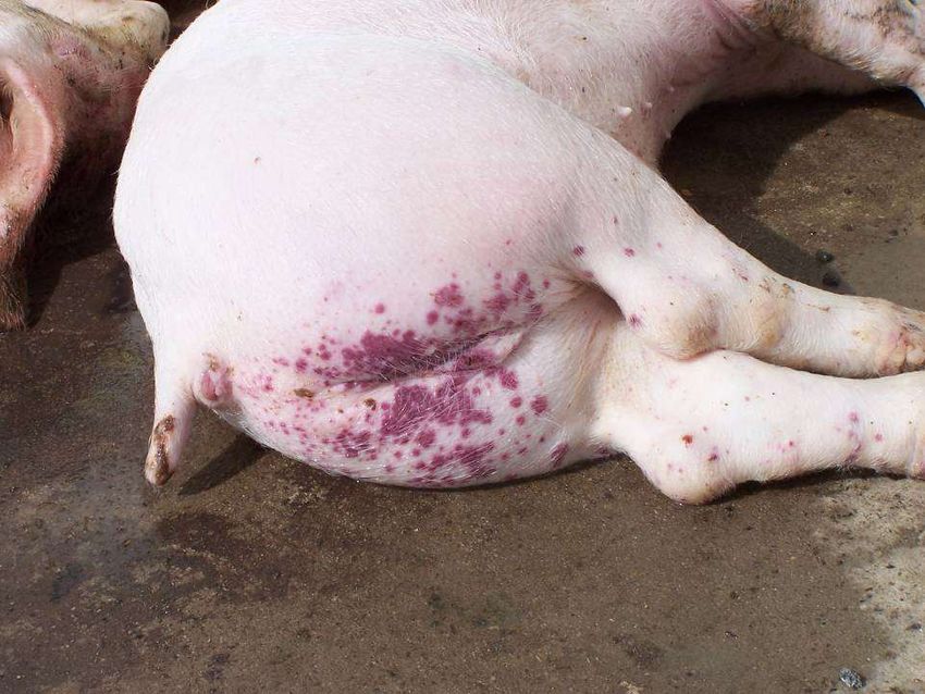
EAD Outbreak

•   Unusually high number of sick animals
•   Unusually high number of deaths
•   Blisters or vesicles on animals’ snout, or feet
•   Unusually high number of lame animals
•   Unusually high number of animals with fevers
•   Unusually high number of animals not eating
•   Unusually high number of recumbent animals
•   Discoloration of the ears, belly, rump, legs, or tail
EAD & Emerging Diseases

• PMWS/PCVAD – PCVAD has emerged in Australia,
  PMWS is still classed as exotic
• Classical Swine Fever
• African Swine Fever
• Nipah virus
• Aujeszky’s Disease (Pseudorabies)
• PRRS
• Swine Influenza
• TGE/PED
• Foot and Mouth Disease
• Japanese Encephalitis
PMWS / PCV2 Associated Disease

Causal Agent      Porcine Circovirus II
                  Circovirus are small non-enveloped DNA virus
                  containing a unique single stranded circular
                  genome
Characteristics   Post Weaning Multi-Systemic Wasting
                  Syndrome (renamed PCV2AD in USA….because
                  of chronic wasting disease in elk)
                  Post weaning from 4 to 16 weeks of age.
                  Especially 8-12 weeks of age (15 -40 kg) as
                  maternal antibodies decline at 8 weeks.
                  Males more susceptible (generally)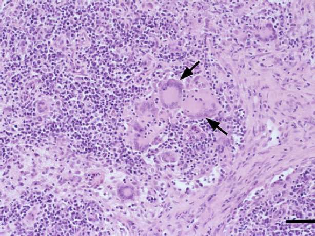
PMWS / PCV2 Associated Disease

Disease versus Infection
PCV2 strain? - The spread of PCV2 a and b worldwide (c, d, e).
PCV2 concentration in tissues determines subclinical vs clinical
disease (as well as the strain of PCV2).
Genetic susceptibility – Large White vs Pietrain/Hampshire as
terminal sire (UK)
Immune system stimulation
    • Activated T cells required for PCV2 replication
    • Virus accumulates in follicular dendritic cells, histiocytes
      and macrophages
    • Immune system no longer responds to infection
      (recognition)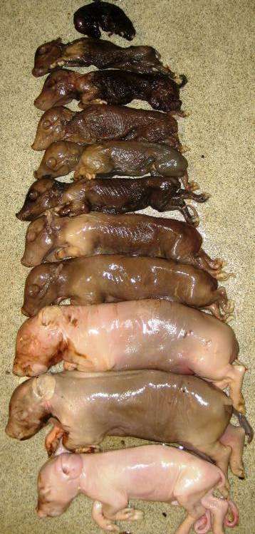
PMWS / PCV2 Associated Disease

Clinical Signs
•   Wasting after successfully weaning
•   Fever
•   Anaemia
•   Jaundice
•   Lymphadenopathy
•   Respiratory Symptoms
•   Gastric ulcers
•   PDNS
•   Doubling of Mortality
•   Poor response to antibiotic therapy
PMWS / PCV2 Associated Disease

Thoughts on “Agent X”
PCV2 capitalizes on inflammatory process
   • Inflammation / immune stimulation can be notable or
      mild
   • Other factors suggested: “PCV2 goes where inflammation
      is”
       o Pestivirus
       o Teschnovirus / Enterovirus
       o Parvovirus
       o Retrovirus
       o Adjuvants
       o MORE…
PMWS / PCV2 Associated Disease

Incubation     Difficult to determine (lots of variables)
Period         4-10 days (maybe)
Pathogenesis   See following slides
Post mortem    Varied
Findings       Lymphadenopathy
               Gastric ulcers (not eating)
               May have mix of the following
               • Loss of myocardial tone
               • Jaundice
               • Intralobular filling
Diagnosis of PCV2AD (PMWS)
Diagnosis Requires all 3 elements
1. Clinical signs/picture
2. Histological evidence of lymphoid depletion
3. Isolation of Virus (IHC)Diagnosis of PCVAD 1. Clinical signs: 2. Histologic lesions: 3. PCV2 infection: Wasting / Depletion of Demonstration of PCV2 weight loss/ ill lymphoid tissues antigen (IHC) and thrift / failure to +/- lymphohistiocytic or characteristic lesions thrive, with or granulomatous without other inflammation in any signs organ
Multinucleated
giant cells
Inclusion bodies PCV2 antigen
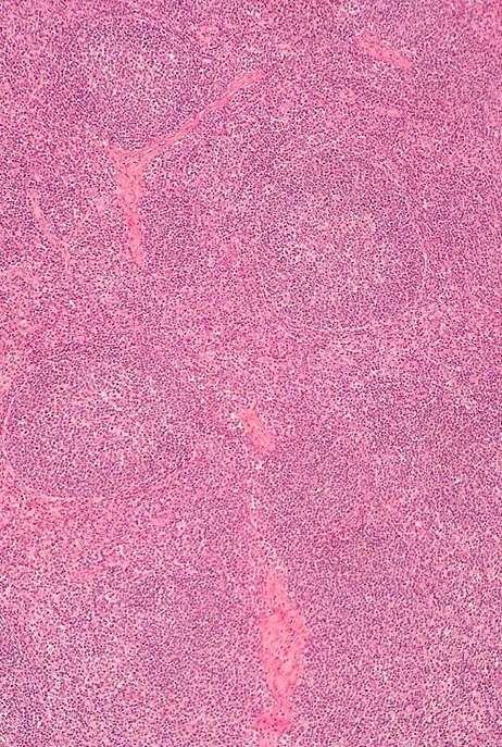
Lymphoid depletion and
granulomatous lymphadenitis Normal lymph node
13PCVAD
PCVAD
mediastinal LN
iliac LN
inguinal LN
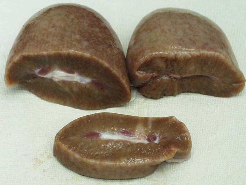
Pigs with PCVAD typically (not always) have enlarged lymph nodes
1618
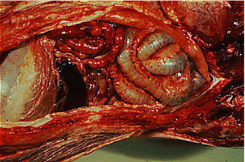
Gastric ulcers are common

       Severe ulcer stage
So this porcine parvovirus…..
isn’t it….?
PCV2 reproductive disorderSubmission for a Multisystemic Disease Investigation
Liver Colon
Lung
Ileum
Heart
Kidney
ADD lymph nodes
Spleen and tonsilCoinfections Immune Host
Differences stimulation susceptibility
between PCV2
isolates
PCV2
PCV2
Infection of Lymphoid depletion + histiocytic
PCV2
lymphoid replacement
tissues
5-10-20%
Low viremia High viremia +
leukopenia
Significant Co-infections
• PPRS
• Porcine Parvovirus +/-Seroconversion
Seroconversion
• Salmonella
• Influenza Systemic
• M. hyopneumonia spread
• L.intracellularis Infection cleared
• +/- H.parasuis 70-80%
• +/- S.suis Clinical
• +/- B.pilosicoli Subclinical MortalityControl Minimize Address Host
Coinfections Immune susceptibility
stimulation
PCV2
PCV2
Infection of Lymphoid depletion + histiocytic
PCV2
lymphoid replacement
tissues
5-10-20%
Low viremia High viremia +
leukopenia
Eliminate or
decrease PCV2
load with PCV2 +/-Seroconversion
vaccines Seroconversion
Systemic
spread
Infection cleared
70-80%
Clinical
Subclinical MortalityPCV2AD
Treatment & Vaccination of progeny
Control Role in vaccinating breeders?
One shot product (s) available in Australia
Two shot options elsewhere in the world
Hygiene is very important (as are the other
components of environmental medicine)
Madec’s 20 Point Principles for PMWS
control
Eradication Has not been demonstrated
Would need good disinfection
Down time?
PCV2 freedom in replacement stock?Madec’s Principles
Acknowledgement

For the use of their photos & information on
PCV2,
many thanks to…………………

•   Howard Hill DVM
•   John Kolb DVM
•   Iowa State University
•   Boehringer Ingleheim
African Swine Fever (ASF)

Causal Agent      Caused by an Asfarviridae (enveloped DNA
                  virus)
                  Some 22 genotypes
Characteristics   First described in Kenya in 1921
& History         (retrospective to 1907)
                  Moved from warthog population into
                  domestic pigs
                  Asymptomatic in warthogs
                  Involves biting arthropods (soft bodied ticks)
                  Spread to Portugal in 1957
                  Eradicated from Europe (apart from Sardinia)
                  Different genotype emerged in Russia in
                  2007
African Swine Fever (ASF)
Epidemiology • The disease can be spread by ticks
(Ornithodoros spp.), fomites, swill
feeding and direct contact with infected
animals (not aerosol)
• Recovered animals remain infective for
at least 6 months
• Virus lasts for a long time in pig products
and the environment
• Different strains – high to low virulence
• Carriers – warthogs, bush pigs, giant
forest hogs and the peccary.
Incubation 4-19 days
PeriodAfrican Swine Fever (ASF)
Pathogenesis Virus replicated in the monocytes &
macrophages of the lymph nodes closest to
the site of infection.
Spreads through blood and lymphatics
Clinical Signs Impossible to distinguish clinically from CSF
• Sudden death
• Fever, off feed
• Necrosis & haemorrhage in lymphoid
tissue (tonsils)
• Skin haemorrhage (ears & flanks)
• Can see laboured breathing, nasal
bleeding (vomiting, constipation &
diarrhoea - blood)African Swine Fever (ASF)
Post Mortem Dependant on virus virulence
Findings • Haemorrhage in spleen, lymph nodes &
myocardium
• Petechial haemorrhages in kidney,
bladder and pleura
• Excess fluid in thoracic cavity &
pericardial sac
Treatment and • The virus is inactivated by approved
Control disinfectants
• No vaccine or treatment
• Notifiable Disease
• Control is by depopulation and site clean
upClassical Swine Fever (CSF)
Causal Agent Caused by a Flaviviridae, genus Pestivirus
(enveloped RNA virus)
Also called Swine Fever or Hog Cholera
Characteristics Severity depends on virus virulence, age of
animal, and immunity of herd.
The virus is quite resistant in the
environment, surviving a couple of days
Other members of the Pestivirus genus can
cause disease in pigs, notably Bovine Viral
Diarrhoea.
The virus is excreted from pigs for 10-20 days
post-infection in large amounts
Carriers – wild boar in parts of EuropeClassical Swine Fever (CSF)
Epidemiology • Pig is the natural host
• Normally pig to pig contact (or uncooked
pig products)
• Excreted in nasal and occular secretions
• Also in urine and faeces
• Wild pig populations can be a reservoir
of infection
• Exotic to Australia, NZ, Canada, USA &
western & central Europe
Incubation 3-7 days (some debate)
PeriodCSF Clinical Signs - Naïve Herds • Initially a few pigs appear drowsy and less active, with some anorexia and they may appear chilled • Within days, pigs will present with a marked fever (41-42•C), sometimes with a reddening of the skin • The pigs develop a conjunctivitis and constipation leading to yellowish diarrhoea • The pigs appear chilled and will huddle together. • A few pigs may convulse before they die
CSF Clinical Signs - Naïve Herds

• Pigs start to die with a spreading purple
  discoloration of the skin.
• Death often occurs some 10 to 20 days post-
  infection
• Pigs which survive will be chronically affected with
  severe retardation of growth and often present
  with arched backs
• In the adult herd, returns, abortions, and an
  increase in mummified and stillborn piglets
CSF Clinical Signs – Established Herds

• Abortion
• Piglets infected from their mothers during
  pregnancy can result in, mummification,
  malformations (may present with a congenital
  tremor with cerebral hypoplasia with CSF),
  stillbirths and weak born piglets.
• Piglets born from CSF infected mothers may
  remain healthy but continually spread the disease
  through out their lives
Classical Swine Fever (CSF)

Post Mortem   The pigs may die so rapidly that there are
Findings      few post-mortem signs
              Multiple haemorrhages through out the
              carcase
              Swollen, oedematous and haemorrhagic
              lymph nodes
              Infarction of the spleen (large areas where
              the blood supply has been cut off resulting
              in blood filled blebs on the surface of the
              spleen)
              In CSF ulceration (button ulcers) can be
              seen in the large intestine
Classical Swine Fever (CSF)
Treatment and • Vaccines – Subunit and modified live
Control vaccines.
• The virus is quite readily inactivated by
approved disinfectants
• Notifiable
• Slaughter out policyClassical Swine Fever (CSF)
Differential Dx • ASF
• PDNS
• Salmonellosis
• Acute Pasteurellosis
• Erysipelas
• Acute septicaemic streptococcal infections
• Thrombocytopaenia
• Warfarin poisoning
• Reproductive diseases
• Other causes of congenital tremorNipah Virus
Causal Agent • A paramyxovirus closely related to the
Hendra virus
Epidemiology • From the village, Sungai Nipah where
the virus was isolated from the first
human victim – a pig farmer with
encephalitis (ZOONOTIC)
• Can affect weaners, growers and
finishers and adults
• Carriers – Pteropus bats (flying foxes)Nipah Virus
Clinical Signs Weaners
• Mild to severe coughing. High morbidity
but low mortality
Adult Pigs
• Moderate to severe respiratory signs
with dyspnoea, convulsions and death.
• Death can occur within several hours.Nipah Virus
Post Mortem & Varying degrees of consolidation of the
Diagnosis lungs, primarily the diaphragmatic lobes
(prominently thickened interlobular septa)
Kidneys show signs of congestion
Other organs normal
Diagnosis from virus isolation and serology
Treatment NoneNipah Virus
Zoonosis • Very fatal to man, out of 258 people
infected 100 died (1998-1999)
• Mild to severe clinical signs,
characterised by fever and headaches of
varying severity
• Patients become drowsy and disoriented
leading to coma.
• Majority of patients developing coma
die.
• Incubation period in man one to three
weeks
• Close contact required with pig or bat.
Not transmitted person to person.Aujeszky’s Disease
Causal Agent Herpes Virus (Alphaherpesvirinae)
Pseudorabies (PRV)
Characteristics Can affect all classes of pig (primary host)
EAD in Australia and many other pig producing countries
Can survive 3 weeks outside the pig
Clinical Signs Piglets
Range of CNS signs (fitting to paddling) inc dog sitting (paralysis)
Sneezing/Coughing
Diarrhoea
High mortality
Weaner/Growers
Sneezing/Coughing
Reduction in CNS signs with increase in respiratory signs
Usually associated with secondary opportunists
Mortality not as high
Stunted/wasted animals
Incubation Can remain dormant in endemic herds for a long time
PeriodAujeszky’s Disease
Route of Carrier pigs
Infection Airborne (3 km)
Fomites – people, equipment, vehicles
Semen (AI)
Feral pigs
Birds?
Pathogenesis Virus will cross the placenta in breeding stock and infect the foetus
Nose to nose between progeny pigs
Replicates in the epithelia of the upper respiratory tract
Deseminates either in free form or via infected leukocytes
Also enters CNS via trigeminal & olfactory nerve endings
Post Mortem Often minimal
Findings Necrotic tonsillitis, laryngitis, tracheitis & sero –fibrinonecrotic
rhinitis
Respiratory lesions associated with other pathogensAujeszky’s Disease
Aujeszky’s Disease

Diagnosis      Clinical picture
               Virus isolation (brain, tonsils, nasal swabs)
               ELISA serology may help but virus isolation better
               PRV will kills non host species such as cats, mice & rats
               Dogs present with signs of rabies (thus the name)
               Cattle & sheep (sometimes horses) suffer from mad itch
               Not a zoonotic disease
Treatment &    No treatment in Australia
Control        Reportable disease
               Vaccination in endemic countries
Porcine Reproductive & Respiratory Syndrome
Causal Agent PRRS virus (Arteriviridae)
RNA virus (Blue Ear)
Two distinct strains European (LV ) & the Nt American (VR -
2332)
Chinese strain is derived from Nth America
Characteristics Appeared in the late 1980s
Presumably entered the swine population from a yet to be
identified wild life species
Exotic to Australia (present in most other pig producing
countries)Porcine Reproductive & Respiratory Syndrome
Clinical Signs Vary due to strain differences, host factors, management
Sows
Fever, anorexia, reproductive (stillborn, weak, mummified,
normal)
Piglets
Weak/sickly, high mortality, CNS, thumping
Weaner/Growers
Dyspnoea, anorexia, lethargy, reduced growth rates
(secondary respiratory infections are common), conjunctivitis
Route of Intranasal, intramuscular, oral, intrauterine & vaginal
Infection Breaks in skin, insect bites
Vertical transmission (third trimester)Porcine Reproductive & Respiratory Syndrome
Incubation Can be as short as 12 hours (virulent strains)
Period
Pathogenesis Replication in lymphoid tissue (thus immunosuppression)
Targets the lung macrophage (kills them for 26 days)
After 7 weeks the lung macrophages become resistant to
PRRSv infection
Post Mortem Respiratory
Findings Interstitial pneumonia (lack of air space histologically)
Enlarged lymph nodes
Diagnosis Clinical picture
Gross and histopathology
Virus isolation (PCR plus other)
SerologyPRRS
Porcine Reproductive & Respiratory Syndrome

Treatment &    None in Australia
Control        Reportable Disease – Eradication
               In endemic countries – ideal is to keep herd negative
               Testing of source herds
               Quarantine (minimum 30 days) & testing prior to release
               Good farm biosecurity (positive pressure ventilation and
               filters)
               Positive herds need a combination of gilt/boar
               acclimatization and vaccination.
               Antibiotics are only supportive in the face of an outbreak
               Eradication can be achieved when known negative
               replacement stock are introduced at a time when the herd
               had stopped shedding virus (PRRS can not persist within an
               immune population)
PRRS (EAD) – eliminating the myths • there is no carrier state • immune sows recover from disease quickly • environmentally unstable so it is easy to kill with any disinfectant, heat etc. • Can survive in water for 8-14 days. •
PRRS – in naïve herd PHASE 1

• Phase 1 (lasts 2 weeks)
  • 5-75% pigs viraemia with lethargy/anorexia
  • “rolling inappetence” – spreads within 3-7 days
    to all groups of pigs
  • Pyrexia (39-41˚C), hyperpneic/dyspneic, 1-2%
    develop cutaneous hyperaemia or cyanosis of
    ears/nose (“blue ear disease”).
  • Increase in returns-to-oestrous, abortions,
    decreased farrowing rate
  • BOARS = lack libido, reduction in semen
    quality, virus in semen.
PRRS – in naïve herd PHASE 2
• Phase 2 (lasts 1-4 months)
• 5-80% sows late term pregnancy abortions
(100-118 days) – premature farrowings
• Pre-weaning piglet mortality up to 60%
• Premature piglets die within a couple of days
• Majority of piglet deaths are first 7 days but continue
afterwards
• Swelling of eyelids
• Emaciation
• Dyspnoea
• Secondary infections!!!PRRS (EAD) diagnosis • PRRS is suspected on the basis of the clinical signs • The presence of PRRS on a unit is confirmed by the use of antibody tests. • However, it can take 2-3 weeks for the antibody level to rise before the test becomes positive. • Unfortunately the antibodies may also disappear 6 months after exposure. • Examination of the lung tissue by histology and special staining techniques (immunoperoxidase) can reveal the organism in the lung • PCR examination of tissues, in particular used for semen examination
PRRS (EAD) treatment

• There is no specific anti viral treatment for PRRS virus
  infection
• The treatment regimes aim to minimise the effect of
  secondary infections.
• Aim to keep the pigs warm and in the draught free
  environment and possibly increase feed density to
  compensate for the anorexia.
• Review the control measures for the secondary infections
  with the practice
• SEW programmes can help to control the spread of the
  disease around the farm and minimise the effect of the
  disease on the farm's economy
• All-in/ all-out and hygiene are essential precursors to
  controlling the disease
PRRS (EAD) control

• Current live vaccines result in excretion from vaccinated
  pigs and therefore cannot be used on PRRS negative
  herds.
• The use of live vaccines in incoming breeding animals in
  PRRS +ve herds helps to maintain farm stability
• The vaccinated stock must be kept separate from the farm
  until shedding has stopped
• Autogenous vaccines from serum or tonsilar scrape
  therapy may be utilised to help gilt and boar introduction
  programmes. These should be restricted to the single
  farm
• Gilts and boars must be stabilised before service so an
  acclimatisation program is essential
PRRS (EAD)

• Killed vaccines generally confer no or little
  protection in naive animals, but it will reduce
  excretion of virus and assist reducing farm
  clinical signs in infected herds.
• Modified live vaccines – these can be very
  variable in response depending on the
  modification carried out.
• Several MLV can cause severe clinical signs
  without field virus. In addition, there can be
  little protection provided for heterolous virus
  strains.
• Allowing sufficient time between vaccination
  and field infection essential part of control.
PRRS (EAD)

• MLV general reduce excretion of virus particles.
• Review fly and mosquito control programmes
• Before purchasing breeding or other incoming
  stock ensure you match serostatus.
• Practice on-farm AI collection, do not rely on a
  commercial AI stud
Swine Influenza

    COVERED IN DISEASES OF THE CHEST LECTURE

• Nasal discharge and puffy eyes
• Fever 40.5-41.5•C
• As the disease progresses loss of weight may be seen
• Mortality is generally low
• The high rectal temperature in breeding stock can result
  in abortions, infertility (a boar can become sub-fertile for
  6 weeks), production of small weak litters and increased
  stillbirths
• Recovery generally starts 5 to 7 days after the first
  clinical signs
Swine influenza
Source: Iowa State UniversityTGE/PED WILL COVER IN GREATER DEPTH IN ENTERIC LECTURES • Both are corona viruses • When first enters a herd: • Piglets less than 21 days generally all die • Weaners unthrifty • Growers, finishers and adults mildly affected • Endemic in herd – post-weaning diarrhoea • Spread by starlings? (trucks?).
Foot and Mouth Disease
Causal Agent Picornovirus (Aphthovirus) (A,O,C, Asia 1 & SAT 1, 2 &3)
             Note other vesicular diseases can resemble FMD
             Swine Vesicular Disease (SVD) caused by Picornovirus
             (Enterovirus)
             Vesicular stomatitis (VS) caused by Rhabdovirus
             (Vesiculovirus)
Incubation     1 to 5 days but can be up to 21 days
Period
Major Clinical Affects all cloven-hoofed animals - pigs, cattle, sheep
Signs          and goats
               Fever to 40.5˚C
               Skin around the snout, lips, tongue, inside the mouth,
               around the coronary band and soft skin on the feet
               becomes whiter (blanched) and vesicles develop
               Lesions in the mouth may not be obvious in the pig
               The animals are lame….very painful (pigs squeal)
               Vesicles may develop on the sow's teats
SAT3
• 7 serotypes:
+++ Strains
SAT2
• No cross-protection
A • Multiple serotypes at
SAT1
the same time
O
C
• Multiple vaccine
Asia1
strains requiredWhere is the world?
Day 1
FMD

Vesicles rupture up to 24 hours after development and if no
secondary infection occurs healing is rapid

                                                              45
Day 3
Foot and Mouth Disease

• With the feet, the hoof may detach, revealing the painful raw
  tissues underneath.
• The hoof can re-grow, but is often deformed. This can take
  several weeks
• The disease affects nearly all susceptible animals, but few
  animals will die specifically with the disease
Day 9                        Day 20

If lesion is at coronary band < 1 week old
Thereafter measure distance from coronary band to lesion
Horn grows at
1mm per week- adults
2mm per week- weaners
Foot and Mouth Disease

• DDx - VS also affects horses
Route of Infection           Rapidly spread through the air, animal contact and vectors,
                             such as clothing, utensils, vehicles
                             Can be spread through meat and meat by-products
                             Spread through semen
                             High humidity, cloud cover and moderate temperatures
                             favour airborne spread (over 20 km)
                             Pigs produce aerosols 3000 times more concentrated than
                             cattle
                             Carrier status occurs in cattle
                             FMD can be excreted in the milk for up to 7 weeks

Diagnosis                    Virus isolation
(dependant on time pig       • Ag ELISA
has been                     • PCR
infected………….and the         Antibody
Government Authorities       • Ab ELISA
will be calling the shots)   • VNT
Principals of FMD Diagnosis
Uninfected FMD Virus infected Recovered (or vaccinated)
Probang
samples
Lesion, swab, probang or Clotted blood samples
clotted blood samples (saliva)
Virus or viral Anti-viral antibodies can
components can be be detected
detected
Live Virus Viral Proteins by LFD or double Viral Nucleic Acid Anti-FMD antibodies can be
by virus isolation antibody sandwich ELISA by RT-PCR detected in serum by ELISA or
in cell cultures VNT
10-30 minutes (LFD)
1-4 days Within a day 5-18 hours (ELISA)
4 hours (ELISA)
2-3 days (VNT)Foot and mouth disease
Diagnosis

• This is generally left to the appropriate
  government body / laboratory to attend the
  suspect farm to collect samples.
• Generally samples collected will include
  vesicular fluid or epithelium from oral,
  nasal or hoof lesions.
Creating an FMD Timeline

1. Based on OLDEST lesion observed in the herd
2. Establish a time window of likely introduction
   of virus using the INCUBATION PERIOD: 1-14
   days most likely 2-5 days
3. Establish a time window of likely spread to
   other units using the PERIOD OF VIRUS
   EXCRETION: 4 days before clinical signs
   most likely 2 day prior first lesion to 4-5 days
   after first clinical signs seen in individual animal
Creating an FMD Timeline
Foot and Mouth Disease

Notifiable                                         Scorched earth
Disease                                            response

Endemic in areas of Africa, Asia, Middle East and South America.
Control – in endemic countries

• For endemic countries – palliative care is the only
  treatment option for affected animals and vaccination is
  available for prevention.
• With vaccination, have to ensure that the oil formulation
  is used (not aqueous) for pigs (cattle respond to either)
  and that the inactivated antigens in the vaccine are
  representative of the circulating viruses in the region.
• Diagnostics can differentiate between vaccinated and
  naturally infected animals.
If an EAD is suspected?

• CONTACT - Department of Agriculture and
  Food veterinary officer or stock inspector,
  or contact the Emergency Disease Watch
  Hotline on 1800 675 888 (Free call 24
  hours).

• AUSVETPLAN – technical response plans
  – Australia’s response to incursion of each
  exotic disease of importance.
So how real is the risk?

• ASF in Belarus, Kenya, central Russia
• CSF in Colombio (South America)
• FMD in Amur (Russia)
• PEDv in America – sweeping across the
  states.
• Others…
Prevention of exotic diseases

• Eliminate or minimise the sources of infection:
  • Live pigs from overseas – banned
  • Semen from overseas - banned
  • Embryos from overseas - banned
  • Meat products from overseas – fresh pork is
    banned
     • Legally imported product has to be cooked and not
       contain bones
     • Feeding of ‘swill’ to pigs is banned
Swill feeding risk – large scale
2013 - A man has been convicted and sentenced in Australia for illegally
importing over 20 tonnes of pork and chicken into Australia from Korea.
• Tim Chapman - First Assistant Secretary, Border Compliance Division
“Given that Korea has had outbreaks of this highly contagious disease,
these meat products posed a potentially significant risk to Australia.
“To put this into perspective, we estimate that a small Foot and Mouth
Disease outbreak, controlled in 3 months, could cost Australia around $7.1
billion, while a large 12 month outbreak would cost $16 billion.”
• The case comes from an investigation by the Department of Agriculture,
Fisheries and Forestry that uncovered large-scale and deliberate illegal
imports of foods across businesses in Brisbane, Melbourne and Sydney.You can also read