Evaluation and Management of Heart Murmurs in Children
←
→
Page content transcription
If your browser does not render page correctly, please read the page content below
Evaluation and Management of Heart
Murmurs in Children
JENNIFER E. FRANK, MD, and KATHRYN M. JACOBE, MD, University of Wisconsin Fox Valley Family Medicine
Residency Program, Appleton, Wisconsin

Heart murmurs are common in healthy infants, children, and adolescents. Although most are not pathologic, a mur-
mur may be the sole manifestation of serious heart disease. Historical elements that suggest pathology include fam-
ily history of sudden cardiac death or congenital heart disease, in utero exposure to certain medications or alcohol,
maternal diabetes mellitus, history of rheumatic fever or Kawasaki disease, and certain genetic disorders. Physical
examination should focus on vital signs; age-appropriate exercise capacity; respiratory or gastrointestinal manifes-
tations of congestive heart failure; and a thorough cardiovascular examination, including features of the murmur,
assessment of peripheral perfusion, and auscultation over the heart valves. Red flags that increase the likelihood of a
pathologic murmur include a holosystolic or diastolic murmur, grade 3 or higher murmur, harsh quality, an abnormal
S2, maximal murmur intensity at the upper left sternal border, a systolic click, or increased intensity when the patient
stands. Electrocardiography and chest radiography rarely assist in the diagnosis. Referral to a pediatric cardiologist
is recommended for patients with any other abnormal physical examination findings, a history of conditions that
increase the likelihood of structural heart disease, symptoms suggesting underlying cardiac disease, or when a specific
innocent murmur cannot be identified by the family physician. Echocardiography provides a definitive diagnosis and
is recommended for evaluation of any potentially pathologic murmur, and for evaluation of neonatal heart murmurs
because these are more likely to be manifestations of structural heart disease. (Am Fam Physician. 2011;84(7):793-
800. Copyright © 2011 American Academy of Family Physicians.)

                                 H
                                                eart murmurs are common                               and can help identify children who are likely
                                                in asymptomatic, otherwise                            to have structural heart disease (Table 2).4,7,10
                                                healthy children. These mur-                             In infants, feeding difficulties may be the
                                                murs are often innocent and                           first sign of congestive heart failure, which is
                                  result from the normal patterns of blood                            present in approximately one-third of infants
                                  flow through the heart and vessels.1 How-                           and children with CHD.4 The most common
                                  ever, a heart murmur may be the sole find-                          symptoms in a series of children presenting
                                  ing in children with structural heart disease;                      to the emergency department with acute
                                  therefore, a thorough evaluation is necessary.                      heart failure included dyspnea (74 percent),
                                                                                                      nausea and vomiting (60 percent), fatigue (56
                                  Incidence and Prevalence                                            percent), and cough (40 percent).12
                                  Congenital heart disease (CHD) may occur                               Exercise tolerance should be assessed in an
                                  in the presence or absence of a heart mur-                          age-appropriate fashion. Parents of infants
                                  mur. The incidence of CHD varies between                            should be asked about their child’s ability to
                                  four and 50 per 1,000 live births.2 One review                      play and the duration and vigor of feeding;
                                  found an incidence of 75 cases per 1,000 live                       parents of older children should compare
                                  births; of these, six cases per 1,000 were                          their child’s ability to participate in team
                                  moderate or severe.3                                                sports with that of peers.4 Chest pain is rarely
                                                                                                      a presenting symptom of cardiac disease in
                                  History                                                             children.13,14 In a pediatric cardiology clinic,
                                  Certain historical features suggest possible                        chest pain or syncope prompted consulta-
                                  structural heart disease (Table 1).1,2,4-11 Car-                    tion in approximately 10 percent of children;
                                  diovascular signs and symptoms can be non-                          only 11 percent of those with chest pain and
                                  specific (e.g., poor feeding, failure to thrive)                    5 percent of those with syncope had cardiac
                                  or specific (e.g., chest pain, palpitations),                       disease.14 A high degree of suspicion is
   Downloaded from the American Family Physician Web site at www.aafp.org/afp. Copyright © 2011 American Academy of Family Physicians. For the private, noncommer-
October cial
        1, 2011                                                                                                                   American               requests. 793
                                                                                                                                                Family Physician
                   ◆ Volume 84, Number 7
             use of one                                                      www.aafp.org/afp
                        individual user of the Web site. All other rights reserved. Contact copyrights@aafp.org for copyright questions and/or permission
Heart Murmurs in Children
Table 1. Historical Findings Suggesting Structural Heart Disease in Children with Heart Murmurs
Historical finding Significance
Family history
CHD More common in children with a first-degree relative who has CHD (three- to 10-fold increased
risk7); high penetrance with ventricular septal defect and mitral valve prolapse
Sudden cardiac death or hypertrophic Increased risk of hypertrophic cardiomyopathy (autosomal dominant pattern)
cardiomyopathy
Sudden infant death syndrome Can be secondary to undiagnosed CHD lesions8
Personal history
Conditions that may coexist with CHD: Certain genetic disorders (e.g., DiGeorge syndrome, velo-cardio-facial syndrome) are associated
Aneuploidy (e.g., trisomy 21, Turner with cardiac malformations
syndrome) Trisomy 21 is associated with an increased risk of atrioventricular septal defects, atrial septal
Connective tissue disorder (e.g., defects, ventricular septal defects, patent ductus arteriosus, and tetralogy of Fallot
Marfan syndrome) Turner syndrome is associated with increased risk of coarctation of the aorta, aortic valve
Inborn error of metabolism stenosis, and left ventricular hypertrophy
Major congenital defects of other Marfan syndrome is associated with mitral valve prolapse, aortic root dilation, and aortic
organ systems insufficiency
Syndrome with dysmorphic features
Frequent respiratory infections Respiratory symptoms may be attributable to heart disease (i.e., congestive heart failure);
enlarged vessels may lead to atelectasis or difficulty clearing respiratory secretions, thereby
promoting infection
Kawasaki disease Leading cause of acquired cardiac disease in children; can cause coronary artery aneurysm and
stenosis9
Rheumatic fever Associated with development of rheumatic heart disease
Prenatal or perinatal history
In utero exposure to alcohol or other Fetal alcohol syndrome is associated with an increased risk of atrial and ventricular septal
toxins defects, and tetralogy of Fallot10
In utero exposure to selective Selective serotonin reuptake inhibitor exposure is associated with a small but statistically
serotonin reuptake inhibitors significant increased risk of mild heart lesions, including ventricular septal defects and bicuspid
or other potentially teratogenic aortic valve (although not all studies found an increased risk11)
medications Lithium exposure is associated with Ebstein anomaly of the tricuspid valve
Valproate (Depacon) exposure is associated with coarctation of the aorta and hypoplastic left
heart syndrome
Intrauterine infection Maternal infections may increase risk of structural heart lesions (e.g., maternal rubella infection is
associated with patent ductus arteriosus and peripheral pulmonary stenosis)
Maternal diabetes mellitus Increased risk of CHD, including transient hypertrophic cardiomyopathy, tetralogy of Fallot,
truncus arteriosus, and double-outlet right ventricle
Preterm delivery CHD is associated with other conditions (e.g., genetic disorders, in utero exposure to toxins) that
can result in preterm birth; 50 percent of newborns weighing less than 3 lb, 5 oz (1,500 g) at
birth have CHD (most commonly patent ductus arteriosus)7
CHD = congenital heart disease.
Information from references 1, 2, and 4 through 11.
necessary to detect underlying cardiac disease in children child’s appearance, activity level, color, and respiratory
who report exertional syncope or chest pain, or who have effort should be assessed, and the neck should be exam-
a family history of hypertrophic cardiomyopathy.1,13,14 ined for prominent vessels, abnormal pulsations, and
bruits.1 Jugular venous distension is rare in children.4
Physical Examination The chest wall should be inspected for abnormalities of
The patient’s vital signs should be compared with age- the sternum, which can be associated with CHD,15 and
established norms (available at http://www.cc.nih.gov/ for abnormal cardiac impulses or thrills.1 The lungs
ccc/pedweb/pedsstaff/age.html), and a focused exami- should be auscultated for abnormal breath sounds such as
nation of the respiratory, cardiovascular, and gastroin- crackles, which may indicate pulmonary congestion, or
testinal systems should be performed5 (Table 32,5-7,10,15,16). wheezing, which may indicate cardiac asthma. Abdom-
Congenital anomalies of other organ systems may be inal examination should focus on the liver location
associated with CHD in up to 25 percent of patients.6 The (seeking abdominal situs) and evaluation for liver
794 American Family Physician www.aafp.org/afp Volume 84, Number 7 ◆ October 1, 2011Heart Murmurs in Children
Table 2. Symptoms Suggesting Cardiac Disease Table 3. Physical Examination of Children
with Heart Murmurs
Symptom or sign Significance
Finding Significance
Cardiovascular
Chest pain May be related to aortic stenosis or Abnormal growth Feeding difficulties may be a sign of
hypertrophic cardiomyopathy (height and weight cardiac disease in newborns and
Cyanosis Structural heart lesion with restricted plotted on growth infants (decreased exercise capacity)
pulmonary blood flow chart) Certain genetic disorders may
Dizziness Multiple potential causes, including increase risk of delayed growth
hypoxia and CHF and CHD
Near-syncope or May be related to aortic stenosis or Abnormal vital signs Arrhythmia, tachycardia, hypoxia,
syncope hypertrophic cardiomyopathy (compared with age- and tachypnea may indicate
adjusted norms) underlying structural heart disease
Palpitations May be related to arrhythmias secondary
to structural heart lesions Blood pressure discrepancy between
upper and lower limbs may
Constitutional
indicate coarctation of the aorta
Developmental Congenital heart lesions are more common (pressure gradient of > 20 mm Hg
delay in children with certain genetic disorders with low blood pressure in the
and syndromes lower extremities)
Diaphoresis May indicate CHF or poor cardiac fitness Adventitial breath Wheezing may be associated
Easily fatigued May indicate CHF, hypoxia, or poor cardiac sounds (e.g., with cardiac asthma; rales may
fitness wheezing, rales, be associated with pulmonary
Poor exercise May indicate CHF, hypoxia, or poor cardiac ronchi, pleural rub) congestion secondary to
tolerance or fitness congestive heart failure
capacity for play Chest contour signaling Defective segmentation of the
Poor growth or May indicate CHF, poor cardiac fitness, or maldevelopment of sternum may occur in children
failure to thrive a genetic disorder or syndrome; poor the sternum15 with CHD
weight gain most commonly reflects Dysmorphic features Certain genetic or congenital
decreased cardiac output or left-to-right conditions increase risk of CHD
shunts with pulmonary hypertension Cardiovascular findings
Respiratory Abnormal S2 Classic finding of wide split fixed
Asthma-like Cardiac asthma resulting from pulmonary S2 with atrial septal defects;
symptoms congestion abnormal S2 may be present in
Chronic cough Atelectasis or difficulty clearing secretions other types of CHD
because of pulmonary vascular congestion Capillary refill Normal peripheral perfusion is less
Dyspnea on May indicate CHF, hypoxia, or poor cardiac than 2 to 3 seconds; delay may
exertion fitness indicate poor perfusion secondary
to diminished cardiac output
CHF = congestive heart failure. Displaced point of Possible structural abnormality or
Information from references 4, 7, and 10.
maximal impulse; ventricular enlargement
precordial impulses
(heaves, lifts, thrills)
Edema Congestive heart failure
Left-sided precordial Cardiac enlargement
enlargement or ascites, which may signal congestive heart bulge
failure.5 S3 or S 4 Can indicate structural heart disease;
The peripheral pulses should be examined for rate, S3 can be a normal finding but
rhythm, volume, and character, and capillary refill time usually disappears when patient
should be less than three seconds.4 The heart should be is upright
auscultated over the tricuspid, pulmonary, mitral, and Substernal heave Right ventricular hypertension
aortic areas with the bell and diaphragm of the stetho- Systolic ejection click Semilunar valvular stenosis
Weak or absent Coarctation of the aorta
scope while the patient is supine, sitting, and standing17
femoral pulses
(Figure 118).Innocent murmurs are produced by the nor-
Gastrointestinal findings
mal flow of blood through the heart. Changing the flow by
Ascites Congestive heart failure
changing the patient’s position (for example, decreasing Hepatomegaly Congestive heart failure
flow to the heart with the Valsalva maneuver) will change Location of liver signals High rate of CHD with abdominal
the intensity of the murmur. Young children should be abdominal situs situs
prompted to push out their abdomen against the exam-
iner’s hand.1 The physician should listen for normal S1 and CHD = congenital heart disease.
S2 ; a wide fixed split S2 is characteristic of an atrial septal Information from references 2, 5 through 7, 10, 15, and 16.
defect.19 Gallops can be a normal finding in adolescents.1
October 1, 2011 ◆ Volume 84, Number 7 www.aafp.org/afp American Family Physician 795Heart Murmurs in Children
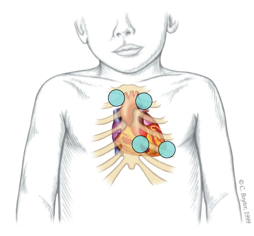
URSB ULSB
Apex
The heart murmur is characterized by its timing dur-
ing the cardiac cycle; its location, quality, intensity, and
pitch (how it sounds); and the presence or absence of
clicks1 (Table 4 5,7,17 and Table 5 20-23). The intensity of heart
murmurs is graded from 1 to 6. Grade 1 murmurs are
barely audible; grade 2 murmurs are faint but can be
heard immediately; grade 3 murmurs can be heard easily
and are moderately loud; grade 4 murmurs can be heard
easily over a wide area but do not have a palpable thrill;
ILLUSTRATION BY C. BOYTER
grade 5 murmurs are loud and have a precordial thrill;
LLSB and grade 6 murmurs are loud enough to hear with the
stethoscope raised off the chest.17,24 Certain characteristics
of the murmur may be considered red flags, prompting
Figure 1. Listening areas for clicks: upper right sternal bor- stronger consideration for structural heart disease. These
der (URSB) for aortic valve clicks; upper left sternal bor- include a holosystolic murmur (odds ratio [OR] of patho-
der (ULSB) for pulmonary valve clicks; lower left sternal logic murmur = 54), grade 3 or higher (OR = 4.8), harsh
border (LLSB), or the tricuspid area, for ventricular septal
quality (OR = 2.4), an abnormal S2 (OR = 4.1), maximal
defects; and the apex for aortic or mitral valve clicks.
intensity at the upper left sternal border (OR = 4.2), a
Reprinted with permission from McConnell ME, Adkins SB III, Hannon DW.
Heart murmurs in pediatric patients: when do you refer? Am Fam Physician. systolic click (OR = 8.3), diastolic murmur, or increased
1999;60(2):560. murmur intensity with standing.6,10,25 A decrease or lack
Table 4. Characteristics of Innocent Heart Murmurs
Type Description Age at detection Can sound like
Aortic systolic Systolic ejection murmur best heard over the aortic valve Older childhood —
murmur into adulthood
Mammary artery High-pitched systolic murmur that can extend into diastole; Rare in adolescence Arteriovenous anastomoses
soufflé* best heard along the anterior chest wall over the breast or patent ductus arteriosus
Peripheral Grade 1 or 2, low-pitched, early- to mid-systolic ejection < 1 year Pulmonary artery stenosis or
pulmonary murmur heard over axilla or back normal breath sounds
stenosis
Pulmonary flow Grade 2 or 3, crescendo-decrescendo, early- to mid-systolic All Atrial septal defect or
murmur murmur peaking in mid-systole; best heard at the left pulmonary valve stenosis
sternal border between the second and third intercostal
spaces; characterized by a rough, dissonant quality;
loudest when patient is supine and decreases when
patient is upright and holding breath
Still murmur Grade 1 to 3, early systolic murmur; low to medium pitch Infancy to Ventricular septal defect
with a vibratory or musical quality; best heard at lower adolescence, or hypertrophic
left sternal border; loudest when patient is supine and often 2 to 6 years cardiomyopathy
decreases when patient stands
Supraclavicular/ Brief, low-pitched, crescendo-decrescendo murmur heard in Childhood to Bicuspid/stenotic aortic
brachiocephalic the first two-thirds of systole; best heard above clavicles; young adulthood valve, pulmonary valve
systolic murmur radiates to neck; diminishes when patient hyperextends stenosis, or coarctation of
shoulders the aorta
Venous hum Grade 1 to 6 continuous murmur; accentuated in diastole; 3 to 8 years Cervical arteriovenous
has a whining, roaring, or whirring quality; best heard fistulas or patent ductus
over low anterior neck, lateral to the sternocleinomastoid; arteriosus
louder on right; resolves or changes when patient is supine
*—Mammary artery soufflé murmur is caused by blood flow in the arteries and veins leading to and from the breasts.
Information from references 5, 7, and 17.
796 American Family Physician www.aafp.org/afp Volume 84, Number 7 ◆ October 1, 2011Table 5. Prevalence and Characteristics of Pathologic Heart Murmurs
Prevalence among
Type of children with
structural congenital heart
heart lesion disease (%) Symptoms and clinical course Characteristics
Ventricular 20 to 25 Small defects: usually asymptomatic Small defects: loud holosystolic murmur at LLSB
septal defect Medium or large defects: CHF, (may not last throughout systole if defect is very
symptoms of bronchial obstruction, small)
frequent respiratory infections Medium and large defects: increased right-to-left
ventricular impulses; thrill at LLSB; split or loud
single S2; holosystolic murmur at LLSB without
radiation; grade 2 to 5; may also hear a grade 1
or 2 mid-diastolic rumble
Atrial septal 8 to 13 Usually asymptomatic and incidentally Grade 2 or 3 systolic ejection murmur best heard at
defect found on physical examination or ULSB; wide split fixed S2; absent thrill; may have a
echocardiography; large defects can grade 1 or 2 diastolic flow rumble at LLSB
be present in infants with CHF
Patent ductus 6 to 11 May be asymptomatic; can cause Continuous murmur (grade 1 to 5) in ULSB
arteriosus easy fatigue, CHF, and respiratory (crescendo in systole and decrescendo into
symptoms diastole); normal S1; S2 may be “buried” in the
murmur; thrill or hyperdynamic left ventricular
impulse may be present
Tetralogy of 10 Onset depends on severity of Central cyanosis; clubbing of nail beds; grade 3
Fallot pulmonary stenosis; cyanosis may or 4 long systolic ejection murmur heard at ULSB;
appear in infancy (2 to 6 months of may have holosystolic murmur at LLSB; systolic
age) or in childhood; other symptoms thrill at ULSB; normal to slightly increased S1;
include hypercyanotic spells or single S2
decreased exercise tolerance
Pulmonary 7.5 to 9 Usually asymptomatic but may have Systolic ejection murmur (grade 2 to 5); heard
stenosis symptoms secondary to pulmonary best at ULSB radiating to infraclavicular regions,
congestion axillae, and back; normal or loud S1; variable S2;
systolic ejection click may be heard at left sternal
border and may vary with respiration
Coarctation of 5.1 to 8.1 Newborns and infants may present Systolic ejection murmur best heard over
the aorta with CHF; older children are usually interscapular region; normal S1 and S2; decreased
asymptomatic or may have leg pain or delayed femoral pulse; may have increased left
or weakness ventricular impulse
Aortic stenosis 5 to 6 Usually asymptomatic; symptoms may Systolic ejection murmur (grade 2 to 5) best heard
include dyspnea, easy fatigue, chest at upper right sternal border with radiation to
pain, or syncope; newborns and carotid arteries; left ventricular heave; thrill at
infants may present with CHF ULSB or suprasternal notch
Transposition 5 Variable presentation depending on Cyanosis; clubbing of nail beds; single S2; murmur
of the great type; may include cyanosis or CHF in may be absent or grade 1 or 2 nonspecific systolic
arteries first week of life ejection murmur; may have a grade 3 or 4
holosystolic murmur at LLSB and mid-diastolic
murmur at apex
Total anomalous 2 to 3 Onset of CHF at 4 to 6 weeks of age Grade 2 or 3 systolic ejection murmur at ULSB;
pulmonary grade 1 or 2 mid-diastolic flow rumble at LLSB;
venous wide split fixed S2
connection
Tricuspid atresia 1.4 Early-onset cyanosis or CHF within the Cyanosis; clubbing of nail beds; normal pulses;
first month of life single S2; holosystolic murmur at LLSB or
midsternal border; murmur may be absent; mid-
diastolic flow murmur at apex may be present
Hypoplastic Rare May be asymptomatic at birth, with Hyperdynamic precordium; single S2; nonspecific
left heart cyanosis and CHF developing with grade 1 or 2 systolic ejection murmur along left
syndrome duct closure sternal border
Truncus Rare Onset of CHF in first few weeks of life; Increased cardiac impulses; holosystolic murmur
arteriosus minimal cyanosis (ventricular septal defect); mid-diastolic rumble
CHF = congestive heart failure; LLSB = lower left sternal border; ULSB = upper left sternal border.
Information from references 20 through 23.
October 1, 2011 ◆ Volume 84, Number 7 www.aafp.org/afp American Family Physician 797Heart Murmurs in Children
are present, and when findings on radiography or elec-
Table 6. The Seven S’s: Key Features trocardiography (ECG) are abnormal.28 Online libraries
of Innocent Murmurs of digital heart sounds are available to familiarize physi-
cians with the characteristics of abnormal heart sounds
Sensitive (changes with child’s position or with respiration) (Table 7).
Short duration (not holosystolic)
Single (no associated clicks or gallops) Role of Diagnostic Testing
Small (murmur limited to a small area and nonradiating) Chest radiography and ECG rarely assist in the diagnosis of
Soft (low amplitude) a heart murmur.5,6,29 Studies in newborns30 and children31
Sweet (not harsh sounding) with asymptomatic murmurs have shown that chest radi-
Systolic (occurs during and is limited to systole) ography does not influence clinical management or assist
Information from reference 27.
with diagnosis. A prospective study of 201 newborns who
were referred to pediatric cardiologists for evaluation of
a heart murmur found that the addition of ECG to clini-
cal assessment did not improve the sensitivity or speci-
of change in the murmur intensity with passive leg eleva- ficity of detecting structural heart lesions.32 In a study of
tion (likelihood ratio [LR] = 8.0) or when the child moves 128 infants and children who were evaluated for heart
from standing to squatting (LR = 4.5) increases the likeli- murmurs, the addition of ECG and chest radiography to
hood of hypertrophic cardiomyopathy.26 cardiac auscultation was more likely to mislead than assist
Characteristics that are more likely to be associated the physician in making the correct diagnosis.33
with an innocent murmur include a systolic (rather than In a study of more than 900 children in a pediatric
diastolic) murmur; soft sound; short duration; musical cardiology clinic who had innocent-sounding mur-
or low pitch; varying intensity with phases of respiration murs, an abnormal finding from the history, physical
and posture (louder in supine position); and murmurs examination, or diagnostic tests (ECG, chest radiog-
that become louder with exercise, anxiety, or fear 17,24 raphy, or pulse oximetry) was 67 percent sensitive but
(Table 6 27). The most common innocent murmur is a only 38 percent specific for the presence of a structural
Still murmur, which is characteristically loudest at the heart lesion in infants younger than six weeks, yielding
lower left sternal border and has a musical or vibratory positive and negative LRs very near 1.0 (i.e., no use-
quality that is thought to represent vibrations of the left ful diagnostic information).28 In infants older than six
outflow tract.1,5 weeks, sensitivity increased to 100 percent, but specific-
Auscultation may be less accurate in younger patients, ity decreased to 28 percent (positive LR = 1.6; negative
when other signs or symptoms of cardiovascular disease LR = 0.026). Thus, this information is helpful for rul-
ing out structural causes of an innocent-
sounding murmur in infants and children
Table 7. Online Resources for Pediatric Cardiac Auscultation older than six weeks, but it is not helpful in
younger infants.
The Auscultation Assistant In two separate populations geographi-
Web site: http://www.wilkes.med.ucla.edu/inex.htm cally remote from a pediatric cardiolo-
Blaufuss Medical Multimedia Laboratories gist, phonocardiography (i.e., digital heart
Web site: http://www.blaufuss.org
sound recordings reviewed by a pediatric
Heart Sounds and Murmurs
Web site: http://www.dundee.ac.uk/medther/Cardiology/hsmur.html cardiologist) had high sensitivity and speci-
Johns Hopkins University Cardiac Auscultatory Recording Database ficity, and good intraobserver agreement
Web site: http://www.murmurlab.com/card6/ (registration required) in distinguishing between innocent mur-
Texas Heart Institute murs and murmurs that were potentially
Web site: http://www.texasheart.org/education/cme/explore/events/ or probably pathologic and that required
eventdetail_5469.cfm
echocardiography.34,35
University of Michigan Heart Sound and Murmur Library
Web site: http://www.med.umich.edu/lrc/psb/heartsounds/index.htm
Indications for Referral
University of Washington Department of Medicine
Demonstrations: Heart Sounds and Murmurs In children and adolescents, the diagnosis of
Web site: http://depts.washington.edu/physdx/heart/demo.html an innocent heart murmur can be made if
four criteria are met: absence of abnormal
798 American Family Physician www.aafp.org/afp Volume 84, Number 7 ◆ October 1, 2011Heart Murmurs in Children
physical examination findings (except for
the murmur); a negative review of systems SORT: KEY RECOMMENDATIONS FOR PRACTICE
(i.e., child is asymptomatic); a history that Evidence
is negative for features that increase the risk Clinical recommendation rating References
of structural heart disease; and characteris-
Structural heart disease is more likely when C 6, 10, 25
tic auscultatory features of a specific inno- the murmur is holosystolic, diastolic, grade 3
cent heart murmur. These criteria are not
2,5
or higher, or associated with a systolic click;
appropriate for newborns or infants younger when it increases in intensity with standing; or
than one year because these patients have when it has a harsh quality.
a higher rate of asymptomatic structural Chest radiography and electrocardiography B 5, 6, 29-33
rarely assist in the diagnosis of heart murmurs
heart disease. When an innocent murmur
36
in children.
cannot be definitively diagnosed, the child Family physicians should order echocardiography B 28, 43
should be referred for echocardiography, to or consider referral to a pediatric cardiologist
a pediatric cardiologist, or both. for newborns with a heart murmur, even if the
child is asymptomatic, because of the higher
A study in Oman found that the preva-
prevalence of structural heart lesions in this
lence of abnormal findings on echocar- population.
diography was not significantly different
between patients referred by pediatric cardi- A = consistent, good-quality patient-oriented evidence; B = inconsistent or limited-
quality patient-oriented evidence; C = consensus, disease-oriented evidence, usual
ologists and those referred by primary care
practice, expert opinion, or case series. For information about the SORT evidence
physicians. However, pediatric cardiolo-
37
rating system, go to http://www.aafp.org/afpsort.xml.
gists more accurately detect structural heart
lesions in newborns and children with heart
murmurs,32,38 and can assist family physicians in the depends on multiple factors, including his or her con-
assessment of a suspicious murmur. For both innocent fidence in the diagnosis. Echocardiography may not be
and pathologic murmurs, referral to a pediatric cardi- required in newborns with a heart murmur if a pedi-
ologist for confirmation or clarification of the diagnosis atric cardiologist has diagnosed an innocent murmur
is associated with decreased parental anxiety.39 with a high degree of confidence32 ; however, it is impor-
tant to consider the relatively high prevalence of struc-
Neonatal Heart Murmurs tural heart disease among asymptomatic newborns
Newborns are at higher risk of having serious struc- with a heart murmur.
tural heart disease that presents as an asymptomatic The evaluation of newborns for CHD may include
murmur.6,10 Approximately 1 percent of newborns have pulse oximetry after 24 hours of life. Clinical exami-
a heart murmur, and 31 to 86 percent of these infants nation of asymptomatic newborns has a sensitivity of
have structural heart disease,40-42 including asymp- 46 percent for detection of CHD; this sensitivity
tomatic newborns. Because of the higher likelihood of increases to 77 percent when clinical examination
structural heart disease in asymptomatic newborns and is combined with pulse oximetry (with a cutoff of
young infants with heart murmurs, referral to a pediat- 94 percent).44
ric cardiologist and/or for echocardiography is recom-
mended.28,42,43 Even potentially life-threatening heart The Authors
defects may not be associated with any initial signs or
JENNIFER E. FRANK, MD, FAAFP, is in private practice at Theda Care
symptoms other than a heart murmur.41,42
Physicians in Neenah, Wis. At the time this article was written, she was
The reported sensitivity for detection of a patho- an assistant professor of family medicine at the University of Wisconsin
logic heart murmur in newborns ranges from 80.5 to School of Medicine and Public Health, Madison, and a faculty member
94.9 percent among pediatric cardiologists, with speci- at the University of Wisconsin Fox Valley Family Medicine Residency Pro-
ficity ranging from 25 to 92 percent.32,43 These varia- gram, Appleton.
tions are significant because the lowest specificity KATHRYN M. JACOBE, MD, is a third-year resident at the University of
corresponds to positive and negative LRs of 1.1 and Wisconsin Fox Valley Family Medicine Residency Program.
0.7, which are uninformative, and the highest specific- Address correspondence to Jennifer E. Frank, MD, FAAFP, 1380 Tul-
ity corresponds to positive and negative LRs of 10 and lar Rd., Neenah, WI 54956 (e-mail: jennifer.frank@thedacare.org).
0.21, which are quite accurate. The ability of a pediatric Reprints are not available from the authors.
cardiologist to accurately identify pathologic murmurs Author disclosure: No relevant financial affiliations to disclose.
October 1, 2011 ◆ Volume 84, Number 7 www.aafp.org/afp American Family Physician 799Heart Murmurs in Children
25. McCrindle BW, Shaffer KM, Kan JS, Zahka KG, Rowe SA, Kidd L. Car-
REFERENCES dinal clinical signs in the differentiation of heart murmurs in children.
Arch Pediatr Adolesc Med. 1996;150(2):169-174.
1. Biancaniello T. Innocent murmurs. Circulation. 2005;111(3):e20-e22.
26. Etchells E, Bell C, Robb K. Does this patient have an abnormal systolic
2. Harris JP. Consultation with the specialist. Evaluation of heart murmurs.
murmur? JAMA. 1997;277(7):564-571.
Pediatr Rev. 1994;15(12):490-494.
27. Bronzetti G, Corzani A. The seven “S” murmurs: an alliteration about
3. Hoffman JI, Kaplan S. The incidence of congenital heart disease. J Am
innocent murmurs in cardiac auscultation. Clin Pediatr (Phila). 2010;
Coll Cardiol. 2002;39(12):1890-1900.
49(7):713.
4. Pelech AN. Evaluation of the pediatric patient with a cardiac murmur.
28. Danford DA, Martin AB, Fletcher SE, Gumbiner CH. Echocardiographic
Pediatr Clin North Am. 1999;46(2):167-188.
yield in children when innocent murmur seems likely but doubts linger.
5. Danford DA. Effective use of the consultant, laboratory testing, and Pediatr Cardiol. 2002;23(4):410-414.
echocardiography for the pediatric patient with heart murmur. Pediatr
29. Yi MS, Kimball TR, Tsevat J, Mrus JM, Kotagal UR. Evaluation of heart
Ann. 2000;29(8):482-488.
murmurs in children: cost-effectiveness and practical implications.
6. Poddar B, Basu S. Approach to a child with a heart murmur. Indian J Pediatr. 2002;141(4):504-511.
J Pediatr. 2004;71(1):63-66.
30. Oeppen RS, Fairhurst JJ, Argent JD. Diagnostic value of the chest radio-
7. Martins P, Dinis A, Canha J, Ramalheiro G, Castela E. Innocent heart graph in asymptomatic neonates with a cardiac murmur. Clin Radiol.
murmurs. Rev Port Cardiol. 2008;27(6):815-831. 2002;57(8):736-740.
8. Weber MA, Ashworth MT, Risdon RA, Brooke I, Malone M, Sebire NJ. 31. Birkebaek NH, Hansen LK, Elle B, et al. Chest roentgenogram in the eval-
Sudden unexpected neonatal death in the first week of life: autopsy uation of heart defects in asymptomatic infants and children with a car-
findings from a specialist centre. J Matern Fetal Neonatal Med. 2009; diac murmur: reproducibility and accuracy. Pediatrics. 1999;103(2):E15.
22(5):398-404.
32. Mackie AS, Jutras LC, Dancea AB, Rohlicek CV, Platt R, Béland MJ. Can
9. Gordon JB, Kahn AM, Burns JC. When children with Kawasaki disease cardiologists distinguish innocent from pathologic murmurs in neo-
grow up: myocardial and vascular complications in adulthood. J Am Coll nates? J Pediatr. 2009;154(1):50-54.
Cardiol. 2009;54(21):1911-1920.
33. Rajakumar K, Weisse M, Rosas A, et al. Comparative study of clinical
10. Frommelt MA. Differential diagnosis and approach to a heart murmur in evaluation of heart murmurs by general pediatricians and pediatric car-
term infants. Pediatr Clin North Am. 2004;51(4):1023-1032. diologists. Clin Pediatr (Phila). 1999;38(9):511-518.
11. Merlob P, Birk E, Sirota L, et al. Are selective serotonin reuptake inhibi- 34. Mahnke CB, Mulreany MP, Inafuku J, Abbas M, Feingold B, Paolillo JA.
tors cardiac teratogens? Echocardiographic screening of newborns with Utility of store-and-forward pediatric telecardiology evaluation in dis-
persistent heart murmur. Birth Defects Res A Clin Mol Teratol. 2009; tinguishing normal from pathologic pediatric heart sounds. Clin Pediatr
85(10):837-841. (Phila). 2008;47(9):919-925.
12. Macicek SM, Macias CG, Jefferies JL, Kim JJ, Price JF. Acute heart failure 35. Germanakis I, Dittrich S, Perakaki R, Kalmanti M. Digital phonocardiog-
syndromes in the pediatric emergency department. Pediatrics. 2009; raphy as a screening tool for heart disease in childhood. Acta Paediatr.
124(5):e898-e904. 2008;97(4):470-473.
13. Kane DA, Fulton DR, Saleeb S, Zhou J, Lock JE, Geggel RL. Needles 36. Koo S, Yung TC, Lun KS, Chau AK, Cheung YF. Cardiovascular symp-
in hay: chest pain as the presenting symptom in children with serious toms and signs in evaluating cardiac murmurs in children. Pediatr Int.
underlying cardiac pathology. Congenit Heart Dis. 2010;5(4):366-373. 2008;50(2):145-149.
14. Geggel RL. Conditions leading to pediatric cardiology consultation in a 37. Venugopalan P, Agarwal AK, Johnston WJ, Riveria E. Spread of heart
tertiary academic hospital. Pediatrics. 2004;114(4):e409-e417. diseases seen in an open-access paediatric echocardiography clinic. Int
15. Andren L, Hall P. Diminished segmentation or premature ossification of J Cardiol. 2002;84(2-3):211-216.
the sternum in congenital heart disease. Br Heart J. 1961;23:140-142.
38. Advani N, Menahem S, Wilkinson JL. The diagnosis of innocent mur-
16. Washington R. Sports cardiology in the adolescent athlete: concerns for murs in childhood. Cardiol Young. 2000;10(4):340-342.
the pediatrician. Pediatr Ann. 2007;36(11):698-702.
39. Giuffre RM, Walker I, Vaillancourt S, Gupta S. Opening Pandora’s box:
17. Pelech AN. The physiology of cardiac auscultation. Pediatr Clin North parental anxiety and the assessment of childhood murmurs. Can J Car-
Am. 2004;51(6):1515-1535. diol. 2002;18(4):406-414.
18. McConnell ME, Adkins SB III, Hannon DW. Heart murmurs in pediatric 40. Bansal M, Jain H. Cardiac murmur in neonates. Indian Pediatr. 2005;
patients: when do you refer? Am Fam Physician. 1999;60(2):558-565. 42(4):397-398.
19. Christensen DD, Vincent RN, Campbell RM. Presentation of atrial septal 41. Rein AJ, Omokhodion SI, Nir A. Significance of a cardiac murmur
defect in the pediatric population. Pediatr Cardiol. 2005;26(6):812-814. as the sole clinical sign in the newborn. Clin Pediatr (Phila). 2000;
20. Syamasundar Rao P. Diagnosis and management of acyanotic heart dis- 39(9):511-520.
ease: part I — obstructive lesions. Indian J Pediatr. 2005;72(6):496-502. 42. Ainsworth S, Wyllie JP, Wren C. Prevalence and clinical significance of
21. Syamasundar Rao P. Diagnosis and management of acyanotic heart dis- cardiac murmurs in neonates. Arch Dis Child Fetal Neonatal Ed. 1999;
ease: part II — left-to-right shunt lesions. Indian J Pediatr. 2005;72(6): 80(1):F43-F45.
503-512. 43. Azhar AS, Habib HS. Accuracy of the initial evaluation of heart murmurs
22. Rao PS. Diagnosis and management of cyanotic congenital heart dis- in neonates: do we need an echocardiogram? Pediatr Cardiol. 2006;
ease: part I. Indian J Pediatr. 2009;76(1):57-70. 27(2):234-237.
23. Syamasundar Rao P. Diagnosis and management of cyanotic congenital 44. Bakr AF, Habib HS. Combining pulse oximetry and clinical examina-
heart disease: part II. Indian J Pediatr. 2009;76(3):297-308. tion in screening for congenital heart disease. Pediatr Cardiol. 2005;
24. Uner A, Doğan M, Bay A, Cakin C, Kaya A, Sal E. The ratio of congenital 26(6):832-835.
heart disease and innocent murmur in children in Van city, the Eastern
Turkey. Anadolu Kardiyol Derg. 2009;9(1):29-34.
800 American Family Physician www.aafp.org/afp Volume 84, Number 7 ◆ October 1, 2011You can also read