Role of inflammatory and liver function markers in assessing the prognosis of patients with COVID 19
←
→
Page content transcription
If your browser does not render page correctly, please read the page content below
WORLD ACADEMY OF SCIENCES JOURNAL 3: 52, 2021

                Role of inflammatory and liver function markers in
                assessing the prognosis of patients with COVID‑19
             SWATHY MOORTHY1, TEENA KOSHY2, MAHESH KUMAR K3 and SANTHI SILAMBANAN4

   Departments of 1General Medicine, and 2Human Genetics, Sri Ramachandra Institute of Higher Education and Research,
            Chennai, Tamil Nadu 600116; 3Department of Physiology and Biochemistry, Government Yoga and
          Naturopathy Medical College and Hospital, Chennai, Tamil Nadu 600106; 4Department of Biochemistry,
             Sri Ramachandra Institute of Higher Education and Research, Chennai, Tamil Nadu 600116, India

                                           Received June 8, 2021; Accepted August 23, 2021

                                                       DOI: 10.3892/wasj.2021.123

Abstract. Severe acute respiratory syndrome corona‑                  counts along with the total leucocyte counts and absolute
virus 2 (SARS‑CoV‑2) caused Coronavirus disease 2019                 lymphocyte counts, NLR, erythrocyte sedimentation rate,
(COVID‑19) in early December, 2019. The disease begins as            D‑dimer, ferritin and AST levels serve to predict the severity
a respiratory disease with varied outcomes, from complete            and prognosis of patients with COVID‑19. AST appears to
recovery to long‑standing complications, such as respiratory         be a more sensitive marker than alanine aminotransferase.
distress, heart ailments and stroke. The present study was           However, the De Ritis ratio, although not as sensitive as AST,
undertaken to identify the derangements in liver function            may provide insight towards morbidity.
and inflammatory status which may be used to diagnose
the severity and thus assess the prognosis of patients with          Introduction
COVID‑19 infection. The present study was conducted at a
tertiary care teaching hospital. The study population included       Coronavirus disease 2019 (COVID‑19) is caused by severe
456 patients with COVID‑19, confirmed by the reverse tran‑           acute respiratory syndrome coronavirus 2 (SARS‑CoV‑2).
scription‑PCR of nasopharyngeal swabs for SARS‑CoV‑2.                The disease began in Wuhan, the capital of Hubei Province in
Data were collected from patient files. The study was                China and soon developed into a global pandemic on March
approved by the institutional ethics committee. Data are             11, 2020. The measures taken to prevent or contain the disease
expressed based on the normality of distribution. Variables          were not successful due to the rapidity of spread within the
were compared and correlation analyses were performed.               individual, as well as within the community (1). The incuba‑
Receiver operating characteristics and the Youden index              tion period of the virus is between 2 days to 2 weeks, during
were applied to obtain cut‑off values. The results revealed          which time, the patient is asymptomatic, yet infectious. By the
an increase in C‑reactive protein (CRP), ferritin, D‑dimer,          time the patients present with symptoms, the severity of the
lactate dehydrogenase (LDH), aspartate aminotransferase              disease can be mild to moderate, depending on the presence of
(AST) and total bilirubin levels. In addition, there was             metabolic disorders, such as obesity, diabetes mellitus, hyper‑
evidence of leukocytosis, neutrophilia, lymphopenia and an           tension and chronic obstructive pulmonary disease, amongst
increased neutrophil‑to‑lymphocyte ratio (NLR). The NLR              others (2).
exhibited an area under the curve of 0.77, with a cut‑off value          When the virus infects the host, a battery of inflam‑
of 2.23 mg/l. Collectively, the findings of the present study        matory responses is initiated to combat the pathogen. The
demonstrate that the CRP, LDH, neutrophil and eosinophil             inflammatory response is not terminated with scavenging
                                                                     the virus alone; it also damages tissues surrounding the area
                                                                     infected by the viral pathogen, as well as organs remote from
                                                                     the place of origin of infection. Thus, COVID‑19‑infected
                                                                     individuals tend to have systemic disease with the involve‑
Correspondence to: Dr Santhi Silambanan, Department of
Biochemistry, Sri Ramachandra Institute of Higher Education and      ment of various organs, such as the heart, liver, central
Research, 1 Mount Poonamallee Road, Ramachandra Nagar, Porur,        nervous system, blood vessels and lungs. The estimation of
Chennai, Tamil Nadu 600116, India                                    the extent of injury to these organs may guide clinicians as
E‑mail: santhisilambanan@sriramachandra.edu.in                       regards the extent or severity of the disease. Based on these
                                                                     features, the appropriate disease management can be initi‑
Key words: SARS‑CoV‑2, COVID‑19, inflammation, lymphopenia,          ated at the appropriate time (1). The present retrospective
neutrophilia, De Ritis ratio, neutrophil‑to‑lymphocyte ratio         study was undertaken to identify the derangements in liver
                                                                     function and inflammatory biomarkers, which may be used
                                                                     to diagnose the severity and thus assess the prognosis of
                                                                     patients with COVID‑19 infections.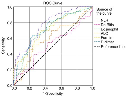
2 MOORTHY et al: Alterations of liver parameters in COVID-19
Patients and methods Ethics statement. The present study was approved by the
Institutional Ethics Committee of Sri Ramachandra Institute
Study design and setting. The present study was a retrospec‑ of Higher Education and Research, with a waiver of informed
tive study performed at the tertiary care teaching hospital, Sri consent of patients, as permitted by the national regulatory
Ramachandra Institute of Higher Education and Research in body (IEC‑NI/20/AUG/75/49, dated 08‑08‑2020). Patient
Chennai, India. The study population included patients with identification was de‑identified reversibly for the purpose of
COVID‑19, between June and August, 2020, aged >18 years, the analysis.
which was confirmed by reverse transcription‑PCR of naso‑
pharyngeal swabs for SARS‑CoV‑2 (assessed at one of the Statistical analysis. Normality distribution was evaluated using
Indian Council of Medical Research‑approved laboratories the the Kolmogorov‑Smirnov test for all variables. Depending
for RNA virus nucleic acid amplification tests). Patients on the presence or absence of the normality of distribution,
discharged against medical advice for whom outcomes were data are expressed as the mean ± standard deviation or the
not known were excluded from the study. median and interquartile range (IQR). Between‑group differ‑
ences of variables were compared using one‑way ANOVA
Classification of study population. The division of the patients followed by Tukey's post hoc test. The association between
into groups was based on peripheral oxygen saturation, respi‑ variables was obtained by Pearson's correlation analysis.
ratory rate and CT scan findings. The study population was Receiver operating characteristics (ROC) curve analysis was
subdivided into three groups as mild, moderate and severe performed to estimate the area under the curve (AUC) with the
based on peripheral oxygen saturation and respiratory rate. 95% confidence interval (CI) and the cut‑off point according
Mild, moderate and severe cases had an oxygen saturation of to the Youden index. All statistical analyses were conducted
≥94, 91‑93 and ≤90%, respectively and respiratory rates of using R statistical software version 4.0.2 (The R Project for
≤23, 24‑29 and ≥30 breaths/min, respectively (3). A CT scan Statistical Computing; https://www.r‑project.org/). P100˚F was considered as laboratory parameters of the study population are presented
febrile. A prolonged hospital duration was considered if the in Table I. The most common presenting clinical features
hospital stay was >10 days. were fever, sore throat, cough, dyspnea, diarrhea, anosmia
and myalgia in the order of occurrence. The patients in the
Data collection. Data collection was carried out by the manual younger age group with an age of 48.85±14.50 years recovered
perusal of inpatient case sheets, investigations of computer‑ completely. Leukocytosis with progressive neutrophilia and
ized patient data systems and the transcription database for lymphopenia, and an increased neutrophil‑to‑lymphocyte
discharge summaries. Details of basic demographic charac‑ ratio (NLR) were noted in patients with mild to severe disease.
teristics, and information for symptoms, such as fever, throat The concentrations of liver function markers in the three study
pain, cough, dyspnea, diarrhea, anosmia and myalgia, hemo‑ groups are presented in Table II. There was highly significant
dynamic parameters upon admission and during the course of increase in AST, ALT and total bilirubin levels from the mild
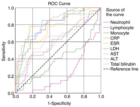
stay, daily clinical assessment, initial laboratory tests, such as to severe disease groups. The ROC curve of hematological
complete blood count, liver function tests [bilirubin, aspartate markers is illustrated in Fig. 1. The AUC was found to be high
aminotransferase (AST), alanine aminotransferase (ALT), for the NLR, eosinophil count and D‑dimer levels. The ROC
alkaline phosphatase, albumin, C‑reactive protein (CRP)] and curves of various inflammatory and liver function markers
the detection of serum ferritin, lactate dehydrogenase (LDH) are presented in Fig. 2. The AUC was found to be high for
and D‑dimer levels were collected. The laboratory parameters the CRP and LDH levels, and the neutrophil count. The AUC
were analyzed using standard accepted methods in the National of the various hematological, inflammatory and liver func‑
Accreditation Board for Testing and Calibration Laboratories tion markers among the three study groups are presented
(NABL) accredited Sri Ramachandra Laboratory services. In in Table III. According to the AUC values, the studied
addition, information regarding chest imaging, medications biomarkers were categorized into four groups for predicting
administered (steroids, heparin, remdesivir and antibiotics), the severity and prognosis of COVID‑19 infection as follows:
oxygen support, organ dysfunction, secondary sepsis and final i) AUC 1.0‑0.8, excellent diagnostic accuracy (CRP, LDH
outcome were collected and deidentified for personal details and neutrophil counts); ii) AUC 0.8‑0.6, very good diagnostic
prior to the analysis. The primary outcomes of interest in the accuracy [NLR, eosinophil count, erythrocyte sedimentation
study were clinical severity, elevated inflammatory marker rate (ESR), D‑dimer, ferritin, total white blood cell (WBC)
levels, high D‑dimer levels, a prolonged hospital duration and counts, absolute lymphocyte counts (ALC) and AST]; iii) AUC
the condition of the patients at discharge. 0.6‑0.5, good diagnostic accuracy (De Ritis and ALT); andWORLD ACADEMY OF SCIENCES JOURNAL 3: 52, 2021 3
Table I. Levels of the inflammatory markers of the study participants.
Mild n=324 Moderate Severe n=66 ANOVA Tukey's post hoc
Parameters (72%) n=66 (14%) (14%) P-value analysis: P‑value
Age (years) 48.85±14.50 56.63±11.5 61.47±11.034 MOORTHY et al: Alterations of liver parameters in COVID-19
Table II. Levels of liver function parameters in the study participants.
Parameters Mild n=324 Moderate n=66 Severe n=66 P‑value Tukey's post hoc analysis P‑value
AST (U/l)a 29 (14‑159) 37 (3‑288) 47 (25‑143) 0.006 Mild‑moderate=0.008
Mild‑severe=0.11
Moderate‑severe=0.82
ALT (U/l)a 26 (8‑128) 29 (10‑313) 73 (20‑148) 0.003 Mild‑moderate=0.17
Mild‑severe=0.02
Moderate‑severe=0.20
De Rittis ratioa 1.21 (0.45‑3.13) 1.31 (0.08‑5.54) 0.97 (0.45‑2.35) 0.56 ‑
Serum albumin (g/dl)b 4.14±0.42 3.70±0.37 3.64±0.57 0.09 Mild‑moderate=0.001
Mild‑severe=0.001
Moderate‑severe=0.99
Total bilirubin (mg/dl)a 0.54 (0.19‑2.30) 0.61 (0.19‑12.83) 0.83 (0.3‑3.1) 0.008 Mild‑moderate=0.04
Mild‑severe=0.28
Moderate‑severe=0.91
Direct bilirubin (mg/dl)a 0.11 (0.04‑9.0) 0.17 (0.06‑6.62) 0.18 (0.05‑1.78) 0.74 ‑
Values of mild, moderate and severe are expressed as median and range; bValues of mild, moderate and severe are expressed as the mean ± SD.
a
AST, aspartate transaminase; ALT, alanine transaminase.
Figure 2. Receiver operating characteristics curves of neutrophil, lympho‑
Figure 1. Receiver operating characteristics curves of eosinophil counts, cyte and monocyte counts, and total WBC counts, ESR, CRP, LDH, AST
ALC, NLR, D‑dimer levels, ferritin levels and the De Ritis ratio. ALC, abso‑ and ALT levels. WBC, white blood cell; CRP, C‑reactive protein; ESR,
lute lymphocyte count; NLR, neutrophil‑to‑lymphocyte ratio. erythrocyte sedimentation rate; LDH, lactate dehydrogenase; AST, aspartate
aminotransferase; ALT, alanine aminotransferase.
markers are presented in Table IV. Of the liver function
parameters examined, the De Ritis ratio exhibited positive Discussion
correlations with total bilirubin and AST, and negative corre‑
lations with serum albumin and ALT. When compared with When COVID‑19 infection progresses to severe disease, it has
the inflammatory markers, the De Ritis ratio exhibited a posi‑ been found to present a systemic manifestation with multiorgan
tive correlation with D‑dimer levels, and negative correlations failure and septicemia, which manifests as acute respiratory
with hemoglobin levels and the WBC count. The results of the distress syndrome, respiratory failure, acute liver injury, renal
correlation between the NLR and other inflammatory markers, injury or failure and cardiovascular manifestations (5). Few
as well as liver function markers are presented in Table V. patients exhibit an inadequate pro‑inflammatory response,
NLR exhibited positive correlations with the neutrophil resulting in severe disease manifestations. This presentation is
count, CRP, ferritin, D‑dimer and LDH levels, and negative known as the cytokine response syndrome (CRS) or cytokine
correlations with the lymphocyte count and monocyte count. storm syndrome (CSS) (1,6). The present study demonstrated
When compared with liver function markers, NLR exhibited a that patients with severe disease were in the age group of
positive correlation with ALT, and a negative correlation with 61.47±11.03 years, during which the onset of comorbid condi‑
serum albumin levels. tions was observed. COVID‑19 has predominantly affectedWORLD ACADEMY OF SCIENCES JOURNAL 3: 52, 2021 5 Table III. AUC of the biochemical parameters of the study participants. Variables AUC SE 95% CI Cut‑off value P‑value Total WBC count 0.64 0.043 0.56 to 0.72 5750 0.002 Neutrophil count 0.828 0.032 0.76 to 0.89 65.4 0.0001 Lymphocyte count 0.178 0.030 0.11 to 0.23 ‑ 0.0001 Monocytes count 0.38 0.052 0.28 to 0.49 ‑ 0.01 Eosinophil count 0.71 0.02 0.66 to 0.76 0.35 0.001 ALC 0.60 0.05 0.48 to 0.71 ‑ 0.08 NLR 0.77 0.45 0.68 to 0.85 2.23 0.001 ESR 0.74 0.041 0.66 to 0.82 22.5 0.001 D‑dimer 0.72 0.05 0.62 to 0.82 0.54 0.001 Ferritin 0.71 0.05 0.61 to 0.81 173.60 0.001 CRP 0.825 0.03 0.76 to 0.88 1.79 0.0001 LDH 0.808 0.036 0.73 to 0.88 240.5 0.0001 AST 0.63 0.07 0.49 to 0.77 71 0.04 ALT 0.56 0.05 0.42 to 0.69 ‑ 0.34 De Ritis ratio 0.51 0.06 0.39 to 0.63 ‑ 0.74 Values in bold font indicate statistically significant differences (P
6                                MOORTHY et al: Alterations of liver parameters in COVID-19

Table IV. Correlation between De Ritis ratio and other param‑        ferritin, D‑dimer and LDH levels. The NLR exhibited a negative
eters in patients with COVID‑19.                                     correlation with lymphocytes, monocytes and serum albumin
                                                                     levels (Table V). In addition, there was significant increase in
Parameters               Correlation coefficient (r)     P‑value     the ESR (P= 0.02; Table I). The AUC was 0.74 with a 95%
                                                                     CI of 0.66‑0.82 (P=0.001) and a cut‑off value of 22.5 mm/h
Hb                                 ‑0.42
WORLD ACADEMY OF SCIENCES JOURNAL 3: 52, 2021 7
Table V. Correlation between NLR and other parameters in in patients (38). Moderate to severe liver damage is character‑
patients with COVID‑19. ized by a De Ritis ratio 1.0 (39,40). According to the study by
Parameters Correlation coefficient (r) P‑value Zinellu et al (39), the median De Ritis ratio was 1.33, similar to
that reported by Yazar et al in their cohort (38). Furthermore,
Hb ‑0.17 0.0002 according to the study by Qin et al (41), the cut‑off value of
Total WBC count 0.55 0.001 the De Ritis ratio was found to be 1.38; patients with a De
Neutrophil count 0.63 0.001 Ritis ratio >1.38 upon admission have a significantly poor
Lymphocyte count ‑0.62 0.001 survival (41). With the hepatic proportion of the AST:ALT
Monocytes count ‑0.35 0.0001 ratio of 2.5:1, the hepatocyte turnover may result in a much
Eosinophil count ‑0.14 0.02 higher amount of AST in serum compared to ALT. AST has
De Ritis ratio ‑0.15 0.06 a shorter half‑life of 18 h compared with ALT, which has a
Serum Albumin ‑0.26 0.001 half‑life of 36 h. Hence, the serum levels of AST and ALT
ESR 0.17 0.007 are fairly similar in healthy individuals. AST is present in the
cytoplasm and mitochondria, whereas ALT is present only in
CRP 0.39 0.001
the cytoplasm, although at a much higher concentration than
Total Bilirubin 0.12 0.12
AST (42). In the present study, the De Ritis ratio exhibited a
Direct Bilirubin ‑0.02 0.79 positive correlation with the total bilirubin, D‑dimer and AST
AST 0.13 0.09 levels, and a negative correlation with the hemoglobin, total
ALT 0.24 0.002 WBC, serum albumin and ALT levels (Table IV).
Ferritin 0.23 0.001 In the present study, there was a significant increase in total bili‑
D‑Dimer 0.22 0.001 rubin levels between mild and moderate cases (P=0.04; Table I).
LDH 0.31 0.001 The serum albumin levels however, did not exhibit a statistically
significant difference between the groups (P=0.09; Table II).
Values in bold font indicate statistically significant differences Albumin is considered to be a negative acute phase reactant with
(P8 MOORTHY et al: Alterations of liver parameters in COVID-19
Such type of correlation was not observed for the De Ritis 5. Falasca K, Ucciferri C, Brandimarte A, Auricchio A,
Pontolillo M, Caiazzo L and Vecchiet J: Clinical characteris‑
ratio. Hence, NLR and AST may be be better indicators of tics and cardiovascular implications of the dead patients for
liver involvement than the De Ritis ratio in predicting systemic COVID‑19. Eur J Inflam 19, 2021.
inflammation associated with liver injury or inflammation in 6. Ali AHM, Mohamed SOO, Elkhidir IHE, Elbathani MEH,
Ibrahim AAH, Elhassan ABE, Salman MST, Elhassan MAM,
patients with COVID‑19. Elnil M and Abuzied AIH: The association of lymphocyte count,
CRP, D‑Dimer, and LDH with severe coronavirus disease 2019
Acknowledgements (COVID‑19): A meta‑analysis. Sudan J Med Sci 15: 9‑23, 2020.
7. Büyükaydın B: The relationship of hemogram and inflammatory
biomarkers to lenght of stay in hospital and clinical course in
Not applicable. patients with COVID‑19. Bezm Sci 8: 7‑14, 2020.
8. Moutchia J, Pokharel P, Kerri A, McGaw K, Uchai S, Nji M and
Goodman M: Clinical laboratory parameters associated with
Funding severe or critical novel coronavirus disease 2019 (COVID‑19):
A systematic review and meta‑analysis. PLoS One 15: e0239802,
No funding was received. 2020.
9. Zhao Y, Nie HX, Hu K, Wu XJ, Zhang YT, Wang MM, Wang T,
Zheng SC, Li XC and Zeng SL: Abnormal immunity of non-
Availability of data and materials survivors with COVID-19: predictors for mortality. Infect Dis
Poverty 9: 108, 2020.
10. Pourabdollah Toutkaboni M, Askari E, Khalili N, Tabarsi P,
The datasets used and/or analyzed during the current study Jamaati H, Velayati AA, Dorudinia A, Rezaei M, Nadji SA,
are available from the corresponding author on reasonable Mohamadnia A and Khalili N: Demographics, laboratory
request. parameters and outcomes of 1061 patients with coronavirus
disease 2019: A report from Tehran, Iran. New Microbe and New
Infect 38: 100777, 2020.
Authors' contributions 11. Kordzadeh‑Kermani E, Khalili H and Karimzadeh I:
Pathogenesis, clinical manifestations and complications of
COVID‑19. Future Microbiol 15: 1287‑1305, 2020.
SM was involved in the conception of the study. SM and TK 12. Thompson S, Bohn MK, Mancini N, Loh TP, Wang CB,
were involved in data collection and compilation. MKK and Grimmler M, Yuen KY, Mueller R, Koch D, Sethi S, et al: Horvath
SS were involved in the analysis of the data. SS and SM were and the IFCC taskforce on COVID‑19. IFCC interim guidelines
on biochemical/hematological monitoring of COVID‑19 patients.
involved in the preparation of the first draft of the article. SM Clin Chem Lab Med 58: 2009‑2016, 2020.
and SS confirm the authenticity of all the raw data. All authors 13. Danwang C, Endomba FT, Nkeck JR, Wouna DLA, Robert A and
have read and approved the final version of the article. Noubiap JJ: A meta‑analysis of potential biomarkers associated
with severity of coronavirus disease 2019 (COVID‑19). Biomark
Res 8: 37, 2020.
Ethics approval and consent to participate 14. Ucciferri C, Vecchiet J and Falasca K: Role of monoclonal anti‑
body drugs in the treatment of COVID‑19. World J Clin Cases 8:
4280‑4285, 2020.
The present study was approved by the Institutional 15. Zenga F, Huangc Y, Guoa Y, Yina M, Chena X, Xiaod L and
Ethics Committee of Sri Ramachandra Institute of Higher Deng G: Association of inflammatory markers with the severity
Education and Research, with a waiver of informed consent of COVID‑19: A meta‑analysis. Int J Infect Dis 96: 467‑474, 2020.
16. Jurado A, Martín MC, Abad‑Molina C, Orduña A, Martínez A,
of patients, as permitted by the national regulatory body Ocaña E, Yarce O, Navas AM, Trujillo A, Fernández L, et al:
(IEC‑NI/20/AUG/75/49, dated 08‑08‑2020). Patient identifica‑ COVID‑19: Age, interleukin‑6, C‑reactive protein, and lympho‑
tion was de‑identified reversibly for the purpose of the analysis. cytes as key clues from a multicentre retrospective study. Immun
Ageing 17: 22, 2020.
17. Khourssaji M, Chapelle V, Evenepoel A, Belk hir L,
Patient consent for publication Yombi JC, van Dievoet MA, Saussoy P, Coche E, Fillée C,
Constantinescu SN, et al: A biological profile for diagnosis
and outcome of COVID‑19 patients. Clin Chem Lab Med 58:
Not applicable. 2141‑2150, 2020.
18. Marfia G, Navone S, Guarnaccia L, Campanella R, Mondoni M,
Competing interests Locatelli M, Barassi A, Fontana L, Palumbo F, Garzia E, et al:
Decreased serum level of sphingosine‑1‑phosphate: A novel
predictor of clinical severity in COVID‑19. EMBO Mol Med 13:
The authors declare that they have no competing interests. e13424, 2021.
19. Hu H, Du H, Li J, Wang Y, Xu X, Wang C, Zhang Y, Zhang G,
Zhao Y, Kang W and Lian J: Early prediction and identifica‑
References tion for severe patients during the pandemic of COVID‑19: A
severe COVID‑19 risk model constructed by multivariate logistic
1. Kenneth Oh: Aberrant cytokine activity in the host immune regression analysis. J Glob Health 10: 020510, 2020.
response to COVID‑19 leads to cytokine release syndrome; 20. Mirmohammadi S, Kianmehr A, Arefi M and Mahrooz A:
Bio-Rad Bulletin_7335; May, 2020. https://www.bio-rad.com/ Biochemical parameters and pathogenesis of SARS‑CoV‑2 infec‑
webroot/web/pdf/lsr/literature/Bulletin_7335.pdf. tion in vital organs: COVID‑19 outbreak in Iran. New Microbes
2. Hanif M, Haider MA, Xi Q, Ali MJ and Ahmed MU: A review New Infect 38: 100792, 2020.
of the risk factors associated with poor outcomes in patients with 21. Alhenc‑Gelas F and Drueke TB: Blockade of SARS‑CoV‑2 infec‑
coronavirus disease 2019. Cureus 12: e10350, 2020. tion by recombinant soluble ACE2. Kidney Int 97: 1091‑1093,
3. Government of India Ministry of Health and Family Welfare: 2020.
Clinical Management Protocol for COVID‑19 (adults). 22. Zhang L, Peng Y, Zheng Q, Jiang L, Tang S and Chen P:
https://www.mohfw.gov.in/pdf/UpdatedDetailedClinicalManage‑ Retrospective analysis of clinical characteristics and laboratory
mentProtocolforCOVID19adultsdated24052021.pdf. Accessed, results of COVID‑19 patients. European Journal of Inflammation.
July 27, 2021. January 2021. doi:10.1177/20587392211011919.
4. Bernheim A, Mei X, Huang M, Yang Y, Fayad ZA, Zhang N, 23. Kho J, Ioannou A, Van den Abbeele K, Mandal AKJ and
Diao K, Lin B, Zhu X, Li K, et al: Chest CT findings in coro‑ Missouris CG: Pulmonary embolism in COVID‑19: Clinical
navirus disease‑19 (COVID‑19): Relationship to duration of characteristics and cardiac implications. Am J Emerg Med 38:
infection. Radiology 295: 200463, 2020. 2142‑2146, 2020.WORLD ACADEMY OF SCIENCES JOURNAL 3: 52, 2021 9
24. Farooqi F, Dhawan N, Morgan R, Dinh J, Nedd K and Yatzkan G: 36. Adhikari IP, Tiwari R and Bala R: Estimation of the De Ritis
Treatment of severe COVID‑19 with Tocilizumab mitigates ratio in the cases of chronic alcoholic liver disease attending
cytokine storm and averts mechanical ventilation during acute OPD of a tertiary health care level institute of Kanpur, UP.
respiratory distress: A case report and literature review. Trop PARIPEX‑Indian J Res 7: 219‑221, 2018.
Med Infect Dis 5: 112, 2020. 37. Ghahramani S, Tabrizi R, Lankarani KB, Kashani SMA,
25. Guillén L, Padilla S, Fernández M, Agulló V, García JA, Rezaei S, Zeidi N, Akbari M, Heydari ST, Akbari H,
Telenti G, García‑Abellán J, Botella Á, Gutiérrez F and Masiá M: Nowrouzi‑Sohrabi P and Ahmadizar F: Laboratory features of
Preemptive interleukin‑6 blockade in patients with COVID‑19. severe vs. non‑severe COVID‑19 patients in Asian populations:
Sci Rep 10: 16826, 2020. A systematic review and meta‑analysis. Eur J Med Res 25: 30,
26. Cavalli G, Farina N, Campochiaro C, De Luca G, Della‑Torre E, 2020.
Tomelleri A and Dagna L: Repurposing of biologic and 38. Yazar H, Kayacan Y and Ozdin M: De Ritis ratio and biochemical
targeted synthetic Anti‑Rheumatic drugs in COVID‑19 and parameters in COVID‑19 patients. Arch Physiol Biochem: 1‑5,
Hyper‑Inflammation: A comprehensive review of available 2020 (Epub ahead of print).
and emerging evidence at the Peak of the Pandemic. Front 39. Zinellu A, Arru F, De Vito A, Sassu A, Valdes G, Scano V,
Pharmacol 11: 598308, 2020. Zinellu E, Perra R, Madeddu G, Carru C, et al: The De Ritis ratio
27. Li H, Liu L, Zhang D, Xu J, Dai H, Tang N, Su X and Cao B: as prognostic biomarker of in‑hospital mortality in COVID‑19
SARS‑CoV‑2 and viral sepsis: Observations and hypotheses. patients. Eur J Clin Invest 51: e13427, 2021.
Lancet 395: 1517‑1520, 2020. 40. Parmar KS, Singh GK, Gupta GP, Pathak T and Nayak S:
28. Ponti G, Maccaferri M, Ruini C, Tomasi A and Ozben T: Evaluation of De Ritis ratio in liver‑associated diseases. Int
Biomarkers associated with COVID‑19 disease progression. Crit J Med Sci Public Health 5: 1783‑1788, 2016.
Rev Clin Lab Sci 57: 389‑399, 2020. 41. Qin C, Wei Y, Lyu X, Zhao B, Feng Y, Li T, Cao H, Yang X,
29. Huang I, Pranata R, Lim MA, Oehadian A and Alisjahbana B: Zhou X, Wang W, et al: High aspartate aminotransferase to
C‑reactive protein, procalcitonin, D‑dimer, and ferritin in severe alanine aminotransferase ratio on admission as risk factor
coronavirus disease‑2019: A meta‑analysis. Ther Adv Respir for poor prognosis in COVID‑19 patients. Sci Rep 10: 16496,
Dis 14: 1753466620937175, 2020. 2020.
30. Weidmann MD, Ofori K and Rai AJ: Laboratory Biomarkers 42. Botros M and Sikaris KA: The de ritis ratio: The test of time.
in the management of patients with COVID‑19. Am J Clin Clin Biochem Rev 34: 117‑130, 2013.
Pathol 155: 333‑342, 2021. 43. Varim C, Yaylaci S, Demirci T, Kaya T, Nalbant A, Dheir H,
31. Gris JC, Perez‑Martin A, Quéré I and Sotto A: COVID‑19 associ‑ Senocak D, Kurt R, Cengiz H and Karacaer C: Neutrophil count
ated coagulopathy: The crowning glory of thrombo‑inflammation to albumin ratio as a new predictor of mortality in patients with
concept. Anaesth Crit Care Pain Med 39: 381‑382, 2020. COVID‑19 infection. Rev Assoc Med Bras (1992) 66 (Suppl 2):
32. Yazdanpanah F, Hamblin MR and Rezaei N: The immune system S77‑S81, 2020.
and COVID‑19: Friend or foe? Life Sci 256: 117900, 2020. 44. Ucciferri C, Caiazzo L, Di Nicola M, Borrelli P, Pontolillo M,
33. Zhang L and Guo H: Biomarkers of COVID‑19 and technologies Auricchio A, Vecchiet J and Falasca K: Parameters associated
to combat SARS‑CoV‑2. Adv Biomark Sci Technol 2: 1‑23, 2020. with diagnosis of COVID‑19 in emergency department. Immun
34. South AM, Brady TM and Flynn JT: ACE2 (Angiotensin‑Converting Inflamm Dis 9: 851‑861, 2021.
Enzyme 2), COVID‑19, and ACE Inhibitor and Ang II (Angiotensin 45. Kabak M, Çil B and Hocanlı I: Relationship between leuko‑
II) Receptor blocker use during the Pandemic: The Pediatric cyte, neutrophil, lymphocyte, platelet counts, and neutrophil to
perspective. Hypertension 76: 16‑22, 2020. lymphocyte ratio and polymerase chain reaction positivity. Int
35. Gong J, Ou J, Qiu X, Jie Y, Chen Y, Yuan L, Cao J, Tan M, Xu W, Immunopharmacol 93: 107390, 2021.
Zheng F, et al: A tool for early prediction of severe Coronavirus
disease 2019 (COVID‑19): A multicenter study using the risk
Nomogram in Wuhan and Guangdong, China. Clin Infect Dis 71: This work is licensed under a Creative Commons
833‑840, 2020. Attribution-NonCommercial-NoDerivatives 4.0
International (CC BY-NC-ND 4.0) License.You can also read