Development and reliability of a novel ultrasonographic joint-specific scoring system for synovitis with reference atlas for patients with ...
←
→
Page content transcription
If your browser does not render page correctly, please read the page content below
Paediatric rheumatology
RMD Open: first published as 10.1136/rmdopen-2021-001581 on 21 April 2021. Downloaded from http://rmdopen.bmj.com/ on August 14, 2021 by guest. Protected by copyright.
ORIGINAL RESEARCH
Development and reliability of a novel
ultrasonographic joint-specific scoring
system for synovitis with reference atlas
for patients with juvenile
idiopathic arthritis
Nina Krafft Sande ,1 Pernille Bøyesen ,1 Anna-Birgitte Aga,1
Hilde Berner Hammer ,2,3 Berit Flatø,1,3 Johannes Roth,4 Vibke Lilleby1
To cite: Sande NK, ABSTRACT
Bøyesen P, Aga A-B, et al. Objective To develop an ultrasonographic image
Key messages
Development and reliability acquisition protocol and a joint-specific scoring system
of a novel ultrasonographic
for synovitis with reference atlas in patients with juvenile
What is already known about this subject?
joint-specific scoring system ►► Ultrasound is an important tool in the evaluation of
idiopathic arthritis (JIA) and to assess the reliability of the
for synovitis with reference joint inflammation but can be difficult to interpret in
atlas for patients with juvenile system.
Methods Seven rheumatologists with extensive children.
idiopathic arthritis. RMD Open
2021;7:e001581. doi:10.1136/ ultrasound experience developed a scanning protocol What does this study add?
rmdopen-2021-001581 and a semiquantitative joint-specific scoring system ►► A novel, reliable joint-specific scoring system for sy-
for B-mode (BM) synovitis for the elbow, wrist, novitis with reference atlas for frequently affected
►► Additional supplemental metacarpophalangeal 2–3, proximal interphalangeal 2–3, joints in JIA was developed.
material is published online only. hip, knee, ankle and metatarsophalangeal 2–3 joints. An
To view, please visit the journal ultrasonographic reference atlas for BM synovitis, divided How might this impact on clinical practice or
online (http://dx.d oi.org/10. in four age groups (2–4, 5–8, 9–12, 13–18 years), and further developments?
1136/r mdopen-2021-001581). power Doppler (PD) activity was then developed. Reliability ►► The combination of a defined joint-specific scoring
was assessed for all joints on still images and in a live system with reference atlas for assessing synovitis
exercise including 10 patients with JIA, calculated by may introduce an intuitive and feasible implementa-
Received 12 January 2021
intraclass correlation coefficient (ICC) and weighted kappa. tion of ultrasound in patients with JIA.
Revised 3 March 2021
Accepted 2 April 2021 Results A scanning protocol and scoring system for
multiple joints with reference atlas composed of images
with four different score levels for BM and PD were (JIA).1 If not properly reversed by treatment
developed. Still image scoring for BM synovitis on joint this inflammation can ultimately destroy the
level showed good to excellent intra-reader reliability (ICC/ joints, explaining why JIA used to be one of
kappa ranges: 0.75–0.95/0.63–0.91) and moderate to the most disabling childhood diseases.1 Over
excellent inter-reader reliability (ICC/kappa ranges: 0.89–
the last 20 years the development of new
0.99/0.50–0.91). Still image scoring for PD activity showed
excellent intra-reader and inter-reader reliability (ICC/ effective drugs has improved the outcome
kappa: 0.96/0.91 and ICC/kappa: 0.97/0.80, respectively). of JIA and reduced the burden of disease for
In the live scoring, inter-reader reliability (ICC/kappa) was the afflicted children.2 3 Still, less than half
moderate to excellent for BM synovitis (0.94/0.51) and PD of these patients achieve sustained inactive
activity (0.91/0.60). disease.4
Conclusion An ultrasonographic image acquisition The challenge of achieving inactive disease
© Author(s) (or their protocol and joint-specific scoring system with reference in JIA relates closely to the challenge of moni-
employer(s)) 2021. Re-use atlas were developed and demonstrated moderate to toring disease activity to detect persistent
permitted under CC BY-NC. No excellent reliability for scoring of synovitis in patients with
commercial re-use. See rights joint inflammation, and step up therapy when
JIA. This can be a valuable tool in clinical practice and
and permissions. Published future research. required. Symptoms and clinical signs of joint
by BMJ. inflammation can be difficult to assess and
For numbered affiliations see interpret in children due to vague complaints
end of article. and clinically challenging anatomical
Correspondence to INTRODUCTION regions.5 6 This emphasises the need for sensi-
Dr Nina Krafft Sande; Persistent joint inflammation is the hall- tive measures of joint inflammation to assess
ninamkrafft@hotmail.com mark feature in juvenile idiopathic arthritis disease activity and treatment response.7
Sande NK, et al. RMD Open 2021;7:e001581. doi:10.1136/rmdopen-2021-001581 1
RMD Open
RMD Open: first published as 10.1136/rmdopen-2021-001581 on 21 April 2021. Downloaded from http://rmdopen.bmj.com/ on August 14, 2021 by guest. Protected by copyright.
Ultrasound is an important tool in the evaluation of ultrasound (5–20 years) developed an image acquisition
joint inflammation and provides a unique possibility for protocol for frequently affected joints in JIA (anterior
systematically assessing all joints in one single bedside elbow, posterior elbow, radiocarpal, midcarpal, MCP2–3
examination.8 9 Ultrasound is well suited for use in chil- (dorsal), proximal interphalangeal (PIP) 2–3 (dorsal and
dren, is relatively cheap and feasible, and does not require volar), hip, knee (suprapatellar recess and lateral parap-
sedation or exposure to ionising radiation. However, ultra- atellar recess), tibiotalar, talonavicular, anterior subtalar,
sound interpretation in children requires thorough knowl- posterior subtalar and metatarsophalangeal (MTP) 2–3
edge of the age- dependent variability in the maturing (dorsal)). Different views for some joints were chosen to
skeleton.8 10–12 The Outcome Measures in Rheumatology provide additional information when scoring synovitis,
(OMERACT) ultrasound paediatric group has started the i.e. the anterior and posterior elbow were considered
process of standardising ultrasound assessments in chil- to be recesses of the same joint. The protocol was built
dren. The group has developed definitions of sonographic on established scanning approaches.13 15 17 18 However,
features of joints and descriptions of scanning approaches since only a few scanning procedures have previously
for the knee, ankle, wrist and second metacarpophalan- been described for children,13 17 the image acquisition
geal (MCP) joints in healthy children, and preliminary protocol was adjusted and further specified to be appli-
ultrasound definitions of synovitis.11 13 14 However, an ultra- cable for paediatric joints through a consensus process
sonographic scoring system has so far not been published driven by literature review, discussions and face- to-
by OMERACT. The use of an ultrasonographic atlas as face meetings including two live exercises where seven
reference for scoring of synovitis in patients with rheu- patients with JIA (ages 3–16 years), who volunteered to
matoid arthritis has shown high reliability,15 but cannot participate, were assessed. The first session was held in
be directly applied to children due to their distinctive May 2018 (NKS, A-BA, HBH, VL), the second in June
anatomy during growth. To our knowledge, two scoring 2018 (NKS, A-BA, HBH, BF, JR, VL). General aspects
systems in JIA exist.16 17 One, the scoring system of paedi- like defining important anatomical landmarks and the
atric synovitis (PedSynS), proposes a single standard optimal position of the patient to acquire a good image
scoring system but does not clearly apply to all joints.16 were discussed. Joint-specific landmarks were included as
The second offers joint- specific scoring for the knee part of the image acquisition protocol to ensure a stand-
but cannot be applied to other joints.17 The two scoring ardised scanning position. For the wrist, the validated
systems have made important contributions to standardise scanning procedure by Collado et al was applied.13 For
the use of ultrasound in patients with JIA. However, they the knee, the scanning procedure published by Ting et
do not fully encompass the heterogeneous joint distribu- al was used.17 For the remaining joints, the image acqui-
tion in these patients. In an effort to further broaden the sition protocol including landmarks was developed
application and feasibility of musculoskeletal ultrasound through the consensus process described above. Finally,
in JIA, we wanted to examine if a joint-specific approach a protocol with specific instructions for each joint was
and an ultrasonographic reference atlas for patients with developed and full consensus among the rheumatolo-
JIA could improve the feasibility of ultrasound examina- gists was reached.
tion and scoring of synovitis in these patients.
The objectives of this study were to develop an ultraso- Collection of still images
nographic image acquisition protocol and a semiquan- In the second step, ultrasonographic images of joints with
titative joint-specific scoring system with an age-divided different degrees of pathology were collected from the
reference atlas for scoring of synovitis in patients with inpatient and outpatient rheumatology clinics at OUH
JIA, and to assess the reliability of the scoring system. according to the predefined image acquisitions. The
images were collected during routine ultrasound exam-
METHODS ination as part of daily clinical practice. Two GE Logiq
The study was performed at Oslo University Hospital S8 ultrasound machines with linear probes (6–15 MHz)
(OUH) at the Department of Rheumatology from and hockey sticks (8–18 MHz) were used to acquire and
January 2018 to October 2020 and conducted through collect the images. Approximately 5000 images were
the following six steps: Development of an ultrasono- obtained and categorised jointwise in four age groups
graphic image acquisition protocol and still image collec- according to age-related changes; 2–4, 5–8, 9–12, 13–18
tion (step 1 and 2), development of a semiquantitative years.13 The images served as a database for the third step.
joint-specific scoring system with reference atlas (step 3
and 4), reliability testing of the scoring system with refer- Development of an ultrasonographic scoring system and
ence atlas including a still image scoring (step 5) and a reference atlas
live exercise (step 6), (flowchart in online supplemental In the third step, the rheumatologists performed a liter-
figure 1). ature review and discussed important aspects related to
synovitis in different joints in patients with JIA. They also
Development of an image acquisition protocol reviewed ultrasonographic images (obtained from the
In the first step, one adult and six paediatric rheuma- database of 5000 images) with different degrees of B-mode
tologists with extensive experience in musculoskeletal (BM) synovitis. They decided for a joint-specific scoring
2 Sande NK, et al. RMD Open 2021;7:e001581. doi:10.1136/rmdopen-2021-001581Paediatric rheumatology
RMD Open: first published as 10.1136/rmdopen-2021-001581 on 21 April 2021. Downloaded from http://rmdopen.bmj.com/ on August 14, 2021 by guest. Protected by copyright.
system because a single standard system did not clearly Then a live exercise performing ultrasound of the joints
apply to all joints. A semiquantitative scoring system included in the image acquisition protocol was done
(0–3) was chosen in accordance with what has previously bilaterally in four patients with JIA (ages 2–15 years). The
been done.16 17 19 Sonographic features of synovitis were assessors were blinded to each other’s scoring and clin-
defined according to the OMERACT ultrasound group.14 ical information, but the patient’s age was known. The
Based on this, the rheumatologists proposed scores for images were scored individually according to the system
different grades and joints and discussed the suggestions and saved for a following discussion concerning the
on teleconferences, mail correspondence and face- to- scoring of the images obtained. Feasibility was assessed by
face meetings. The nomenclature ‘mild, moderate and the time spent performing the ultrasound examination
severe’ was included in the joint-specific scoring system and scoring of pathology defined to be within 30 min,
to further elaborate the severity of the findings. Through and the tolerance of the examined children.
this dynamic process a preliminary four-point semiquan- The definitions and scores were thoroughly discussed
titative joint-
specific scoring system for BM synovitis, during the still image scoring and live scoring exercises.
ranging from grade 0 (normal) to grade 3 (severe) for The main sources of initial disagreement concerned the
each joint was developed and full consensus reached development of suitable scores for the subtalar, wrist
among the rheumatologists. The scoring system for the and finger joints, where the distribution of synovial
knee was built on a newly published system that displayed hypertrophy/effusion was discussed in detail. The rheu-
good reliability, and was in accordance with our aim of matologists agreed on the use of percentages to differ-
developing a joint-specific scoring system.17 entiate grades in some joints and that the terms ‘without
Scoring of Doppler activity was applied to Doppler overall convex shape’ and ‘clearly convex shape’ could
signals detected within synovial hypertrophy (BM score distinguish between grades 2 and 3 in the MCP, PIP and
>grade 0), harmonising with the joint-specific scoring MTP joints. They also found that scoring of the knee
system for BM synovitis, and in accordance with the defi- joint (suprapatellar recess) in the youngest children
nitions developed by the OMERACT ultrasound group could underestimate the degree of pathology due to
where Doppler signals must be detected within synovial their relatively shorter femur and made adjustments in
hypertrophy to be considered as a sign of synovitis.14 the protocol. In case of disagreement, ultrasonographic
The scoring system for Doppler activity followed Ting et images were reviewed and scoring feasibility discussed to
al.17 Power Doppler (PD) was chosen instead of colour reach harmonisation of the scoring definitions. During
Doppler because the participating rheumatologists used these exercises the system was adjusted, and the refer-
PD in their daily clinical practice and had more experi- ence atlas improved accordingly by including images that
ence with this method.
satisfied the criteria to the scoring system. Finally, 100%
In the fourth step, NKS selected BM images from the
consensus was reached among the rheumatologists for
collection of 5000 images in the database that best corre-
the scoring system and atlas.
sponded to the scoring system for each joint in the four
age ranges to establish an age-divided reference atlas for
scoring of BM synovitis. All selected images were reviewed Reliability testing of the scoring system with reference atlas
and approved by the research group. The reference atlas on still images
was finally composed of 224 characteristic BM images (14 The fifth step was conducted in December 2019/January
joint regions with four different score levels in four age- 2020 where the same group (NKS, PB, VL, JR) performed
groups). Representative PD images were selected by NKS an intra-reader and inter-reader reliability study. From
from the database of 5000 images without accounting for the database of 5000 images, NKS selected 370 ultra-
an equal distribution of images from all age groups to sonographic still images of joints with different degrees
develop a reference atlas for scoring of PD activity. Even- of BM synovitis from patients with JIA (ages 2–18 years),
tually, the reference atlas consisted of 51 distinctive PD consisting of at least 20 images from each joint included
images (13 joint regions (the hip was not included as PD in the scoring system for BM synovitis. The number of
signals are rarely found in this joint) with four different images per age varied for each joint, but every joint had
score levels). Due to missing images for some grades in images from all four age groups. The images were scored
our database, two BM and seven PD images were added jointwise with the novel scoring system and atlas for BM
to the atlas by JR working at a collaborating centre using synovitis as reference. The rheumatologists were blinded
GE Logiq E9 ultrasound machines with linear probes to each other’s scoring and clinical information. The
(6–15 MHz) or hockey sticks (8–18 MHz). images were rearranged for a second round of scoring
During a meeting in October 2019 the feasibility and that was done at least 2 weeks later. To assess reliability of
face validity of the system with reference atlas were tested the scoring system and atlas for PD activity, three rheu-
by four rheumatologists (NKS, PB, VL, JR) in a scoring matologists (NKS, PB, VL) scored 37 ultrasonographic
exercise consisting of 69 ultrasonographic still images of still images of joints with different degrees of PD activity
joints with different degrees of pathology from patients selected by NKS from the database of 5000 images. After
with JIA (ages 2–18 years). The images were randomly 3 weeks, the images were rearranged, and a second PD
selected by NKS from the database of 5000 images. scoring was performed.
Sande NK, et al. RMD Open 2021;7:e001581. doi:10.1136/rmdopen-2021-001581 3RMD Open
RMD Open: first published as 10.1136/rmdopen-2021-001581 on 21 April 2021. Downloaded from http://rmdopen.bmj.com/ on August 14, 2021 by guest. Protected by copyright.
Reliability testing of the scoring system with reference atlas RESULTS
in a live exercise An ultrasonographic image acquisition protocol for
The sixth step was a live scoring exercise including 10 frequently affected joints in JIA was first established
consecutive patients fulfilling the International League (table 1). A semiquantitative joint-specific scoring system
of Associations for Rheumatology (ILAR) criteria for (table 2) with an age-divided reference atlas for scoring
JIA,20 attending the paediatric rheumatology clinic at of BM synovitis and a reference atlas for scoring of PD
OUH in the period September/October 2020. Signed activity in patients with JIA was then developed. The
informed consent was obtained by patients and/ atlas is shown with example illustrations in figure 1A-D
or parents. Each patient was assessed bilaterally by and figure 2. For the full scoring system and atlas see
three rheumatologists (NKS, PB, VL) within 1–2 days, online supplementary file. Feasibility assessment showed
performing ultrasound of the joints included in the that the time spent on the ultrasound examination and
image acquisition protocol using a GE Logiq S8 machine scoring of pathology was attainable within 30 min and was
with linear probe (6–15 MHz) and standardised settings well tolerated by the participants.
for BM and PD (pulse repetition frequency 0.6 kHz,
frequency 7.7 MHz and low wall filter). The rheumatol- Reliability for BM synovitis scoring on still images
ogists were blinded to each other’s scoring and clinical Intra-reader reliability for BM synovitis scoring on still
information and had digital and printed versions of the images on a joint level was good to excellent (smICC
scoring system and atlas available during the examina- range 0.75–0.95 and weighted kappa range 0.63–0.91).
tions. The assessed joints were used to derive separate Inter-reader reliability for BM synovitis scoring on still
BM synovitis and PD activity sum scores. For the joints images, assessed by avmICC (range) for each joint was
assessed from two views, one view was selected to avoid excellent (0.89–0.99) and weighted kappa (range) was
increased weighting of these joints. The ultrasound moderate to excellent (0.50–0.91). Jointwise results for
sum score included the anterior elbow, radiocarpal, the BM synovitis still image scoring are presented in
midcarpal, MCP2–3 dorsal, PIP2–3 volar, hip, knee table 3.
(suprapatellar recess), tibiotalar, talonavicular, anterior
subtalar and MTP2–3 dorsal joints. Reliability for PD activity scoring on still images
Intra-reader reliability for PD activity scoring on still
images from all joints included in the scoring exercise
Patient and public involvement
combined was excellent (smICC (SD) 0.96 (0.03) and
Patients have not actively participated in the planning of
weighted kappa (SD) 0.91 (0.06)). Inter- reader relia-
this study. However, the patients included in the study and
bility for PD activity scoring on still images from all joints
the Norwegian Rheumatism Association have endorsed
included in the scoring exercise combined, was good
the project and voiced that this is of true interest to the
to excellent (avmICC (95% CI) 0.97 (0.94 to 0.98) and
patient community, especially concerning the national
weighted kappa (SD) 0.80 (0.06)).
and international disparity in the use of ultrasound.
Study results will be disseminated to patients and the
Reliability for BM synovitis and PD activity scoring in live
public through the patient organisation’s website and
exercise
newsletter. Ten patients, seven girls and three boys were included
in the live scoring exercise with a median age (range)
Statistics 7.5 years (3–10 years). Seven patients had oligoarthritis
Descriptive statistics were calculated as mean (range) (70%), three patients had rheumatoid factor- negative
and median (range), as appropriate. Reliability testing polyarthritis (30%). Median disease duration (range) was
in the still image exercise was performed on a joint level 10 months (0–65 months). Five patients were treated with
for BM scoring, and on all joints included in the scoring methotrexate, one with etanercept, one with nonsteroidal
exercise combined for PD scoring. In the live exercise, anti-
inflammatory drug (NSAID)s and three patients
separate ultrasound sum scores for BM synovitis and PD were without systemic treatment. The number of patients
activity were calculated and used for reliability testing. assessed in each age group was two in group 1 (ages
Reliability was calculated by intraclass correlation coef- 2–4 years), five in group 2 (ages 5–8 years) and three in
ficient (ICC, absolute-agreement, two-way mixed-effects group 3 (ages 9–12 years). Ultrasound findings of a BM
model) and weighted kappa with linear weights.21 Intra- synovitis score ≥1 were present in 29 of 280 joint regions
reader reliability was calculated as single measure (sm) (10.4%) and a PD activity score ≥1 in 13 of the 29 joints
ICC and Cohen’s weighted kappa, reported as mean with BM synovitis (44.8%). The most frequently affected
(SD) between readers. Inter-reader reliability was calcu- joints (number) were the knee (9), anterior elbow (5)
lated as average measure (avm) ICC, with 95% CI and and anterior subtalar (3) for BM synovitis, and the radi-
Light’s weighted kappa (SD). ICC and kappa values ocarpal (4) and knee (3) for PD activity. Mean (range)
0.2–0.4 were considered fair, 0.41–0.6 moderate, 0.61– ultrasound sum scores for BM synovitis and PD activity
0.8 good and >0.81 excellent. Analyses were performed were 7.5 (5.8–9.6) and 2.2 (1.8–2.9), respectively. Inter-
using SPSS V.27. reader reliability (avmICC (95% CI)) for BM synovitis
4 Sande NK, et al. RMD Open 2021;7:e001581. doi:10.1136/rmdopen-2021-001581Paediatric rheumatology
RMD Open: first published as 10.1136/rmdopen-2021-001581 on 21 April 2021. Downloaded from http://rmdopen.bmj.com/ on August 14, 2021 by guest. Protected by copyright.
Table 1 Ultrasonographic image acquisition protocol for frequently affected joints in juvenile idiopathic arthritis
Regions Image acquisition protocol
General The scanning will be done bilaterally. The left side of the screen is proximal, the right side distal. The probe will
remarks be moved across the joint for the specified scans. Scoring of BM and PD should be done at the area of the
maximal distension of the synovial recess and the maximum amount of PD while keeping the bony landmarks
clearly in view. PD will only be done when the BM score is 1 or more. The Doppler box should be placed to
cover the entire joint and extend to the top of the image to be aware of reverberation artefacts.
Anterior The subject will be in a supine position, but the scanning can also be done with the subject on the parents’
elbow lap. The elbow should be in full extension and supination of the lower arm for a longitudinal anterior scan of
the elbow (humeroradial) joint.
Landmarks: (1) The distal humerus and (2) The radius
Posterior The subject will be in a supine position, but the scanning can also be done with the subject on the parents’
elbow lap. The elbow should be flexed at 90 degrees with the forearm resting on the stomach. A longitudinal
posterior scan of the elbow (humeroulnar) joint.
Landmarks: (1) The distal humerus and (2) The olecranon (ulna)
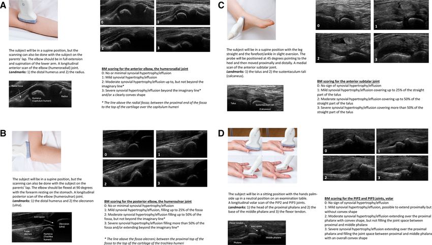
Radiocarpal The subject will be in a sitting position, the hands palm-side down in a neutral position on an examination
and midcarpal table and resting the elbow on the table. A longitudinal dorsal scan of the radiocarpal and midcarpal joints at
the sagittal midline of the wrist, including the distal radius, the lunate and the capitate bone.
Landmarks: (1) The distal end of diaphysis and epiphysial cartilage of radius and (2) The dorsal recess of the
radiocarpal and midcarpal joints and over them (3) A compartment of the extensor tendons according to the
area imaged
13
MCP2–3, The subject will be in a sitting position with the hands palm-side down in a neutral position on an examination
dorsal table. A longitudinal dorsal scan of the MCP2 and MCP3 joints.
Landmarks: (1) The head of metacarpal bone (2/3 of the image) and (2) The base of proximal phalanx (1/3 of
the image)
PIP2–3, dorsal The subject will be in a sitting position with the hands palm-side down in a neutral position on an examination
table. A longitudinal dorsal scan of the PIP2 and PIP3 joints.
Landmarks: (1) The head of proximal phalanx (2/3 of the image) and (2) The base of middle phalanx (1/3 of
the image)
PIP2–3, volar The subject will be in a sitting position with the hands palm-side up in a neutral position on an examination
table.
A longitudinal volar scan of the PIP2 and PIP3 joints.
Landmarks: (1) The head of the proximal phalanx, (2) The base of the middle phalanx and (3) The flexor
tendon
Hip The subject will be in a supine position with the hip in a neutral position, slightly externally rotated. A
longitudinal anterior scan parallel to the femoral neck of the hip joint.
Landmarks: (1) The femoral head and (2) The femoral neck.
Knee, The subject will be in a supine position. The knee should be flexed at 30 degrees, and images taken after the
suprapatellar subject completes flexion and extension three times. A longitudinal scan of the suprapatellar joint space. For
recess the youngest subjects the patella should fill 1/3 of the image to compensate for the relatively shorter femur (to
not underestimate the scoring).
Landmarks: (1) The proximal third of the patella and (2) A clearly visualised quadriceps tendon
17
Knee, lateral The subject will be in a supine position. The knee should be flexed at 30 degrees. For the lateral parapatellar
parapatellar recess the image will be obtained with the probe in transverse position over the mid-patella with both the
recess patella and femur in view.
Landmarks: (1) The superior edge of the patella and (2) The femoral condyle
17
Tibiotalar The subject will be in a supine position with the knee at 90 degrees flexion and the foot sole-side down. A
longitudinal scan of the tibiotalar joint.
Landmarks: (1) The distal end of the tibia and (2) The talus
Talonavicular The subject will be in a supine position with the knee at 90 degrees flexion and the foot sole-side down. A
longitudinal scan of the talonavicular joint.
Landmarks: (1) The talus and (2) The navicular bone
Continued
Sande NK, et al. RMD Open 2021;7:e001581. doi:10.1136/rmdopen-2021-001581 5RMD Open
RMD Open: first published as 10.1136/rmdopen-2021-001581 on 21 April 2021. Downloaded from http://rmdopen.bmj.com/ on August 14, 2021 by guest. Protected by copyright.
Table 1 Continued
Regions Image acquisition protocol
Anterior The subject will be in a supine position with the forefoot/ankle in slight eversion. The probe will be positioned
subtalar at 45 degrees pointing to the heel and then moved proximally and distally. A medial scan of the anterior
subtalar joint.
Landmarks: (1) The talus and (2) The sustentaculum tali (calcaneus)
Posterior The subject will be in a supine position with the forefoot/ankle in slight inversion. The probe will be positioned
subtalar along the sinus tarsi perpendicular to the sole, and then moved posteriorly. If no distension is seen, the image
will be taken visualising the joint with the peroneus tendons. A lateral scan of the posterior subtalar joint.
Landmarks: (1) The talus and (2) The calcaneus
MTP2–3 The subject will be in a supine position with the knee at 90 degrees flexion and the foot sole-side down. A
dorsal longitudinal dorsal scan of the MTP2 and MTP3 joints.
Landmarks: (1) The head of metatarsal bone (2/3 or the image) and (2) The base of the proximal phalanx (1/3
of the image)
BM, B-mode; MCP, metacarpophalangeal; MTP, metatarsophalangeal; PD, power Doppler; PIP, proximal interphalangeal.
and PD activity ultrasound sum scores were 0.94 (0.72 was first scored with the scoring system presented by
to 0.99) and 0.91 (0.74 to 0.97), respectively. Weighted Ting et al.17 However, in the smallest children we discov-
kappa (SD) was 0.51 (0.09) for BM synovitis and 0.60 ered that this system could underestimate the degree of
(0.12) for PD activity. pathology due to their relatively shorter femur. In the
image acquisition protocol, we therefore added that for
the youngest children the patella should fill a third of
DISCUSSION
the image on the ultrasound screen when scoring for
For the first time we present an ultrasonographic image
acquisition protocol and a semiquantitative joint-specific pathology.
scoring system for synovitis with an age-divided reference The variable sonoanatomy in the growing child may
atlas for BM synovitis and a reference atlas for PD activity lead to pitfalls even when performed by experienced
for frequently affected joints in patients with JIA. The rheumatologists, and there is a lack of published age-
present study demonstrated overall moderate to excel- specific and joint- specific imaging data in the litera-
lent reliability. ture. The images used in this study were selected from
The image acquisition protocol ensured a standardised our database consisting of approximately 5000 ultraso-
ultrasound examination in the practical sessions and live nographic images. These images were collected during
scoring exercises. Some of the views were adapted and routine ultrasound examination as part of daily clinical
adjusted from the OMERACT ultrasound paediatric practice from patients with JIA attending our inpatient
group.13 Their scanning approaches showed to be appli- and outpatient clinics. We therefore believe that our
cable in children regardless of age. In our study, the image selection of images is representative of the patients with
acquisition protocol was easily learnt and highly feasible, JIA seen in clinical practice. The comprehensive ultraso-
probably because of the thorough descriptions and illus- nographic atlas consisting of 224 BM images of normal
trative ultrasound images with important anatomical and inflamed joints divided in four age groups and 51
landmarks for each joint. As the pattern of joint involve- images with semiquantitative scores for the presence of
ment seems to be of prognostic importance,22 23 a stan- PD activity, enables the sonographer to recognise age-
dardised and systematic ultrasound examination might specific and joint-specific ultrasonographic findings of
be able to improve assessment of disease activity and indi- synovitis and to score ultrasound images according to the
vidualise treatment in patients with JIA. best possible match in the reference atlas. The combi-
The novel scoring system proposes joint-specific scores nation of a defined joint- specific scoring system with
for frequently affected joints in JIA. A single standard reference atlas for assessing synovitis may introduce an
paediatric scoring system may have some limitations in intuitive and feasible implementation of ultrasound in
that it does not clearly apply to all joints.16 For instance, patients with JIA.
grade 2 and grade 3 BM synovitis in the PedSynS is partly The still image scoring exercise demonstrated
defined by whether or not the joint recess is extending moderate to excellent reliability for all joints. At joint
over the bone diaphysis.16 The score may be difficult to level, the scoring of BM synovitis in our study showed the
use for joints adjacent to short bones without diaphysial highest ICC and kappa values for the posterior elbow,
bone structures. A joint-specific scoring system for the knee, tibiotalar, anterior subtalar and the MTP joints
knee in patients with JIA was recently developed and (table 3). The good reliability for the scoring of the knee
demonstrated good reliability.17 This provided the basis was in accordance with data reported by Ting et al.17
for our further development of joint-specific scores for The subtalar joint is one of the most difficult joints
frequently affected joints in JIA. The suprapatellar recess to assess clinically in the ankle, but this joint is often
6 Sande NK, et al. RMD Open 2021;7:e001581. doi:10.1136/rmdopen-2021-001581Paediatric rheumatology
RMD Open: first published as 10.1136/rmdopen-2021-001581 on 21 April 2021. Downloaded from http://rmdopen.bmj.com/ on August 14, 2021 by guest. Protected by copyright.
Table 2 Ultrasonographic semiquantitative joint-specific scoring system for BM in juvenile idiopathic arthritis
Joint Semiquantitative scoring system, BM
Anterior elbow 0: No or minimal synovial hypertrophy/effusion
1: Mild synovial hypertrophy/effusion
2: Moderate synovial hypertrophy/effusion up to, but not beyond the imaginary line*
3: Severe synovial hypertrophy/effusion beyond the imaginary line* and a clearly convex shape
* The line above the radial fossa; between the proximal end of the fossa to the top of the cartilage over the
capitulum humeri
Posterior elbow 0: No or minimal synovial hypertrophy/effusion
1: Mild synovial hypertrophy/effusion, filling up to 25% of the fossa
2: Moderate synovial hypertrophy/effusion filling up to 50% of the fossa, but not beyond the imaginary line*
3: Severe synovial hypertrophy/effusion filling more than 50% of the fossa and/or extending beyond the
imaginary line*
* The line above the fossa olecrani; between the proximal end of the fossa to the top of the cartilage of the
trochlea humeri
Radiocarpal 0: No sign of synovial hypertrophy/effusion
and midcarpal 1: Mild synovial hypertrophy/effusion
2: Moderate synovial hypertrophy/effusion up to, but not beyond the imaginary line*
3: Severe synovial hypertrophy/effusion with a convex shape extending beyond the imaginary line* and can
push up the extensor tendons
* The line between the top of the cartilage of the distal end of the radius to the top of the cartilage of the
capitate (just proximal to the CMC joint)
MCP2–3, dorsal 0: No sign of synovial hypertrophy/effusion
1: Mild synovial hypertrophy/effusion but not beyond the imaginary line*
2: Moderate synovial hypertrophy/effusion extending beyond the imaginary line*, but without overall convex
shape
3: Severe synovial hypertrophy/effusion extending beyond the imaginary line* with a clearly convex shape
* The line between the top of the cartilage of the distal end of the metacarpal to the top of the cartilage of
the proximal end of the phalanx
PIP2–3, dorsal 0: No sign of synovial hypertrophy/effusion
1: Mild synovial hypertrophy/effusion but not beyond the imaginary line*
2: Moderate synovial hypertrophy/effusion extending beyond the imaginary line*, but without overall convex
shape
3: Severe synovial hypertrophy/effusion extending beyond the imaginary line* with a clearly convex shape
*The line between the top of the cartilage of the distal end of the proximal phalanx to the top of the cartilage
of the proximal end of the middle phalanx
PIP2–3, volar 0: No sign of synovial hypertrophy/effusion
1: Mild synovial hypertrophy/effusion, possible to extend proximally but without convex shape
2: Moderate synovial hypertrophy/effusion extending over the proximal phalanx with convex shape, but not
filling the joint space between proximal and middle phalanx
3: Severe synovial hypertrophy/effusion extending over the proximal phalanx and filling the joint space
between proximal and middle phalanx with an overall convex shape
Hip 0: No sign of synovial hypertrophy/effusion
1: Mild synovial hypertrophy/effusion, but just a ‘slit’ of fluid between the two layers of the capsule
2: Moderate synovial hypertrophy/effusion leading to a straight line/minimal convex shape of the capsule
3: Severe synovial hypertrophy/effusion with a clearly convex shape, the effusion can also extend proximally
over the femoral head
Knee, 0: ‘Slit’ of fluid/synovium without elevation of the prepatellar fat pad but with only minimal extension beyond
suprapatellar the prepatellar fat pad
recess 1: Mild synovial hypertrophy/effusion with elevation of the prepatellar fat pad and extension proximally
50%
of the visualised portion of the quadriceps tendon
3: Significant distension of the suprapatellar recess throughout the image, and with the most proximal
portion of the synovial recess being >50% of the maximum distension of the recess
17
Continued
Sande NK, et al. RMD Open 2021;7:e001581. doi:10.1136/rmdopen-2021-001581 7RMD Open
RMD Open: first published as 10.1136/rmdopen-2021-001581 on 21 April 2021. Downloaded from http://rmdopen.bmj.com/ on August 14, 2021 by guest. Protected by copyright.
Table 2 Continued
Joint Semiquantitative scoring system, BM
Knee, lateral 0: Empty parapatellar recess but a minimal bulge of synovial hypertrophy/effusion may be found extending
parapatellar to the patellofemoral joint line
recess 1: Synovial hypertrophy/effusion filling 2/3 of the full area of the parapatellar recess and clearly pushing
up the retinaculum
17
Tibiotalar 0: No sign of synovial hypertrophy/effusion in the tibiotalar joint, but possible to have a minimal amount of
fluid in the concave neck of the talus
1: Mild synovial hypertrophy/effusion filling the gap between the tibia and the talus and in the concave neck
of the talus, but not continuously over the talus
2: Moderate synovial hypertrophy/effusion filling 50% of the area between the tibia, the talus and the
imaginary line* or beyond the imaginary line*
* The line between the top of the cartilage of the distal end of the tibia and the top of the cartilage of the
talar head
Talonavicular 0: No sign of synovial hypertrophy/effusion
1: Mild synovial hypertrophy/effusion but not beyond the imaginary line*
2: Moderate synovial hypertrophy/effusion extending beyond the imaginary line* and proximal with a
concave or straight shape
3: Severe synovial hypertrophy/effusion extending beyond the imaginary line* and over the talus with a
convex shape clearly pushing up the joint capsule
* The line between the top of the cartilage of the head of the talus to the top of the cartilage of the navicular
bone
Anterior 0: No sign of synovial hypertrophy/effusion
subtalar 1: Mild synovial hypertrophy/effusion covering up to 25% of the straight part of the talus
2: Moderate synovial hypertrophy/effusion covering up to 50% of the straight part of the talus
3: Severe synovial hypertrophy/effusion covering more than 50% of the straight part of the talus
Posterior 0: No sign of synovial hypertrophy/effusion
subtalar 1: Mild synovial hypertrophy/effusion filling the gap between the talus and the calcaneus
2: Moderate synovial hypertrophy/effusion extending beyond the talus and the calcaneus but not with a
convex shape
3: Severe synovial hypertrophy/effusion extending beyond the talus and the calcaneus with a convex shape
MTP2–3 dorsal 0: No sign of synovial hypertrophy/effusion
1: Mild synovial hypertrophy/effusion but not beyond the imaginary line*
2: Moderate synovial hypertrophy/effusion extending beyond the imaginary line*, but without overall convex
shape
3: Severe synovial hypertrophy/effusion extending beyond the imaginary line* with a clearly convex shape
* The line between the top of the cartilage of the distal end of the metatarsal to the top of the cartilage of the
proximal end of the phalanx
BM, B-mode; CMC, carpometacarpal; MCP, metacarpophalangeal; MTP, metatarsophalangeal; PIP, proximal interphalangeal.
involved in patients with JIA.6 24 25 To our knowledge, process of defining age-related vascularisation of joints
image acquisitions and scoring systems for the paedi- in healthy children,13 26 and developed preliminary defi-
atric anterior and posterior subtalar joints have not been nitions of synovitis in children which define that Doppler
published before. We therefore included them in our signals must be detected within synovial hypertrophy to
scanning protocol and scoring system, which is also in be considered as a sign of synovitis.14 Our reference atlas
accordance with suggestions by others.25 In our study, the
for scoring of PD activity might improve the feasibility
joint-specific scoring with illustrative images in the refer-
of this ultrasonographic feature, but further studies are
ence atlas of the anterior and posterior subtalar joints
showed good to excellent reliability. needed regarding the detection of abnormal vascularisa-
Interpretation of PD signals in children is complicated tion in the paediatric joint.
due to a variable degree of physiological blood flow within Inter-reader reliability on live scoring has only been
the joint that can easily be misinterpreted as inflamma- reported in few JIA studies. The live scoring of 10 patients
tion. The OMERACT ultrasound group has started the with JIA in this study demonstrated good reliability, which
8 Sande NK, et al. RMD Open 2021;7:e001581. doi:10.1136/rmdopen-2021-001581Paediatric rheumatology
RMD Open: first published as 10.1136/rmdopen-2021-001581 on 21 April 2021. Downloaded from http://rmdopen.bmj.com/ on August 14, 2021 by guest. Protected by copyright.
Figure 1 (A–D) Description of ultrasound examination and scoring of B-mode (BM) synovitis from the ultrasonographic
BM reference atlas in juvenile idiopathic arthritis (JIA). (A) The elbow joint, longitudinal anterior scan (2–4 years). (B) The
elbow joint, longitudinal posterior scan (5–8 years). (C) The anterior subtalar joint, medial scan (9–12 years). (D) The proximal
interphalangeal (PIP)2 and PIP3 joints, longitudinal volar scan (13–18 years).
is comparable to the results presented by Magni-Manzoni we experienced that the COVID-19 situation made it
et al.19 impossible to conduct a large scoring exercise including
Limitations of this study are the low number of patients more patients and readers. However, we wished to test the
included in the live scoring exercise, that only three scoring system in a live setting and made adaptations to
sonographers participated and that they only scanned our project within these limitations. Previous studies have
the patients once. At the time of our live scoring exercise, shown that inclusion of 10 patients may yield sufficient
Figure 2 Description of ultrasound examination and scoring of power Doppler (PD) activity for the wrist; radiocarpal and
midcarpal joints (longitudinal dorsal scan) from the ultrasonographic PD reference atlas in juvenile idiopathic arthritis (JIA).
Sande NK, et al. RMD Open 2021;7:e001581. doi:10.1136/rmdopen-2021-001581 9RMD Open
RMD Open: first published as 10.1136/rmdopen-2021-001581 on 21 April 2021. Downloaded from http://rmdopen.bmj.com/ on August 14, 2021 by guest. Protected by copyright.
Table 3 Intra-reader and inter-reader reliability for B-mode (BM) synovitis scoring on still images in juvenile idiopathic arthritis
(JIA)
Intra-reader reliability Inter-reader reliability
Cohen’s weighted Light’s weighted
smICC kappa kappa
Regions No. images Mean (SD) Mean (SD) avmICC (95% CI) Mean (SD)
Anterior elbow 25 0.90 (0.03) 0.81 (0.06) 0.96 (0.92 to 0.98) 0.72 (0.09)
Posterior elbow 27 0.93 (0.04) 0.88 (0.06) 0.96 (0.93 to 0.98) 0.76 (0.05)
Radiocarpal 28 0.79 (0.10) 0.67 (0.12) 0.93 (0.87 to 0.96) 0.61 (0.10)
Midcarpal 28 0.89 (0.05) 0.79 (0.08) 0.96 (0.93 to 0.98) 0.73 (0.11)
MCP2–3, dorsal 20 0.75 (0.07) 0.63 (0.07) 0.89 (0.79 to 0.95) 0.50 (0.10)
PIP2–3, dorsal 20 0.87 (0.05) 0.77 (0.07) 0.94 (0.88 to 0.98) 0.64 (0.11)
PIP2–3, volar 30 0.85 (0.07) 0.72 (0.10) 0.95 (0.92 to 0.98) 0.72 (0.10)
Hip 26 0.92 (0.05) 0.84 (0.08) 0.96 (0.93 to 0.98) 0.75 (0.08)
Knee, suprapatellar recess 24 0.95 (0.01) 0.91 (0.02) 0.98 (0.96 to 0.99) 0.86 (0.05)
Knee, lateral parapatellar recess 27 0.88 (0.09) 0.81 (0.13) 0.95 (0.91 to 0.98) 0.70 (0.04)
Tibiotalar 26 0.94 (0.04) 0.90 (0.07) 0.98 (0.96 to 0.99) 0.83 (0.05)
Talonavicular 22 0.87 (0.12) 0.83 (0.13) 0.95 (0.90 to 0.98) 0.69 (0.09)
Anterior subtalar 27 0.95 (0.04) 0.91 (0.07) 0.99 (0.97 to 0.99) 0.91 (0.04)
Posterior subtalar 20 0.86 (0.09) 0.74 (0.08) 0.95 (0.90 to 0.98) 0.75 (0.10)
MTP2–3, dorsal 20 0.94 (0.04) 0.89 (0.08) 0.96 (0.93 to 0.99) 0.79 (0.11)
avmICC, average measure ICC; ; ICC, intraclass correlation coefficient; MCP, metacarpophalangeal; MTP, metatarsophalangeal; PIP, proximal
interphalangeal; smICC, single measure ICC.
power for reliability testing.15 27 We found it feasible for to be considered as a sign of synovitis, which will be
three dedicated rheumatologists to do a live scoring exer- clearly identified first by using the scoring system and
cise implemented in our daily clinical practice by consec- age-divided atlas for BM synovitis as reference. In addi-
utively including 10 patients with JIA disease flare in need tion, we will continuously include images in our database
of hospital admission. In this setting, the inter-reader reli- and aspire to include the best possible reference images
ability was good, suggesting that the scoring system with for all grades in the reference atlas.
atlas is a reliable tool. Examination of potential variability The strengths of the study are the unique collection
in the reliability of the scoring system with respect to age of ultrasonographic images with different degrees of
or disease activity was beyond the scope of this study and pathology in four age ranges and the approach to define
should be addressed in future research. individual scores for a substantial number of joints in
Another limitation is the lack of comparison with patients with JIA.
healthy subjects. The main target of this study was to In conclusion, this study presents an ultrasonographic
develop an ultrasonographic scoring system with refer- image acquisition protocol and a semiquantitative joint-
ence atlas for patients with JIA and to test the reliability of specific scoring system for synovitis with reference atlas
the system. The study was not designed to compare ultra- in patients with JIA. The study demonstrated moderate
sonographic findings in healthy children with patients to excellent reliability when used in assessments on still
with JIA. However, results from available musculoskel- images as well as on patients. We expect that the system
etal ultrasound studies highlighting findings in healthy can be a valuable tool for clinicians and future research.
children were taken into account in the process.11–13 26 A Future studies are needed for further validation of the
comparison of ultrasonographic findings in healthy chil- scoring system with atlas, such as association to clinical
dren with patients with JIA according to the presented measures of disease activity and the system’s sensitivity to
scoring system could be a future study of interest. change.
Other limitations are that we did not have images of
all grades in the atlas, and that the reference atlas for Author affiliations
1
Department of Rheumatology, Oslo University Hospital, Oslo, Norway
scoring of PD activity was not age- divided. However, 2
Department of Rheumatology, Diakonhjemmet Hospital, Oslo, Norway
the main goal for the PD reference atlas is to illustrate 3
Faculty of Medicine, University of Oslo, Oslo, Norway
different grades of PD signals for each joint and not the 4
Division of Pediatric Dermatology and Rheumatology, Children's Hospital of
age variability. Furthermore, in accordance with the defi- Eastern Ontario and University of Ottawa, Ottawa, Ontario, Canada
nitions developed by the OMERACT ultrasound group,14
PD signals must be detected within synovial hypertrophy
10 Sande NK, et al. RMD Open 2021;7:e001581. doi:10.1136/rmdopen-2021-001581Paediatric rheumatology
RMD Open: first published as 10.1136/rmdopen-2021-001581 on 21 April 2021. Downloaded from http://rmdopen.bmj.com/ on August 14, 2021 by guest. Protected by copyright.
Acknowledgements The authors thank David Swanson, Researcher (statistician), management of juvenile idiopathic arthritis in clinical practice. Ann
PhD, for statistical advice, and Øystein Horgmo, senior photographer, for taking Rheum Dis 2015;74:1946–57.
photographs of probe positioning to the ultrasonographic atlas. The authors also 8 Lanni S, Wood M, Ravelli A, et al. Towards a role of ultrasound
thank all the patients and their families who contributed to this study. in children with juvenile idiopathic arthritis. Rheumatology
2013;52:413–20.
Contributors NKS designed the study, made substantial contributions to 9 Windschall D, Malattia C. Ultrasound imaging in paediatric
acquisition, analysis and interpretation of data and writing the manuscript. PB rheumatology. Best Pract Res Clin Rheumatol 2020;34:101570.
and VL designed the study, and made substantial contributions to acquisition, 10 Magni-Manzoni S, Malattia C, Lanni S, et al. Advances and
interpretation of data and writing the manuscript. JR participated in the study challenges in imaging in juvenile idiopathic arthritis. Nat Rev
design, made substantial contributions to acquisition, interpretation of data and Rheumatol 2012;8:329–36.
writing the manuscript. A-BA, HBH and BF participated in the study design and 11 Roth J, Jousse-Joulin S, Magni-Manzoni S, et al. Definitions for the
made substantial contributions to data interpretation and writing the manuscript. sonographic features of joints in healthy children. Arthritis Care Res
2015;67:136–42.
Funding The authors have not declared a specific grant for this research from any 12 Windschall D, Trauzeddel R, Haller M, et al. Pediatric
funding agency in the public, commercial or not-for-profit sectors. musculoskeletal ultrasound: age- and sex-related normal B-mode
Competing interests HBH reports personal fees from Lilly, personal fees from findings of the knee. Rheumatol Int 2016;36:1569–77.
Novartis, personal fees from AbbVie, personal fees from Roche, outside the 13 Collado P, Vojinovic J, Nieto JC, et al. Toward standardized
submitted work. musculoskeletal ultrasound in pediatric rheumatology: normal age-
related ultrasound findings. Arthritis Care Res 2016;68:348–56.
Patient consent for publication Not required. 14 Roth J, Ravagnani V, Backhaus M, et al. Preliminary definitions for
Ethics approval The study was done in accordance with the Declaration of the sonographic features of synovitis in children. Arthritis Care Res
Helsinki and was approved by the Norwegian Regional Committee for Medical 2017;69:1217–23.
15 Hammer HB, Bolton-King P, Bakkeheim V, et al. Examination of intra
and Health Research Ethics (REK 2018/805) and the Data Protection Officer at
and interrater reliability with a new ultrasonographic reference atlas
Oslo University Hospital, Norway (18/11742 and 18/12493). Image storage was
for scoring of synovitis in patients with rheumatoid arthritis. Ann
approved by the Data Protection Officer at OUH (18/11742). Rheum Dis 2011;70:1995–8.
Provenance and peer review Not commissioned; externally peer reviewed. 16 Collado P, Naredo E, Calvo C, et al. Reduced joint assessment vs
comprehensive assessment for ultrasound detection of synovitis in
Data availability statement Data are available upon reasonable request to the juvenile idiopathic arthritis. Rheumatology 2013;52:1477–84.
corresponding author. 17 Ting TV, Vega-Fernandez P, Oberle EJ, et al. Novel ultrasound image
Open access This is an open access article distributed in accordance with the acquisition protocol and scoring system for the pediatric knee.
Creative Commons Attribution Non Commercial (CC BY-NC 4.0) license, which Arthritis Care Res 2019;71:977–85.
permits others to distribute, remix, adapt, build upon this work non-commercially, 18 Backhaus M, Burmester GR, Gerber T, et al. Guidelines for
and license their derivative works on different terms, provided the original work is musculoskeletal ultrasound in rheumatology. Ann Rheum Dis
properly cited, appropriate credit is given, any changes made indicated, and the 2001;60:641–9.
19 Magni-Manzoni S, Epis O, Ravelli A, et al. Comparison of clinical
use is non-commercial. See: http://c reativecommons.org/licenses/by-nc/4.0/.
versus ultrasound-determined synovitis in juvenile idiopathic
arthritis. Arthritis Rheum 2009;61:1497–504.
ORCID iDs
20 Petty RE, Southwood TR, Manners P, et al. International League
Nina Krafft Sande http://orcid.org/0000-0003-3461-9525 of associations for rheumatology classification of juvenile
Pernille Bøyesen http://orcid.org/0000-0002-9751-5144 idiopathic arthritis: second revision, Edmonton, 2001. J Rheumatol
Hilde Berner Hammer http://o rcid.org/0000-0001-7317-8991 2004;31:390–2.
21 Hallgren KA. Computing inter-rater reliability for observational
data: an overview and tutorial. Tutor Quant Methods Psychol
2012;8:23–34.
REFERENCES 22 Eng SWM, Aeschlimann FA, van Veenendaal M, et al. Patterns of
1 Ravelli A, Martini A. Juvenile idiopathic arthritis. Lancet joint involvement in juvenile idiopathic arthritis and prediction of
2007;369:767–78. disease course: a prospective study with multilayer non-negative
2 Ringold S, Angeles-Han ST, Beukelman T, et al. 2019 American matrix factorization. PLoS Med 2019;16:e1002750.
College of Rheumatology/Arthritis Foundation guideline for the 23 Flatø B, Lien G, Smerdel A, et al. Prognostic factors in juvenile
treatment of juvenile idiopathic arthritis: therapeutic approaches for rheumatoid arthritis: a case-control study revealing early predictors
non-systemic polyarthritis, sacroiliitis, and Enthesitis. Arthritis Care and outcome after 14.9 years. J Rheumatol 2003;30:386–93.
Res 2019;71:717–34. 24 Hendry GJ, Gardner-Medwin J, Steultjens MPM, et al. Frequent
3 Steigerwald KA, Ilowite NT. Novel treatment options for juvenile discordance between clinical and musculoskeletal ultrasound
idiopathic arthritis. Expert Rev Clin Pharmacol 2015;8:559–73. examinations of foot disease in juvenile idiopathic arthritis. Arthritis
4 Shoop-Worrall SJW, Kearsley-Fleet L, Thomson W, et al. How Care Res 2012;64:441–7.
common is remission in juvenile idiopathic arthritis: a systematic 25 Lanni S, Bovis F, Ravelli A, et al. Delineating the application of
review. Semin Arthritis Rheum 2017;47:331–7. ultrasound in detecting synovial abnormalities of the Subtalar joint in
5 Rooney ME, McAllister C, Burns JFT. Ankle disease in juvenile juvenile idiopathic arthritis. Arthritis Care Res 2016;68:1346–53.
idiopathic arthritis: ultrasound findings in clinically swollen Ankles. J 26 Windschall D, Collado P, Vojinovic J, et al. Age-Related
Rheumatol 2009;36:1725–9. vascularization and ossification of joints in children: an international
6 Pascoli L, Wright S, McAllister C, et al. Prospective evaluation pilot study to test multiobserver ultrasound reliability. Arthritis Care
of clinical and ultrasound findings in ankle disease in juvenile Res 2020;72:498–506.
idiopathic arthritis: importance of ankle ultrasound. J Rheumatol 27 Haavardsholm EA, Ostergaard M, Ejbjerg BJ, et al. Reliability and
2010;37:2409–14. sensitivity to change of the OMERACT rheumatoid arthritis magnetic
7 Colebatch-Bourn AN, Edwards CJ, Collado P, et al. EULAR-PReS resonance imaging score in a multireader, longitudinal setting.
points to consider for the use of imaging in the diagnosis and Arthritis Rheum 2005;52:3860–7.
Sande NK, et al. RMD Open 2021;7:e001581. doi:10.1136/rmdopen-2021-001581 11You can also read