Imaging of endometrial osseous metaplasia-an uncommon but treatable cause of infertility
←
→
Page content transcription
If your browser does not render page correctly, please read the page content below
Wani et al. Middle East Fertility Society Journal         (2020) 25:35
https://doi.org/10.1186/s43043-020-00045-0
                                                                                                                                      Middle East Fertility
                                                                                                                                          Society Journal

 RESEARCH                                                                                                                                           Open Access

Imaging of endometrial osseous
metaplasia—an uncommon but treatable
cause of infertility
Abdul Haseeb Wani1* , Arshed Hussain Parry2, Imza Feroz3, Majid Jehangir1 and Masarat Rashid4

  Abstract
  Background: Endometrial osseous metaplasia (EOM) is an uncommon condition characterised by metaplastic
  transformation of endometrial tissue into osteoblasts (mature or immature bone in the endometrium).
  Etiopathogenesis of EOM is explained by multiple putative mechanisms like dystrophic calcification, metaplastic
  ossification, retained foetal bones after abortions and genito-urinary tuberculosis. EOM has varied clinical
  presentation ranging from patient being asymptomatic to secondary infertility. Although hysteroscopy is the gold
  standard for its diagnosis and treatment, non-invasive imaging comprising chiefly of ultrasonography (USG) is
  increasingly becoming the mainstay of diagnosis. We aim to present the imaging findings in EOM to acquaint
  radiologists and gynaecologists with this condition to avert misdiagnosis of this uncommon yet treatable cause of
  infertility.
  Results: Mean age of patients was 31.4 ± 5.4 (S.D) years. USG revealed linear or tubular densely echogenic
  endometrium with posterior acoustic shadowing in all the 14 patients. MRI in 3 patients revealed diffuse or patchy
  areas of T1W and T2W hypointense signal intensity with unilateral (n = 2) and bilateral (n = 1) ovarian cysts. One
  patient who underwent CT scan revealed dense endometrial calcification. Histopathologic examination (HPE) revealed
  lamellar (n = 6) or trabecular (n = 4) bone within endometrium (EOM) and inflammatory cells with calcification in four
  patients (calcific endometritis). Twelve patients conceived after dilatation and curettage within 15 months.
  Conclusion: Familiarity with the imaging appearances of EOM is indispensable to clinch this diagnosis and avert
  misdiagnosis of this rare but potentially treatable cause of infertility. USG is usually sufficient for diagnosis. MRI and CT
  are only supplementary tools in difficult clinical scenarios.
  Keywords: Endometrium, Osseous metaplasia, Ultrasonography, Endometrial calcification, Dilatation and curettage

Background                                                                             foetal bones after spontaneous/missed/incomplete or
Etymologically endometrial osseous metaplasia (EOM)                                    therapeutic abortion, chronic inflammation (genito-urin-
meaning metaplastic transformation of endometrial tis-                                 ary tuberculosis) and extended post-abortion estrogenic
sue into osteoblasts is an uncommon condition charac-                                  treatment being the most plausible ones [2–4]. EOM has
terised by the presence of mature or immature bone in                                  varied clinical presentation ranging from patient being
the endometrium [1]. Various theories have been pro-                                   asymptomatic to secondary infertility [5–7]. Within this
pounded to explain its aetiology with dystrophic calcifi-                              wide spectrum, patients can also present with menstrual
cation, metaplastic ossification, heterotopia, retained                                irregularities (menorrhagia or oligomenorrhea) and re-
                                                                                       current abortions [8, 9]. Although hysteroscopy is the
* Correspondence: soberseeb@gmail.com                                                  gold standard for its diagnosis and treatment, non-
1
 Department of Radiodiagnosis, Government Medical College, Srinagar,                   invasive imaging comprising chiefly of ultrasound is in-
Jammu and Kashmir 190010, India
                                                                                       creasingly becoming the mainstay of diagnosis and is
Full list of author information is available at the end of the article

                                       © The Author(s). 2020 Open Access This article is licensed under a Creative Commons Attribution 4.0 International License,
                                       which permits use, sharing, adaptation, distribution and reproduction in any medium or format, as long as you give
                                       appropriate credit to the original author(s) and the source, provide a link to the Creative Commons licence, and indicate if
                                       changes were made. The images or other third party material in this article are included in the article's Creative Commons
                                       licence, unless indicated otherwise in a credit line to the material. If material is not included in the article's Creative Commons
                                       licence and your intended use is not permitted by statutory regulation or exceeds the permitted use, you will need to obtain
                                       permission directly from the copyright holder. To view a copy of this licence, visit http://creativecommons.org/licenses/by/4.0/.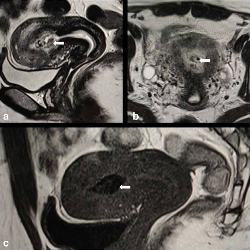
Wani et al. Middle East Fertility Society Journal (2020) 25:35 Page 2 of 10
gaining wider acceptance for initial diagnosis due to its infertility is defined as failure to conceive after a previ-
easy availability, non-invasive nature and lack of ionising ous conception. Informed consent was taken from all
radiation [10]. Given the rarity of this condition there is the patients with institutional review board approval ob-
sparse literature available on EOM particularly the imaging tained for the study. Relevant clinical history, detailed
features. Whatever miniscule literature is present that is in general physical examination, and local examination
the form of case reports. We aim to present the imaging were recorded. All the biochemical parameters were re-
findings in EOM to acquaint radiologists and gynaecolo- corded including liver function tests, renal function tests,
gists with this condition in order to avert misdiagnosis of fasting blood sugar, TSH, T3, T4, FSH, LH, FSH/LH ra-
this uncommon yet treatable cause of infertility. tio, CA-125 and prolactin levels.
All the patients were subjected to preliminary transab-
Methods dominal ultrasound (TAS) examination of pelvis. Four-
This was prospective descriptive single-centre study cov- teen patients were suspected to have EOM on the basis
ering a study period of 3 and half years from April 2016 of TAS findings and were included in this study. These
to December 2019. During the study period, a total of patients subsequently underwent transvaginal ultrasound
411 infertile women comprising of 198 (48.2%) patients (TVS) to confirm the findings of TAS. Patients with his-
of primary infertility and 213 (51.8%) of secondary infer- tory of intra-uterine contraceptive device insertion in
tility attended the out-patient clinic of our hospital. Pa- past were excluded from the study. TAS examination of
tients were considered infertile when they failed to the pelvis was performed using LOGIQ P5 (GE Health-
conceive after unprotected and regular sexual inter- care, Chicago, IL, USA) ultrasound machine with convex
course for a period of 1 year. Primary infertility refers to low frequency ultrasound probe 4C (1.6 to 4.6 MHz) in
inability to conceive even once whereas secondary all patients. TVS examination was performed using 3C8
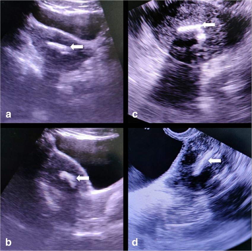
Fig. 1 Transabdominal axial (a) and sagittal (b) sonogram in a 27-year-old infertile patient revealing thick linear hyperechogencity (arrows) in
endometrial canal within the uterine corpus. Transvaginal axial (c) and longitudinal (d) ultrasound images confirming the findings of
transabdominal ultrasound. Subsequent HPE proved the diagnosis of EOM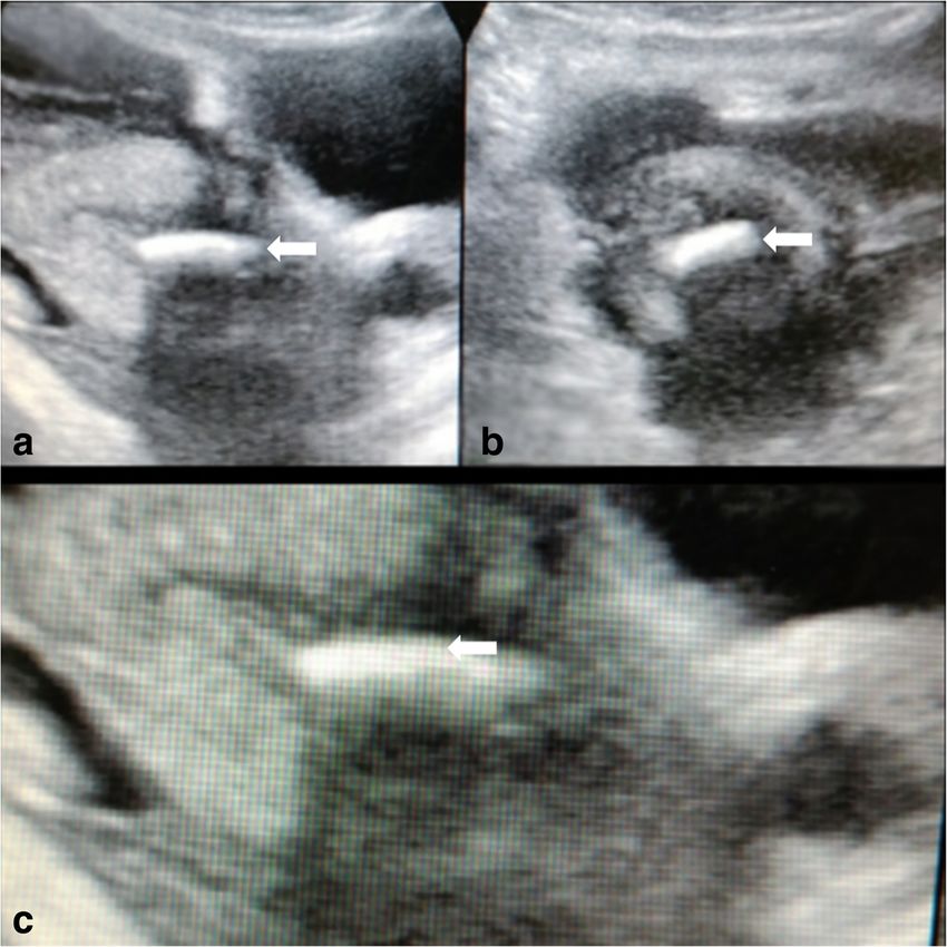
Wani et al. Middle East Fertility Society Journal    (2020) 25:35                                                                          Page 3 of 10

 Fig. 2 Sagittal (a), axial (b) and magnified sagittal (c) transabdominal ultrasound images in a 32-year-old patient with previous history of post-
 abortal instrumentation showing linear hyperechogencity with posterior acoustic shadowing in endometrial cavity of lower uterine
 corpus (arrows)

endo-cavitary probe (3 to 8MHz). Longitudinal and                             average period of 15 months (range 9 months to 24
transverse ultrasound images of uterus, endometrial lin-                      months) to monitor the response to endometrial curet-
ing and bilateral adnexae were obtained. Three patients                       tage in the form of future conception.
had an additional magnetic resonance imaging (MRI) of
pelvis and one patient additionally underwent computed                        Results
tomographic (CT) abdomen. MRI was performed in                                Mean age of patients was 31.4 ± 5.4 (S.D) years. Second-
these three patients owing to preliminary USG examin-                         ary infertility and primary infertility were the chief com-
ation showing complex ovarian cysts in addition to USG                        plaints in 12/14 (85.7%) and 2/14 (14.3%), respectively.
findings of EOM. One patient who underwent CT abdo-                           Menstrual irregularities were present in 5 (35.7%) pa-
men had pulmonary tuberculosis with recurrent abdom-                          tients. Pelvic pain was associated in 4 (28.6%) patients.
inal pain. All these 14 patients finally underwent                            Recurrent abdominal pain was seen in 1 patient. Ten
hysteroscopic examination with dilatation and curettage.                      (71.4%) patients gave a history of previous conception
The retrieved uterine tissue was put in 10% formalin,                         followed by spontaneous missed abortion at 9 to 17
and the vial was transported to histopathological labora-                     weeks of gestational age followed by dilatation and cur-
tory. The tissue was kept for overnight fixation in forma-                    ettage. However, in remaining four (28.6%) patients, his-
lin. Subsequently, the tissue was grossed and processed                       tory of previous abortion or dilatation and curettage was
routinely. The slides obtained were stained with haema-                       not forthcoming. Two patients in addition to being sec-
toxylin and eosin (H&E) and subjected to microscopic                          ondarily infertile presented with vaginal discharge. His-
examination under scanner, low power and high power                           tory of previously diagnosed pulmonary tuberculosis was
view. . Patients were subsequently followed for an                            forthcoming in one patient; however, the patient had left
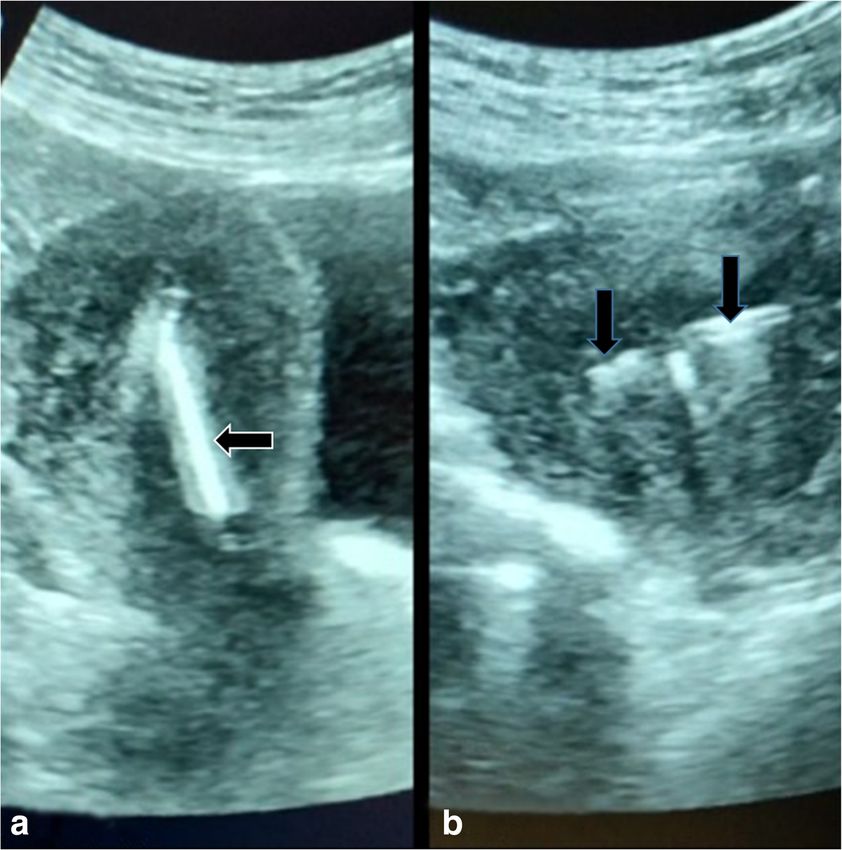
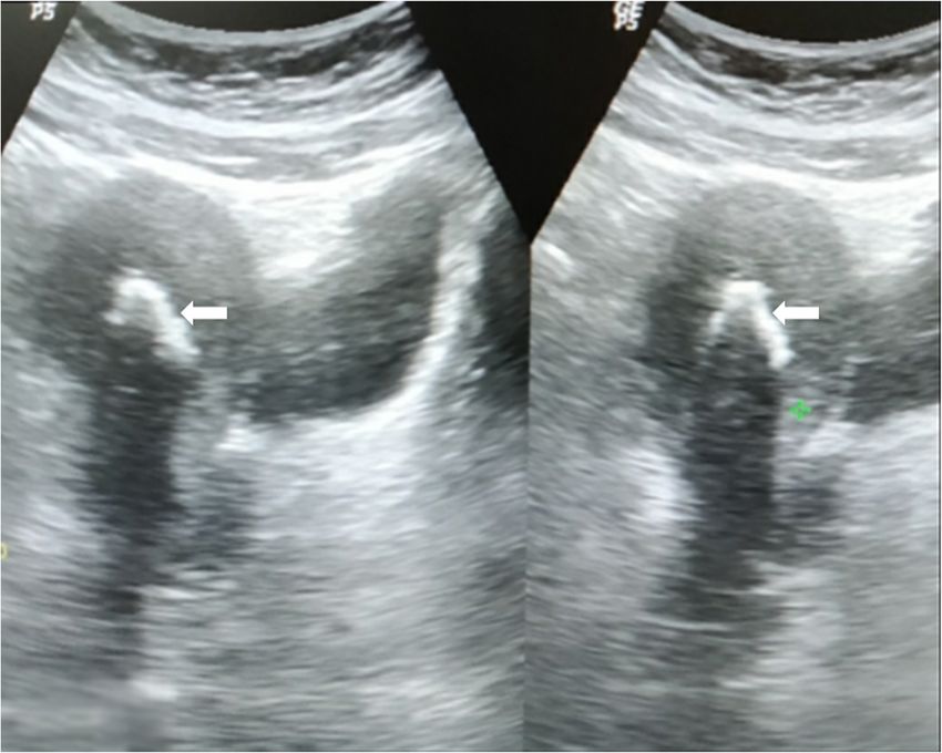
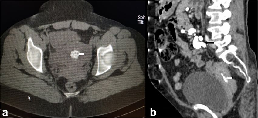
Wani et al. Middle East Fertility Society Journal    (2020) 25:35                                                                         Page 4 of 10

anti-tubercular treatment (ATT) midway. There was no                         no cervical fibroid or mass. Pertinent negative USG
history of intra-uterine contraception device (IUCD) in-                     findings were absence of hydrometra or hydrosalpinx.
sertion in any patient. The overall physical examination                     Eleven patients had normal bilateral ovaries with nor-
was unremarkable and bimanual pelvic examination was                         mal ovarian volume and folliculometry. Two patients
normal in 12 patients. Two patients with vaginal dis-                        had unilateral complex ovarian cysts. One patient
charge had mild cervical tenderness on per vaginal                           with vaginal discharge had bulky uterus with bilateral
examination. The routine laboratory investigations, hor-                     adnexal complex cystic lesions and mild free fluid in
monal and biochemical profile of all patients were within                    pouch of Douglas.
normal range as follows: T4 (SIU-standard international                         On the basis of history, clinical examination and ultra-
unit) 114.04 ± 6.36, T3 (SIU) 2.29 ± 0.24, TSH (SIU) 4.44                    sound findings, a presumptive diagnosis of endometrial
± 0. 73, fasting blood sugar levels 86 ± 9 mg/dl, serum bili-                osseous metaplasia (EOM) was made in all the 14 pa-
rubin levels 0.8 ± 0.2 mg/dl, albumin level 5.5 ± 0.4 g/L,                   tients. Three patients with additional finding of complex
serum creatinine level 0.75 ± 0.2 mg/dl, blood urea nitro-                   adnexal cysts on USG were further examined by non-
gen level 19.3 ± 4.2 mg/dl, FSH levels on day 3 of men-                      contrast MRI pelvis which revealed scattered areas of
strual cycle 11.23 ± 3.63 mIU/ml, LH levels 9.1 ± 1.2                        T2-weighted (T2W) hypointense signal intensity within
mIU/ml, LH/FSH ratio < 1, prolactin level 18 ± 4 ng/ml,                      the background hyperintense endometrium in all cases.
testosterone levels 30 ± 3 ng/dl and CA-125 levels 19 ± 3                    Corresponding T1W images showed hypointense signal
U/ml. The semen analysis of the husband was normal in                        intensity in all the three patients (Fig. 4). Two patients
each case. Four patients had undergone hysterosalpingog-                     showed unilateral ovarian cysts with hyperintense T1W
raphy (HSG) in previous 1 year and all four had normal                       and T2W signal intensity with non-suppression of signal
peritoneal spill of contrast suggesting tubal patency.                       on T1W fat-sat images suggesting a diagnosis of ovarian
  Longitudinal and transverse ultrasound of the pelvis                       haemorrhagic cysts. One patient had bilateral complex
revealed well defined, linear or tubular echogenic area                      adnexal cysts with T1W hypointense and T2W hyperin-
with dense posterior acoustic shadowing within the                           tense signal intensity with free fluid in pelvis. Subse-
endometrial canal of uterine corpus in nine cases                            quent polymerase chain reaction (PCR) of menstrual
(Figs. 1, 2, and 3) which extended to the upper part                         blood detected nucleic acid of acid-fast bacilli (AFB) in
of the cervix along the endocervical canal in the                            this patient thus confirming genital tuberculosis. CT of
remaining five cases. The anterior and posterior myo-                        abdomen, done in one patient for recurrent abdominal
metrial walls were normal in morphology with normal                          pain, revealed multiple calcified mesenteric and retro-
echotexture in all cases. No fibroid or adenomyosis                          peritoneal nodes in addition to dense endometrial ossifi-
was seen. Cervix was mildly bulky in one patient with                        cation with attenuation values similar to spongy bone

 Fig. 3 Sagittal split-screen transabdominal ultrasound images in a 28-year-old infertile patient revealing curvilinear echogenic area in endometrial
 canal with dense posterior acoustic shadowing (arrows). Subsequent HPE revealed calcific endometritis
Wani et al. Middle East Fertility Society Journal    (2020) 25:35                                                                           Page 5 of 10

(Fig. 5). With the preliminary sonographic diagnosis of                       Discussion
EOM, all the 14 patients underwent hysteroscopy with                          The putative hypothesis for the genesis of endometrial
dilatation and curettage. Hysteroscopy revealed patchy                        osseous metaplasia is an area of active research. Various
or diffuse ivory white ossified endometrium (Fig. 6) in                       theories have been propounded which may also explain
11 patients whereas 3 patients showed shaggy disco-                           the varied names by which it has been labelled since its
loured endometrium. Histopathological examination of                          early description in 1884 by Virchow who attributed the
the retrieved endometrial tissue revealed non-specific                        formation of ossified tissue within endometrium to de
lymphocytic infiltrate with interspersed lamellar bone in                     novo differentiation of fibroblasts into osteoblasts [11].
six (42.8%), trabecular bone in four (28.5%) patients with                    Nine out of 10 (90%) of HPE proven EOM had a history
scanty endometrial glands and stroma in various hormo-                        of abortion followed by dilatation and curettage in our
nal phases confirming the diagnosis of osseous metapla-                       study whereas 1 patient had no previous history of abor-
sia of endometrium (Figs. 7 and 8). Remaining four                            tion or uterine instrumentation. Post-abortion instru-
(28.5%) patients showed chronic inflammatory (granulo-                        mentation was the most common putative aetiology of
cytic) infiltrate with specks of calcification within the                     EOM in our study. Chronic inflammation of endomet-
endometrium suggesting diagnosis of calcific endometritis.                    rium due to post-abortal tissue likely stimulates release
These four patients were given a diagnosis of endometrial                     of various inflammatory mediators causing metaplastic
tuberculosis and were started on ATT. On follow-up, 11                        transformation of stromal cells into osteoblasts [12].
patients conceived in the subsequent 15-month follow-up                       Why this metaplastic transformation occurs in very few
period whereas 1 patient conceived after 20 months and 2                      patients is a matter of speculation and active research.
have not conceived till the end of study.                                     To account for this discrepancy, superoxide dismutase

 Fig. 4 Fast spin echo T2W sagittal (a) and axial (b) images of uterus in a 29-year-old infertile patient reveal patchy areas of hypointensity (arrows)
 within the endometrial canal in the uterine body. T1W sagittal (c) image reveals markedly hypointense and thick endometrial signal (arrows)
 compared to the myometrium. Subsequent HPE proved the diagnosis of EOM
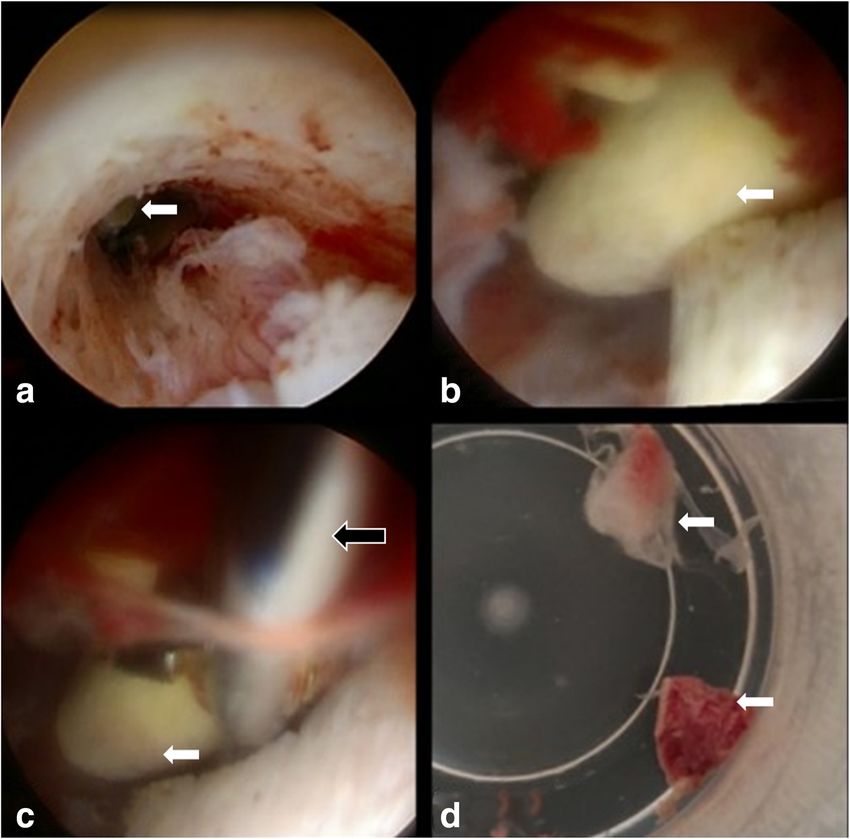
Wani et al. Middle East Fertility Society Journal    (2020) 25:35                                                                         Page 6 of 10

 Fig. 5 Axial (a) and sagittal (b) non-contrast CT images of pelvis and lower abdomen in a 30-year-old infertile patient with a history of old
 pulmonary tuberculosis and recurrent abdominal pain revealing dense endometrial calcification (white block arrows) with Hounsfield attenuation
 of 520 HU similar to adjacent pelvic bone and calcified retroperitoneal nodes (black block arrow). Subsequent HPE clinched the diagnosis of
 calcific endometritis

 Fig. 6 Serial hysteroscopic images of Fig. 1 patient showing distant (a) and close-up (b) view of ossified (ivory bone) endometrial tissue (white
 block arrows). c Hysteroscopy biopsy forceps (black block arrow) within endometrial cavity during retrieval of bony endometrial bits (d)
Wani et al. Middle East Fertility Society Journal   (2020) 25:35                                                          Page 7 of 10

                                                                        perhaps lead to chronic exposure of multipotential stro-
                                                                        mal cells to these inflammatory factors eventually lead-
                                                                        ing to transformation of these endometrial stromal cells
                                                                        into osteoblasts [13]. The mean duration between post-
                                                                        abortion instrumentation and detection of endometrial
                                                                        ossification in our study was 5.1 ± 1.8 years justifying
                                                                        the time required for metaplastic transformation. Six
                                                                        cases had first trimester abortion in our study and three
                                                                        had history of early second trimester abortion. It is also
                                                                        proposed that ossification of the foetal skeleton reaches
                                                                        a certain level in second trimester with foetal skeleton
                                                                        serving as a source of calcium for subsequent endomet-
                                                                        rial ossification [14]. Pathologically minimal adjacent
                                                                        endometrial reaction with endochondral ossification may
                                                                        help to distinguish osseous metaplasia from retained
                                                                        foetal osteoblastic tissue after an abortion [1, 14].
                                                                           Three out of four patients in whom HPE was consist-
                                                                        ent with calcific endometritis did not have any history of
                                                                        abortion or instrumentation where as one patient had a
 Fig. 7 Low power (10×) view photomicrograph showing                    previous second trimester abortion. Dystrophic calcifica-
 endometrial glands and stroma (white block arrows) infiltrated by
                                                                        tion of retained and necrotic tissues may explain the
 lamellar (black block arrows) bone (EOM). Endometrial stroma is also
 infiltrated by lymphocytes. Areas of haemorrhage are also seen         endometrial calcification in genital tuberculosis with
                                                                        overlapping contribution of chronic endometritis [15].
                                                                        Calcific endometritis is histologically distinct from EOM.
system concept was put forth. Superoxide dismutase sys-                 Calcific endometritis shows abundant inflammatory cells
tem is a protective system and has a key role in endo-                  and interspersed calcific foci. On the contrary, EOM has
metrial differentiation. It is probable that post-abortion              an endogenous development with metaplastic transform-
endometrial inflammation due to retained gestational                    ation of stromal cells into trabecular or lamellar bone
tissues stimulates release of various factors like tumour               [13]. However, on USG calcific endometritis is indistin-
necrosis factor from the mononuclear phagocytes. Defi-                  guishable from EOM as both show linear or tubular
cient superoxide dismutase in endometrium may                           echogenic endometrium with posterior acoustic shadow-
                                                                        ing. This may be confined to corpus uteri or may extend
                                                                        into endocervical canal. Endometrial tuberculosis causing
                                                                        endometrial calcification is sonographically indistinguishable
                                                                        from EOM; however, past history of pulmonary tuberculosis,
                                                                        presence of complex adnexal cystic lesions and pelvic ascites
                                                                        are pointers towards the diagnosis of endometrial tubercu-
                                                                        losis. On histology abundance of chronic inflammatory
                                                                        granulomatous infiltrates with interspersed foci of calcifica-
                                                                        tion is characteristic of endometrial tuberculosis. Addition-
                                                                        ally, detection of AFB-specific nucleic acid in menstrual
                                                                        blood or vaginal discharge may be found in these cases.
                                                                           Other common differential diagnosis of EOM on USG
                                                                        is intra-uterine contraceptive device but the history is
                                                                        usually forthcoming about its insertion. IUCD is usually
                                                                        discrete linear echogenic structure (Fig. 9) with separate
                                                                        wings in upper uterine body in axial plane which helps
                                                                        in discriminating between IUCD and EOM. Less com-
                                                                        mon differential diagnosis includes retained foetal tissue.
                                                                        Rare differential diagnosis of EOM on USG includes
                                                                        uterine teratomas, mixed mesodermal tumours and ma-
 Fig. 8 High power (40×) view photomicrograph showing single            lignant mixed Mullerian tumours. However, presence of
 endometrial gland (white block arrow) and stroma infiltrated by
                                                                        mass like morphology with associated mass effect is a
 bony trabeculae (black block arrow).
                                                                        distinguishing feature.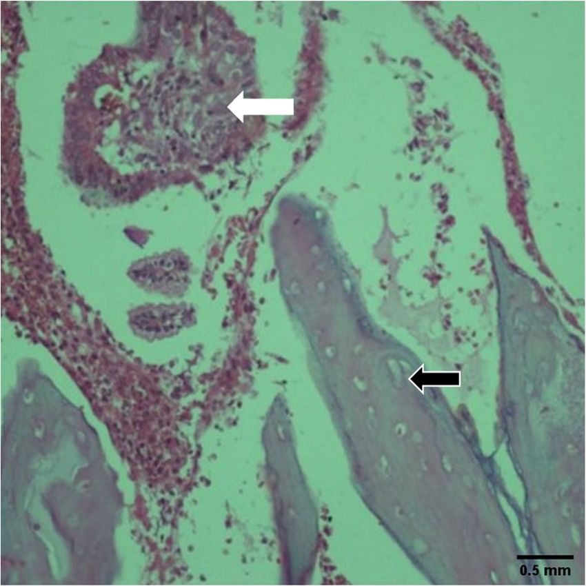
Wani et al. Middle East Fertility Society Journal        (2020) 25:35                                                                                Page 8 of 10

 Fig. 9 Sagittal (a) and axial (b) transabdominal ultrasound images of intra-uterine Contraceptive device (black block arrows) mimicking endometrial ossification

   MRI examination revealing loss of normal T2W hyper-                             history of uterine instrumentation related to pregnancy
intense endometrial signal with T1W hypointense signal                             termination was present in 88% patients. The compiled
may serve as a complementary tool in establishing a                                review concluded that infertility (56.2%) was the most
diagnosis of EOM. However, MRI of pelvis is not war-                               common presenting symptom followed by menstrual ir-
ranted for the primary diagnosis of EOM in our view, as                            regularities (19.8%), vaginal discharge (6.4%), pelvic pain
it has poor sensitivity and specificity for detecting ossifi-                      (7.9%) and dysmenorrhea (2.6%). Some patients were
cation in addition to being expensive and selectively                              asymptomatic and were found incidentally. The USG
available [16]. However, MRI may be required in pa-                                findings were more or less similar in the reported cases
tients with uncertain or additional uterine (adenomyosis                           with presence of linear echogenic structure within endo-
or mass) or adnexal (endometriosis or suspected genital                            metrial canal. They also reported that a majority of pa-
tuberculosis) findings on USG. CT scan though having                               tients conceived spontaneously after treatment.
an extremely high sensitivity for detection of ossification                           Majority of reported cases of endometrial ossification
is of limited value due to significant radiation exposure                          have an antecedent history of pregnancy loss. But the re-
to the patient.                                                                    ported cases do not elaborate whether endometrial ossi-
   The exact prevalence of EOM is unknown. Based on a                              fication was a retained foetal bone or de novo bone
compendious review of literature spanning almost a cen-                            formation. Cayuela E et al. attempted to unravel this
tury from 1928 to 2016, Khan SN et.al concluded that a                             mystery by performing genetic analysis of the retrieved
total of 293 cases have been reported in literature [17].                          endometrial bone and the woman, and concluded that
Most of the knowledge about EOM has emanated from                                  the genetic markers of endometrial bone completely
case reports and small case series. However, the wide                              matched that of woman thus leading to the conclusion
use of USG has resulted in increased detection of this                             that endometrial bone is not retained foetal bone but
condition. In this review, they found that antecedent                              true de novo osseous metaplasia [18].
Wani et al. Middle East Fertility Society Journal     (2020) 25:35                                                                        Page 9 of 10

  Infertility is a common presentation in EOM as was                     Acknowledgements
seen in all patients in the present study. The relationship              Acknowledgment of grants, disclosures or other assistance:
                                                                         I, Wani Abdul Haseeb, have no disclosures regarding grants or other
between infertility and EOM is not well established.                     assistance.
However, reactive endometritis caused by metaplastic                     I, Parry Arshed Hussain, have no disclosures regarding grants or other
bone may interfere with blastocyst implantation [15].                    assistance.
                                                                         I, Feroz Imza, have no disclosures regarding grants or other assistance.
Lewis et al. reported 50% reduction in local prostaglan-                 I, Jehangir Majid, have no disclosures regarding grants or other assistance.
din concentration after therapeutic dilatation and curet-                I, Rashid Masarat, have no disclosures regarding grants or other assistance.
tage in these cases supporting inflammation as a
contributing factor in causing infertility [17]. Another                 Authors’ contributions
                                                                         WA, PA and JM performed, analysed and interpreted the ultrasound,
plausible explanation for associated infertility is osteo-               computed tomography (CT) and magnetic resonance images. FI performed
blastic bone acting as a foreign body, a mechanism simi-                 the histopathological examination of specimen and was also involved in
lar to intra-uterine contraceptive device preventing                     obtaining hysteroscopic specimen. RM performed hysteroscopies, dilatation
                                                                         and curettage and provided lab, hormonal and biochemical parameters. All
conception [12, 18, 19]. Complete removal of this meta-                  authors were involved in manuscript preparation and literature research. All
plastic tissue restores fertility [12, 18, 19]. Uterine curet-           the authors have read and approved the manuscript.
tage or hystercopically guided removal of endometrial
ossification is the treatment of choice [20, 21]. Recently,              Funding
                                                                         No funding was required for this study as it was the part of infertility
ultrasound-guided hysteroscopic removal of bone has                      evaluation as per the institutional protocol. The patients paid themselves the
been recommended due to its good visibility which helps                  nominal fee for the procedure.
in complete removal of the metaplastic tissue embedded
even in adjacent myometrium [12, 22, 23]. Genital tuber-                 Availability of data and materials
                                                                         All the data and materials were obtained from patients registered in our
culosis was likely the cause of endometrial calcification                hospital.
in four of our patients amongst which one had history of
pulmonary tuberculosis. In endemic country like ours                     Ethics approval and consent to participate
where genital tuberculosis is so common, it must be in-                  IRB statement: This study was duly approved by the Institutional Review
                                                                         Board (IRB). The study didn’t require a reference number. No animal
cluded in the diagnostic workup of endometrial                           participants were used in this study. Informed verbal consent in the
calcification.                                                           vernacular language was obtained from illiterate patients and written
  The present study is the largest study in terms of                     consent in patients who could read.
number of EOM patients reported. However, the single-
                                                                         Consent for publication
centre design is a limitation of our study. The present
                                                                         None
study may not reflect the actual incidence of EOM be-
cause the patients in our study were symptomatic for in-                 Competing interests
fertility and attended infertility clinic. As a result, it will          We declare that we have no (financial and non-financial) competing
miss those cases who are not infertile, who have other                   interests.

symptoms like pelvic pain, vaginal discharge or men-                     Author details
                                                                         1
strual irregularity or who may not be attempting                          Department of Radiodiagnosis, Government Medical College, Srinagar,
conception.                                                              Jammu and Kashmir 190010, India. 2Department of Radiodiagnosis,
                                                                         Sher-i-Kashmir Institute of Medical Sciences, Srinagar, Jammu and Kashmir
                                                                         190010, India. 3Department of Pathology, Sher-i-Kashmir Institute of Medical
                                                                         Sciences, Srinagar, Jammu and Kashmir 190010, India. 4Department of
Conclusion                                                               Obstetrics and Gynecology, Directorate of Health Services, Srinagar, Jammu
In nutshell, endometrial osseous metaplasia is a rare and                and Kashmir 190010, India.
treatable cause of secondary infertility with ultrasound
                                                                         Received: 15 May 2020 Accepted: 2 December 2020
being the best non-invasive diagnostic imaging modality.
Post-abortion instrumentation is the most common aeti-
ology; however, genital tuberculosis must be included in                 References
the diagnostic workup especially in endemic zones. The                   1. Garg D, Bekker G, Akselrod F. Endometrial osseous metaplasia: an unusual
                                                                             cause of infertility. BMJ Case Rep. 2015;2015:bcr2015209523.
actual pathogenesis is debatable and area of active re-                  2. Ceccacci L, Clancy G (1981) Endometrial ossification: report of an additional
search. Ultrasound-guided hysteroscopic removal/dilata-                      case. Am J Obstet Gynecol 141:103–104
tion and curettage is the treatment of choice and usually                3. Acharya U, Pinion SB, Parkin DE, Hamilton MPR (1993) Osseous metaplasia
                                                                             of the endometrium treated by hysteroscopic resection. Br J Obstet
curative for infertility.                                                    Gynaecol 100:391–392
                                                                         4. AbdullGaffar B, AlMulla A (2020 Mar) Endometrial calcifications. Int J Surg
Abbreviations                                                                Pathol 1:1066896920909425
ATT: Anti-tubercular treatment; CT: Computed tomography;                 5. Jamal I, Gupta RK, Raj S, Bhadani PP (2020) Endometrial and cervical osseous
EOM: Endometrial osseous metaplasia; HPE: Histopathologic examination;       metaplasia with extramedullary haematopoiesis presenting as amenorrhea:
HSG: Hysterosalpingography; IUCD: Intra-uterine contraception device;        Report of three cases. Indian J Pathol Microbiol 63(2):295
MRI: Magnetic resonance imaging; S.D: Standard deviation; TAS: Trans-    6. Rodriguez BD, Adamson GD (1993) Hysteroscopic treatment of ectopic
abdominal sonography; TVS: Trans-vaginal sonography; USG: Ultrasound         intrauterine bone A case report. J Reprod Med 38:515–520
Wani et al. Middle East Fertility Society Journal (2020) 25:35 Page 10 of 10
7. Guerra LF, Pessanha LB, Oliveira GA, Melo AM, Braga FS, Souza RS (2016
Feb) Endometrial osseous metaplasia: sonographic, radiological and
histopathological findings. Radiol Bras 49(1):62–63
8. DL SJ (2020) Endometrial Osseous metaplasia associated with secondary
infertility-Report of 2 cases. University Journal of Surgery and Surgical
Specialities 31:6(1)
9. Herbold DR, Magrane DM (1986) Calcifications of the benign endometrium.
Arch Pathol Lab Med 10:666
10. Lainas T, Zorzovilis I, Petsas G, Alexopoulou E, Lainas G (2004) Ioakimidis T:
Osseous metaplasia: case report and review. FertilSteril 82:1433–1435
11. Sood A, Lewis E, Grey B, Mohiyiddeen L (2019) Novel management of
endometrial osseous metaplasia. World J Surg Surgical Res 2:1164
12. Bahceci M, Demirel LC (1996) Osseous metaplasia of the endometrium: a
rare cause of infertility and its hysteroscopic management. Hum Reprod 11:
2537–2539
13. Rosa-e-Silva JC, Barcelos ID, Navarro PA, de Sá Rosa AC, Nogueira AA, Ferriani
RA (2009 Dec) Osseous metaplasia of the endometrium associated with
infertility: a case report and review of the literature. J Med Case Rep 3(1):1–4
14. Perino A, Mangione D, Svelato A, Forlani F, Gargano F, Incandela D, Coppola
MA, Venezia R (2013 Jan) Chronic renal failure and endometrial osseous
metaplasia: a hypothetical pathway. Acta Obstet Gynecol Scand 92(1):118–119
15. Tyagi SP, Saxena K, Rizvi R, Langley FA (1979) Foetal remnants in theuterus
and their relation to other uterine heterotopia. Histopathology. 3(4):339–345
16. Marcus SF, Bhattacharya J, Williams G, Brinsden P, Hamou J (1994)
Endometrial ossification: a case of secondary infertility. Report of two cases.
Am J ObstetGynecol 170:1381–1383
17. Khan SN, Modi M, Hoyos LR, Imudia AN, Awonuga AO (2016 Jul) Bone in the
endometrium: A review. International journal of fertility & sterility 10(2):154
18. Cayuela E, Perez-Medina T, Vilanova J, Alejo M, Cañadas P (2009 Apr 1) True
osseous metaplasia of the endometrium: the bone is not from a fetus. Fertil
Steril 91(4):1293–12e1
19. Lewis V, Khan-Dawood F, King M, Beckham C (1990) Dawood MY:Retention
of intrauterine fetal bone increases menstrual prostaglandins.
ObstetGynecol 75:561–563
20. Chotaliya AB, Saifi SG, Achuthan G (2018) Sonographic appearance of
endometrial osseous metaplasia. J Clin Ultrasound 46(8):536–539
21. Umashankar T, Patted S, Handigund RS (2010) Endometrial osseous
metaplasia: Clinicopathological study of a case and literature review. Journal
of human reproductive sciences 3(2):102
22. Madaan M, Suman S, Sharma R, Kapoor N, Garg P, Raj SS (2015) Osseous
metaplasia of the endometrium and successful hysteroscopic resection: a
report of two cases and a review of the literature. Asian J Endosc Surg 8(1):
63–66
23. Roach MK, Thomassee MS (2015) An incidental finding of endometrial
osseous metaplasia during office hysteroscopy. Am J Obstet Gynecol 212(3):
402–4e1
Publisher’s Note
Springer Nature remains neutral with regard to jurisdictional claims in
published maps and institutional affiliations.You can also read