Lymphoplasmacytic Sclerosing Pancreatitis (Autoimmune Pancre-atitis): Evaluation with Multidetector CT1
←
→
Page content transcription
If your browser does not render page correctly, please read the page content below
Note: This copy is for your personal non-commercial use only. To order presentation-ready
          copies for distribution to your colleagues or clients, contact us at www.rsna.org/rsnarights.

EDUCATION EXHIBIT                                                                                                                             157

                            Lymphoplasmacytic
                            Sclerosing Pancreatitis
                            (Autoimmune Pancre-
                            atitis): Evaluation with
                            Multidetector CT1
                            Satomi Kawamoto, MD ● Stanley S. Siegelman, MD ● Ralph H. Hruban,
    ONLINE-ONLY
        CME                 MD ● Elliot K. Fishman, MD
      See www.rsna          Lymphoplasmacytic sclerosing pancreatitis is a form of chronic pancreati-
      .org/education        tis characterized by a mixed inflammatory infiltrate that centers on the
      /rg_cme.html.
                            pancreatic ducts. It is a cause of benign pancreatic disease that can clini-
                            cally mimic pancreatic cancer. Preoperative detection of lymphoplasma-
      LEARNING              cytic sclerosing pancreatitis is important because patients usually respond
     OBJECTIVES
                            to steroid therapy. Patients with lymphoplasmacytic sclerosing pancreatitis
    After reading this
    article and taking      are often referred for computed tomography (CT) when they are sus-
   the test, the reader     pected of having a pancreatic or biliary neoplasm; therefore, it is important
     will be able to:
䡲 List the clinical and     to search for potential findings suggestive of lymphoplasmacytic sclerosing
pathologic features of      pancreatitis when typical findings of a pancreatic or biliary neoplasm are
lymphoplasmacytic
sclerosing pancreati-       not found. Typical CT findings include diffuse or focal enlargement of the
tis.                        pancreas without dilatation of the main pancreatic duct. Focal enlarge-
䡲 Discuss the spec-         ment is most commonly seen in the head of the pancreas, and the involved
trum of CT appear-
ances of lymphoplas-        pancreas on contrast material– enhanced CT images may be isoattenuat-
macytic sclerosing          ing relative to the rest of the pancreas, or hypoattenuating, especially dur-
pancreatitis.
                            ing the early postcontrast phase. Thickening and contrast enhancement of
䡲 Describe the CT
features that may           the wall of the common bile duct and gallbladder may reflect inflamma-
allow lymphoplasma-         tory infiltrate and fibrosis associated with lymphoplasmacytic sclerosing
cytic sclerosing pan-
creatitis to be differ-     pancreatitis. There are several features seen at CT that may help to differ-
entiated from pan-          entiate lymphoplasmacytic sclerosing pancreatitis from pancreatic cancer,
creatic cancer.
                            such as diffuse enlargement of the pancreas with minimal peripancreatic
                            stranding in patients with obstructive jaundice, an absence of significant
    TEACHING                pancreatic atrophy, and an absence of significant main pancreatic duct
    POINTS
    See last page
                            dilatation. When these findings are encountered, clinical, other imaging,
                            and serologic data should be evaluated.
                            ©
                             RSNA, 2008

Abbreviation: ERCP ⫽ endoscopic retrograde cholangiopancreatography

RadioGraphics 2008; 28:157–170 ● Published online 10.1148/rg.281065188 ● Content Codes:
1From   the Russell H. Morgan Department of Radiology and Radiological Science (S.K., S.S.S., E.K.F.) and Department of Pathology, Sol Goldman
Pancreatic Cancer Research Center (R.H.H.), Johns Hopkins Medical Institutions, JHOC 3235A, 601 N Caroline St, Baltimore, MD 21287. Recipi-
ent of a Certificate of Merit award for an education exhibit at the 2005 RSNA Annual Meeting. Received November 7, 2006; revision requested April
18, 2007, and received June 21; accepted June 28. All authors have no financial relationships to disclose. Address correspondence to S.K. (e-mail:
skawamo1@jhmi.edu).
©
    RSNA, 2008
158 January-February 2008 RG f Volume 28 ● Number 1
Introduction
In recent years, the concept of autoimmune pan-
creatitis has been established to refer to a special
type of chronic pancreatitis with unique clinical
and histologic manifestations. Autoimmune pan-
creatitis can be defined as a chronic inflammatory
process of the pancreas that is caused by an auto-
immune mechanism (1,2). The morphologic hall-
marks are periductal infiltration by lymphocytes
and plasma cells and granulocytic epithelial le-
sions with consequent destruction of the duct epi-
thelium and venulitis (3). Therefore, autoim-
mune pancreatitis has been morphologically
described as lymphoplasmacytic sclerosing pan-
creatitis, non-alcoholic duct-destructive chronic
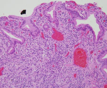
pancreatitis (4,5), or chronic sclerosing pancreati- Figure 1. Photomicrograph (original magnification,
tis (6). The terms autoimmune pancreatitis and ⫻40; hematoxylin-eosin stain) of a pancreatic duct
sclerosing pancreatitis have been used interchange- specimen from a patient with lymphoplasmacytic scle-
rosing pancreatitis shows dense inflammatory infiltrates
ably (7).
with associated fibrosis surrounding the pancreatic duct.
The gross morphologic alterations produced
by lymphoplasmacytic sclerosing pancreatitis may
simulate malignancy, with characteristics such as ing.” These features may lead to surgical resec-
masslike enlargement of the pancreatic head tion because of the suspicion that the lesion is
and/or irregular narrowing of the pancreatic duct carcinomatous (7). The inflammatory process
and stricture of the common bile duct. However, may involve either the entire pancreas, or it may
lymphoplasmacytic sclerosing pancreatitis re- be limited to only a portion of the pancreas.
Teaching sponds to oral steroid therapy, with reversible When the inflammatory process involves only one
Point improvement of pancreatic morphology and func- portion of the pancreas, that segment is most of-
tion (1,8 –10). Thus, when findings typical of a ten the pancreatic head. In a minority of cases,
pancreatic or biliary neoplasm are not seen on however, the inflammatory process is concen-
computed tomographic (CT) images, it is impor- trated in the body or in the tail of the pancreas
tant to search for characteristics suggestive of (7). It is not known how frequently and to what
lymphoplasmacytic sclerosing pancreatitis and extent the entire pancreas is affected in lympho-
allow accurate diagnosis and appropriate therapy plasmacytic sclerosing pancreatitis (3).
to occur. Microscopic findings of autoimmune pancre-
The purpose of this article is to discuss and atitis are consistent with lymphoplasmacytic scle-
illustrate the spectrum of appearances of lym- rosing pancreatitis. In pathologic specimens,
phoplasmacytic sclerosing pancreatitis on CT dense lymphoplasmacytic infiltrate centered
images. Pathologic and clinical features of lym- around the medium- and large-sized interlobular
phoplasmacytic sclerosing pancreatitis, CT tech- pancreatic ducts is seen (11) (Fig 1). Although
niques, and findings with other imaging modali- the inflammatory infiltrate consists mainly of lym-
ties are also presented. CT features that may be phocytes and plasma cells, it also contains some
potentially useful in differentiating lymphoplas- macrophages and occasionally neutrophilic and
macytic sclerosing pancreatitis from pancreatic eosinophilic granulocytes (11). Immunocyto-
cancer are discussed and illustrated. chemical typing of the lymphocytes reveals that
Most of the patients that are discussed in this most of them are CD8- and CD4-positive T lym-
article had no serologic parameters measured, phocytes, with B lymphocytes present to a lesser
and diagnosis was based on pathologic analysis. degree (3). The lumen of inflamed pancreatic
Therefore, in this article, we use the term lym- ducts is encompassed by infiltrate and is nar-
phoplasmacytic sclerosing pancreatitis. rowed by infolding of the epithelium (3). A dis-
tinctive venulitis is also seen.
Pathologic Features of Lympho- When the pancreas is only slightly affected, the
plasmacytic Sclerosing Pancreatitis inflammation centers almost entirely on the
At gross examination, the involved pancreas is ducts; in severely affected pancreata, the inflam-
firm or hard and may be enlarged or “mass form- matory process involves the acinar parenchyma in
addition to the ducts and leads to diffuse sclerosis
(3). The acinar cells are replaced by inflammatoryRG f Volume 28 ● Number 1 Kawamoto et al 159
ing pancreatitis. Okazaki et al (13) reported that
among 620 patients with chronic pancreatitis, 30
of them (5%) had lymphoplasmacytic sclerosing
pancreatitis. In a recent Italian multicenter study
on the epidemiology of chronic pancreatitis that
involved 21 centers and enrolled 282 patients suf-
fering from chronic pancreatitis over 2 years, au-
toimmunity was recognized as an associated fac-
tor in 23 patients (6%) (7). The reported mean
age of 41 patients with lymphoplasmacytic scle-
rosing pancreatitis was 62.2 years (range, 32–76
years) (14). Another study reported that the mean
age of 53 patients with lymphoplasmacytic scle-
rosing pancreatitis was 56 years (range, 14 –77
years) (15).
Figure 2. Photomicrograph (original magnification, Clinically, lymphoplasmacytic sclerosing pan-
⫻40; hematoxylin-eosin stain) of a surgical specimen of creatitis commonly manifests as obstructive jaun-
the common bile duct shows dense mixed infiltrate and dice with no or only mild abdominal pain, weight
fibrosis surrounding the common bile duct. loss, and recent-onset diabetes in elderly patients.
Acute attacks seen in severe or acute pancreatitis
do not usually occur (13). Obstructive jaundice is
cells and fibrosis, and the lobular architecture of often caused by stenosis of the intrapancreatic
the pancreas is almost lost (3). The peripancreatic common bile duct (7) and is seen in 63%–75% of
and peribiliary lymph nodes are enlarged and patients with lymphoplasmacytic sclerosing pan-
show follicular hyperplasia (3). creatitis (13–15). Diabetes mellitus is also often
It is also well known that patients with lym- associated with lymphoplasmacytic sclerosing
phoplasmacytic sclerosing pancreatitis display pancreatitis and has a reported frequency of less
distal common bile duct strictures and inflamma- than 20%– 68% (7,13). Because these signs over-
tion, features that overlap with those of primary lap with those of pancreatic cancer, lymphoplas-
sclerosing cholangitis. Abraham et al (12) re- macytic sclerosing pancreatitis has frequently
ported that among 20 patients with lymphoplas- been misdiagnosed as pancreatic cancer. Harda-
macytic sclerosing pancreatitis treated with pan- cre et al (16) reported that between pancreatic
creaticoduodenectomy, inflammatory infiltrates cancer and lymphoplasmacytic sclerosing pancre-
were seen in the extrapancreatic common bile atitis, there were no statistically significant differ-
duct in 60%, in the intrapancreatic common bile ences in the rates of abdominal pain, weight loss,
duct in 84.2% (Fig 2), and in the gallbladder in jaundice, preoperative carcinoembryonic antigen,
60%. However, inflammatory and sclerosing or CA19-9 levels. Analysis of a large series involv-
changes of the intrahepatic bile duct, which are ing more than 1200 patients who underwent pan-
typical findings in primary sclerosing cholangitis, creaticoduodenectomy with a presumed preop-
are uncommon in lymphoplasmacytic sclerosing erative diagnosis of pancreatic cancer, periampul-
pancreatitis (3). Moreover, unlike typical primary lary neoplasm, or cholangiocarcinoma revealed
sclerosing cholangitis, biliary lesions in lympho- that 2.2%–2.4% of patients had pathologic fea-
plasmacytic sclerosing pancreatitis usually im- tures consistent with lymphoplasmacytic scleros-
prove with administration of steroids. These ing pancreatitis (16,17).
findings suggest that the mechanism of the devel- Elevation of serum gamma globulin or immu-
opment of biliary lesions in lymphoplasmacytic noglobulin G (IgG) concentration and the pres-
sclerosing pancreatitis may differ from that of ence of some autoantibodies are often observed.
typical primary sclerosing cholangitis (3,13). Serum IgG4 concentrations were reported to be
significantly and specifically high in patients with
Clinical Presentation of Lympho- lymphoplasmacytic sclerosing pancreatitis and are
plasmacytic Sclerosing Pancreatitis closely associated with disease activity (18). Ha-
Lymphoplasmacytic sclerosing pancreatitis is a mano et al (18) reported that the accuracy, sensi-
rare disorder, and the exact prevalence is un- tivity, and specificity of elevated serum concentra-
known. Ito et al (8) reported that among 161 pa- tions of IgG4 in the diagnosis of this disease, with
tients with chronic pancreatitis evaluated with a cutoff value of 135 mg/dL, were 97%, 95%,
endoscopic retrograde pancreatography, three
patients (1.86%) had lymphoplasmacytic scleros-160 January-February 2008 RG f Volume 28 ● Number 1
Figure 3. Venous phase axial (a, b) and coronal (c) re-
formatted CT images show mild diffuse enlargement of
the pancreas with minimal peripancreatic stranding. The
distal common bile duct shows smooth beaklike stenosis in
the region of the pancreatic head with dilatation of the upper
common bile duct. Mild thickening and contrast material
enhancement of the common bile duct wall are also seen
(arrows in b and c). Pathologic analysis revealed an intense
lymphoplasmacytic process involving the common bile duct.
and 97% respectively. However, other groups
reported lower sensitivity (19 –21). Ghazale et al
(21) reported that serum concentrations of IgG4
were elevated in 10% of pancreatic cancer pa-
tients (13 of 135), but that only 1% had serum
IgG4 levels of ⬎280 mg/dL, compared with 53%
of patients with lymphoplasmacytic sclerosing
pancreatitis. one-third the length of the entire pancreas),
Occasionally, patients with lymphoplasmacytic (b) laboratory data demonstrate abnormally el-
sclerosing pancreatitis have extrapancreatic le- evated levels of serum gamma globulin and/or
sions that are seen in other autoimmune diseases, IgG or the presence of autoantibodies, and (c) fi-
including Sjögren syndrome, sclerosing cholangi- brotic change with dense lymphoplasmacytic in-
tis, primary biliary cirrhosis, interstitial nephritis, filtration is noted in the pancreas at histopatho-
sialoadenitis, enlarged mediastinal or cervical logic examination. A diagnosis of lymphoplasma-
lymph nodes, ulcerative colitis, and retroperito- cytic sclerosing pancreatitis can be established if
neal fibrosis (22). The incidence of associated all of the criteria are present or if criterion a is
extrapancreatic autoimmune lesions is reported to present with either criterion b or criterion c (23).
range from 19% to more than 50% (7,15,22). Recently, these criteria have been modified by the
These extrapancreatic autoimmune diseases may Japan Pancreas Society to allow diagnosis of auto-
be recognized at the time of diagnosis or they may immune pancreatitis to include focal pancreatic
develop later (7). The presence of an associated mass and focal pancreatic duct stricture (24). Re-
autoimmune disease may help in the diagnosis of cently, other groups have also proposed diagnos-
lymphoplasmacytic sclerosing pancreatitis (7). tic criteria for autoimmune pancreatitis (19,25).
Lymphoplasmacytic sclerosing pancreatitis is
difficult to diagnose. In 2002, the Japan Pancreas CT Technique
Society proposed the following diagnostic criteria CT examinations were performed with multide-
for autoimmune pancreatitis: (a) pancreatic im- tector CT scanners, including a Siemens Volume
aging studies show diffuse enlargement of the Zoom scanner (4 ⫻ 1 mm collimation), a Sie-
pancreas and diffuse narrowing of the main pan- mens Sensation 16 scanner (16 ⫻ 0.75 mm colli-
creatic duct with an irregular wall (more than mation), and a Siemens Sensation 64 scanner
(64 ⫻ 0.6 mm collimation) (Siemens MedicalRG f Volume 28 ● Number 1 Kawamoto et al 161 Figure 4. (a, b) Venous phase axial images show mild enlargement of the pancreatic head, with a stent present in the common bile duct. Nondilated main pancreatic duct is seen in the body and tail (arrowheads in a). Focal hypoat- tenuating lesions are seen in both kidneys (arrows in b). Although biopsy of the lesions was not performed, follow-up CT showed that these lesions became less obvious. (c, d) Contrast-enhanced chest CT scans (d, lung window set- ting) show a left hilar mass with bulky mediastinal adenopathy. Results of biopsy performed after mediastinoscopy and limited anterior thoracotomy revealed anthracotic lymph nodes with fibrosis in the mediastinal lymph nodes, fi- brous tissue of chronic inflammation in the mediastinal soft tissue, and mild chronic inflammation and pleural fibro- sis in the left upper lobe. Solutions, Malvern, Pa). The data were recon- All image data were reconstructed with the structed to obtain 1.25-mm section thickness at body soft tissue algorithm and sent to the work- 1-mm intervals (0.25-mm overlap) with the Sie- station (Leonardo, Siemens Medical Solutions). mens Volume Zoom Scanner and 0.75-mm sec- InSpace software (Siemens Medical Solutions) tion thickness at 0.5-mm intervals (0.25-mm was used for data analysis, which was the volume overlap) with the Siemens Sensation 16 and 64 imaging application for interactive viewing of vol- scanners. After fasting for at least 2–3 hours, each ume data available on the Leonardo workstation. patient ingested 750 –1000 mL of water over a 15–20 minute period before scanning was begun. CT Findings of Lympho- Arterial and venous phase images were acquired plasmacytic Sclerosing Pancreatitis at 25 seconds and 50 – 60 seconds from the start of intravenous administration of contrast mate- Enlargement of the Pancreas rial. We injected 120 mL of iohexol (Omnipaque Typically, CT images show diffuse enlargement of 350; Amersham Health, Princeton, NJ) through the pancreas (Fig 3). Focal enlargement may also be Teaching the peripheral venous line at a rate of 3 mL/sec. seen, particularly in the pancreatic head (Fig 4), but Point Other scanning parameters included 120 kV and also in the body or tail (5) (Fig 5). The presence 150 –200 mAs.
162   January-February 2008                                                         RG f Volume 28       ●   Number 1

      Figure 5. Venous phase oblique axial (a) and coronal (b) reformatted CT images show focal enlargement of
      the pancreatic tail with a distinct area of decreased attenuation (arrows). Minimal stranding is seen around the
      enlarged pancreatic tail.

Figure 6. Venous phase axial (a) and coronal (b) reformatted CT images show a diffusely hypoattenuating pan-
creas that appears to be normal in size. The pancreas appears featureless, and the normal lobular appearance is ef-
faced. A stent is seen in the common bile duct.

of multiple masses has also been reported (26). In            was focal enlargement, and the incidence of dif-
prior studies with CT, diffuse enlargement of the             fuse enlargement ranged from 56% to 100%
pancreas was more commonly encountered than                   (9,27–31). Sahani et al (27) reported that among
                                                              25 patients with lymphoplasmacytic sclerosing
                                                              pancreatitis who underwent helical CT examina-
RG f Volume 28 ● Number 1 Kawamoto et al 163
Figure 7. Venous phase axial (a) and coronal (b, c) re-
formatted CT images show diffuse enlargement of the en-
tire pancreas. The pancreas appears featureless, and the
normal lobular appearance is effaced. There is minimal
peripancreatic stranding, and a stent is seen in the com-
mon bile duct.
phoplasmacytic sclerosing pancreatitis (25). Atro-
phy of the pancreas is not usually seen (28), al-
though the normal lobular appearance of the pan-
creas may be effaced and the gland may appear
featureless in the involved region (32) (Figs 6, 7).
Contrast Enhancement Pat-
tern of the Pancreatic Parenchyma
The reported contrast enhancement pattern of
the involved pancreas seen in lymphoplasmacytic
sclerosing pancreatitis is variable. Previous stud-
tion, 14 showed a diffusely enlarged pancreas, ies reported that on arterial phase or early post-
seven had smooth focal enlargement or a masslike contrast phase images, the involved portion of the
appearance in the pancreatic head and/or the un- pancreas appears hypoattenuating compared with
cinate process, and four had a pancreas that ap- unaffected pancreatic parenchyma and occasion-
peared to be normal in size (Fig 6). Kamisawa et ally has a distinct margin (9,33) (Fig 5). On ve-
al (28) reported that among 17 patients with lym- nous phase images, the involved portion of the
phoplasmacytic sclerosing pancreatitis, 10 had pancreas may remain hypoattenuating (33) or
diffuse enlargement and seven had segmental en- may become nearly isoattenuating compared with
largement of the pancreatic head. However, an-
other study reported that diffuse enlargement was
seen in only six of 22 patients (27%) with lym-164 January-February 2008 RG f Volume 28 ● Number 1
Figure 8. (a, b) Arterial phase axial (a) and coro-
nal (b) reformatted CT images show enlargement of the
pancreatic head with a distinct area of decreased attenua-
tion within the enlarged pancreatic head (arrowheads).
The body and tail of the pancreas are relatively atrophic.
(c) Venous phase axial image shows that the hypoattenu-
ating area seen in the pancreatic head on arterial phase
images becomes nearly isoattenuating relative to adjacent
pancreatic parenchyma. A stent (arrow in a and c) is seen
in the common bile duct.
unaffected pancreatic parenchyma (Figs 8, 9).
However, a discrete area of differential contrast
enhancement may not be observed on arterial or
venous phase images in the areas of focal enlarge-
ment, and the pancreas may show homogeneous
contrast enhancement (27,30,32,34). In previous
CT studies, the area of focal enlargement may
appear homogeneously isoattenuating relative to reported to become homogeneously isoattenuat-
the pancreas in 25%–71% of patients, or it may ing or hyperattenuating compared with the sur-
appear hypoattenuating in 29%–75% of patients rounding pancreatic parenchyma (31,34,35).
(27,34). When delayed phase images are obtained However, another study reported that the in-
at several minutes after intravenous administra- volved lesion remains hypoattenuating compared
tion of contrast material, the involved portion is with unaffected pancreatic parenchyma on de-
layed phase images (33). These differences may
be attributed to the different CT techniques and
the degree of inflammatory infiltrate and fibrosis
of the involved pancreas.RG f Volume 28 ● Number 1 Kawamoto et al 165
Figure 9. (a, b) Arterial phase oblique axial (a) and
coronal (b) reformatted CT images show enlargement of
the pancreatic head with a distinct area of decreased at-
tenuation within the enlarged pancreatic head (black ar-
rows). The main pancreatic duct (white arrow) is mini-
mally dilated (3.8 mm) proximal to the enlarged portion.
(c) Venous phase axial image shows that a hypoattenuat-
ing area seen in the pancreatic head on arterial phase im-
ages becomes nearly isoattenuating (arrows) relative to
adjacent pancreatic parenchyma.
creas (Fig 9). However, significant dilatation of
the main pancreatic duct, a characteristic of pan-
creatic ductal adenocarcinoma, is not found in
lymphoplasmacytic sclerosing pancreatitis.
Other Pancreatic/
Peripancreatic Findings
Minimal peripancreatic stranding, which may
Narrowing of the Pancreatic Duct simulate mild edematous acute pancreatitis, is
Thin-section CT with multiplanar reformation often seen at CT (27,33) (Figs 3, 7). A capsule-
helps to delineate the main pancreatic duct. In like low-attenuation rim has also been described
patients with lymphoplasmacytic sclerosing pan- in 12%– 80% of reported cases of lymphoplasma-
creatitis, the main pancreatic duct is diffusely or cytic sclerosing pancreatitis (Fig 10) (19,27,28,
segmentally narrowed. On CT images, the main 30 –32). On delayed phase images, a capsulelike
pancreatic duct may be seen as a small nondilated low-attenuation rim may show subtle delayed en-
Teaching
duct, or it may appear attenuated, particularly in hancement (31,33).
Point
the area of the pancreatic enlargement. Mild dila-
tation of the main pancreatic duct may also be
seen proximal to the enlarged portion of the pan-166 January-February 2008 RG f Volume 28 ● Number 1
Figure 10. Venous phase oblique axial (a), coronal (b, c), and sagittal (d) reformatted CT images show mild dif-
fuse enlargement of the pancreas with a capsulelike low-attenuation rim (arrows in a and b). The splenic vein is sur-
rounded by the capsulelike rim and is narrowed (arrowhead in d). A stent is present in the distal common bile duct,
and the proximal common bile duct is dilated. Pathologic analysis revealed prominent lymphoplasmacytic infiltrate
involving the distal common bile duct.
In contrast to other forms of chronic pancreati- tery and the superior mesenteric artery— has not
tis, lymphoplasmacytic sclerosing pancreatitis been reported. At conventional angiography,
does not commonly manifest with parenchymal however, irregular narrowing and encasement of
calcifications and pseudocysts (3,28,36,37). the small peripancreatic arteries were reported in
However, intraductal calcifications may occur late up to 57% of cases (28,35). Poor opacification of
in the course of the disease (9,31,33,38), and for- portal or splenic veins due to stenosis or obstruc-
mation of pseudocysts associated with lympho- tion with collateral veins was also reported in 23%
plasmacytic sclerosing pancreatitis has also been of cases at conventional angiography (28).
reported (39). Enlarged peripancreatic lymph nodes may also
Major pancreatic vascular involvement is un- be observed on CT images. Sahani et al (27) re-
common in lymphoplasmacytic sclerosing pancre- ported that nine of 25 patients with lymphoplas-
atitis compared with pancreatic adenocarcinoma macytic sclerosing pancreatitis had enlarged
(27), although cases with venous occlusion or peripancreatic nodes that measured more than 1
narrowing, particularly the splenic vein, have cm in diameter on the short axis on CT scans.
been reported (9,30,32) (Fig 10). Major arterial
involvement—such as narrowing of the celiac ar- Biliary Tract Findings
Stricture of the common bile duct is often seen in
Teaching
patients with lymphoplasmacytic sclerosing pan- Point
creatitis, particularly in patients whose pancreatic
head is affected, with a reported frequency of
33%–90% (5,27,28,30,31,40). At endoscopicRG f Volume 28 ● Number 1 Kawamoto et al 167
Figure 11. Venous phase axial (a, b) and coronal (c)
reformatted CT images show mild diffuse enlargement of
the pancreas with minimal peripancreatic stranding. The
pancreas appears featureless, and the normal lobular ap-
pearance is effaced. The distal common bile duct shows
smooth beaklike stenosis in the region of the pancreatic
head (arrow in c) with dilatation of the proximal common
bile duct. Mild thickening and contrast enhancement of
the common bile duct wall are present (arrowheads in b
and c). At pathologic analysis, the common bile duct also
showed marked chronic inflammation and fibrosis.
wall have been described, and these findings cor-
respond to the inflammatory infiltrate and fibrosis
observed microscopically (Figs 3, 11) (30,32,42).
However, when a biliary stent is present at the
time of CT scanning, these biliary tract findings
may be obscured due to pneumobilia or changes
related to stent placement.
Extrapancreatic Findings
retrograde cholangiopancreatography (ERCP), Involvement with multiple organ systems has
smooth stenosis of the distal common bile duct been reported, and these findings may be seen on
localized in the pancreatic head, with dilatation of CT images. Such changes include, but are not
the more proximal common bile duct, is the most limited to, retroperitoneal fibrosis, renal involve-
common finding in patients with lymphoplasma- ment, lung disease, and mediastinal adenopathy
cytic sclerosing pancreatitis (27,41) (Figs 3, 11). (27,43).
Narrowing of the intrapancreatic common bile
duct is thought to be induced mainly by compres- Findings at
sion of the swollen pancreas (7). Coronal three- Other Imaging Modalities
dimensional or reformatted CT images help to Patients with lymphoplasmacytic sclerosing pan-
delineate smooth, beaklike stenosis of the com- creatitis typically show diffuse or segmental nar-
mon bile duct in the intrapancreatic portion and rowing of the main pancreatic duct at ERCP. The
dilatation of the proximal common bile duct (Figs secondary branches are usually not visualized (7–9).
3, 11). Stenosis or strictures of the proximal and MR imaging may reveal diffuse pancreatic en-
middle extrahepatic bile duct and the intrahepatic largement with hypointensity on T1-weighted
bile duct—findings that resemble those seen in images. The low-attenuation capsulelike rim de-
primary sclerosing cholangitis— have also been scribed at CT is hypointense on T2-weighted im-
observed at ERCP and magnetic resonance (MR) ages and shows delayed contrast enhancement,
cholangiopancreatography (7,27,28). which suggests fibrous tissue rather than a fluid
At CT, thickening and enhancement of the
gallbladder wall and/or the common bile duct168 January-February 2008 RG f Volume 28 ● Number 1
collection or phlegmon (31). MR cholangiopan- lymphoplasmacytic sclerosing pancreatitis and
creatography may show stenosis of the bile ducts pancreatic cancer can be extremely difficult (48).
mainly in the intrapancreatic area, which results The mass may also be seen in the body or tail of
in dilatation of the proximal biliary tract (7). Scle- the pancreas, and it may simulate pancreatic can-
rosing changes of the intrahepatic bile ducts or cer. CT findings more typically seen in pancreatic
common bile duct that are similar to primary scle- cancer than in lymphoplasmacytic sclerosing pan-
rosing cholangitis are sometimes observed (7,44). creatitis include (a) significant dilatation of the
Although stenosis of the main pancreatic duct main pancreatic duct proximal to the narrowed Teaching
Point
may be observed, determining whether it is due to segment, (b) atrophy of the pancreatic paren-
lymphoplasmacytic sclerosing pancreatitis or to chyma proximal to the mass or focal enlargement,
pancreatic carcinoma may be difficult with MR and (c) involvement of the major peripancreatic
cholangiopancreatography (40). vessels. In patients with segmental narrowing of
At ultrasonographic (US) examination, the the main pancreatic duct due to lymphoplasma-
pancreas appears hypoechoic and diffusely en- cytic sclerosing pancreatitis, the main pancreatic
larged with a so-called sausagelike appearance duct proximal to the segmental narrowing typi-
(8,9,28). It may also appear as a hypoechoic mass cally shows minimal or no dilatation (19,28). Pre-
in the affected site (5,28). Endoscopic US may vious studies that compared ERCP images of fo-
show diffuse hypoechoic pancreatic enlargement cal lymphoplasmacytic sclerosing pancreatitis and
or a focal, irregular hypoechoic mass (20). Stric- those of pancreatic cancer showed that the caliber
ture of the common bile duct in the pancreatic of the main pancreatic duct proximal to the stric-
head, widespread bile duct wall thickening, and ture is smaller in patients with lymphoplasmacytic
diffuse strong enhancement of the bile duct sys- sclerosing pancreatitis (⬍4 mm in 67% of pa-
tem, including the gallbladder, proximal bile tients and 4 – 6 mm in 33%) than in those with
duct, and distal bile duct, have also been seen pancreatic ductal adenocarcinoma (⬍4 mm in
with endoscopic US when contrast material is 4%, 4 – 6 mm in 22%, and ⬎6 mm in 74%)
used (41). Farrell et al (20) reported that findings (30,35,49). Major peripancreatic vascular in-
from endoscopic US-guided fine-needle aspira- volvement, particularly arterial involvement, is an
tion may support the diagnosis of lymphoplasma- uncommon CT finding in lymphoplasmacytic
cytic sclerosing pancreatitis in combination with sclerosing pancreatitis. Atrophy of the pancreas
endoscopic US findings and clinical data. proximal to the mass is often seen in patients with
Gallium scintigraphy (45,46) and fluorine 18 pancreatic cancer, but it is usually not found in
fluorodeoxyglucose positron emission tomogra- lymphoplasmacytic sclerosing pancreatitis (28).
phy (47) have been reported to show increased Although the acinar parenchyma becomes atro-
uptake in the affected site of the pancreas during phic in lymphoplasmacytic sclerosing pancreati-
the active stage of the disease, and such findings tis, it is replaced with fibrous tissue (36), which
may be confused with the increased uptake seen probably explains why the overall size of the pan-
in pancreatic cancer or lymphoma. creas does not usually change. Fine-needle aspira-
tion biopsy may remain a necessary step to con-
CT Features to Help firm the diagnosis (7).
Differentiate Pancreatic Cancer Smooth narrowing of the distal common bile
Lymphoplasmacytic sclerosing pancreatitis has duct can be seen in patients with lymphoplasma-
several typical characteristics on CT scans that cytic sclerosing pancreatitis; however, pancreatic
may be useful in differentiating the condition cancer may display similar findings. Wakabayashi
from pancreatic cancer. When diffuse enlarge- et al (35) reported that there were no significant
ment of the pancreas with mild peripancreatic differences among patients with focal lympho-
stranding is seen on CT images of patients with plasmacytic sclerosing pancreatitis and those with
obstructive jaundice without clinical features of pancreatic cancer in terms of the frequency of
acute pancreatitis, lymphoplasmacytic sclerosing common bile duct stenosis and its character and
pancreatitis should be considered as a potential length as seen with ERCP.
diagnosis. Clinical, serologic, and other imaging Procacci et al (33) reported that CT findings
studies should be carefully evaluated. can be used to correctly diagnose lymphoplasma-
In patients with focal enlargement of the pan- cytic sclerosing pancreatitis with an accuracy of
creas, diagnosing lymphoplasmacytic sclerosing 92.5%. They evaluated seven patients with lym-
pancreatitis is more challenging. When a patient phoplasmacytic sclerosing pancreatitis and 20
with obstructive jaundice presents with a “mass” patients with other pancreatic diseases (acute or
in the pancreatic head, differentiating between chronic pancreatitis and adenocarcinoma of the
pancreas). The criteria for a diagnosis of lym-
phoplasmacytic sclerosing pancreatitis includedRG f Volume 28 ● Number 1 Kawamoto et al 169
focal or diffuse enlargement of the pancreas, pos- 4. Ectors N, Maillet B, Aerts R, et al. Non-alcoholic
sible capsulelike rim, possible stenosis or com- duct destructive chronic pancreatitis. Gut 1997;
41(2):263–268.
plete obstruction of the biliary duct, and possible 5. Van Hoe L, Gryspeerdt S, Ectors N, et al. Nonal-
stenosis (either focal or diffuse) of the main pan- coholic duct-destructive chronic pancreatitis: im-
creatic duct. The affected pancreatic parenchyma aging findings. AJR Am J Roentgenol 1998;
was isoattenuating relative to the spleen and adja- 170(3):643– 647.
cent unaffected parenchyma on unenhanced CT 6. Sood S, Fossard DP, Shorrock K. Chronic scleros-
ing pancreatitis in Sjogren’s syndrome: a case re-
images and hypoattenuating on arterial phase, port. Pancreas 1995;10(4):419 – 421.
venous phase, and delayed phase images (33). 7. Pearson RK, Longnecker DS, Chari ST, et al.
Procacci and colleagues reported one false-nega- Controversies in clinical pancreatology: autoim-
tive diagnosis of lymphoplasmacytic sclerosing mune pancreatitis— does it exist? Pancreas 2003;
pancreatitis in a case of chronic pancreatitis su- 27(1):1–13.
8. Ito T, Nakano I, Koyanagi S, et al. Autoimmune
perimposed with lymphoplasmacytic sclerosing pancreatitis as a new clinical entity: three cases of
pancreatitis that showed diffuse calcifications in autoimmune pancreatitis with effective steroid
the pancreas. They also recorded one false-posi- therapy. Dig Dis Sci 1997;42(7):1458 –1468.
tive diagnosis for lymphoplasmacytic sclerosing 9. Furukawa N, Muranaka T, Yasumori K, Matsu-
pancreatitis in a case of mild edematous acute bayashi R, Hayashida K, Arita Y. Autoimmune
pancreatitis: radiologic findings in three histologi-
pancreatitis with imaging findings similar to those cally proven cases. J Comput Assist Tomogr 1998;
of lymphoplasmacytic sclerosing pancreatitis. 22(6):880 – 883.
10. Kawa S, Hamano H. Autoimmune pancreatitis
Conclusions and bile duct lesions. J Gastroenterol 2003;38(12):
In conclusion, lymphoplasmacytic sclerosing pan- 1201–1203.
11. Abraham SC, Leach S, Yeo CJ, et al. Eosinophilic
creatitis is a unique clinical entity that is becom- pancreatitis and increased eosinophils in the pan-
ing more frequently recognized in clinical prac- creas. Am J Surg Pathol 2003;27(3):334 –342.
tice. Because patients with lymphoplasmacytic 12. Abraham SC, Cruz-Correa M, Argani P, Furth
sclerosing pancreatitis often present with obstruc- EE, Hruban RH, Boitnott JK. Lymphoplasma-
tive jaundice, their disease has been frequently cytic chronic cholecystitis and biliary tract disease
in patients with lymphoplasmacytic sclerosing
misdiagnosed as pancreatic cancer. However, be- pancreatitis. Am J Surg Pathol 2003;27(4):441–
cause lymphoplasmacytic sclerosing pancreatitis 451.
responds to steroid therapy, it is important to rec- 13. Okazaki K. Autoimmune pancreatitis: etiology,
ognize this entity in order to avoid surgery. Char- pathogenesis, clinical findings and treatment—the
acteristics seen on CT images that suggest lym- Japanese experience. JOP 2005;6(1 suppl):89 –96.
14. Kawa S, Ota M, Yoshizawa K, et al. HLA
phoplasmacytic sclerosing pancreatitis include DRB10405-DQB10401 haplotype is associated
diffuse or focal enlargement of the pancreas, ab- with autoimmune pancreatitis in the Japanese
sence of significant pancreatic atrophy, absence of population. Gastroenterology 2002;122(5):1264 –
substantial main pancreatic duct dilatation, and 1269.
in some cases a rim of low attenuation surround- 15. Zamboni G, Luttges J, Capelli P, et al. His-
topathological features of diagnostic and clinical
ing the pancreas. However, diagnosis of this dis- relevance in autoimmune pancreatitis: a study on
ease can be difficult, especially with patients who 53 resection specimens and 9 biopsy specimens.
present with a focal mass or a masslike enlarge- Virchows Arch 2004;445(6):552–563.
ment of the pancreas. When these CT findings 16. Hardacre JM, Iacobuzio-Donahue CA, Sohn TA,
are encountered, lymphoplasmacytic sclerosing et al. Results of pancreaticoduodenectomy for
lymphoplasmacytic sclerosing pancreatitis. Ann
pancreatitis should be considered as a potential Surg 2003;237(6):853– 858; discussion 858 – 859.
diagnosis, and clinical, other imaging, and sero- 17. Weber SM, Cubukcu-Dimopulo O, Palesty JA, et
logic data should be carefully evaluated. Fine- al. Lymphoplasmacytic sclerosing pancreatitis:
needle aspiration biopsy may remain a necessary inflammatory mimic of pancreatic carcinoma. J
step for excluding malignancy and for final defini- Gastrointest Surg 2003;7(1):129 –137; discussion
137–129.
tive diagnosis. 18. Hamano H, Kawa S, Horiuchi A, et al. High se-
rum IgG4 concentrations in patients with scleros-
References ing pancreatitis. N Engl J Med 2001;344(10):732–
1. Yoshida K, Toki F, Takeuchi T, Watanabe S, 738.
Shiratori K, Hayashi N. Chronic pancreatitis 19. Kim KP, Kim MH, Kim JC, Lee SS, Seo DW,
caused by an autoimmune abnormality: proposal Lee SK. Diagnostic criteria for autoimmune
of the concept of autoimmune pancreatitis. Dig chronic pancreatitis revisited. World J Gastroen-
Dis Sci 1995;40(7):1561–1568. terol 2006;12(16):2487–2496.
2. Okazaki K, Chiba T. Autoimmune related pancre- 20. Farrell JJ, Garber J, Sahani D, Brugge WR. EUS
atitis. Gut 2002;51(1):1– 4. findings in patients with autoimmune pancreatitis.
3. Kloppel G, Luttges J, Sipos B, Capelli P, Zamboni Gastrointest Endosc 2004;60(6):927–936.
G. Autoimmune pancreatitis: pathological find-
ings. JOP 2005;6(1 suppl):97–101.170 January-February 2008 RG f Volume 28 ● Number 1
21. Ghazale A, Chari ST, Smyrk TC, et al. Value of creatitis with focal pancreatic swelling or mass for-
serum IgG4 in the diagnosis of autoimmune pan- mation: comparison with so-called tumor-forming
creatitis and in distinguishing it from pancreatic pancreatitis and pancreatic carcinoma. Am J Gas-
cancer. Am J Gastroenterol 2007;102(8):1646 – troenterol 2003;98(12):2679 –2687.
1653. 36. Abraham SC, Wilentz RE, Yeo CJ, et al. Pancre-
22. Ohara H, Nakazawa T, Sano H, et al. Systemic aticoduodenectomy (Whipple resections) in pa-
extrapancreatic lesions associated with autoim- tients without malignancy: are they all ‘chronic
mune pancreatitis. Pancreas 2005;31(3):232–237. pancreatitis’? Am J Surg Pathol 2003;27(1):110 –
23. Diagnostic criteria for autoimmune pancreatitis by 120.
the Japan Pancreas Society. J Jpn Pancreas Soc 37. Notohara K, Burgart LJ, Yadav D, Chari S, Smyrk
2002;17:585–587. TC. Idiopathic chronic pancreatitis with periduc-
24. Okazaki K, Kawa S, Kamisawa T, et al. Clinical tal lymphoplasmacytic infiltration: clinicopatho-
diagnostic criteria of autoimmune pancreatitis: logic features of 35 cases. Am J Surg Pathol 2003;
revised proposal. J Gastroenterol 2006;41(7):626 – 27(8):1119 –1127.
631. 38. Takayama M, Hamano H, Ochi Y, et al. Recur-
25. Chari ST, Smyrk TC, Levy MJ, et al. Diagnosis of rent attacks of autoimmune pancreatitis result in
autoimmune pancreatitis: the Mayo Clinic experi- pancreatic stone formation. Am J Gastroenterol
ence. Clin Gastroenterol Hepatol 2006;4(8): 2004;99(5):932–937.
1010 –1016; quiz 1934. 39. Nishimura T, Masaoka T, Suzuki H, Aiura K,
26. Ohana M, Okazaki K, Hajiro K, Kobashi Y. Mul- Nagata H, Ishii H. Autoimmune pancreatitis with
tiple pancreatic masses associated with autoimmu- pseudocysts. J Gastroenterol 2004;39(10):1005–
nity. Am J Gastroenterol 1998;93(1):99 –102. 1010.
27. Sahani DV, Kalva SP, Farrell J, et al. Autoim- 40. Kamisawa T, Chen PY, Tu Y, et al. MRCP and
mune pancreatitis: imaging features. Radiology MRI findings in 9 patients with autoimmune pan-
2004;233(2):345–352. creatitis. World J Gastroenterol 2006;12(18):
28. Kamisawa T, Egawa N, Nakajima H, Tsuruta K, 2919 –2922.
Okamoto A, Kamata N. Clinical difficulties in the 41. Hyodo N, Hyodo T. Ultrasonographic evaluation
differentiation of autoimmune pancreatitis and in patients with autoimmune-related pancreatitis. J
pancreatic carcinoma. Am J Gastroenterol 2003; Gastroenterol 2003;38(12):1155–1161.
98(12):2694 –2699. 42. Nikfarjam M, Muralidharan V, Christophi C,
29. Nishino T, Toki F, Oyama H, Shimizu K, Shira- Tang H, Clouston D. Autoimmune pancreatitis.
tori K. Long-term outcome of autoimmune pan- ANZ J Surg 2002;72(6):450 – 452.
creatitis after oral prednisolone therapy. Intern 43. Brennan D, Pedrosa I. Lymphoplasmacytic scle-
Med 2006;45(8):497–501. rosing pancreatitis. AJR Am J Roentgenol 2005;
30. Yang DH, Kim KW, Kim TK, et al. Autoimmune 185(5):1367–1368; author reply 1368.
pancreatitis: radiologic findings in 20 patients. Ab- 44. Eerens I, Vanbeckevoort D, Vansteenbergen W,
dom Imaging 2006;31(1):94 –102. Van Hoe L. Autoimmune pancreatitis associated
31. Irie H, Honda H, Baba S, et al. Autoimmune pan- with primary sclerosing cholangitis: MR imaging
creatitis: CT and MR characteristics. AJR Am J findings. Eur Radiol 2001;11(8):1401–1404.
Roentgenol 1998;170(5):1323–1327. 45. Horiuchi A, Kaneko T, Yamamura N, et al. Auto-
32. Kawamoto S, Siegelman SS, Hruban RH, Fish- immune chronic pancreatitis simulating pancreatic
man EK. Lymphoplasmacytic sclerosing pancre- lymphoma. Am J Gastroenterol 1996;91(12):
atitis with obstructive jaundice: CT and pathology 2607–2609.
features. AJR Am J Roentgenol 2004;183(4):915– 46. Saegusa H, Momose M, Kawa S, et al. Hilar and
921. pancreatic gallium-67 accumulation is characteris-
33. Procacci C, Carbognin G, Biasiutti C, et al. Auto- tic feature of autoimmune pancreatitis. Pancreas
immune pancreatitis: possibilities of CT character- 2003;27(1):20 –25.
ization. Pancreatology 2001;1(3):246 –253. 47. Nakamoto Y, Saga T, Ishimori T, et al. FDG-
34. Wakabayashi T, Kawaura Y, Satomura Y, et al. PET of autoimmune-related pancreatitis: prelimi-
Clinical study of chronic pancreatitis with focal nary results. Eur J Nucl Med 2000;27(12):1835–
irregular narrowing of the main pancreatic duct 1838.
and mass formation: comparison with chronic 48. Pezzilli R, Casadei R, Calculli L, Santini D. Auto-
pancreatitis showing diffuse irregular narrowing of immune pancreatitis: a case mimicking carcinoma.
the main pancreatic duct. Pancreas 2002;25(3): JOP 2004;5(6):527–530.
283–289. 49. Inoue K, Ohuchida J, Ohtsuka T, et al. Severe lo-
35. Wakabayashi T, Kawaura Y, Satomura Y, et al. calized stenosis and marked dilatation of the main
Clinical and imaging features of autoimmune pan- pancreatic duct are indicators of pancreatic cancer
instead of chronic pancreatitis on endoscopic ret-
rograde balloon pancreatography. Gastrointest
Endosc 2003;58(4):510 –515.
This article meets the criteria for 1.0 AMA PRA Category 1 Credit . To obtain credit, see www.rsna.org/education
TM
/rg_cme.html.RG Volume 28 • Volume 1 • January-February 2008 Kawamoto et al Lymphoplasmacytic Sclerosing Pancreatitis (Autoimmune Pancreatitis): Evaluation with Multidetector CT Satomi Kawamoto, MD, et al RadioGraphics 2008; 28:157–170 ● Published online 10.1148/rg.281065188 ● Content Codes: Page 158 However, lymphoplasmacytic sclerosing pancreatitis responds to oral steroid therapy, with reversible improvement of pancreatic morphology and function. Page 161 Typically, CT images show diffuse enlargement of the pancreas. Focal enlargement may also be seen, particularly in the pancreatic head, but also in the body or tail. Page 165 On CT images, the main pancreatic duct may be seen as a small nondilated duct, or it may appear attenuated, particularly in the area of the pancreatic enlargement. Page 166 Stricture of the common bile duct is often seen in patients with lymphoplasmacytic sclerosing pancreatitis, particularly in patients whose pancreatic head is affected, with a reported frequency of 33%–90%. Page 168 CT findings more typically seen in pancreatic cancer than in lymphoplasmacytic sclerosing pancreatitis include (a) significant dilatation of the main pancreatic duct proximal to the narrowed segment, (b) atrophy of the pancreatic parenchyma proximal to the mass or focal enlargement, and (c) involvement of the major peripancreatic vessels. In patients with segmental narrowing of the main pancreatic duct due to lymphoplasmacytic sclerosing pancreatitis, the main pancreatic duct proximal to the segmental narrowing typically shows minimal or no dilatation.
You can also read