Silent hypoxia: higher NO in red blood cells of COVID-19 patients
←
→
Page content transcription
If your browser does not render page correctly, please read the page content below
Mortaz et al. BMC Pulmonary Medicine     (2020) 20:269
https://doi.org/10.1186/s12890-020-01310-8

 RESEARCH ARTICLE                                                                                                                                   Open Access

Silent hypoxia: higher NO in red blood cells
of COVID-19 patients
Esmaeil Mortaz1, Majid Malkmohammad2*, Hamidreza Jamaati3, Parisa Adimi Naghan3,
Seyed MohamadReza Hashemian3, Payam Tabarsi1, Maohammad Varahram1, Hamidreza Zaheri3,
Efsun Gonca Uğur Chousein4, Gert Folkerts5 and Ian M. Adcock6,7

  Abstract
  Background: Severe acute respiratory syndrome coronavirus 2 (SARS-CoV-2) that causes coronavirus disease 2019
  (COVID-19) has spread to almost 100 countries, infected over 31 M patients and resulted in 961 K deaths worldwide
  as of 21st September 2020. The major clinical feature of severe COVID-19 requiring ventilation is acute respiratory
  distress syndrome (ARDS) with multi-functional failure as a result of a cytokine storm with increased serum levels of
  cytokines. The pathogenesis of the respiratory failure in COVID-19 is yet unknown, but diffuse alveolar damage with
  interstitial thickening leading to compromised gas exchange is a plausible mechanism. Hypoxia is seen in the
  COVID-19 patients, however, patients present with a distinct phenotype. Intracellular levels of nitric oxide (NO) play
  an important role in the vasodilation of small vessels. To elucidate the intracellular levels of NO inside of RBCs in
  COVID-19 patients compared with that of healthy control subjects.
  Methods: We recruited 14 COVID-19 infected cases who had pulmonary involvement of their disease, 4 non-
  COVID-19 healthy controls (without pulmonary involvement and were not hypoxic) and 2 hypoxic non-COVID-19
  patients subjects who presented at the Masih Daneshvari Hospital of Tehran, Iran between March–May 2020. Whole
  blood samples were harvested from patients and intracellular NO levels in 1 × 10⁶ red blood cells (RBC) was
  measured by DAF staining using flow cytometry (FACS Calibour, BD, CA, USA).
  Results: The Mean florescent of intensity for NO was significantly enhanced in COVID-19 patients compared with
  healthy control subjects (P ≤ 0.05). As a further control for whether hypoxia induced this higher intracellular NO, we
  evaluated the levels of NO inside RBC of hypoxic patients. No significant differences in NO levels were seen
  between the hypoxic and non-hypoxic control group.
  Conclusions: This pilot study demonstrates increased levels of intracellular NO in RBCs from COVID-19 patients.
  Future multi-centre studies should examine whether this is seen in a larger number of COVID-19 patients and
  whether NO therapy may be of use in these severe COVID-19 patients.
  Keywords: COVID-19, NO, Hypoxia

* Correspondence: mmalekmohammad@yahoo.com
2
 Tracheal Disease Research Center, National Research Institute of
Tuberculosisand Lung Diseases (NRITLD), Shahid Beheshti University of
Medical Science, Tehran, Iran
Full list of author information is available at the end of the article

                                        © The Author(s). 2020 Open Access This article is licensed under a Creative Commons Attribution 4.0 International License,
                                        which permits use, sharing, adaptation, distribution and reproduction in any medium or format, as long as you give
                                        appropriate credit to the original author(s) and the source, provide a link to the Creative Commons licence, and indicate if
                                        changes were made. The images or other third party material in this article are included in the article's Creative Commons
                                        licence, unless indicated otherwise in a credit line to the material. If material is not included in the article's Creative Commons
                                        licence and your intended use is not permitted by statutory regulation or exceeds the permitted use, you will need to obtain
                                        permission directly from the copyright holder. To view a copy of this licence, visit http://creativecommons.org/licenses/by/4.0/.
                                        The Creative Commons Public Domain Dedication waiver (http://creativecommons.org/publicdomain/zero/1.0/) applies to the
                                        data made available in this article, unless otherwise stated in a credit line to the data.
Mortaz et al. BMC Pulmonary Medicine (2020) 20:269 Page 2 of 6 Background and functional eNOS-like enzyme (RBC NOS). RBC The coronavirus (severe acute respiratory syndrome NOS is localized in the RBC membrane and cytoplasm coronavirus 2, SARS-CoV-2) that causes coronavirus and has similar properties to eNOS in terms of phos- disease 2019 (COVID-19) has spread to almost 100 phorylation sites controlling enzymatic activity and its countries, infected over 31 million patients and resulted dependence on intracellular calcium and L-arginine con- in 961 K deaths worldwide as of 21st September 2020 centrations for its activity [11]. [1]. The major clinical feature is acute respiratory distress Transfer of NO from SNO–Hb to the membrane- syndrome (ARDS) with a key complication being heart bound anion exchanger (AE1) is required for transfer of and multi-functional failure. Normal blood oxygen satur- NO out of the RBC and is dependent on both the SNO– ation levels are at least 95% and this decreases in most Hb state (T or R) and the SNO–Hb concentration. lung diseases including pneumonia and is further de- Therefore, the ability of SNO–Hb to transfer NO to AE1 creased in the presence of stiff or oedematous lungs whilst or other proteins such as glutathione are limiting factors increasing levels of carbon dioxide are usually seen in in respiratory efficiency. The kinetics and allosteric regu- COVID-19 patients with pneumonia [2]. Thus, many lation of Hb nitrosylation by oxygen and pH are consist- COVID-19-infected patients with pulmonary involvement ent with the physiologic mechanisms that modulate have hypoxia and dyspnea as important hallmarks of dis- tissue blood flow, namely acidosis and hypoxemia and ease. Many COVID-19 patients are often alert and feeling tissue hypoxia leads to NO generation by the RBC via relatively well and can easily talk despite the respiratory SNO–protein transfer of NO activity [12]. In addition, system being unable to sufficiently oxygenate blood a state insults such as cellular stress activates RBC NOS, leading known as ‘happy’ or silent hypoxia’ [3]. to NO release and vasodilation of vessel segments under Red blood cells (RBCs) are highly adapted cells for hypoxic conditions. Together, this supports a prominent blood gas transport. At the high oxygen tensions (PO2) role of RBC-derived NO in the regulation of local blood prevailing in the pulmonary system, the blood is nor- flow [13]. mally completely saturated with oxygen and hemoglobin Therefore, the erythrocrine function of RBCs i.e. the (Hb) will formed an R structure. When the blood enters release of bioactive molecules including NO, NO metab- the microcirculation, the PO2 is attenuated promoting olites, and ATP are likely to be important in tissue pro- oxygen dissociation from hemoglobin and a shift to the tection and regulation of cardiovascular homeostasis by T form [4]. RBCs. Despite this clear role of NO in vasodilation, there Clinical examination of severe cases of COVID-19 pa- is little evidence regarding the role of NO in COVID-19 tients shows a decreased ratio of arterial oxygen partial particularly in silent hypoxic patients. To examine the pressure to fractional inspired oxygen (PaO2:FiO2 ratio) hypothesis that NO is important in regulating vasodila- with concomitant hypoxia and tachypnea in most cases tion during hypoxia in these subjects we studied intra- [5]. Nitric oxide (NO) plays a key role in controlling the cellular levels of NO in COVID-19 patients. vascular system by regulating vascular tone and blood flow following activation of soluble guanylate cyclase Methods (sGC) within the vascular smooth muscle. NO also con- We examined 14 COVID-19 infected cases who had trols mitochondrial oxygen consumption by inhibiting lung involvement of their disease, 4 non-COVID-19 cytochrome c oxidase [6]. RBCs have long been consid- healthy controls (without lung involvement and who ered as powerful scavengers of endothelial cell-derived were not hypoxic) and 2 hypoxic non-COVID-19 pa- NO, participating in systemic NO metabolism mainly by tients subjects who presented at the Masih Daneshvari limiting NO bioavailability [7]. RBCs passing through Hospital of Tehran, Iran between March–May 2020. All the microcirculation sense tissue oxygen conditions via COVID-19 infected cases were diagnosed based on the their degree of deoxygenation and couple this informa- World Health Organization (WHO) interim guidance. tion to the release of vasodilatory compounds including Patients were confirmed positive for COVID-19 nucleic ATP and NO to enhance blood flow to hypoxic tissues acid in the respiratory samples via real-time reverse-tran- [8]. NO is a free radical and has a critical pathophysio- scriptase polymerase-chain-reaction (RT-PCR) or serum logical role in infectious diseases. specific antibodies and chest imaging including chest X ray Intracellular NO within RBCs is derived from three and CT sacn. Demographic data of all participants is pre- sources: a) entry from the cell exterior by binding to the sented in Table 1. Red blood cells were isolated from 3 ml highly conserved β-globin chain cysteine 93 residue to whole blood cells with EDTA used as an anticoagulant. form bioactive S-nitrosohemoglobin (SNO–Hb) [9], b) Whole blood was diluted 400-fold with FACS buffer (BSA formation from nitrite entering RBC due to the reduc- and PBS) and then stained with the NO-specific probe, 4- tive potential of deoxyhemoglobin [10] and c) intracellu- amino-5-methylamino-2′, 7′-difluorofluorescein (DAF-FM lar production of NO by RBC derived from an active DA) (BD Pharmingin, catalog 566,663, USA). Intracellular
Mortaz et al. BMC Pulmonary Medicine (2020) 20:269 Page 3 of 6
Table 1 Demographic information of all participants (COVID-19 and control groups)
IgG IgM RT-PCR Mortalility Radialogy Ventilation PCO2 O2/S Sex AGE
COVID-19
– – – – – – 41 92/97 M 39 Control
– – – – – – 40 98/97 M 65 Control
– – – – – – 40 97/98 F 44 Control
– – – – – – 42 94/98 F 54 Control
+ + ++ MV 56 69/86 F 80 Patient 1
+ + +++ MV 28 34/69 M 77 Patient 2
+ – ++ – 47 61/87 M 34 Patient 3
+ + ++ MV 44 26/51 F 62 Patient 4
+ + ++ MV 47 43/65 M 72 Patient 5
+ + – + ++ – 63 36/63 M 69 Patient 6
+ + – + ++ MV 47.5 47/82 M 60 Patient 7
+ + – – ++ – 55 48/80 M 41 Patient 8
+ + – – ++ MV 58.6 36/69 M 66 Patient 9
+ + – – ++ – 45 98/98 M 61 Patient 10
+ + – + ++ NIV 41.4 81/82.5 F 74 Patient 11
+ + – – ++ – 41 40.5/74.6 F 58 Patient 12
+ + – – ++ – 44 84/97 F 81 Patient 13
– – + + ++ MV 49 44/78 F 21 Patient 14
Abbreviation used: MV Mechanical Ventilation, NIV Non-invasive Ventilation
In radiology: -, negative; +, Unilateral Ground glass opacity (GGO)/Consolidation, ++, Bilateral GGO/Consolidation; +++, ARDS
NO was detected by flow cytometry (FCM, BD FACS histogram (Fig. 1d). This showed a significant increase in
Calibour) using DAF-FM DA dye as a flurochrome. Follow- intracellular NO levels in RBCs from COVID-19 subjects
ing incubation of RBC cell suspensions for 30 min with the (P ≤ 0.05).
dye (10 μM) a fluorescent signal was detected that corre- To determine whether hypoxia itself may be respon-
sponded to the level of intracellular NO. sible for the increased levels of intracellular NO in
COVID-19 patient’s RBC, we examined intracellular NO
Results levels of RBC from hypoxic patients without COVID-19
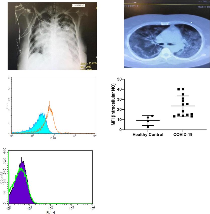
Chest X ray and CT imaging of the lungs shows signifi- (due to COPD and emphysema). As depicted Fig. 1e, no
cant changes in the lungs with bilateral alveolar diffusion significant increase of intracellular NO was seen in hyp-
confirming consolidation of the lungs in these patients oxic patients compared to non-hypoxic controls.
(Fig. 1 a and b). In addition, patients with severe
COVID-19 had higher serum lactate dehydrogenase (in- Discussion
dicating tissue damage), C-reactive protein peaks (indi- We demonstrate increased levels of intracellular NO in
cating inflammation) and lower counts of infection- RBC from COVID-19 subjects. This is not due to the
fighting lymphocytes than those with milder disease. No presence of hypoxia per se but may afford protection
significant differences were found in the levels of white against the hypoxia seen in COVID-19 patients. During
blood cells, creatinine kinase (measuring muscle inflam- health, constitutive NO production in RBCs is largely
mation) or in lactic acid (measure of muscle oxygen NOS-dependent, whereas in hypoxic conditions NO pro-
levels) (Table 1). Having confirmed the clinical and duction may involve nitrite reduction by deoxyhemoglo-
radiographic features of COVID-19 in these subjects we bin carbonic anydrase and/or eNOS itself [14].
then examined the levels of intracellular NO within their RBC-derived NO causes the vasodilation of small vessels
circulation RBCs. allowing oxygen to be readily released to tissues. In our
Isolated RBC were stained with DAF and intracellular study, intracellular RBC NO of COVID-19 patients is
NO determined. COVID-19 patients had a rightward significantly higher than in healthy controls and this may
shift in the FACs plot reflecting higher intracellular enable the release of oxygen to tissues resulting in the
levels of NO (Fig. 1c). The mean florescent intensity clinical manifestation of silent hypoxia in these patients.
(MFI) was calculated for each subject and plotted as a Pronounced arterial hypoxemia without proportionalMortaz et al. BMC Pulmonary Medicine (2020) 20:269 Page 4 of 6 Fig. 1 a Representative chest X ray of a COVID-19 patient on mechanical ventilation showing bilateral consolidations (red arrows). b Spiral CT scan of a representative COVID-19 patient indicating multiple bilateral patchy ground glass infiltration. c Red blood cells were preincubated with 5 mM of DAF for 20 min at 37 °C in PBS containing 1% BSA in the dark. Intracellular NO was determined by FACS analysis. A representative histograph from one out of 14 COVID-19 positive patients and 4 healthy controls is shown. d The mean fluorescent intensity (MFI) of all the subjects in each group is presented (*p < 0.05 using Student’s t-test). e Representative histogram of intracellular NO from RBCs of a single hypoxic non-COVID-19 patient signs of respiratory distress is reported in COVID-19 pa- The mechanism(s) underlying NO generation inside tients [15–18]. For example, Tobin and colleagues recently RBC is not well understood. However, acidosis, hypox- reported three cases of silent hypoxemia with a PaO2 ran- emia and tissue hypoxia lead to NO generation by RBC ging between 36 and 45 mmHg in the absence of in- via SNO–protein transfer of NO activity [20, 21]. The ef- creased alveolar ventilation [16]. ficiency of NO produced by RBC NOS to promote vaso- However, the mechanism(s) underlying this silent hyp- dilation is not well described however perfusion of blood oxia have not been explored despite the need to under- vessel segments with pre-sheared RBC suspensions stand why some COVID-19 patients are able to continue caused a significant dilation under hypoxic conditions, with their normal daily activities despite often pro- but not high oxygen, levels [22]. Vasodilation was abol- nounced hypoxia [19]. ished by pre-incubation of the RBC suspension with the Many theories have been proposed to account for this NOS inhibitor L-NAME. These findings support the silent hypoxia. For example, silent hypoxia may be due concept that RBC-derived NO has a functional role in to the differential effect of O2 and CO2 on gas exchange the regulation of local blood flow [22]. Moreover, shear which may produce a relative preservation of the lungs’ stress induces ATP release from hypoxic RBC as a con- ability to excrete CO2 despite falling O2 levels. Since the sequence of their role as O2 sensors [21]. body is better able to detect changes in CO2 than O2, Since NO is a pulmonary vasodilator and also has anti- the relatively normal CO2 levels may attenuate any drive viral activity against coronavirus strains it is likely that to increase the patients breathing rate despite the pres- exogenous NO treatment may be effective in COVID-19 ence of low oxygen levels and thereby prevent the sensa- subjects. There is no evidence that direct oxygen therapy tion of shortness of breath. is beneficial in the management of breathlessness in
Mortaz et al. BMC Pulmonary Medicine               (2020) 20:269                                                                                      Page 5 of 6

severe COVID-19 patients but our data suggests that                              Consent for publication
NO therapy may be beneficial in COVID-19 patients                                All participants has consent for publication form.

with hypoxia [23].
                                                                                 Competing interests
  Autoimmune hemolytic anemia (AIHA) was recently                                The authors have no conflicts of interest to declare.
described in COVID-19 patients [24, 25]. AIHA causes
platelet cell death and RBCs can also modulate platelet                          Author details
                                                                                 1
                                                                                   Mycobacteriology Research Center, National Research Institute of
activity directly through either chemical signalling or                          Tuberculosis and Lung Diseases (NRITLD), Masih Daneshvari Hospital, Shahid
direct RBC-platelet interactions. In this way RBCs pro-                          Beheshti University of Medical Sciences, Tehran, Iran. 2Tracheal Disease
mote platelet aggregation and degranulation by releasing                         Research Center, National Research Institute of Tuberculosisand Lung
                                                                                 Diseases (NRITLD), Shahid Beheshti University of Medical Science, Tehran,
ATP and ADP under low pO2, low pH and in response                                Iran. 3Chronic Respiratory Diseases Research Center, National Research
to mechanical deformation [26, 27]. In addition, the re-                         Institute ofTuberculosis and Lung Diseases (NRITLD), Shahid Beheshti
lease of extracellular hemoglobin can also cause platelet                        University of Medical Sciences, Tehran, Iran. 4University of Health Sciences
                                                                                 Turkey, Yedikule Chest Diseases and Thoracic Surgery, Education and
activation by lowering NO bioavailability [28]. Thus, our                        research Hospital, Department of pulmonology, Istanbul, Turkey. 5Division of
current finding and evidence for hemolysis in patients                           Pharmacology, Utrecht Institute for Pharmaceutical Sciences, Faculty of
may account for the microvascular coagulation seen in                            Science, Utrecht University, Utrecht, Netherlands. 6Cell and Molecular Biology
                                                                                 Group, Airways Disease Section, Faculty of Medicine, National Heart and
COVID-19 patients. We were unable to explore the                                 Lung Institute, Imperial College London, London, UK. 7Priority Research
mechanism(s) causing the accumulation of intracellular                           Centre for Asthma and Respiratory Disease, Hunter Medical Research
NO in RBC of COVID-19 patients in this study but this                            Institute, University of Newcastle, Newcastle, NSW, Australia.

will be the focus of future research.                                            Received: 3 July 2020 Accepted: 9 October 2020
  In summary, COVID-19 patients show higher levels of
NO inside RBC compared to non-COVID-19 hypoxemic
patients. Whether higher levels of intracellular NO in-                          References
                                                                                 1. Nile SH, Nile A, Qiu J, Li L, Jia X, Kai G. COVID-19: Pathogenesis, cytokine
side RBC of COVID-19 infected patients drive the unex-                               storm and therapeutic potential of interferons. Cytokine Growth Factor Rev.
pected silent hypoxia phenotype needs to be examined                                 2020;53:66–70.
in future clinical studies using NO donors in hypoxemic                          2. Pedersen SF, Ho Y-C. SARS-CoV-2: a storm is raging. J Clin Invest. 2020;
                                                                                     130(5):2202–5. https://doi.org/10.1172/JCI137647.
COVID-19 patients.                                                               3. Yuki K, Fujiogi M, Koutsogiannaki S. COVID-19 pathophysiology: A review.
                                                                                     Clin Immunol. 2020;215:108427. https://doi.org/10.1016/j.clim.2020.108427.
                                                                                 4. Soy M, Keser G, Atagündüz P, Tabak F, IşıkAtagündüz SK. Cytokine storm in
Conclusions                                                                          COVID-19: pathogenesis and overviewof anti-inflammatory agents used in
This pilot study shows that elevated levels of intracellu-                           treatment. Clin Rheumatol. 2020;39:2085–94.
lar NO may mask the effects of hypoxia in COVID-19                               5. Wang D, Hu B, Hu C, Zhu F, Liu X. Clinical characteristics of 138 hospitalized
                                                                                     patients with 2019 novel coronavirus-infected pneumonia in Wuhan, China.
patients that presents as a silent hypoxic state. Further
                                                                                     JAMA. 2020;7(323):1061–9.
studies are required to confirm this but the data suggests                       6. Chen G, Wu D, Guo W, Cao Y, Huang D, Wang H, et al. Clinical and
that trials of NO therapy or NO donors may be useful in                              immunological features of severe and moderate coronavirus disease 2019. J
                                                                                     Clin Invest. 2020;130(5:2620–9. https://doi.org/10.1172/JCI137244.
treating severe COVID-19 patients with hypoxia.
                                                                                 7. Cameron MJ, Bermejo-Martin JF, Danesh A, Muller MP, Kelvin DJ. Human
                                                                                     immunopathogenesis of severe acute respiratory syndrome (SARS). Virus
Abbreviations
                                                                                     Res. 2008;133:13–9.
ARDS: Acute Respiratory Distress Syndrome (ARDS); COVID-19: coronavirus
                                                                                 8. Magro G. SARS-CoV-2 and COVID-19: is interleukin-6 (IL-6) the ‘culprit lesion’
disease 2019 (COVID-19; FACS: Fluorescence-activated cell sorting; MFI: Mean
                                                                                     of ARDS onset? What is there besides Tocilizumab? SGP130Fc. Cytokine X.
florescent of intensity; NO: Nitric oxide; RBC: Red blood cells
                                                                                     2020;2(2):100029. https://doi.org/10.1016/j.cytox.2020.100029.
                                                                                 9. Han H, Ma Q, Li C, Liu R, Zhao L, Wang W, Zhang P, Liu X, Gao G, Liu F,
Acknowledgements                                                                     Jiang Y, Cheng X, Zhu C, Xia Y. Profiling serum cytokines in COVID-19
We acknowledge all study participants who are alive and remember those               patients reveals IL-6 and IL-10 are disease severity predictors. Emerg
patients who died of COVID-19.                                                       Microbes Infect. 2020;9(1):1123–30. https://doi.org/10.1080/22221751.2020.
                                                                                     1770129.
Authors’ contributions                                                           10. Zhang Y, Wang X, Li X, Xi D, Mao R, Wu X, Cheng S, Sun X, Yi C, Ling Z, Ma
EM performed the experiments and initial data analysis. MM, HRJ, PANN,               L, Ning Q, Fang Y, Sun B, Wu D. Potential contribution of increased soluble
SMRH, PT, MV and HZ provided the patients and samples. EGC, GF and IMA               IL-2R to lymphopenia in COVID-19 patients. Cell Mol Immunol. 2020;17(8):
critically reviewed and revised the manuscript. All authors reviewed the final       878–80. https://doi.org/10.1038/s41423-020-0484-x.
version and approved submission.                                                 11. Grivennikov SI, Tumanov AV, Liepinsh DJ, et al. Distinct and nonredundant
                                                                                     in vivo functions of TNF produced by T cells and macrophages/neutrophils:
Funding                                                                              protective and deleterious effects. Immunity. 2005;22:93–104.
This study was supported by the authors own funds.                               12. Tay MZ, Poh CM, Rénia L, Paul A, MacAry, Lisa F, Ng P. The trinity of COVID-
                                                                                     19: immunity, inflammation and intervention, vol. 20; 2020. p. 363–74.
Availability of data and materials                                               13. Horiuchi T, Mitoma H, Harashima S-i, Tsukamoto H, Shimoda T.
The data will be available upon written request.                                     Transmembrane TNF-α: Structure, function and interaction with anti-TNF
                                                                                     agents. Rheumatology (Oxford, England). 2010;49:1215–28.
Ethics approval and consent to participate                                       14. Ware CF, Crowe PD, Vanarsdale TL, Andrews JL, Grayson MH, Jerzy R, et al.
The study was approved by Ethical committee of Masih Daneshvari Hospital             Tumor necrosis factor (TNF) receptor expression in T lymphocytes.
IR (SBMU.NRITLD.REC.1399.123) and written consent was obtained from the              Differential regulation of the type I TNF receptor during activation of resting
study participants or their next of kin.                                             and effector T cells. J Immunol. 1991;147:4229–38.
Mortaz et al. BMC Pulmonary Medicine (2020) 20:269 Page 6 of 6
15. Tobin MJ, Laghi F, Jubran A. Why COVID-19 silent hypoxemia is baffling top
physicians. Am J Respir Crit Care Med. 2020; Available from: http://www.
ncbi.nlm.nih.gov/pubmed/32539537.6; [cited 2020 Jun 23].
16. Couzin-Frankel J. The mystery of the pandemic’s‘happy hypoxia’. Science.
2020;368(6490):455–6. https://doi.org/10.1126/science.368.6490.455.
17. Wilkerson RG, Adler JD, Shah NG, Brown R. Silent hypoxia: a harbinger of
clinical deterioration in patients with COVID-19. Am J Emerg Med. 2020; W.
B. Saunders; Available from:https://pubmed.ncbi.nlm.nih.gov/32471783/.8;.
18. Allali G, Marti C, Grosgurin O, Morélot-Panzini C, Similowski T. Adler D.Dyspnea:
the vanished warning symptom of COVID-19 pneumonia. J MedVirol. 2020;
jmv.26172 Wiley; [cited 2020 Jul 4]. Available from:https://onlinelibrary.wiley.
com/doi/abs/10.1002/jmv.26172).
19. Doctor A, Platt R, Sheram ML, Eischeid A, McMahon T, Maxey T, et al.
Hemoglobin conformation couples erythrocyte S-nitrosothiol content to O2
gradients. Proc Natl Acad Sci U S A. 2005;102:5709–14.
20. Singel DJ, Stamler JS. Chemical physiology of blood flow regulation by red
blood cells: the role of nitric oxide and S-nitrosohemoglobin. Annu Rev
Physiol. 2005;67:99–145.
21. Ulker P, Gunduz F, Meiselman HJ, Baskurt OK. Nitric oxide generated by red
blood cells following exposure to shear stress dilates isolated small
mesenteric arteries under hypoxic conditions. Clin Hemorheol Microcirc.
2013;54:357–69.
22. Ellsworth ML, Ellis CG, Goldman D, Stephenson AH, Dietrich HH, Sprague RS.
Erythrocytes: oxygen sensors and modulators of vascular tone. Physiology
(Bethesda). 2009;24:107–16.
23. Pimentel-Muinos FX, Seed B. Regulated commitment of TNF receptor
signaling: a molecular switch for death or activation. Immunity. 1999;11:
783–93.
24. Lazarian G, Quinquenel A, Bellal M, et al. Autoimmune hemolytic anemia
associated with Covid-19 infection. Br J Haematol. 2020. https://doi.org/10.
1111/bjh.16794 [published online ahead of print, 2020 may 6.
25. Lopez C, Kim J, Pandey A, Huang T, DeLoughery TG. Simultaneous onset of
COVID-19 and autoimmune hemolytic anemia. Br J Haematol. 2020. https://
doi.org/10.1111/bjh.16786.
26. Reimers RC, Sutera SP, Joist JH. Potentiation by red blood cells of shear-
induced platelet aggregation: relative importance of chemical and physical
mechanisms. Blood. 1984;64:1200–6.
27. Sprague RS, Ellsworth ML, Stephenson AH, et al. ATP: the red blood cell link
to NO and local control of the pulmonary circulation. Am J Phys. 1996;271:
H2717–22.
28. Helms CC, Marvel M, Zhao W, et al. Mechanisms of hemolysis-associated
platelet activation. J Thromb Haemost. 2013;11:2148–54.
Publisher’s Note
Springer Nature remains neutral with regard to jurisdictional claims in
published maps and institutional affiliations.You can also read