IMMUNOGENICITY AND PROTECTIVE ECACY OF BBV152: A WHOLE VIRION INACTIVATED SARS COV-2 VACCINE IN THE SYRIAN HAMSTER MODEL - RESEARCH SQUARE
←
→
Page content transcription
If your browser does not render page correctly, please read the page content below
Immunogenicity and protective e cacy of BBV152:
a whole virion inactivated SARS CoV-2 vaccine in
the Syrian hamster model
Sreelekshmy Mohandas
Indian Council of Medical Research-National Institute of Virology, Pune, Maharashtra, India, Pin-411021
Pragya D Yadav ( hellopragya22@gmail.com )
Indian Council of Medical Research-National Institute of Virology, Pune, Maharashtra, India, Pin-411021
Anita Shete
Indian Council of Medical Research-National Institute of Virology, Pune, Maharashtra, India, Pin-411021
Priya Abraham
Indian Council of Medical Research-National Institute of Virology, Pune, Maharashtra, India, Pin-411021
Krishna Mohan
Bharat Biotech International Limited, Genome Valley, Hyderabad, Telangana, India, Pin-500 078
Gajanan Sapkal
Indian Council of Medical Research-National Institute of Virology, Pune, Maharashtra, India, Pin-411021
Chandrashekhar Mote
Department of Veterinary Pathology, Krantisinh Nana Patil College of Veterinary Science, Shirwal,
Maharashtra, India, Pin-412801
Dimpal Nyayanit
Indian Council of Medical Research-National Institute of Virology, Pune, Maharashtra, India, Pin-411021
Nivedita Gupta
Indian Council of Medical Research, V. Ramalingaswami Bhawan, P.O. Box No. 4911, Ansari Nagar, New
Delhi, India, Pin-110029
V K Srinivas
Bharat Biotech International Limited, Genome Valley, Hyderabad, Telangana, India, Pin-500 078
Manoj Kadam
Indian Council of Medical Research-National Institute of Virology, Pune, Maharashtra, India, Pin-411021
Abhimanyu Kumar
Indian Council of Medical Research-National Institute of Virology, Pune, Maharashtra, India, Pin-411021
Rajlaxmi Jain
Indian Council of Medical Research-National Institute of Virology, Pune, Maharashtra, India, Pin-411021
Triparna Majumdar
Indian Council of Medical Research-National Institute of Virology, Pune, Maharashtra, India, Pin-411021
Gururaj Deshpande
Page 1/22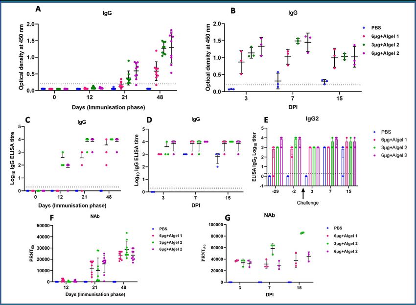
Indian Council of Medical Research-National Institute of Virology, Pune, Maharashtra, India, Pin-411021
Savita Patil
Indian Council of Medical Research-National Institute of Virology, Pune, Maharashtra, India, Pin-411021
Prasad Sarkale
Indian Council of Medical Research-National Institute of Virology, Pune, Maharashtra, India, Pin-411021
Deepak Patil
Indian Council of Medical Research-National Institute of Virology, Pune, Maharashtra, India, Pin-411021
Raches Ella
Bharat Biotech International Limited, Genome Valley, Hyderabad, Telangana, India, Pin-500 078
Sai D Prasad
Bharat Biotech International Limited, Genome Valley, Hyderabad, Telangana, India, Pin-500 078
Sharda Sharma
Indian Council of Medical Research-National Institute of Virology, Pune, Maharashtra, India, Pin-411021
Krishna M Ella
Bharat Biotech International Limited, Genome Valley, Hyderabad, Telangana, India, Pin-500 078
Samiran Panda
Indian Council of Medical Research, V. Ramalingaswami Bhawan, P.O. Box No. 4911, Ansari Nagar, New
Delhi, India, Pin-110029
Balram Bhargava
Indian Council of Medical Research, V. Ramalingaswami Bhawan, P.O. Box No. 4911, Ansari Nagar, New
Delhi, India, Pin-110029
Research Article
Keywords: SARS-CoV-2, Inactivated vaccine, Hamster, Neutralizing antibodies, Real time RT-PCR
DOI: https://doi.org/10.21203/rs.3.rs-76768/v1
License: This work is licensed under a Creative Commons Attribution 4.0 International License.
Read Full License
Page 2/22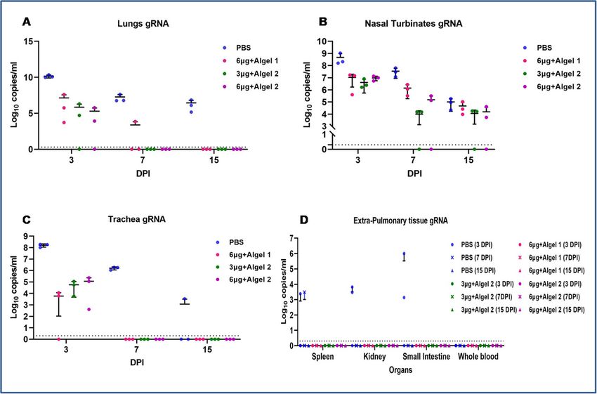
Abstract
The availability of a safe and effective vaccine would be the eventual measure to deal with SARS-CoV-2
threat. Here, we have developed and assessed the immunogenicity and protective e cacy of an
inactivated SARS-CoV-2 vaccine (BBV152) in hamsters. Three dose vaccination regime with three
formulations of BBV152 induced signi cant titres of SARS-CoV-2 speci c IgG and neutralizing
antibodies. The formulation with imidazoquinoline adsorbed on alum adjuvant remarkably generated a
quick and robust immune response. Th1 biased immune response was demonstrated by the detection
of IgG2 antibodies. Post-SARS-CoV-2 infection, vaccinated hamsters did not show any histopathological
changes in the lungs. The protection of the hamsters was evident by the rapid clearance of the virus from
lower respiratory tract, reduced virus load in upper respiratory tract, absence of lung pathology and robust
humoral immune response. These ndings con rm the immunogenic potential of BBV152 and further
protection of hamsters challenged with SARS-CoV-2.

Highlights
   BBV152, an inactivated vaccine induced potent humoral immune response in hamsters
   Th1 biased immune response was elicited by BBV152
   BBV152 protected Syrian hamsters from SARS-CoV–2 pneumonia

Introduction
Since the rst report in December 2019, severe acute respiratory syndrome coronavirus–2 (SARS-CoV–2)
has spread at an alarming rate and has infected more than 24 million people until August 31st 2020
(World Health Organization, 2020). Globally, scienti c communities are actively engaged and trying to
develop suitable vaccine candidates and speci c antiviral therapies against this virus. Vaccination is the
most signi cant pharmaceutical intervention for the prevention of any infectious disease impacting the
health of communities worldwide. Accelerated efforts are being taken for the development of a safe and
effective vaccine worldwide which is evident by the number of vaccine candidates under preclinical and
clinical evaluation. According to the World Health Organization draft landscape document on COVID–19
candidate vaccines published on 23rd August 2020, 33 and 143 vaccine candidates are under clinical and
preclinical studies respectively (World Health Organization, 2020).

Currently along with the conventional vaccine development platforms, advanced technologies are being
used for the development of vaccines against SARS-CoV–2 like messenger RNA, DNA, viral vectors,
recombinant subunit proteins, virus-like particles etc (Pandey et al., 2020). Despite the advances in
vaccine design technologies, the development of an inactivated vaccine remains the most simple and
relatively less expensive approach to produce a safe and effective vaccine. Inactivated vaccines have
been used effectively to curb many infectious diseases in the past (Sanders et al., 2015). Recently, two
inactivated SARS-CoV–2 vaccine candidates have shown promising results in preclinical trials (Gao et al.,
2020, Wang et al., 2020). Among various small laboratory animal models, Syrian hamsters was found to
                                                 Page 3/22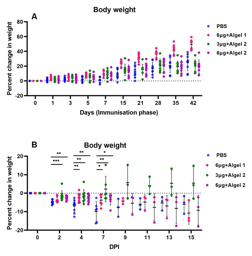
be a suitable model for SARS-CoV–2 research, as the virus has been shown to replicate both in the upper
and lower respiratory tract (Mohandas et al., 2020). The pulmonary pathology and lung viral load
coincides with weight loss in hamsters during the rst week of infection (Chan et al., 2020), and can be
used for the assessment of protective e cacy of vaccine candidates. This animal model has been
successfully used to evaluate medical countermeasures for SARS-CoV–2 by multiple research groups
(Luan et al., 2020, Chan et al., 2020, Imai et al., 2020).

We have developed a whole virion inactivated vaccine candidate (BBV152) using β-propiolactone (BPL)
inactivation method. The vaccine candidate along with aluminum hydroxide adjuvant alone or with
aluminum hydroxide chemisorbed with imidazoquinoline was found to be immunogenic and safe in the
preclinical studies on laboratory mice, rats and rabbits. Here, we report the immunization of Syrian
hamsters with the vaccine candidate (BBV152) and evaluation of protective e cacy against SARS-CoV–
2 by carrying out virus challenge experiment.

Results
Optimization of SARS-CoV–2 challenge dose in Syrian
hamsters
For assessment of the SARS-CoV–2 challenge dose after immunization a 10 fold serial dilutions of 105.5
Median Tissue Culture Infectious Dose (TCID50) were used. One hundred microliter from each of the 5
dilutions (105.5, 104.4,103.5, 102.5, and 101.5) was inoculated intranasally in 5 groups of 6 hamsters each.
On 3 day post infection (DPI), 3 hamsters each from all the 5 groups were sacri ced and the lung viral
titres were measured. Virus load was found to be similar in all the hamsters by real time RT-PCR
irrespective of the virus dilutions inoculated. Further to con rm this nding virus titration was performed
on the same lung samples and TCID50 titre ranging between 105.48 to 105.58 was observed in all samples
on 3DPI (Figure S1). Similarly on day 14, the remaining hamsters were sacri ced and lung samples were
found to show a titre of 102.5 TCID 50/ ml in all the animals, indicating that viral inoculum could induce
disease in all hamsters (Figure S1). On histopathological analysis, lungs samples of the hamsters
inoculated with the 105.5TCID50 and 104.5 TCID50 dilutions collected on 3DPI showed mild in ammatory
changes indicating beginning of pneumonia, whereas other groups showed minimal or no changes. On
14 DPI, the lung pathological changes were found minimal in all groups indicating recovery from
infection. Even though the lung viral titres in all groups were similar irrespective of the dose of virus
inoculums, the lung pathological changes indicated the rapid induction of pneumonic changes in 105.5
and 104.5 dilutions. Hence, a virus dilution dose of 105.5 TCID50 was used for virus challenge.

Clinical observations during BBV152 immunization period
and post SARS- CoV–2 challenge
                                                   Page 4/22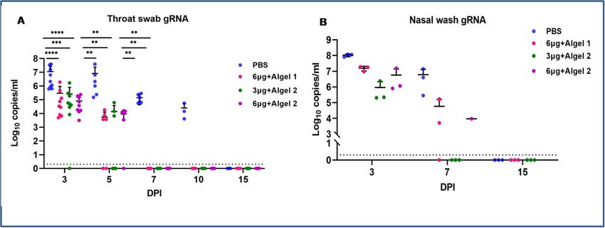
We immunized four groups of 6–8 week old Syrian hamsters (9 hamsters in each group), with phosphate
buffered saline (group I), 6µg of BBV152 with Algel1 (group II), 3µg of BBV152 with Algel2 (group III) and
6µg of BBV152 with Algel2 (group IV) (Figure 1). All the hamsters received two booster doses on day 14
and day 35. Rectal temperature remained within the normal range and no clinical signs were observed in
all the groups throughout the immunization period. The body weight increased until 7 weeks post-
immunization (Figure 2A), but following SARS-CoV–2 infection on day 50, decrease in body weight was
observed in all the groups (Figure 2B). However, the percentage decrease in vaccinated groups were lesser
compared to the group I. The decrease in body weight of group III hamsters was less as compared to
other groups (Figure 2).

Inactivated whole virion vaccine candidates induced
speci c IgG /neutralizing antibody and Th1 biased immune
response
Anti-SARS-CoV–2 IgG antibody response was detected by 3 weeks in 8/9 hamsters of group IV with an
average OD of 0.62, 8/9 hamsters in group III with an average OD of 0.42 and in 2/9 hamsters (average
OD = 0.285) of group II. On day 48, IgG antibody response was found to be increasing in the vaccinated
groups with an average OD of 1.32 in group IV (9/9 hamsters), 1.2 in group III (9/9 hamsters), 0.55 in
group II (9/9 hamsters) (Figure 3A, C). All the animals in group I remained negative for IgG antibody
during immunization period whereas post virus challenge, 2/3 hamsters showed IgG positivity by 7 DPI
and 3/3 by 14 DPI (average OD = 0.29) in the group I (Figure 3B). In the vaccinated groups, an increasing
trend with an average OD of 0.84, 0.97 and 0.91 was observed on days 3, 7 and 15 DPI respectively
(Figure 3D). No signi cant difference was observed in the IgG antibody response post-infection in group
III and IV (Figure 3A -E).

IgG antibody response in vaccinated hamsters was further characterized to determine the IgG subclass
pro les. On sub-typing IgG2 was detected in all the IgG antibody positive samples whereas it was
negative for IgG1 during immunization and post-infection phase (Figure 3E). All three formulations of
vaccine candidates signi cantly induced IgG2 with an increasing trend post-infection indicating a Th1
biased immune response (Figure 3 A-E).

Neutralizing antibody (NAb) started appearing in the immunized groups at 3rd week of immunization and
increased till 7th week with highest titre (mean = 28810) in group III (Figure 3F). After virus infection the
highest titre of NAb (mean = 85623) was seen in group III animals on 15 DPI. Group I did not show NAb
response during immunization phase and after virus infection till 15 DPI (Figure 3G).

Detection of SARS-CoV–2 genomic RNA (gRNA) in swabs/
organ samples post virus challenge
                                                   Page 5/22
SARS-CoV–2 viral genome copy number in throat swabs (TS) of group I were signi cantly high compared
to other groups on 3, 5 and 7 DPI (Figure 4A). The viral gRNA in group I persisted till 10 DPI, whereas it
was cleared in all the vaccinated groups by 5 DPI. Higher copy numbers of viral gRNA were detected in
the nasal washings of group I (Figure 4B).
Lungs (Figure 5A), Nasal turbinates (Figure 5B) and trachea (Figure 5C) of group I showed higher viral
gRNA copy number compared to other groups on 3 and 7 DPI. Trachea was cleared of viral gRNA by 7
DPI in all the groups. Complete gRNA clearance was observed from lungs of group III and IV on 7 DPI and
from group II by 15 DPI. Nasal turbinates viral gRNA persisted in all the groups till 15 DPI, but with lower
copy numbers in vaccinated groups compared to group I. No viral subgenomic (sg) RNA was detected in
TS, nasal wash, nasal turbinate or trachea of animals of vaccinated groups. However viral sgRNA was
detected in lungs (3/3), trachea (1/3) and nasal turbinate (1/3) in group I animals on 3 DPI. The spleen,
kidney and small intestine of hamsters of group I showed viral gRNA positivity on 3 DPI (Figure 5D).
Virus titration
Lungs, nasal turbinate and TS samples of group I showed an average titre of 106, 105.5 and 104
TCID50/ml respectively on 3 DPI. In the vaccinated groups III and IV, nasal turbinates showed an average
titre of 105 and 104 TCID50/ml on 3 DPI, whereas the TS and lungs titre was found considerably less
(102.5 TCID50/ml) compared to group I. In contrast group II did not show live virus titre in any of the
specimens on 3, 7 and 15 DPI. On 7 DPI only nasal turbinates of group I showed virus titre whereas
vaccinated groups were negative. This correlates with the decreasing trend of gRNA and sgRNA in
immunized groups after 3 DPI.
Pathological and Immuno-histochemistry ndings in lungs
post virus inoculation
The lungs of the vaccinated groups appeared normal on 3, 7 and 15 DPI (Figure 6B, 6C and 6D) on gross
pathology whereas on 7 DPI the lungs of group I showed diffuse areas of consolidation and congestion
(Figure 6A). On histopathological examination, lung sections from group I animals showed congestion,
haemorrhages, exudations in the alveoli, mononuclear cell in ltration in the alveolar interstitium and
pneumocyte hyperplasia on 3 and 7 DPI (Figure 6E, 6F). Occasionally, loss of bronchiolar epithelium was
also observed. By 15 DPI, bro-elastic proliferation with collagen deposition at alveolar epithelial lining
were observed in the lungs of group I (Figure 6G). Vaccinated group animals did not show any
histopathologic evidence of pneumonia except few congestive foci on 3 DPI (Figure 6H, 6I and 6J). The
viral antigen could be detected in alveolar type - II pneumocytes and macrophages on 3, 7 and 15 DPI in
the lungs of group I animals (Figure 7A, 7B and 7C) whereas only focal positivity was detected in the
vaccinated groups on 3 DPI. On 7 and 15 DPI viral antigen was not detected in the lung sections of
vaccinated hamsters from all the groups on immunohistochemistry (IHC) (Figure 7D, 7E and 7F).
Page 6/22Cytokine pro le after virus infection
After challenge with the virus vaccinated groups did not have any signi cant elevation of cytokines i.e
TNF-α, IL–4, IL–10, IL–6, IFN-γ and IL–12 whereas in control group I increased level of IL–12 was
detected on 3 DPI which further reduced on 7 and 15 DPI (Figure S2).
Discussion
Considering the pandemic scenario of COVID–19 worldwide a safe and effective vaccine is the most
appropriate and effective method for control of the rapid spread of this disease. Globally several research
groups are working on the development of vaccine candidates for COVID–19. Some of them have
progressed to the clinical trials (Doremalen et al., 2020; Mercado et al., 2020; Corbett et al., 2020; Patel et
al., 2020; Gao et al., 2020; Wang et al., 2020). Preclinical research in animal models is an important step
in evaluating the immunogenicity and protective e cacy of vaccine candidates. It aids in assessing the
host response and ndings on histopathology helps in evaluating the safety of the vaccine candidates
before progressing to clinical trials. Multiple animal models have been used to evaluate the e cacy of
SARS-CoV–2 vaccine candidates. Syrian hamster (Mesocricetus auratus) is one such model which has
been used in diverse research studies on SARS-CoV–2 and seems to be the appropriate model as it
mimics the human disease in comparison to other animals (Mohandas et al., 2020, Luan et al., 2020,
Chan et al., 2020, Imai et al., 2020; Wang et al., 2019). Here we report the immunogenicity and protective
e cacy of the three-dose vaccination regimen of inactivated SARS-CoV–2 vaccine candidate in the
hamster model.
SARS-CoV–2 speci c IgG titres were observed from 3rd week post-immunization in 90% of animals of
Group III and IV, indicating the faster and higher immunogenic potential of BBV152 with
imidazoquinoline. By 7 weeks all the vaccinated groups showed seroconversion however an increased
titre was observed in group III and IV compared to group II. Detection of IgG2 antibodies in the immunized
groups indicates Th1 biased immune response. Neutralizing antibodies was elevated in all the vaccinated
groups post SARS-CoV–2 challenge. Similarly Roberts et al. also reported a signi cant increase in the
antibody titres following the two-dose vaccination regimen of inactivated whole virus SARS-CoV vaccine
in hamsters (Roberts et al., 2010). Compared to the placebo group, rapid virus clearance was observed in
the vaccinated groups from the upper and lower respiratory tract except for nasal turbinates. Viral gRNA
in the nasal turbinates persisted till day 15 in all groups. However gRNA copy number was lower in
vaccinated groups compared to placebo. Gross and histopathological examination of the lungs of the
placebo group had evidence of interstitial pneumonia in the placebo group on 3 and 7 DPI. However
vaccinated groups had no evidence of gross and histopathological changes indicating the protective
e cacy of BBV152. Lower viral gRNA copy number at 3 DPI, absence of lung pathology and high titers of
neutralizing antibodies post-infection demonstrate the protective e cacy of BBV152 in immunised
hamsters.
Page 7/22Star Methods
Key Resource table
Page 8/22Reagent/Resource Source Identi er
Experimental models: Cell lines
Vero CCL-81 ATCC Cat#ATCC-CCL-81
Experimental models: Organisms/strains
Hamsters: Syrian Indian Council of --
hamsters Medical Research-
National Institute of
Nutrition, Hyderabad,
India
Virus strain
NIV-2020-770 ICMR-National Institute hCoV-19/India/770/2020|EPI_ISL_420545|
of Virology, Pune
hCoV19/India/2020770/2020|EPI_ISL_420546|
Antibodies
Biotin mouse anti- BD Biosciences Cat#554007
Armenian and Syrian
Hamster IgG1
Biotin mouse anti- BD Biosciences Cat#554025
Armenian and Syrian
Hamster IgG2
Anti-mice HRP Dako Cat #P044701-2
Chemicals
Imidazoquinoline Viro Vax
Alum Brenntag
b-Propionolactone Ferak
Minimum Essential Thermo Fisher Cat # 11534466
Medium (MEM) Scienti c
Dulbecco's Modi ed Sigma-Aldrich Cat # D5796
Eagle Medium
Foetal Bovine serum Sigma-Aldrich Cat # F4135
Penicillin/Streptomycin Sigma-Aldrich Cat # P4333
Skimmed milk powder Difco Cat #232100
3,3′,5,5′- Clinical science product Cat#01016-1-1000
Tetramethylbenzidine
Page 9/22(TMB) substrate
Critical Commercial Assays
Hamster Interleukin Immunotag Cat #ITE150025, ITE150027,ITE150010,
ELISA Kit ITE150004,ITE150058,ITE150028,ITE150030
MagMAX™ Thermo Fisher Cat #A42352
Scienti c
Viral/Pathogen Nucleic
Acid Isolation Kit
Software
PRISM GraphPad software Version 8
Lead Contact
Further information and requests for resources and reagents should be directed to the Lead Contact, Dr
Pragya D Yadav (hellopragya22@gmail.com).
Materials Availability
This study did not generate new unique reagents.
Data and Code Availability Statement
The published article includes all datasets generated or analysed during this study.
Experiment model and subject details
Cells and virus
Vero CCL81 cells were grown in Minimum Essential Media (MEM) (Thermo sher Scienti c, USA)
supplemented with 2% foetal bovine serum (FBS) (Sigma-Aldrich, USA). SARS-CoV-2 strain, NIV-2020-770
isolated from a patient's throat/ nasal swab sample at Indian Council of Medical Research- National
Institute of Virology, Pune was passaged up to three times in Vero CCL81 cells before use (Sarkale et al.,
2020). The infectious virus titre was found to be 106.5 tissue culture infective dose 50 (TCID50)/ml.
Ethics statement
Page 10/22The study was approved by the Institutional Project Review Committee, Institutional Animal Ethics
Committee and Institutional Biosafety Committee of Indian Council of Medical Research-National
Institute of Virology, Pune. All the experiments were performed as per the guidelines laid down by the
Committee for the Purpose of Control and Supervision of Experiments in Animals (CPCSEA, 2003).
Immunization of hamsters
Thirty-six, 6- 8 week old, female Syrian hamsters were divided into four groups, viz., Group I, II, III, and IV of
9 hamsters each. The hamsters were housed in individually ventilated cages with ad libitum food and
water. Group I was administered with phosphate-buffered saline (PBS), group II with 6µg of vaccine
candidate along with Algel 1, group III with 3µg of vaccine candidate with Algel 2, and group 4 with 3µg of
vaccine candidate with Algel 2. Animals of each group were immunized with 0.1 ml of PBS/vaccine
formulations intramuscularly in the left hind leg under iso urane anaesthesia 0, 14, and 35 days. Post
immunization hamsters were observed daily for clinical signs and injection site reaction. Rectal
temperature was monitored every 24 hours for 3 days post-immunization and weekly thereafter. Body
weight was measured every alternate day for the rst week and weekly thereafter. The hamsters were bled
on day 12, 21, and 48 post-immunization to check for antibody response.
Dose optimization study in hamsters
For optimization, of the intranasal virus challenge dose, SARS-CoV-2 dilutions ranging from 101.5
TCID 50/0.1ml to 105.5 TCID 50/ml were inoculated in 5 groups of 6 Syrian hamsters each in containment
facility. Three hamsters from each group were sacri ced on days 3 and 14 post-inoculation to check for
viral load in the lungs by real-time RT-PCR/virus titration and lung pathology.
Challenge study in hamsters
The immunized hamsters were challenged with 0.1 ml of 105.5 TCID50 SARS-CoV-2 virus intranasally on
the eighth-week post-immunization (day 50) in the containment facility of ICMR-National Institute of
Virology, Pune under iso urane anaesthesia. Throat swabs were collected in 1 ml virus transport media
on every alternate day post-inoculation for viral load estimation. Three hamsters from each group were
euthanized on 3, 7 and 15 DPI to collect throat swab, nasal wash, rectal swab, blood and organ samples
for viral RNA estimation, titration, histopathology, and immunological analysis.
Method details
Inactivated SARS-CoV-2 whole virion vaccine (BBV152)
Page 11/22SARS-CoV-2 strain (NIV-2020-770) isolated at ICMR-NIV, Pune was propagated in Vero CCL81 cells and
harvested on observation of cytopathic effect in the cells. At BBIL BPL (Ferak, Germany) was added to the
virus harvest following ltration and stabilization of the harvest using a buffer. The mixture was kept at 2-
8˚C with continuous stirring for 24 hrs and was further hydrolysed by incubating at 37˚C for 2hrs. Column
chromatography was used for further puri cation and the process intermediate was concentrated to
prepare the whole virion vaccine. Two different antigen concentrations (3 µg and 6 µg) and 2 adjuvants
namely Algel 1 (Alum) and Algel 2 (TLR 7/8 (imidazoquinoline) agonist adsorbed alum) in combinations
were used for the study. The vaccine formulations evaluated in the study were 6 µg antigen with Algel1, 3
µg with Algel 2, and 6 µg with Algel 2.
Enzyme-linked Immunosorbent Assay
Ninety-six well microtitre plates were coated with 1:10 diluted inactivated SARS-CoV-2 antigen with
carbonate buffer (pH 9.5) overnight at 4 °C. Subsequently, wells were blocked with liquid plate sealer
(CANDOR Bioscience GmbH, Germany) for two hours at room temperature (25-30°C). The wells were
washed 5 times with phosphate-buffered saline with 0.05% Tween 20 (PBS-T) and were incubated at
37°C for one hour with 100μl of diluted hamster serum samples (1: 100). Negative control was added to
each plate. After 5 washes with PBS-T, anti-hamster IgG antibodies 1:3000 (Thermoscienti c, USA) were
added and incubated for 1 hour at 37°C. Following 5 washes with PBS-T, 100 μl of substrate, 3’,3’5,5’-
tetramethylbenzidine (TMB) was added to each well. The colour reactions were developed for 10 minutes
and after termination, absorbance was measured at 450 nm. Serum IgG titres were determined by testing
serial 10-fold dilutions of each sample, starting from 1:100 dilution. Titre values were determined as the
highest dilution at which the optical density was more than 0.2 and positive/negative (P/N) ratio above
1.5.
For antibody sub-typing, plates were coated with antigen and blocked as described earlier. Hamster
serum, 1:100 diluted in 1% bovine serum albumin in 1× PBST was added to each well and incubated for 1
hour at 37°C. After washing the plates 5 times with 1× PBST wells were probed with biotinylated anti-
Syrian hamster IgG1 /IgG2 antibodies diluted at 1:10000 (BD biosciences, USA) and incubated for 1 hour
at 37°C. After washing, the plates were incubated with Streptavidin-horseradish peroxidase 1:8000
(Thermo-scienti c, USA) for 30 minutes at 37°C. The reaction was read as described earlier.
Plaque Reduction Neutralization test
The assay was performed as described by Gururaj et al., 2020. Brie y, a four-fold serial dilution of
hamster serum samples was mixed with an equal amount of virus suspension and incubated at 37°C for
1 hour. Further 0.1 ml of the mixture was inoculated in a 24-well tissue culture plate containing a
con uent monolayer of Vero CCL-81 cells. The plate was incubated at 37°C for 60 min and overlay
medium (2% carboxymethyl cellulose with 2% FBS in 2X MEM) was added to the cell monolayer, which
Page 12/22was further incubated at 37°C in 5% CO2 incubator for 4-5 days and PRNT 50 titres were calculated as
described earlier.
Cytokine analysis
The serum cytokine levels (TNF-α, IFN-γ, IL-4, IL-6, IL-10 and IL-12) were assessed in hamsters post
challenge at 3, 7, and 15 days. An ELISA based commercial assay (Immunotag, USA) was used for the
hamster speci c cytokine quantitation. For this, plates pre-coated with hamster speci c cytokine antibody
were used and a streptavidin based HRP system was used for detection and the absorbance was
measured at 450 nm.
Real-time RT-PCR for detection of genomic and
subgenomic viral RNA
Nasal wash, throat swab and rectal swab samples were collected in 1ml viral transport medium and
weighed organ samples (lungs, nasal turbinate, trachea, spleen, kidney and intestine) were triturated in 1
ml sterile tissue culture media, using a tissue homogenizer (Eppendorf, Germany) and 200 µl of the
homogenate/ swab specimens were used for further RNA extraction using MagMAX™ Viral/Pathogen
Nucleic Acid Isolation Kit as per the manufacturer’s instructions. Real-time RT-PCR was performed for E
and RdRp2 gene for SARS-CoV-2 as well as for detection of sgRNA of E gene using published primers
(Choudhary et al., 2020 and Wolfelet al., 2020)
Virus titration
The lungs, nasal turbinate and throat swab samples from 3, 7, and 15 DPI were used for virus titration in
Vero CCL81 cells. 100 ml of the sample was added onto a 24-well plate with Vero CCL81 monolayers and
incubated for one hour at 37°C. The media was removed and the cell monolayer was washed with
PBS. The plate was incubated with maintenance media with 2% FBS in a CO2 incubator. The plate was
examined daily for any cytopathic effects (CPE) using an inverted microscope (Nikon, Eclipse Ti,
Japan). The culture supernatant from the wells showing CPE was con rmed by real-time RT-PCR (Gururaj
et al., 2020).
Histopathology and immunohistochemistry
Lungs samples collected during necropsy were xed in 10% neutral buffered formalin. The tissues were
processed by routine histopathological techniques for hematoxylin and eosin staining. Duplicate sections
were taken for immunohistochemical evaluation. An in-house developed anti-SARS-CoV-2 mouse
polyclonal serum was used as the primary antibody for detection. The tissue sections were rehydrated
Page 13/22and antigen retrieval was performed using 0.3% hydrogen peroxide in methanol. The slides were
incubated with 1: 500 dilution of primary antibody for an hour and an anti-mouse HRP antibody (Dako,
USA) was used as a secondary antibody. For detection, 3, 3’-diaminobenzidine tetrahydrochloride
substrate, and hydrogen peroxide were used.
Data analysis
For analysis of the data, Graphpad Prism version 8.4.3 software was used. The statistical signi cance
was assessed using the Kruskal-Wallis test Dunn's multiple comparisons test. Two-tailed Mann-Whitney
test was performed between the control and the vaccinated groups if the p-value for the Kruskal-Wallis
test was found to be signi cant; p-values less than 0.05 were considered to be statistically signi cant.
Declarations
Acknowledgments
Authors acknowledge the support received from and laboratory team which includes Dr. Himanshu
Kaushal,Deepak Mali, Rashmi Gunjikar, Rajen Lakra, Shreekant Baradkar, Pranita Gawande, Ganesh
Chopade, Manjunath Holleppanavar, Ratan More, Darpan Phagiwala, Chetan Patil, Sanjay Thorat, Ratan
More, Madhav Acharya, Malvika Salave, Ashwini Baghmare, Ciyona, Sapna Gawande, Nitin Deshpande
and Poonam Bodke and Deepika Chawdhary, Mayur Mohite, Vishal Gaikwad and Nandkumar Sharma of
ICMR-National Institute of Virology, Pune, India. We acknowledge the excellent support of Dr Sapan
Kumar Behra, Dr Shashi Kanth Muni, Dr Rajaram Ravikrishnan, Dr Brunda Ganneru from Bharat Biotech
International Limited, Genome Valley, Hyderabad, Telangana, India.
Author’s Contribution
SM, PDY, KM, PA, NG and BB conceived and designed the study. KME, RE, VKS and SDP performed
vaccine design and production. PDY, SM performed the planning of animal experiments. SM, PDY, MK,
AK, CM performed the animal experimentation. PDY, AS, GS, GD, DN, DYP performed the laboratory work
planning and data analysis. GD, SP, RJ, TM, SS, PS performed sample processing in the laboratory. PDY,
SM, DN, AS and DYP have drafted the manuscript. PDY, SM, AS, DYP, KM, PA, NG, SP and
BB substantively revised it. All authors reviewed the manuscript and agree to its contents.
Declaration of interests
The authors declare no competing interests.
Page 14/22Lead Contact
Further information and requests for resources and reagents should be directed to the Lead Contact, Dr
Pragya D Yadav (hellopragya22@gmail.com).
Materials Availability
This study did not generate new unique reagents.
Data and Code Availability Statement
The published article includes all datasets generated or analysed during this study.
Ethics statement
The study was approved by the Institutional Project Review Committee, Institutional Animal Ethics
Committee and Institutional Biosafety Committee of Indian Council of Medical Research-National
Institute of Virology, Pune. All the experiments were performed as per the guidelines laid down by the
Committee for the Purpose of Control and Supervision of Experiments in Animals (CPCSEA, 2003).
References
1. Chan JF, Zhang AJ, Yuan S, Poon VK, Chan CC, Lee AC, Chan WM, Fan Z, Tsoi HW, Wen L, Liang R.
Simulation of the clinical and pathological manifestations of Coronavirus Disease 2019 (COVID-19)
in golden Syrian hamster model: implications for disease pathogenesis and transmissibility. Clinical
Infectious Diseases. 2020 Mar 26.
2. Chan, J.F.W., Zhang, A.J., Yuan, S., Poon, V.K.M., Chan, C.C.S., Lee, A.C.Y., Chan, W.M., Fan, Z., Tsoi,
H.W., Wen, L. et al. (2020). Simulation of the clinical and pathological manifestations of Coronavirus
Disease 2019 (COVID-19) in golden Syrian hamster model: implications for disease pathogenesis
and transmissibility. Clin Infect Dis.
3. Corbett, K.S., Flynn, B., Foulds, K.E., Francica, J.R., Boyoglu-Barnum, S., Werner, A.P., Flach, B.,
O’Connell, S., Bock, K.W., Minai, M. et al. (2020). Evaluation of the mRNA-1273 Vaccine against
SARS-CoV-2 in Nonhuman Primates. N Engl J Med.
4. CPCSEA (2003). CPCSEA guidelines for laboratory animal facility. Indian J Pharmacol, 35(4), 257-
274.
Page 15/225. Gao Q, Bao L, Mao H, Wang L, Xu K, Yang M, Li Y, Zhu L, Wang N, Lv Z, Gao H. (2020). Development
of an inactivated vaccine candidate for SARS-CoV-2. Science.
6. Imai, M., Iwatsuki-Horimoto, K., Hatta, M., Loeber, S., Halfmann, P.J., Nakajima, N., Watanabe, T., Ujie,
M., Takahashi, K., Ito, M. et al. (2020). Syrian hamsters as a small animal model for SARS-CoV-2
infection and countermeasure development. Proceedings of the National Academy of
Sciences, 117(28),16587-16595.
7. Luan J, Lu Y, Jin X, Zhang L. (2020) Spike protein recognition of mammalian ACE2 predicts the host
range and an optimized ACE2 for SARS-CoV-2 infection. Biochemical and biophysical research
communications.
8. Mercado, N.B., Zahn, R., Wegmann, F., Loos, C., Chandrashekar, A., Yu, J., Liu, J., Peter, L., McMahan,
K., Tostanoski, L.H. et al. (2020). Single-shot Ad26 vaccine protects against SARS-CoV-2 in rhesus
macaques. Nature 1-11.
9. Mohandas, S., Jain, R., Yadav, P.D., Shete-Aich, A., Sarkale, P., Kadam, M., Kumar, A., Deshpande, G.,
Baradkar, S., Patil, S. et al. (2020). Evaluation of the susceptibility of mice & hamsters to SARS-CoV-2
infection. Indian Journal of Medical Research, 151(5), p.479.
10. Pandey, S. C., Pande, V., Sati, D., Upreti, S., Samant, M. (2020). Vaccination strategies to combat novel
corona virus SARS-CoV-2. Life sciences 256, 117956.
11. Patel, A., Walters, J., Reuschel, E.L., Schultheis, K., Parzych, E., Gary, E.N., Maricic, I., Purwar, M.,
Eblimit, Z., Walker, S.N. et al. (2020). Intradermal-delivered DNA vaccine provides anamnestic
protection in a rhesus macaque SARS-CoV-2 challenge model. bioRxiv.
12. Roberts, A., Lamirande, E.W., Vogel, L., Baras, B., Goossens, G., Knott, I., Chen, J., Ward, J.M., Vassilev,
V. and Subbarao, K. (2010). Immunogenicity and protective e cacy in mice and hamsters of a β-
propiolactone inactivated whole virus SARS-CoV vaccine. Viral immunology 23(5), 509-519.
13. Rogers, T.F., Zhao, F., Huang, D., Beutler, N., Burns, A., He, W.T., Limbo, O., Smith, C., Song, G., Woehl, J.
and Yang, L., 2020. Isolation of potent SARS-CoV-2 neutralizing antibodies and protection from
disease in a small animal model. Science.
14. Sanders B, Koldijk M, Schuitemaker H. Inactivated viral vaccines. In Vaccine analysis: Strategies,
principles, and control 2015 (pp. 45-80). Springer, Berlin, Heidelberg.
15. Sia, S.F., Yan, L.M., Chin, A.W., Fung, K., Choy, K.T., Wong, A.Y., Kaewpreedee, P., Perera, R.A., Poon, L.L.,
Nicholls, J.M. et al. (2020). Pathogenesis and transmission of SARS-CoV-2 in golden
hamsters. Nature 583(7818), 834-838.
16. van Doremalen, N., Lambe, T., Spencer, A., Belij-Rammerstorfer, S., Purushotham, J.N., Port, J.R.,
Avanzato, V.A., Bushmaker, T., Flaxman, A., Ulaszewska, M. et al. (2020). ChAdOx1 nCoV-19 vaccine
prevents SARS-CoV-2 pneumonia in rhesus macaques. Nature 1-8.
17. Wang H, Zhang Y, Huang B, Deng W, Quan Y, Wang W, Xu W, Zhao Y, Li N, Zhang J, et al. (2020).
Development of an inactivated vaccine candidate, BBIBP-CorV, with potent protection against SARS-
CoV-2. Cell. Aug 6;182(3):713-21.
Page 16/2218. Wang, Y., Miao, J., Chard, L. and Wang, Z. (2019). Syrian hamster as an animal model for the study
on infectious diseases. Frontiers in immunology 10, 2329.
19. World Health Organization (2020). Coronavirus disease (COVID-19) outbreak situation. Available
from: https://www.who.int/emergencies/diseases/novel-coronavirus-2019
20. World Health Organization (2020). Draft landscape of COVID-19 candidate vaccines-28 August 2020.
Available from: https://www. who. int/who-documents-detail/draft-landscape-of-covid-19-candidate-
vaccines.
Figures
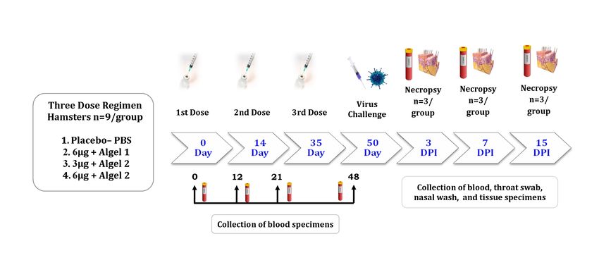
Figure 1
Schematic presentation of experiments in hamsters: Three doses of placebo were administered to the
rst group of the animals which were controls in the study. Three different inactivated SARS-CoV-2
vaccine formulations (3 doses ) were administered to the three groups of animals. All the animals were
challenged at 15 days after the third dose. Samples were collected at different time points of
immunization period and post-infection as indicated in the gure. Necropsy was performed for three
hamsters from each group at 3, 7, and 15 DPI.
Page 17/22Figure 2
Percent body weight gain/loss in hamsters. A. Percentage of body weight gain in hamsters during the
immunization period. B. Percent difference in body weight in hamsters post SARS-CoV-2 challenge. Mean
along with standard deviation (SD) is depicted in the scatter plot. The statistical signi cance was
assessed using the Kruskal-Wallis test followed by the two-tailed Mann-Whitney test between the two
groups; p-values less than 0.05 were considered to be statistically signi cant.
Page 18/22Figure 3
Humoral response in vaccinated animals: (A) IgG antibody response for all groups of animals observed
on 12, 21 and 48 days. (B) IgG antibody response at post-infection (3, 7 and 15 DPI) for all groups of
animals. (C) Comparison of IgG antibody titres between groups on 12, 21 and 48 days. (D) Comparison
of IgG antibody titres between groups post virus challenge at 3, 7 and 15 DPI. (E). Comparison of IgG2
antibody titres between groups during immunization period at 21 and 48 days and post virus challenge at
3,7,15 DPI. (F). Comparison of NAb titres response during a three-dose vaccine regime for all groups of
animals observed on 12, 21 and 48 days (G). Comparison of NAb titres response post virus challenge on
3, 7 and 15 DPI. Mean along with standard deviation (SD) is depicted in the scatter plot. The statistical
signi cance was assessed using the Kruskal-Wallis test followed by the two-tailed Mann-Whitney test
between the two groups; p-values less than 0.05 were considered to be statistically signi cant. The dotted
lines indicate the limit of detection of the assay.
Page 19/22Figure 4
Log10 plot for the genomic viral RNA detection in throat swab and nasal wash after virus challenge.
Genomic viral RNA load in (A) Throat Swab collected at 3, 5, 7, 10, and 15 DPI for the all groups (B) Nasal
wash collected at 3, 7, and 15 DPI for the all groups. Mean along with standard deviation (SD) is depicted
in the scatter plot. The statistical signi cance was assessed using the Kruskal-Wallis test followed by the
two-tailed Mann-Whitney test between the two groups; p-values less than 0.05 were considered to be
statistically signi cant. The dotted lines indicate the limit of detection of the assay.
Page 20/22Figure 5
Log10 plot of the genomic viral RNA detection in the respiratory tract and extra pulmonary specimens.
Genomic viral RNA load in (A) Lung, (B) Nasal turbinates (C) Trachea (D) Extra pulmonary organs at 3, 7,
and 15 DPI. Mean along with standard deviation (SD) is depicted in the scatter plot. The statistical
signi cance was assessed using the Kruskal-Wallis test followed by the two-tailed Mann-Whitney test
between the two groups; p-values less than 0.05 were considered to be statistically signi cant. The dotted
lines indicate the limit of detection of the assay.
Figure 6
Gross and histopathological observations of lungs in hamsters post virus inoculation. A) Lungs of
hamster from group I on 7 DPI showing diffuse areas of consolidation and congestion in the left and
right lower lobe with few congestive foci in right upper lobe. Lungs from B) group II, C) group III and D)
group IV showing normal gross appearance on 7 DPI. E) Lung tissue from group I on 3 DPI showing acute
in ammatory response with diffuse alveolar damage, hemorrhages, in ammatory cell in ltration (black
arrow), hyaline membrane formation (white arrow) and accumulation of eosinophilic edematous exudate
(star). F) Lung tissue of group I on 7 DPI showing acute interstitial pneumonia with marked alveolar
Page 21/22damage, thickening of alveolar and accumulation of mononuclear cells and macrophages (white arrow),
and lysed erythrocytes in the alveolar luminal space (star). G) Lung tissue from group I on 15 DPI
depicting interstitial pneumonia with marked thickening of alveolar septa with type-II pneumocyte
hyperplasia and bro-elastic proliferation with collagen deposition at alveolar epithelial lining (white
arrow). Lung section from group II showing no evidence of disease H) on 3 DPI few congestive foci I) on
7 DPI J) on 15 DPI.
Figure 7
Immunohistochemistry ndings in lungs of hamsters post virus challenge. Left panel depicts group
treated with placebo and the right panel shows vaccinated group II animals. Lung section from group I
showing viral antigen A) on 3 DPI in alveolar type-II pneumocytes (black arrow) and in alveolar
macrophages (white arrow) B) on 7 DPI in alveolar type-II pneumocytes (black arrow) C) on 15 DPI in
type-II alveolar pneumocytes (black arrow). Lung section from group II showing viral antigen D) on 3 DPI
in alveolar macrophages (black arrow) E) on 7 DPI in alveolar epithelium and alveolar macrophages F) on
15 DPI in the alveolar epithelium and alveolar macrophages.
Supplementary Files
This is a list of supplementary les associated with this preprint. Click to download.
SupplementaryFigureS1.png
SupplementaryFigureS2.png
Page 22/22You can also read