LOW DOSE INOCULA OF SARS-COV-2 ALPHA VARIANT TRANSMITS MORE EFFICIENTLY THAN EARLIER VARIANTS IN HAMSTERS - NATURE
←
→
Page content transcription
If your browser does not render page correctly, please read the page content below
ARTICLE
                  https://doi.org/10.1038/s42003-021-02640-x                OPEN

                  Low dose inocula of SARS-CoV-2 Alpha variant
                  transmits more efficiently than earlier variants in
                  hamsters
                  Bobo Wing-Yee Mok1, Honglian Liu1, Shaofeng Deng1, Jiayan Liu1, Anna Jinxia Zhang 1, Siu-Ying Lau1,
                  Siwen Liu1, Rachel Chun-Yee Tam1, Conor J. Cremin1, Timothy Ting-Leung Ng2, Jake Siu-Lun Leung 2,
                  Lam-Kwong Lee2, Pui Wang1, Kelvin Kai-Wang To 1, Jasper Fuk-Woo Chan1, Kwok-Hung Chan1,
                  Kwok-Yung Yuen 1, Gilman Kit-Hang Siu2 & Honglin Chen 1 ✉
1234567890():,;

                  Emerging variants of SARS-CoV-2 have been shown to rapidly replace original circulating
                  strains in humans soon after they emerged. There is a lack of experimental evidence to
                  explain how these natural occurring variants spread more efficiently than existing strains of
                  SARS-CoV-2 in transmission. We found that the Alpha variant (B.1.1.7) increased competitive
                  fitness over earlier parental D614G lineages in in-vitro and in-vivo systems. Using hamster
                  transmission model, we further demonstrated that the Alpha variant is able to replicate and
                  shed more efficiently in the nasal cavity of hamsters than other variants with low dose and
                  short duration of exposure. The capability to initiate effective infection with low inocula may
                  be one of the key factors leading to the rapid transmission of emerging variants of SARS-
                  CoV-2.

                  1 Department
                             of Microbiology and State Key Laboratory for Emerging Infectious Diseases, Li Ka Shing Faculty of Medicine, The University of Hong Kong,
                  Hong Kong SAR, China. 2 Department of Health Technology and Informatics, Faculty of Health and Social Sciences, The Hong Kong Polytechnic University,
                  Hong Kong SAR, China. ✉email: hlchen@hku.hk

                  COMMUNICATIONS BIOLOGY | (2021)4:1102 | https://doi.org/10.1038/s42003-021-02640-x | www.nature.com/commsbio                                       1
ARTICLE COMMUNICATIONS BIOLOGY | https://doi.org/10.1038/s42003-021-02640-x
I
n late 2020, a novel SARS-CoV-2 variant of concern (VOC), SARS-CoV-2 Alpha variant replicated more efficiently in the
VOC 202012/01 (lineage B.1.1.7) was identified in the United nasal cavity of hamsters than earlier D614G lineage with
Kingdom. This B.1.1.7 (Alpha) variant containing multiple enhanced transmission in hamsters. Next, we set up a Syrian
mutations in spike1 has become dominant in the UK and is now hamster infection study to evaluate if the Alpha variant exhibits
rapidly spreading across multiple countries2. It is thought that higher infectivity in vivo. 6-8-week-old male Syrian hamsters
this VOC has the potential to spread more quickly and with were intranasally infected with different variants of 1000 PFU per
higher mortality than the pandemic to date3. Recently, using inoculum, as indicated in Fig. 3a. Infectious viral titres in upper
multiple behavioural and epidemiological data sources, Davies (nasal) and lower (pulmonary) tissues were measured on four
et al. estimated that the VOC 202012/01 variant (lineage B.1.1.7) consecutive days after infection. All viruses tested replicated to
has a 43–90% higher reproduction number than pre-existing similar titres in nasal turbinate and lung tissues of infected
variants in England4. In another study, Davies et al. found that hamsters. This result is consistent with two recent studies which
among specimens collected in the UK in early 2021, higher also found no significant alteration in infectious viral titres in
concentrations of virus RNA were detected in nasopharyngeal samples collected from nasal washes, throat swabs and lungs from
swabs from B.1.1.7 infected individuals, as measured by Ct values hamsters infected with different SARS-CoV-2 variants16,17.
from qRT-PCR testing5. However, there is a lack of experimental Given that hamsters are highly susceptible to SARS-CoV-2
evidence to explain how the Alpha variant is able to spread more infection18, intranasal infection with high-titre inocula may
quickly than pre-existing variants. hamper discrimination of differences in the infectivity and
The Alpha variant of SARS-CoV-2 harbours 21 nonsynon- replication efficiency of variants19. In fact, by titrating the
ymous point mutations and three deletions in comparison to the infection dosage (10-fold dilution) of the inocula administered to
reference genome (accession number: NC_0.45512.2)6. Of these, hamsters, we observed that viral replication in nasal tissues of
eight mutations and two deletions are in the spike protein, which infected hamsters had already plateaued with infection doses of
interacts with the host cell receptor, angiotensin-converting 100 PFU and upwards, even on day one post-infection
enzyme 2 (ACE2), and mediates virus entry into host cells7. (Supplementary Figure 1). Humans are exposed to varying doses
These spike mutations include the deletion ΔH69/ΔV70, which of infectious particles during SARS-CoV-2 transmission and
has arisen in multiple independent lineages and is suggested to some exposures may fail to establish effective infection due to
associate with increased infectivity and evasion of the immune insufficient infectious particles. We reasoned that SARS-CoV-2
response8; the substitution N501Y, which enhances binding affi- variants, which can initiate effective infection with fewer
nity for the human ACE2 receptor and might affect viral infectious particles are likely to transmit more efficiently than
transmissibility9–11; and the mutation P681H, which is adjacent other variants requiring more infectious particles. To test this, we
to the S1/S2 furin cleavage site in spike and might have an impact performed hamster infection and transmission studies using only
on viral infectivity12,13. Interestingly, two other VOCs, P1 and 10 PFU per inoculum (as illustrated in Supplementary Figure 2).
B.1.351, also contain the N501Y mutation14,15. Interestingly, infectious viral loads in nasal turbinates of hamsters
The aim of this study is to provide experimental evidence to were found to be significantly higher on day one post-infection
explain why the Alpha variant has increased transmissibility with the Alpha variant compared to the other strains, whereas
among the human population, compared to earlier D614G var- similar viral loads were observed in lungs of all infected hamsters,
iants. Our results demonstrated that the Alpha variant exhibits except for those inoculated with HK-95, which exhibits higher
increased competitive fitness over earlier D614 variants in Calu-3 viral titres in lungs, although with large variations between
cells and in hamsters. Moreover, with low dose inocula, the Alpha replicates (Fig. 3b). Accordingly, the Alpha variant also showed
variant initiates more robust infectious in the upper respiratory enhanced transmissibility to naive hamsters compared with HK-
tracts of hamsters and transmits more efficiently than earlier 405 (Fig. 3c). Surprisingly, we observed a striking histological
D614G variants. difference between the Alpha variant infected lung (10 PFU;
Donor) and the Alpha variant recipient lung at day 4
postinfection, with more severe pathological changes observed
Results and discussion in the donor hamsters. Also, immunohistochemistry staining
SARS-CoV-2 Alpha variant showed enhance replication fitness showed extensive viral antigen in donor lung tissue in the
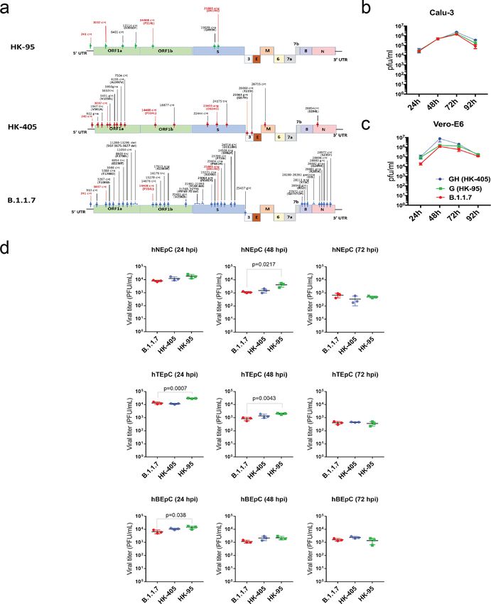
compared with earlier D614G variants. A recent study indicated bronchiolar epithelium and alveoli, while the lung of recipient
that the SARS-CoV-2 variant of concern (VOC) carrying the hamster showed viral antigen only in the bronchiolar epithelium
501Y (B.1.1.7 or the Alpha variant) mutation showed no higher (Fig. 3d). This result demonstrated that the route of exposure
infectivity in Huh-7, Vero, and LLC-MK2 cells than ancestral (direct inoculation vs indirect contact transmission) and initial
D614G variants16. Likewise, we did not observe replication of the dosage of the viral inoculum can largely affect the response of
Alpha variant to be significantly enhanced over that of the pre- animals to infection. However, no apparent difference of body
existing D614G variants (HK-95, collection date: 2020-05-15 and weight loss (Fig. 4a), viral gRNA copies in nasal wash (from 2 dpi
HK-405, collection date: 2020-12-08) (Fig. 1a) at any of the onwards) (Fig. 4b) and histopathology (Fig. 4c & d) was observed
selected time-points in Vero-E6, Calu-3 cells and human primary between HK-405 and the Alpha variants, indicating that the
airway cell lines (Fig. 1b–d). However, we did observe that the pathogenicity of the Alpha variants is comparable to the pre-
Alpha variant dominates in competitive fitness assays. These existing D614G variants.
comparisons of replication fitness between the Alpha variant and
earlier circulating strains were performed in Calu-3 cells and in
hamsters through simultaneous co-infection at a 1:1 ratio with Calu3 responses to SARS-CoV-2 Alpha variant drive less
the Alpha variant and early variants of the D614G lineage (HK- macrophage activation. Although we observed viral load differ-
405 or HK-95). After three rounds of consecutive passage at 72-h ence in hamster nasal cavities on day 1 postinfection with low dose
intervals in cells or 1 day postinfection in hamsters, the Alpha inoculum, almost no replication difference of the variants was
variant became the dominant population (Fig. 2), suggesting that measured in all tested cell cultures. We speculated that this dis-
those new substitutions in the Alpha variant enhance SARS-CoV- crepancy might be explained by the innate immune micro-
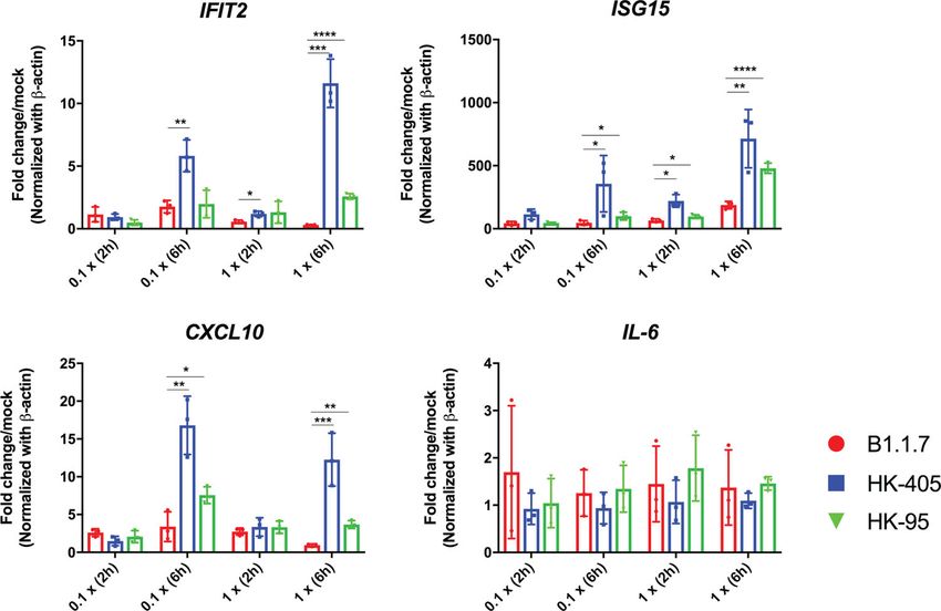
2 replication fitness in in vitro and in vivo systems. environment in which crosstalk between the epithelial and
2 COMMUNICATIONS BIOLOGY | (2021)4:1102 | https://doi.org/10.1038/s42003-021-02640-x | www.nature.com/commsbio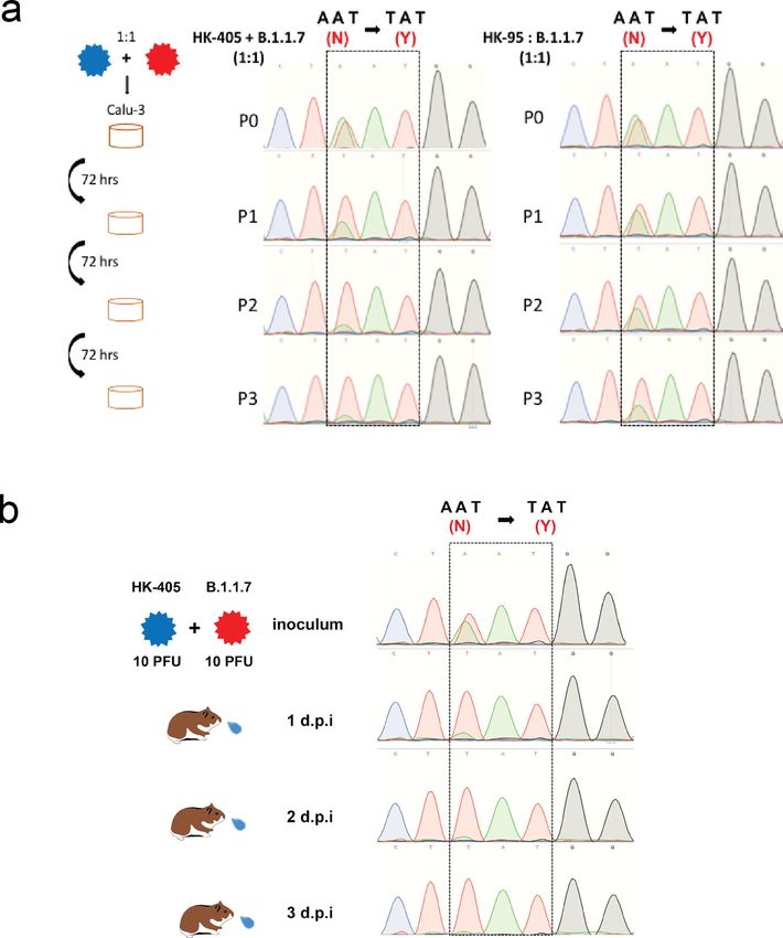
COMMUNICATIONS BIOLOGY | https://doi.org/10.1038/s42003-021-02640-x                                                                        ARTICLE

Fig. 1 Viral growth kinetics of Sars-CoV-2 D614G variants. a Genomic sequence comparison of the D614G variants used in this study to the reference
genome (accession number: NC_0.45512.2). Common SNPs in all thee strains are shown in red. b and c Growth curves of B.1.1.7, HK-405 and HK-95 in
Calu-3 cells and Vero-E6 at a MOI = 0.01 and MOI = 0.001, respectively. Virus-infected cells were cultured at 37 °C and supernatants then harvested at
the indicated time points and subjected to plaque assay in Vero E6 cells to determine the virus titre. Error bars represent mean ± SD (n = 3 per group for
each timepoint). d Comparison of 24, 48 and 72 h titres between B.1.1.7, HK-405 and HK-95 infected primary nasal (hNEpC), trachel (hTEpC) and
bronchial (hBEpC) epithelial cells (PromoCell) at MOI = 0.1. Triplicated titres of the viruses in the cultures from the cell lines were analysed by Student’s
t-test. Horizontal lines indicate the mean ± SD of viral titre per group.

COMMUNICATIONS BIOLOGY | (2021)4:1102 | https://doi.org/10.1038/s42003-021-02640-x | www.nature.com/commsbio                                               3
ARTICLE                                                                 COMMUNICATIONS BIOLOGY | https://doi.org/10.1038/s42003-021-02640-x

Fig. 2 In-vitro and in-vivo competitive fitness assay. Sanger sequencing chromatograms of spike gene fragments amplified from viral samples in the
competition assay. a Cell cultures were infected with a 1:1 mixture of two variants, as indicated, at an MOI of 0.1. The supernatants were serially passaged
three times in Calu-3 cells. 901 bp fragments containing residue 501 (boxed) were amplified from the vRNA of individual samples collected from each
passage (P) and sequenced. HK-95 and HK-405 contain 501N, B.1.1.7 contains 501Y. Initial inoculum was stated as P0 and P1-P3 indicated number of
passages. b Hamsters were inoculated with a mixture of 10 PFU of each of the two viruses (HK-405 and B.1.1.7). Nasal washes were collected daily for
three consecutive days and the vRNA of individual samples collected and the inoculum were sequenced.

immune cells occurs. Variants acquire different abilities to                   Alpha variant might acquire enhanced immune antagonism to
antagonise the host innate immune system, which could directly                 support a higher replication rate in in vivo systems.
affect its own dissemination. During the initial stage of infection,              SARS-CoV-2 VOCs have been emerging in many countries in
the infected epithelial cells along the nasal cavity could express             the past few months, and it is crucial to establish relevant
stimulatory factors to recruit and trigger immune cells to release             experimental models to characterise existing and new variants in
inflammatory signals to prime neighbouring uninfected cells                     terms of transmissibility, disease severity and vaccine efficacy, and
response to infection. To imitate this situation, we treated THP-1             to evaluate therapeutic interventions. In this report, by using a
cells with filtered, virus-free conditioned media supernatants from             lower infectious dose, we demonstrate that the Alpha variant
different SARS-CoV-2 infected Calu-3 cells. It has been proposed               exhibits higher infectivity and/or replication efficiency in the
that increased expression of the key viral innate antagonists, like            nasal epithelium. Our data, albeit based on only one of the VOCs,
Orf9b and Orf6, might contribute to immune evasion of the Alpha                show that novel VOCs can transmit more efficiently than other
variant20. Consistently, we demonstrated that the immune sti-                  pre-existing strains with fewer infectious viral particles. Further
mulatory activity of conditioned media in the Alpha variant                    work to compare transmission ability with other emerging VOCs
infected Calu-3 cells was much attenuated than that of the earlier             and investigation of routes of transmission are required. A
D614G variants (Fig. 5), suggesting that the viral mutations in the            better understanding of SARS-CoV-2 transmission dynamics is

4                           COMMUNICATIONS BIOLOGY | (2021)4:1102 | https://doi.org/10.1038/s42003-021-02640-x | www.nature.com/commsbio
COMMUNICATIONS BIOLOGY | https://doi.org/10.1038/s42003-021-02640-x                                                                            ARTICLE

important for designing appropriate strategies for a more effective            (ATCC; CRL-15786). All experiments involving SARS-CoV-2 viruses were con-
response to emerging variants of SARS-CoV-2.                                   ducted in a Biosafety Level-3 laboratory. For animal challenge, viral stocks were
                                                                               prepared after two serial passages of isolated virus in Vero E6 cells in Dulbecco’s
                                                                               Modified Eagle Medium (DMEM) (Thermo Fisher Scientific) supplemented with
Methods                                                                        5% foetal bovine serum (Thermo Fisher Scientific), and 100 IU penicillin G/ml and
Viruses. The SARS-CoV-2 isolates HK-95 (MT835143), HK-405 (MW856793),          100 ml streptomycin sulphate/ml (Thermo Fisher Scientific), and were divided into
B.1.1.7 (MW856794) and HK-15 (MT835141) were isolated from specimens           aliquots. The aliquots were then sequenced, and viral titres were determined by
obtained from four laboratory-confirmed COVID-19 patients using Vero E6 cells   plaque assay using Vero E6 cells. All viruses used in this study were from the same

COMMUNICATIONS BIOLOGY | (2021)4:1102 | https://doi.org/10.1038/s42003-021-02640-x | www.nature.com/commsbio                                                    5
ARTICLE COMMUNICATIONS BIOLOGY | https://doi.org/10.1038/s42003-021-02640-x
Fig. 3 In-vivo infection studies. a and b Replication efficiency of different SARS-CoV-2 variants in nasal turbinates and lungs of hamsters. Hamsters were
infected with different SARS-CoV-2 variants, as indicated. Viral titres in nasal turbinates and lungs were determined by plaque assay (PFU/ml). a Hamsters
(14 per variant virus group) were each inoculated intranasally with 50 ul of virus stock containing 1000 PFU of virus. Three to four hamsters from each
group were euthanized on each of the four consecutive days following infection for viral titration. b Hamsters (4-5 per group) were each inoculated
intranasally with 50 ul of virus stock containing 10 PFU of virus. One non-D614G lineage variant (HK-15 (MT835141)) and three D614G lineage variants
(HK-405, B.1.1.7 and HK-95) were used. Hamsters were euthanized on one day post-infection for viral titration. Horizontal lines indicate the mean (±SD) of
viral titre per group. Statistical significance was calculated by Student’s t-test; * denotes p < 0.05, *** denotes p < 0.0005 and ns denotes non-significant. c
Viral titres in nasal washes collected from exposed hamsters on day 2 and day 4 post-exposure. The number of positive hamsters in both exposure groups
on day 4 postexposure (HK-405 vs. B.1.1.7 = 0 of 8 vs. 3 of 8), * CT value ≥35. d Representative images of H&E (upper) and immunohistochemistry
(lower) stained hamster lung tissues taken on day 4 post-infection. The right panels were uninfected hamster lung showing normal structure and negative
of viral antigen. H&E images in the left panel was B.1.1.7 infected lung (10 PFU; Donor) showing lung consolidation with massive alveoli infiltration and
localised haemorrhage (open arrows). The bronchiolar lumens filled with cell debris (solid arrows) and thickened alveolar wall. Images of
immunohistochemistry-stained SARS-Cov-2 nucleoprotein (brown) in donor lung tissue showed extensive viral antigen expression in the bronchiolar
epithelium (open arrows) and alveoli (solid arrows). While the lung of recipient hamster (middle) only showed viral antigen in the bronchiolar epithelium
(open arrows). Scale bars = 200 µM.
Fig. 4 Characterisation of hamsters infected with low dose inocula of B.1.1.7 and HK-405. a Body weight loss of hamsters infected with low dose inocula
(10 PFU) of B.1.1.7 and HK-405. b Viral genome copies in nasal washes of the hamsters infected with B.1.1.7 and HK-405 collected on day 1 to 4
postinfection. Individual data points and mean ± SD are shown. c Histological score for lungs of infected hamsters on 4 dpi. d Representative images of
H&E-stained hamster lung (left superior lobes) taken on 1 and 4 dpi. On 1 dpi, both viruses infected lung showed alveolar congestion. But on 4 dpi, diffuse
alveolar infiltration, exudation and localized haemorrhage were observed after both viruses infection. Bronchiolar luminal cell debris (solid arrows) and
vascular infiltration (open arrows) were also observed. Scale bars = 200 µM.
batch of aliquots and used without freeze-thawing. Viral RNAs were obtained from reaction (PCR), as described in the ARTIC network (https://artic.network/ncov-
the supernatants of infected cells and then isolated using the QIAamp RNA Viral 2019). The PCR contained a pool of 98 primer pairs, which generated overlapping
kit (Qiagen) and subjected to whole viral genome sequencing. 400-bp amplicons across the entire genome of SARS-CoV-2 (accession no:
NC_045512) (primer sequences are shown in Supplementary Table 1). The final
PCR mastermix (25 µL) included 2.5 µL of cDNA, 5.0 µL of 5X Q5 Reaction Buffer,
Whole viral genome sequencing. Viral RNA was extracted from the supernatant 0.5 µL of 10 mM dNTP mix, 0.25 µL of Q5 Hot Start DNA Polymerase (New
of the infected Vero E6 cells using the QIAamp RNA Viral kit (Qiagen). 140ul of England Biolabs) and 3.6 µL of 10 µM primer pool 1 or 2, plus 13.15 µL of
the sample was treated with 560 µl of AVL, containing carrier RNA, and then nuclease-free water. The mixtures were incubated at 98 °C for 30 s, followed by 35
mixed with 560 µl of absolute ethanol. The sample was then transferred into cycles at 98 °C for 15 s and 65 °C for 5 min. The PCR amplicons were then purified
QiaAmp spin column, centrifuged, washed with wash buffer provided and then using 1X Agencourt AMPure XP beads (Beckman Coulter).
eluted with 20 ul of DEPC-treated water. Viral RNA was then treated using the A total of 100 ng of multiplex PCR amplicons were subjected to library
TURBO DNA-free Kit (ThermoFisher Scientific) to remove residual host DNA, preparation and dual-indexing using a KAPA HyperPrep Kit and a Unique Dual-
and then reverse-transcribed using SuperScript IV reverse transcriptase (Invitro- Indexed Adapter Kit (Roche Applied Science) according to the manufacturer’s
gen). The viral cDNA was then enriched through multiplex tiling polymerase chain instructions. Ligated libraries were then enriched by six-cycle PCR amplification,
6 COMMUNICATIONS BIOLOGY | (2021)4:1102 | https://doi.org/10.1038/s42003-021-02640-x | www.nature.com/commsbioCOMMUNICATIONS BIOLOGY | https://doi.org/10.1038/s42003-021-02640-x ARTICLE
Fig. 5 Calu-3 cells response to B.1.1.7 drive less macrophage activation. M0 THP1 cells were treated with filtered, virus-free conditioned media
supernatants (prepared from Calu-3 cells infected with different variants as indicated) at dilution 0.1x (1:10 dilution with DMEM) and 1x (no dilution) for 2
or 6 h. Expression of immune genes were then measured by qPCR analysis. Data shown are mean ±SD, n = 3, statistical comparison are made by Student’s
t test.
followed by purification and size selection using AMPure XP beads (Beckman double-layered divider to allow free airflow (70 air changes per hour). After two
Coulter). The pooled libraries were sequenced using the MiSeq Reagent Kit V2 hours of exposure, each naivë hamsters (n = 8 per group) was placed in a new
Nano on an Illumina MiSeq System. The Illumina MiSeq sequencing reads were individual cage. To assess viral replication in nasal turbinates, we determined the
then demultiplexed and mapped to the reference genome (accession number: virus load in the nasal wash specimens collected from exposed hamsters on day 2
NC_0.45512.2) using Samtools v1.7. Variants were called with Freebayes v1.0.0 and day 4 after transmission. Viral RNA was extracted from 70 μl of each nasal
(https://arxiv.org/abs/1207.3907) with the haploid setting, with a minimum base wash sample.
quality and depth of coverage of Q30 and 50x, respectively.
Hamster infection. Female golden Syrian hamsters, aged 4–6 or 6–8 weeks old, In-vitro competitive fitness assay. Calu-3 cells in Dulbecco’s Modified Eagle
were obtained from the LASEC, Chinese University of Hong Kong via the Centre Medium (DMEM) (Thermo Fisher Scientific) supplemented with 5% foetal bovine
for Comparative Medicine Research at the University of Hong Kong (HKU). All serum (Thermo Fisher Scientific), and 100 IU penicillin G/ml and 100 ml strepto-
experiments were performed in a Biosafety Level-3 animal facility at the LKS mycin sulphate/ml (Thermo Fisher Scientific) were infected with MOI of 0.1 of B.1.1.7
Faculty of Medicine, HKU. Hamsters were housed in ventilated isolator cages and another variant of the D614G lineage, either HK-95 or HK-405 mixture at 1:1
(IsoCage N, Tecniplast) at a temperature of 21 °C, the humidity of 70%, and 12:12 ratio. Following 1 h incubation, the cultures were washed thrice with PBS and cultured
dark/light cycles, with access to food and water ad libitum. Housing conditions and for 3 days. To passage the progeny viruses, the virus samples were continuously
experimental procedures were approved by the Committee on the Use of Live passaged three times in Calu-3 cells. Viral RNAs were obtained from the supernatants
Animals in Teaching and Research, HKU. Hamsters were anaesthetised with of infected cells and then isolated using the QIAamp RNA Viral kit (Qiagen). A 901 bp
ketamine (150 mg/kg) and xylazine (10 mg/mg) via intraperitoneal injection and fragment containing the N501Y site was amplified from each RNA sample by RT-PCR
then intranasally inoculated with 50 µl of diluted virus stock. All hamsters were using primer set: 5’- GAAGTCAGACAAATCGCTCCAG-3’ and 5’-GCAACT-
euthanized by intraperitoneal injection of pentobarbital at 200 mg/kg. Body weights GAATTTTCTGCACCA-3’. The amplicon was purified by NucleoSpin® Gel and PCR
were monitored daily for 4 days. Nasal washes were collected from hamsters daily Clean-Up (Takara) for Sanger sequencing.
for 4 consecutive days. Total nucleic acid was extracted from 70 ul of sample fluid
using the QIAamp RNA Viral kit (Qiagen). In brief, sample was treated with 280 µl
of AVL, containing carrier RNA, followed by 280 µl of 100% ethanol. The sample Treatment of THP-1 cells with virus-free conditioned medium. The human
was then transferred into QiaAmp spin column, centrifuged, washed with buffer monocytic cell line THP-1 was maintained in RPMI medium supplemented with
provided and then eluted with 10 µl of DEPC-treated water. 7.4 µl RNA was used 10% FBS, NEAA, L-glutamate, sodium pyruvate and penicillin/streptomycin. To
for reverse transcription using MultiScribe Reverse Transcriptase (Thermofisher). differentiate into M0 macrophage-like cells, THP-1 cells were cultured at 100,000
cDNA was subsequently used for real-time qPCR using TB Green Premix Ex Taq cells per well in a 96-well plate in the presence of 50 ng/ml phorbol 12-myristate
(Tli RNase H Plus) (Takara). For the in vivo co-infection experiment, viral RNA 13-acetate (PMA) for 2 days, followed by washing out in PMA-free medium for
from nasal washes was collected for sanger sequencing. All Hamsters were 2 days. To generate virus-free conditioned media, Calu-3 cells were mock-infected
euthanized at 4 dpi, lung right lobes (superior, middle, and inferior) or nasal or infected with Sars-Cov-2 variants at 0.01 MOI, and supernatant were harvested
turbinates were homogenised in 1 ml of PBS. After centrifugation at 12,000 rpm for at 48 h postinfection, filtered using Amicon Ultra 100 K (Millipore). Then, M0
10 min, the supernatant was harvested, and viral titres were determined by plaque THP-1 cells were exposed to the conditioned medium. RNA from treated THP-1
assay using Vero E6 cells. Lung left superior lobes were fixed in 4% paraf- cells were extracted using RNAzol (Takara). Expression of immune genes were
ormaldehyde and then processed for paraffin embedding. The 4 µm tissue sections then measured by qRT-PCR analysis. Data shown are mean and standard deviation
were stained with haematoxylin and eosin for histopathological examination. of 3 replicates per condition. P-values were determined by Student’s t-test.
Immunohistology was performed using mouse anti-coronavirus nucleocapsid
antibody (1:200, 40143-MM05, Sinobiological). Images were obtained with an
Olympus BX53 semimotorised fluorescence microscope using cellSens imaging
342 software. RT-qPCR. RNA was extracted using RNAzol (Takara) and cDNA was synthesised
using PrimeScript RT Reagent Kit with gDNA Eraser (Takara). RT–qPCR was
performed using TB Green Premix Ex Taq (Takara). The PCR program consisted
Transmission experiment. Three 4–6-week-old hamsters were inoculated intra- of a pre-incubation for 10 min at 95 °C, followed by 40 cycles of denaturation for
nasally with 10 PFU (50 μl) of virus. At 24 h after infection, each infected hamster 10 sec at 95 °C, annealing for 10 sec at 60 °C and extension for 10 sec at 72 °C. Host
was placed in a specially designed cage inside a ventilated cage. Two to three naive gene expression was determined using the 2-ΔΔCt method and normalised to beta-
hamsters were placed on the other side of the cage with 5 cm separation by a actin expression. RT-qPCR primers used in this study are listed in Table 1.
COMMUNICATIONS BIOLOGY | (2021)4:1102 | https://doi.org/10.1038/s42003-021-02640-x | www.nature.com/commsbio 7ARTICLE COMMUNICATIONS BIOLOGY | https://doi.org/10.1038/s42003-021-02640-x
12. Hoffmann, M., Kleine-Weber, H. & Pohlmann, S. A Multibasic Cleavage Site
Table 1 Primers used for RT-qPCR.
in the Spike Protein of SARS-CoV-2 Is Essential for Infection of Human Lung
Cells. Mol. Cell 78, 779–784 (2020).
Target Primer sequence 13. Seyran, M. et al. The structural basis of accelerated host cell entry by SARS-
CoV-2†. Febs j. 2, 15651 (2020).
human_beta-actin Fwd: 5’-GGAAATCGTGCGTGACATTA-3'
14. Faria, N. R., Claro, I. M., Candido, D. Genomic characterisation of an
Rev: 5’-AGGAGGAAGGCTGGAAGAG-3' emergent SARS-CoV-2 lineage in Manaus: preliminary findings. https://
human_IFIT2 Fwd: 5’-AGGCTTTGCATGTCTTGG-3' virological.org/t/genomic-characterisation-of-an-emergent-sars-cov-2-
Rev: 5’-GAGTCTTCATCTGCTTGTTGC-3' lineage-in-manaus-preliminary-findings/586 (2021).
human_Cxcl10 Fwd: 5’-GTGGCATTCAAGGAGTACCTC-3' 15. Tegally, H. et al. Detection of a SARS-CoV-2 variant of concern in South
Rev: 5’-TGATGGCCTTCGATTCTGGATT-3' Africa. Nature 592, 438–443 (2021).
human_IL6 Fwd: 5’-CCAGGAGAAGATTCCAAAGATG-3' 16. Abdelnabi, R. et al. Comparing infectivity and virulence of emerging SARS-
Rev: 5’-GGAAGGTTCAGGTTGTTTTCTG-3' CoV-2 variants in Syrian hamsters. EBioMedicine 68, 103403 (2021).
human_ISG15 Fwd: 5’-CGCAGATCACCCAGAAGATCG-3' 17. Mohandas, S. et al. Comparison of the pathogenicity and virus shedding of
Rev: 5’-TTCGTCGCATTTGTCCACCA-3' SARS CoV-2 VOC 202012/01 and D614G variant in hamster model. bioRxiv.
https://doi.org/10.1101/2021.02.25.432136 (2021).
nCov-spike Fwd: 5’-GAAGTCAGACAAATCGCTCCAG-3'
18. Chan, J. F. et al. Simulation of the clinical and pathological manifestations of
Rev: 5’-GCAACTGAATTTTCTGCACCA-3' Coronavirus Disease 2019 (COVID-19) in golden Syrian hamster model:
implications for disease pathogenesis and transmissibility. Clin. Infect. Dis. 3,
2428–2446 (2020).
Statistics and reproducibility. Graphpad prism Version 7.05. (GraphPad) was 19. Imai, M. et al. Syrian hamsters as a small animal model for SARS-CoV-2
used for all statistical analysis. The number of animals and independent experi- infection and countermeasure development. Proc. Natl Acad. Sci. USA 117,
ments that were performed is indicated in the legends to figures. Statistical sig- 16587–16595 (2020).
nificance was determined using Student’s t-test. P values of ≤0.05 were considered 20. Thorne, L. G. et al. Evolution of enhanced innate immune evasion by the
as statistically significant. SARS-CoV-2 B.1.1.7 UK variant. bioRxiv. https://doi.org/10.1101/
2021.06.06.446826 (2021).
Reporting Summary. Further information on research design is available in the Nature
Research Reporting Summary linked to this article.
Acknowledgements
The authors would like to thank Dr Jane Rayner for critical reading and editing of the
Data availability manuscript. This study is partly supported by the Theme-Based Research Scheme (T11/
Source data for all figures are provided in Supplementary Data file 1. Sequencing data of 707/15 and T11-709/21-N), the Collaborative Research Fund (C5110-20GF) and General
all the viral strains used in the current study were deposited in Genbank with the Research Fund (17107019) of the Research Grants Council, Innovation and Technology
following accession number: HK-95 (MT835143), HK-405 (MW856793), B.1.1.7 Fund of Innovation and Technology Commission, the Government of the Hong Kong
(MW856794) and HK-15 (MT835141). Special Administrative Region, China, and the Sanming Project of Medicine in Shenzhen,
China (No. 290 SZSM201911014).
Code availability
No in-house computer code or script was used in this study. The programs used for Author contributions
analysis of sequencing data are open-source and published, e.g. Samtools v1.7 and B.W.M. designed the project, performed experiments and wrote the manuscript. H.L.,
Freebayes v1.0.0. S.D., J.L, A.J.Z., S.Y.L., S.L., R.C.T., C.J.C., P.W. performed experiments. T.T.N., J.S.L.,
L.L., G.K.S were responsible for viral sequencing and deposition. H.C. interpreted the
results and wrote the manuscript, which was approved by K.K.T., J.F.C., K.C, K.Y.
Received: 3 August 2021; Accepted: 1 September 2021;
Competing interests
The authors declare no competing interests.
References
1. England Public Health. Investigation of novel SARS-CoV-2 Variants of Additional information
Supplementary information The online version contains supplementary material
Concern. https://www.gov.uk/government/publications/investigation-of-
available at https://doi.org/10.1038/s42003-021-02640-x.
novel-sars-cov-2-variant-variant-of-concern-20201201 (2020).
2. O’Toole, A., Hill, V., Pybus, O. G. Tracking the international spread of SARS-
Correspondence and requests for materials should be addressed to Honglin Chen.
CoV-2 lineages B.1.1.7 and B.1.351/501Y-V2. https://virological.org/t/
tracking-the-international-spread-of-sars-cov-2-lineages-b-1-1-7-and-b-1- Peer review information Communications Biology thanks the anonymous reviewers for
351-501y-v2/592 (2021). their contribution to the peer review of this work. Primary Handling Editor: George
3. Grint, D. J. et al. Case fatality risk of the SARS-CoV-2 variant of concern Inglis.
B.1.1.7 in England. Eur. Surveill. 26, 2100256 (2021).
4. Davies, N. G. et al. Estimated transmissibility and impact of SARS-CoV-2
Reprints and permission information is available at http://www.nature.com/reprints
lineage B.1.1.7 in England. Science 9, 6538 (2021).
5. Davies, N. G. et al. Increased mortality in community-tested cases of SARS-
Publisher’s note Springer Nature remains neutral with regard to jurisdictional claims in
CoV-2 lineage B.1.1.7. Nature 593, 270–274 (2021).
published maps and institutional affiliations.
6. Rambaut, A. et al. Preliminary genomic characterisation of an emergent
SARS-CoV-2 lineage in the UK defined by a novel set of spike mutations.
https://virological.org/t/preliminary-genomic-characterisation-of-an-
emergent-sars-cov-2-lineage-in-the-uk-defined-by-a-novel-set-of-spike- Open Access This article is licensed under a Creative Commons
mutations/563 (2020) Attribution 4.0 International License, which permits use, sharing,
7. Starr, T. N. et al. Deep mutational scanning of SARS-CoV-2 receptor binding adaptation, distribution and reproduction in any medium or format, as long as you give
domain reveals constraints on folding and ACE2 binding. Cell 3, 1295–1310 appropriate credit to the original author(s) and the source, provide a link to the Creative
(2020). Commons license, and indicate if changes were made. The images or other third party
8. Kemp, S. et al. Recurrent emergence of SARS-CoV-2 spike deletion H69/V70 material in this article are included in the article’s Creative Commons license, unless
and its role in the Alpha variant B.1.1.7. Cell Rep. 29, 109292 (2021). indicated otherwise in a credit line to the material. If material is not included in the
9. Tian, F. et al. Mutation N501Y in RBD of Spike Protein Strengthens the article’s Creative Commons license and your intended use is not permitted by statutory
Interaction between COVID-19 and its Receptor ACE2. bioRxiv. https:// regulation or exceeds the permitted use, you will need to obtain permission directly from
doi.org/10.1101/2021.02.14.431117 (2021). the copyright holder. To view a copy of this license, visit http://creativecommons.org/
10. Liu, Y. et al. The N501Y spike substitution enhances SARS-CoV-2 licenses/by/4.0/.
transmission. bioRxiv. https://doi.org/10.1101/2021.03.08.434499 (2021).
11. Dejnirattisai, W. et al. The antigenic anatomy of SARS-CoV-2 receptor
binding domain. Cell 15, 2183–2200 (2021). © The Author(s) 2021
8 COMMUNICATIONS BIOLOGY | (2021)4:1102 | https://doi.org/10.1038/s42003-021-02640-x | www.nature.com/commsbioYou can also read