RAPID DIRECT NUCLEIC ACID AMPLIFICATION TEST WITHOUT RNA EXTRACTION FOR SARS-COV-2 USING A PORTABLE PCR THERMOCYCLER - MDPI
←
→
Page content transcription
If your browser does not render page correctly, please read the page content below
G C A T
T A C G
G C A T
           genes
Article
Rapid Direct Nucleic Acid Amplification Test Without
RNA Extraction for SARS-CoV-2 Using a Portable
PCR Thermocycler
Soon Keong Wee , Suppiah Paramalingam Sivalingam                and Eric Peng Huat Yap *
 Lee Kong Chian School of Medicine, Nanyang Technological University, Singapore 308232, Singapore;
 wees0021@e.ntu.edu.sg (S.K.W.); spsuppiah@ntu.edu.sg (S.P.S.)
 * Correspondence: ericyap@ntu.edu.sg

 Received: 9 May 2020; Accepted: 16 June 2020; Published: 18 June 2020

 Abstract: There is an ongoing worldwide coronavirus disease 2019 (Covid-19) pandemic caused by
 severe acute respiratory syndrome coronavirus 2 (SARS-CoV-2). At present, confirmatory diagnosis
 is by reverse transcription polymerase chain reaction (RT-PCR), typically taking several hours and
 requiring a molecular laboratory to perform. There is an urgent need for rapid, simplified, and
 cost-effective detection methods. We have developed and analytically validated a protocol for
 direct rapid extraction-free PCR (DIRECT-PCR) detection of SARS-CoV-2 without the need for
 nucleic acid purification. As few as six RNA copies per reaction of viral nucleocapsid (N) gene
 from respiratory samples such as sputum and nasal exudate can be detected directly using our
 one-step inhibitor-resistant assay. The performance of this assay was validated on a commercially
 available portable PCR thermocycler. Viral lysis, reverse transcription, amplification, and detection
 are achieved in a single-tube homogeneous reaction within 36 min. This minimizes hands-on time,
 reduces turnaround-time for sample-to-result, and obviates the need for RNA purification reagents.
 It could enable wider use of Covid-19 testing for diagnosis, screening, and research in countries and
 regions where laboratory capabilities are limiting.

 Keywords: Point-of-care; Diagnostics; DIRECT-PCR; portable PCR; SARS-CoV-2; Covid-19

1. Introduction
     A novel coronavirus disease 2019 (Covid-19) caused by severe acute respiratory syndrome
coronavirus 2 (SARS-CoV-2) was reported in Wuhan, China in December 2019 [1]. The World Health
Organization (WHO) has declared Covid-19 a pandemic, triggering various travel restrictions, border
control, quarantine and social distancing measures in many countries [2]. Despite these interventions,
the pandemic continues to spread globally, with its epicenter moving across continents [3], including in
countries with well-developed public health systems and aggressive implementation of measures [4].
There is also a possibility of a second wave of cases with lifting of the lock-down measures. For instance,
Singapore, which reported its first imported case on 23rd January 2020 and the first local transmission
on 4th February 2020 [5,6], saw a ten-fold increase in cumulative cases over 21 days in early April [7].
     The first SARS-CoV-2 genome was published and deposited in NCBI database as Wuhan-Hu-1,
GenBank accession number MN908947 on 14 January 2020 [8]. This allowed several laboratories
around the world, including our laboratory, to develop nucleic acid amplification tests (NAAT) to
detect SARS-CoV-2 genetic materials [9]. Currently, there are many quantitative RT-PCR (RT-qPCR)
based NAAT being developed for SARS-CoV-2 [10]. These target various viral genes including the
nucleocapsid (N), polyprotein (ORF1ab), spike (S), and envelope (E) gene region of the positively
stranded 29.9 kb RNA virus. These tests are widely used to screen suspected Covid-19 patients,

Genes 2020, 11, 664; doi:10.3390/genes11060664                                  www.mdpi.com/journal/genes
Genes 2020, 11, 664                                                                                2 of 13

returning travelers from outbreak areas, close contacts of cases, and healthy individuals who may
be asymptomatic carriers of the virus [5,11]. Typically, two respiratory samples such as sputum
or nasal, throat, and nasopharyngeal swabs collected consecutively over two days are tested to
confirm a diagnosis or to confirm recovery [5]. Diagnostic testing is recognized as a rate-limiting step,
and there is a global need to ramp up laboratory capacity, in both well developed and low income
countries [4,12]. This is worsened by the possibility that asymptomatic individuals in the community
could transmit infection, therefore increasing the need to screen healthy individuals or those with mild
symptoms [13–15].
     Quantitative RT-PCR is being deployed as the primary method for SARS-CoV-2 detection in
research and hospital laboratories on account of its single-molecule sensitivity, ease of assay design,
and availability of reagents. However, there are several technical challenges. These tests require
RNA extraction, followed by amplification and detection. Current state-of-the-art PCR typically
require 70 min for RNA extraction and a further 90 min for amplification [15,16]. Highly trained
technical staff, costly equipment (costing USD 20K–50K), and facilities are required. These factors
contribute to longer turnaround-time, costs of manpower, capital, and consumables, as well as risks
of carryover contamination and biosafety risks when handling clinical samples. Currently there is
a limited supply of extraction reagents and test kits worldwide [16,17]. In addition, asymptomatic
individuals have also led to pre-symptomatic transmission in the community which further increases
demand for laboratory testing [13]. These factors have motivated us to explore ways to simplify
and shorten the protocols, without significant compromise to the high sensitivity and specificity of
RT-qPCR. We therefore propose a novel method, which we termed direct rapid extraction-free PCR
(DIRECT-PCR) for Covid-19 diagnosis.
     PCR directly from crude samples without nucleic acid purification has been attempted before
using inhibitor-resistant enzymes, modified buffers and additives in the mastermix. Our group [18]
and others [19] have previously detected whole dengue virus in a single tube reaction containing
serum and plasma in up to 8% (v/v). Direct amplification from samples has also been reported for the
detection of other RNA viruses, including African Chikungunya virus [20], noroviruses [21], and bovine
viral diarrhea virus [22] and from a variety of matrices, including serum, throat swab and feces [23].
However, the presence of PCR inhibitors, such as mucin and proteins, poses a challenge for direct
amplification from respiratory samples [24], and there are limited studies amplifying coronaviruses
directly from such specimens. We have developed a DIRECT-PCR protocol using widely used and
validated PCR primers, established its analytical performance with both DNA and RNA templates in
respiratory samples, and transferred the protocols from benchtop to a portable thermocycler.

2. Materials and Methods

2.1. Samples and Controls Used for Assay Development
     For biosafety purposes, synthetic nucleic acids rather than whole virus templates were used for
the development of this assay. As SARS-CoV-2 is a positive stranded RNA coronavirus, we synthesized
single-stranded RNA (ssRNA) of the amplicon sequence (99 bp) for SARS-CoV-2 nucleocapsid (N)
gene (Integrated DNA Technologies, San Diego, CA, USA). Plasmid DNA containing the N gene
of SARS-CoV-2, MERS-CoV, and SARS-CoV were also used as positive controls (2019-nCoV RUO
Plasmid Controls, Integrated DNA Technologies, San Diego, CA, USA). ssRNA of human ribonuclease
P (RP) gene amplicon sequence (65 bp) was synthesized for use as internal control (Integrated DNA
Technologies, San Diego, CA, USA).

2.2. Collection and Processing of Sputum and Nasal Exudate Samples
     Approximately 1 mL of SARS-CoV-2-negative sputum and nasal exudate from a healthy adult
research team member was collected by hypersaline inhalation, and used for spike-in of ssRNA and
plasmid controls. Briefly, the respiratory samples were mixed in 1:1 (v/v) ratio with Sputasol (Oxoid,
Genes 2020, 11, 664                                                                                  3 of 13

Basingstoke, England) and vortexed for 5 min to remove the viscosity to allow direct addition to the
PCR reaction mix.

2.3. Primer and Probes
     All primers and probes targeting SARS-CoV-2 ORF1ab and N genes were previously published by
the Chinese Center for Disease Control and Prevention, Beijing, China [25,26]. The human Ribonuclease
P (RP) gene primer and probe were previously published by US Centers for Disease Control and
Prevention (CDC) protocol and was added as an internal control to detect the presence of human RNA
in the samples [27]. All oligonucleotides, primers, and probes were commercially purchased from
Integrated DNA Technologies (Integrated DNA Technologies, San Diego, CA, USA).

2.4. RT-qPCR and DIRECT-PCR
      Quantitative RT-PCR was initially performed in monoplex single-tube reaction mixture using
two different separate mastermixes. For standard RT-qPCR, PCR master mix containing Invitrogen
SuperScript™ III Platinum™ One-Step RT-qPCR Kit (Life Technologies, Austin, TX, USA) mastermix,
400 nM of forward and reverse primers, 200 nM of FAM-based probe, and 16 U of RNaseOUT™
Recombinant Ribonuclease Inhibitor (Life Technologies, Austin, TX, USA) was used. For DIRECT-PCR,
it was performed using a PCR enhancer and inhibitor-resistant enzymes (Direct One-Step S/P RT-qPCR
TaqProbe Kit, VitaNavi Technology LLC, Ballwin, MO, USA), supplemented with 400 nM of forward
and reverse primers, 200 nM of FAM-based probe, and 16 U of RNaseOUT™ Recombinant Ribonuclease
Inhibitor (Life Technologies, Austin, TX, USA). Two microliters of ten-fold diluted RNA template in
duplicates was added in a total volume of 20 µL. DNase/RNase-free water was used as the non-template
control (NTC). All reactions were completed in a 96-well plate format (MicroAmp™ Fast Optical
96-Well Reaction Plate with Barcode, 0.1 mL, Applied Biosystems, Beverly, MA, USA). The RT-qPCR
assays were performed under the following conditions: reverse transcription at 50 ◦ C for 15 min and
initial denaturation at 95 ◦ C for 1 min, 45 cycles of denaturation at 95 ◦ C for 10 s and annealing at
55 ◦ C for 45 s using a standard benchtop real-time thermocycler (StepOnePlus Real-Time PCR System,
Applied Biosystems, Beverly, MA, USA). A specimen was considered positive if the amplification
curve crossed the threshold line within 40 quantification cycle (Cq < 40).

2.5. DIRECT-PCR of SARS-CoV-2 N Gene in Sputum and Nasal Exudate
     All samples used for the spike-in experiments were freshly prepared. Briefly, 8.5 µL of sputum and
nasal exudates sample mix with Sputasol were aliquoted. RNaseOUT™ Recombinant Ribonuclease
Inhibitor (0.5 µL (20 U)) (Life Technologies, Austin, TX, USA) was added to each aliquot and inverted to
mix. Meantime, the RNA template was ten-fold serially diluted in DNase/RNase-free water to achieve
8 orders of magnitude. Next, 1 µL of diluted RNA template was spiked into the sputum and nasal
exudate mix forming a total volume of 10 µL. Subsequently, 2 µL of RNA spiked-matrices containing
42.5% sputum and nasal exudate respectively were directly added into the 20 µL reaction volume
of inhibitor-resistant PCR reaction mix containing a PCR enhancer and inhibitor-resistant enzymes
(Direct One-Step S/P RT-qPCR TaqProbe Kit, VitaNavi Technology LLC, Ballwin, MO, USA), forward
and reverse primers, FAM-based probe, and RNaseOUT™ Recombinant Ribonuclease Inhibitor (Life
Technologies, Austin, TX, USA) as described above. RNA template in DNase/RNase-free water was
used as positive control. A sputum only and nasal exudate only sample containing Sputasol were also
added as a blank non-template control (NTC) for the respective matrices. The reaction mixture was
incubated at 50 ◦ C for 15 min, denatured at 95 ◦ C for 1 min, followed by 45 cycles of denaturation
at 95 ◦ C for 10 s and annealing at 55 ◦ C for 45 s on a standard benchtop real-time thermocycler
(StepOnePlus Real-Time PCR System, Applied Biosystems, Beverly, MA, USA).
     The lowest limit of detection (LoD) was determined using SARS-CoV-2 N RNA template that
was ten-fold serially diluted in SARS-CoV-2-negative sputum and nasal exudate samples. LoD was
defined as the last dilution in which quantification cycle (Cq ) value could be detected in all replicates.
Genes 2020, 11, 664 4 of 13
The linear range correlation between the theoretical log copy number calculated from the concentration
molarity of synthetic nucleic acids and Cq value was established applying a best-fit line to the data by
linear regression analysis. PCR efficiency (E) was calculated from the slope of the linear equation.
2.6. Optimization of Fast DIRECT-PCR Assay
The DIRECT-PCR assay was modified in order to further reduce turnaround time and reagent cost.
The final reaction volume was reduced from 20 µL to 10 µL, while reducing the number of cycles from
45 to 40, reducing reverse transcription (RT) step from 15 min to 5 min, reducing initial denaturation
from 1 min to 30 s and reducing annealing duration from 45 s to 15 s. Ten-fold serially diluted RNA
spike-in matrices described above were used in duplicates to evaluate the modified assay to determine
a fast protocol for detection of SARS-CoV-2. Briefly, an aliquot of 1 µL of template was added in the
10 µL PCR reaction mix mentioned above. Similarly, the plasmid controls of N gene from SARS-CoV-2,
MERS-CoV and SARS-CoV were ten-fold serially diluted in DNase/RNase-free water to achieve 6
orders of magnitude, respectively. Subsequently, 1 µL of diluted plasmid template was spiked into the
sputum and nasal exudate mix containing Sputasol and RNaseOUT forming a total volume of 10 µL
as described above. The internal control using human Ribonuclease P (RP) gene was evaluated by
the DIRECT-PCR of human RP in the sputum and nasal exudate using the assay described. Serially
diluted human RP RNA template in water was used as the positive control. MERS-Cov and SARS-Cov
plasmid controls were added as negative controls. The amplification was performed on a standard
benchtop real-time thermocycler (StepOnePlus Real-Time PCR System, Applied Biosystems, Beverly,
MA, USA).
The detailed protocol of DIRECT-PCR for SARS-CoV-2 is available as Supplementary Information
(Supplementary S1).
2.7. Performance of Portable Real-Time Thermocycler
DIRECT-PCR assays were validated on the portable thermocycler (MyGo Mini, IT-IS Life Science
Ltd, Dublin, Ireland) using the same monoplex single-tube protocols described above. The portable
thermocycler is lightweight at less than 2 kg with a dimension of 12 cm (width) by 12 cm (depth) by
16 cm (height). It can perform up to 16 reactions using standard 0.1 mL clear qPCR tubes with 10 to
100 µL reaction volume each. The performance of this portable qPCR thermocycler was evaluated by
comparing the LoD and amplification efficiency (E) of DIRECT-PCR assays.
2.8. Statistical Analysis
The limit of detection (LoD) was determined by plotting quantification cycle (Cq) against log10
copy number concentration. Correlation coefficient (R2 ) was calculated by linear regression analysis.
Amplification efficiency (E) was calculated from the slope of the log-linear curve using the given
equation: E = −1 + 10(−1/slope) . The slopes and intercepts of the linear regression lines were tested for
statistical significance using Analysis of Covariance (ANCOVA) (GraphPad Prism 6). Repeatability
and reproducibility of the RT-qPCR assay was determined by analysing the mean values and standard
deviations of Cq values.
3. Results
3.1. Determination of LoD and Amplification Efficiency (E)
Using synthetic RNA template of the SARS-CoV-2 N gene target, the limit of detection (LoD)
of standard RT-qPCR was assessed to be 120 RNA copies per reaction with a quantification cycle
(Cq ) mean value of 40.67 ± 0.29. However, no amplification was observed when RNA was spiked in
sputum and nasal exudate using this standard mastermix. Using a PCR inhibitor resistant mastermix,
the LoD of DIRECT-PCR was determined to be 120 RNA copies per reaction with a Cq mean value
of 38.48 ± 0.57 at the 7th order of magnitude (Table 1). The lowest Cq was observed at 17.37 ± 0.04,Genes 2020, 11, 664 5 of 13
which corresponded to 1.2 × 10⁸ RNA copies per reaction (Figure 1A). The DIRECT-PCR assay was
completed in 72 min on the benchtop thermocycler. The control assay has amplification efficiency (E)
of 84.94% with a correlation coefficient (R2 ) of 0.9884 (Figure 1D). No amplification was observed for
the non-template control (NTC) using water.
Figure 1. Amplification plot of SARS-CoV-2 N gene ssRNA control in (A) water, (B) spiked in sputum,
and (C) spiked in nasal exudate conducted in the same thermocycler run. Numbers indicated the log10
copy number of the template present. No amplification was observed in the NTC. (D) Comparison of
DIRECT-PCR assay amplification of SARS-CoV-2 N gene ssRNA control (blue line), N gene spiked in
sputum (red line) and N gene spiked in nasal exudate (black line). Templates were ten-fold diluted
in 8 orders of magnitude. Two microliters of template was used in 20 µL of PCR mastermix on the
benchtop thermocycler. The amplification efficiency (E) was determined by plotting of mean Cq values
against log10 copy number calculated using theoretical molarity of templates.
Next, the DIRECT-PCR assay was evaluated using RNA spiked in sputum and nasal exudate
matrices. The amplification of spiked RNA with PCR inhibition tolerance was observed with the
concentration of sputum and nasal exudate at 4.25% (v/v) in the PCR reaction. In RNA-spiked sputum
samples, the sensitivity was greater than the control at 12 RNA copies per reaction with a Cq of 38.79
(Table 1). The assay was reproducible within a relative mean standard deviation of 0.47. The lowest Cq
was observed at 16.22 ± 0.24 which corresponded to 1.2 × 10⁸ RNA copies per reaction (Figure 1B).
When RNA was spiked in nasal exudate, we found the LoD of 12 RNA copies per reaction with a Cq
of 38.72 (Table 1). These LoDs were achieved in one out of two duplicates tested. The lowest Cq in
nasal exudate was observed at 15.18 ± 0.36 which corresponded to 1.2 × 10⁸ RNA copies per reaction
(Figure 1C). The PCR efficiency revealed lower amplification efficiency (E) when spiked into sputum
(82.62%) and nasal exudate (81.27%) compared to control while the correlation coefficient (R2 ) at 0.9944
and 0.9986 for sputum and nasal exudate, respectively. The linear regression lines had similar slopes
(p-value = 0.6503) while the intercepts were significantly different (p-value < 0.0001) (Figure 1D).Genes 2020, 11, 664 6 of 13
Table 1. Limit of detection (LoD) in copies per PCR reaction volume and PCR efficiency of DIRECT-PCR for SARS-CoV-2 on benchtop and portable thermocyclers.
Benchtop Thermocycler Portable Thermocycler
Mastermix
Volume per Mastermix Template Matrix LoD LoD
PCR PCR Efficiency PCR Efficiency
Reaction Used (Cq Mean ± R2 (Cq Mean ± R2
(%) (%)
(µL) S.D) S.D)
Invitrogen
Water 84.30 86.26
SuperScript™ III
120 120
RT-qPCR 20 Platinum™ ssRNA Sputum No amplification 0.9994 No amplification 0.9985
(40.67 ± 0.29) (38.70 ± 0.10)
One-Step Nasal
No amplification No amplification
RT-qPCR Kit Exudate
120 120
VitaNavi Direct Water 84.94 0.9884 88.35 0.9729
(38.48 ± 0.57) (36.99ˆ)
One-Step S/P
12 12
DIRECT-PCR 20 RT-qPCR ssRNA Sputum 82.62 0.9944 88.64 0.9924
(38.79ˆ) (38.10ˆ)
TaqProbe Kit
Nasal 12 1200
81.27 0.9986 77.45 0.9976
Exudate (38.72ˆ) (36.47 ± 0.23)
600 600
Water 81.72 0.9859 89.37 0.9639
(39.42ˆ) (36.63ˆ)
ssRNA
6 600
Sputum 76.03 0.9824 85.52 0.9784
(39.28ˆ) (36.25 ± 0.46)
VitaNavi Direct Nasal 60 60
Fast 69.23 0.9865 81.08 0.9775
One-Step S/P Exudate (39.34ˆ) (36.90ˆ)
DIRECT-PCR 10
RT-qPCR 2 20
Water 119.25 0.9896 113.17 0.9669
TaqProbe Kit (39.75ˆ) (36.56 ± 0.26)
2 20
Sputum 101.91 0.9932 100.25 0.9756
Plasmid (38.93ˆ) (35.40 ± 1.35)
Nasal 20 20
96.72 0.9784 107.33 0.9931
Exudate (38.02 ± 0.55) (36.66 ± 0.08)
ˆ One out of two duplicates positive.Genes 2020, 11, 664 7 of 13
3.2. Evaluation of Fast DIRECT-PCR Assay
To reduce the duration and cost of the DIRECT-PCR assay, we modified the total reaction volume
to 10 µL and duration of cycling conditions with the addition of 1 µL template. Amplification of RNA
template using the fast DIRECT-PCR demonstrated detection at 600 RNA copies per reaction (Cq of
39.42) in contrast to 120 RNA copies per reaction (Cq of 38.48 ± 0.57) detected on the DIRECT-PCR
assay. This optimized fast DIRECT-PCR was completed in 36 min, as compared to 72 min for the
DIRECT-PCR assay. The LoD was similar at 6 (Cq of 39.28) and 60 RNA copies per reaction (Cq of
39.34) when spiked into sputum and nasal exudate samples, respectively (Table 1). Using positive
control plasmids encoding the N gene of SARS-CoV-2, the LoD was found to be 2 copies per reaction
(Cq of 39.75) using the benchtop thermocycler. LoD of 2 and 20 copies per reaction was observed when
the plasmids were spiked into sputum and nasal exudate samples with Cq of 38.93 and Cq mean of
38.02 ± 0.55, respectively, over four orders of magnitude (Figure 2). No amplification was observed
for the negative controls using MERS-CoV and SARS-CoV plasmids. The human ribonuclease P (RP)
primers were used as an internal control to amplify human RP RNA present in the crude samples,
indicating that the DIRECT-PCR detected the presence of human RNA. Using crude sputum and nasal
exudate directly, the mean Cq values for human RP was 26.12 ± 0.24 and 27.76 ± 0.81, respectively, on
the benchtop thermocycler.
Figure 2. Fast DIRECT-PCR assay amplification efficiency of SARS-CoV-2 N gene plasmid spiked
in sputum (red line) and N gene plasmid spiked in nasal exudate (black line) using the benchtop
thermocycler (solid line) and the portable thermocycler (dotted line). Templates were ten-fold diluted
in 6 orders of magnitude. One microliter of template was used in 10 µL of PCR mastermix.
3.3. Performance of Portable Thermocycler
A commercially available portable qPCR thermocycler was validated by comparing to the benchtop
thermocycler using the fast DIRECT-PCR assay of RNA and plasmid templates spiked in sputum
and nasal exudate. The amplification efficiency of RNA control showed that there was no significant
difference (p-value = 0.4344) between benchtop and the portable thermocycler. Both thermocyclers
had similar LoD at 600 RNA copies per reaction (Table 1). When using RNA spike-in sputum, the
portable thermocycler had LoD of 600 RNA copies per reaction (Cq of 36.25 ± 0.46) compared to six
RNA copies per reaction when using the benchtop thermocycler (Table 1). On the other hand, both
thermocyclers had the same LoD of 60 RNA copies per reaction when using RNA spike-in nasal
exudate (Table 1). Using plasmid controls, there was no significant difference (p-value = 0.7799) in
amplification efficiency between benchtop thermocycler and portable thermocycler (Figure 2). Next,Genes 2020, 11, 664 8 of 13
using plasmids encoding the N gene of SARS-CoV-2 as control, the LoD was found to be 2 and 20 copies
per reaction when using plasmid template and plasmid spike-in sputum for benchtop thermocycler
and portable thermocycler, respectively. Both thermocyclers had similar sensitivity of 20 copies per
reaction when using plasmid spike-in nasal exudate (Table 1). In terms of duration, the reaction took
49 min on the portable thermocycler and 36 min on benchtop thermocycler.
4. Discussion
Quantitative RT-PCR of SARS-CoV-2 RNA is currently the method of choice for the diagnosis of
Covid-19. In our DIRECT-PCR method, we used inhibitor-resistant enzymes and reagents to eliminate
the RNA extraction step. The use of synthetic RNA as template obviated the need to handle viral nucleic
acids. With fast DIRECT-PCR protocol, we achieved further reduction in assay time by optimizing the
thermocycling conditions. We also validated the analytical performance of DIRECT-PCR on spiked
crude samples and demonstrated its use on a portable platform.
Current RT-qPCR for SARS-CoV-2 involves RNA purification as part of the pre-PCR sample
preparation procedure and at least four to six hours are needed for time-to-results in most
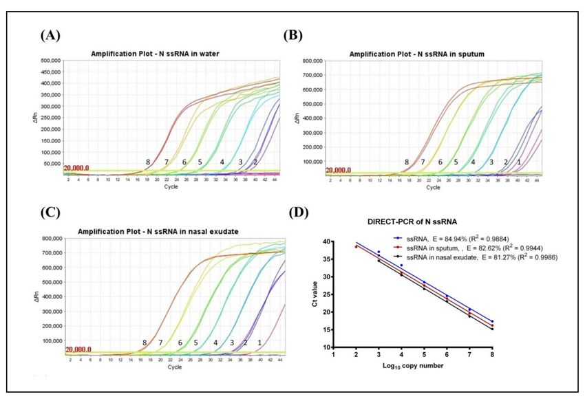
laboratories [16]. When the cycling conditions were optimized in our fast DIRECT-PCR with small
reaction volumes (10 µL), and shorter RT and annealing durations, we were able to amplify SARS-CoV-2
in less than an hour (Figure 3). Furthermore, there are cost savings on nucleic acid extraction kits,
and the availability of reagents themselves may be limiting when laboratory capacity is ramped up
and demand is increased globally. Moreover, DIRECT-PCR is a single-tube homogeneous reaction
that reduces hands-on time and biosafety risk for laboratory personnel, as well as the likelihood for
carryover contamination. One caveat, however, for fast DIRECT-PCR of samples with low viral load is
that of sampling error, as only 1 µL of sample is used. Nucleic acid extractions, on the other hand,
serve to concentrate RNA from typical sample volumes of 150–300 µL, although their yield can also
be low and variable. Therefore, where samples are expected to have low counts near to the limit of
detection, DIRECT-PCR with larger volume reactions (25 µL) to include higher template volume may
be necessary to reduce risk of false negatives.
Figure 3. Workflow of fast DIRECT-PCR protocol for the direct detection of SARS-CoV-2. Covid-19
infection can be confirmed in less than an hour after sample collection and addition to mastermix for
amplification and detection.Genes 2020, 11, 664 9 of 13
Reduction in amplification efficiency is a common concern in DIRECT-PCR from crude samples
(e.g., respiratory samples, blood, and serum). The presence of PCR inhibitors can decrease the sensitivity
and accuracy of pathogen detection through interfering with polymerase activity, degradation of
nucleic acids and efficient cell lysis [24]. A variety of methods have been developed to overcome such
inhibition, including inhibitor tolerant polymerases, additives, and buffer modification. In our study,
we used one commercially available formulation that tolerated PCR inhibition in sputum and nasal
exudates as well as blood/serum/urine (data not shown), and it is not unlikely that other formulations
could be used as well in our study. While amplification directly from sputum and nasal exudate
reduced the efficiency of PCR compared to water controls, there was no net effect on the threshold
of detection.
Nasopharyngeal swabs and sputum have been suggested to be effective clinical samples for
diagnosis [28,29], and we have assessed direct PCR using these matrices. It should be feasible to extend
this to other common samples used for Covid-19 testing, such as swabs of the throat and endotracheal
tubes, and bronchoalveolar lavage fluid. Currently, our assay has successfully directly amplified from
sputum and nasal exudate respiratory samples. If viral transport medium is used, it will need to
be assessed for PCR inhibition. These mediums should ideally be in smaller volumes so that there
is a less dilution effect on the sample concentration. Although we have focused our DIRECT-PCR
protocols on commonly used primer–probe sets developed by the China CDC and US CDC that target
the SARS-CoV-2 N gene as well as ORF1ab gene and the internal control RP, we expect direct PCR to
be similarly useful for other viral primer targets and PCR amplicons developed for Covid-19 [8,9],
as well as for multiplexed amplification of several targets in the same tube. Nevertheless, there may be
other inhibitors of PCR that were not present in our samples that we tested, and clinical validation
with a larger series is needed to exclude this possibility.
Quantitation of viral nucleic acid in samples by RT-qPCR has been used in studies to correlate
viral load with severity, prognosis and transmissibility. The Cq values in patients’ samples can range
from 19 to late cycles close to 40 during the course of infection. The nasal viral load has been shown
to peak within days of symptom onset according to small study of throat and nasal swabs from 17
patients [30]. Using RNA as template, our DIRECT-PCR assay has a dynamic range of over 7 orders of
magnitude with mean Cq range from 17 to 38, indicating it is reproducibly quantitative over a wide
range, including very high viral titers. This method could be useful in future studies, such as to assess
viral survivability, decontamination methods, and preventive intervention.
We have observed, even in spiked water samples, that amplification efficiencies and the Cq values
obtained at the limit of detection are dependent on factors such as the reagent mastermix, the model of
thermocycler, the reaction volumes, and the cycling protocols used (Table 1). This variability would
be even more significant in a clinical setting, with the added confounding factors, such as type of
sample, sampling method, dilution in transport buffers, duration and conditions of storage, and the
reagents and protocols for RNA extraction, that are difficult to standardize across healthcare settings
and laboratories. While removing the RNA extraction step could help to reduce this variability in
RNA yield and quality, variability in sample type and sampling procedure still renders the entire
assay non-quantitative. Therefore, we caution overinterpretation of Cq values, especially in the
absence of in-house quality control and calibration data for each combination of protocol, reagents
and thermocycler. These technical factors may also partly explain some of the false negatives and
temporal variability in viral RNA shedding reported in various studies, that use fixed criteria Cq < 37
for positive, Cq between 37 to 40 as inconclusive for repeat testing and Cq > 40 as negative. In fact, our
data demonstrates that as viral RNA concentrations approach the threshold of detection, our assay
LoD has Cq mean value of > 37 and may exceed 40 (Table 1). As negative controls in these probe-based
assays remain consistently negative, it should be possible to report Cq > 37 as positive. Therefore,
to achieve better standardization of quantitative results, we propose that instead of reporting Cq ,
laboratories may report the equivalent viral copy number in per unit sample volume, derived from
running serial dilutions of viral or RNA controls in-house.Genes 2020, 11, 664 10 of 13
Our DIRECT-PCR assay has been demonstrated on a commercially available portable thermocycler
that weighs less than 2 kg, potentially allowing PCR to be performed outside molecular biology
laboratories, in mobile laboratories, and in low resource settings. There are many real-time fieldable
thermocyclers available that are smaller, portable, and less costly than benchtop ones, and for three of
these we have evaluated perform similar to benchtop ones (data not shown). Although the optical
detection modules and their fluorescence sensitivity vary between designs, the overall sensitivity of
our assay was similar in both types of thermocyclers tested (Table 1). Thermocycling parameters could
also be optimized for rapidity (Figure 3). As the need to transport samples to central laboratories could
prolong the availability of test results to beyond 24 h [16], use of portable thermocyclers, coupled
with appropriate training and quality control procedures, could allow the use of RT-qPCR nearer to
the patients and in primary healthcare settings [12]. Such rapid point-of-care (POC) tests for use at
community level were identified by a WHO expert group as a key research priority [31].
Though it is unlikely that DIRECT-PCR would completely replace conventional RNA extraction
with RT-qPCR in large laboratories as a confirmatory diagnosis, the trade-off in reliability for portability,
ease of use and cost-effectiveness could be useful for patient screening and public health surveillance
and in settings with limited laboratory resources and facilities [4]. DIRECT-PCR for Covid-19 could
address the specific requirements and challenges of delivering molecular genetic testing at the POC
setting. POC diagnostics could play an important role in the detection, diagnosis and control of this
pandemic [17], such as for more timely diagnosis in primary healthcare facilities, and to complement
real-time fever surveillance and screening. Several technologies have been proposed and used to
enable molecular testing at POC, including isothermal amplification methods that do not require
such thermocyclers, like loop-mediated isothermal amplification (LAMP) [12]. However, given the
widespread validation of existing SARS-CoV-2 PCR primer/probe sets, as well as the likely need to
modify amplification primers and probes as the viral sequences evolve in the future, we argue that
performing PCR using our DIRECT-PCR method is a more generic and adaptable approach. Indeed,
while this manuscript was under review, other efforts to develop an extraction-free protocol to detect
SARS-CoV-2 were reported [32–40].
5. Conclusions
Direct amplification of SARS-CoV-2 viral RNA from samples without RNA purification has
been developed, reducing hands-on-time, time-to-results, and costs. Viral lysis, reverse transcription,
amplification, and detection are achieved in a single-tube homogeneous reaction taking less than an
hour, and they can be performed on a portable thermocycler. Analytical validation was performed
with sputum and nasal exudate. The DIRECT-PCR assay has a high sensitivity of six RNA copies per
reaction and is quantitative over a dynamic range of 7 orders of magnitude. This method may be
useful during the current global Covid-19 pandemic in situations where resources are constrained or
where timely results are needed.
Supplementary Materials: The following are available online at http://www.mdpi.com/2073-4425/11/6/664/s1,
Supplementary Information S1.
Author Contributions: Conceptualization, S.K.W., S.P.S., and E.P.H.Y.; methodology, S.K.W., S.P.S., and E.P.H.Y.;
investigation, S.K.W. and S.P.S.; formal analysis, S.K.W., S.P.S., and E.P.H.Y.; funding acquisition, E.P.H.Y.;
supervision, E.P.H.Y.; visualization, S.K.W., S.P.S., and E.P.H.Y.; writing—original draft preparation, S.K.W. and
S.P.S.; writing—review and editing, S.K.W., S.P.S., and E.P.H.Y. All authors have read and agreed to the published
version of the manuscript.
Funding: This research is supported by the National Research Foundation, Prime Minister’s Office, Singapore
under its Competitive Research Programme (CRP13-2014-01, E.P.H.Y.) and Lee Kong Chian School of Medicine,
Nanyang Technological University Singapore Start-Up Grant (E.P.H.Y.).
Conflicts of Interest: The authors declare no conflict of interest. The funders had no role in the design of the
study; in the collection, analyses, or interpretation of data; in the writing of the manuscript; or in the decision to
publish the results.Genes 2020, 11, 664 11 of 13
References
1. Zhu, N.; Zhang, D.; Wang, W.; Li, X.; Yang, B.; Song, J.; Zhao, X.; Huang, B.; Shi, W.; Lu, R.; et al. A Novel
Coronavirus from Patients with Pneumonia in China, 2019. New Engl. J. Med. 2020, 382, 727–733. [CrossRef]
[PubMed]
2. WHO. WHO Director-General’s Opening Remarks at the Media Briefing on COVID-19 - 11 March 2020.
Available online: https://www.who.int/dg/speeches/detail/who-director-general-s-opening-remarks-at-the-
media-briefing-on-covid-19---11-march-2020 (accessed on 12 March 2020).
3. WHO. COVID-19 Dashboard. Available online: https://covid19.who.int/ (accessed on 16 April 2020).
4. Beeching, N.J.; E Fletcher, T.; Beadsworth, M.B.J. Covid-19: Testing times. BMJ 2020, 369, m1403. [CrossRef]
[PubMed]
5. Ng, Y.; Li, Z.; Chua, Y.X.; Chaw, W.L.; Zhao, Z.; Er, B.; Pung, R.; Chiew, C.J.; Lye, D.C.; Heng, D.; et al.
Evaluation of the Effectiveness of Surveillance and Containment Measures for the First 100 Patients with
COVID-19 in Singapore — January 2–February 29, 2020. MMWR. Morb. Mortal. Wkly. Rep. 2020, 69, 307–311.
[CrossRef]
6. Wong, J.E.L.; Leo, Y.S.; Tan, C.C. COVID-19 in Singapore—Current Experience. JAMA 2020, 323, 1243.
[CrossRef]
7. MOH. Updates on COVID-19 (Coronavirus Disease 2019) Local Situation. Available online: https://www.
moh.gov.sg/covid-19 (accessed on 16 April 2020).
8. Zhang, Y.-Z. Novel 2019 Coronavirus Genome. Available online: http://virological.org/t/novel-2019-
coronavirus-genome/319 (accessed on 22 March 2020).
9. WHO. Coronavirus Disease (COVID-19) Technical Guidance: Laboratory Testing for 2019-nCoV in Humans.
Available online: https://www.who.int/emergencies/diseases/novel-coronavirus-2019/technical-guidance/
laboratory-guidance (accessed on 26 March 2020).
10. NUS. SSHSPH COVID-19 Science Report; Saw Swee Hock School of Public Health, National University of
Singapore: Singapore, 2020.
11. WHO. Laboratory testing for 2019 Novel Coronavirus (2019-nCoV) in Suspected Human Cases;
WHO/COVID-19/Laboratory/2020.5; World Health Organization: 2020. Available online: https://www.who.
int/publications/i/item/10665-331501 (accessed on 20 April 2020).
12. Nguyen, T.; Bang, D.D.; Wolff, A. 2019 Novel Coronavirus Disease (COVID-19): Paving the Road for Rapid
Detection and Point-of-Care Diagnostics. Micromachines 2020, 11, 306. [CrossRef] [PubMed]
13. Wei, W.E.; Li, Z.; Chiew, C.J.; Yong, S.E.; Toh, M.P.; Lee, V.J. Presymptomatic Transmission of SARS-CoV-2 —
Singapore, January 23–March 16, 2020. MMWR. Morb. Mortal. Wkly. Rep. 2020, 69, 411–415. [CrossRef]
14. He, X.; Lau, E.H.; Wu, P.; Deng, X.; Wang, J.; Hao, X.; Lau, Y.C.; Wong, J.Y.; Guan, Y.; Tan, X.; et al. Temporal
dynamics in viral shedding and transmissibility of COVID-19. Nat. Med. 2020, 26, 672–675. [CrossRef]
15. Gudbjartsson, D.F.; Helgason, A.; Jonsson, H.; Magnusson, O.T.; Melsted, P.; Norddahl, G.L.;
Saemundsdottir, J.; Sigurdsson, A.; Sulem, P.; Agustsdottir, A.B.; et al. Spread of SARS-CoV-2 in the
Icelandic Population. New Engl. J. Med. 2020, 382, 2302–2315. [CrossRef]
16. Sheridan, C. Fast, portable tests come online to curb coronavirus pandemic. Nat. Biotechnol. 2020, 38, 515–518.
[CrossRef]
17. Patel, R.; Babady, N.E.; Theel, E.S.; Storch, G.A.; Pinsky, B.A.; George, K.S.; Smith, T.C.; Bertuzzi, S. Report
from the American Society for Microbiology COVID-19 International Summit, 23 March 2020: Value of
Diagnostic Testing for SARS–CoV-2/COVID-19. mBio. 2020, 11. [CrossRef]
18. Sivalingam, S.P.; Ayi, T.C.; Mustaffa, S.B.; Loo, S.; Tan, B.H.; Yap, E.P.H. Direct RT-PCR detection of Dengue
1-4 virus from crude samples for rapid point-of-care diagnosis. In Proceedings of the SingHealth Duke-NUS
Scientific Congress, Singapore, 23–24 September 2016.
19. Mehta, N.; Perrais, B.; Martin, K.; Kumar, A.; Hobman, T.C.; Cabalfin-Chua, M.N.; Donaldo, M.E.;
Painaga, M.S.S.; Gaite, J.Y.; Tran, V.; et al. A Direct from Blood/Plasma Reverse Transcription–Polymerase
Chain Reaction for Dengue Virus Detection in Point-of-Care Settings. Am. J. Trop. Med. Hyg. 2019, 100,
1534–1540. [CrossRef]
20. Pastorino, B.; Bessaud, M.; Grandadam, M.; Murri, S.; Tolou, H.J.; Peyrefitte, C.N. Development of a
TaqMan®RT-PCR assay without RNA extraction step for the detection and quantification of African
Chikungunya viruses. J. Virol. Methods 2005, 124, 65–71. [CrossRef] [PubMed]Genes 2020, 11, 664 12 of 13
21. Nishimura, N.; Nakayama, H.; Yoshizumi, S.; Miyoshi, M.; Tonoike, H.; Shirasaki, Y.; Kojima, K.; Ishida, S.
Detection of noroviruses in fecal specimens by direct RT-PCR without RNA purification. J. Virol. Methods
2010, 163, 282–286. [CrossRef] [PubMed]
22. Bachofen, C.; Willoughby, K.; Zadoks, R.; Burr, P.; Mellor, D.; Russell, G.C. Direct RT-PCR from serum enables
fast and cost-effective phylogenetic analysis of bovine viral diarrhoea virus. J. Virol. Methods 2013, 190, 1–3.
[CrossRef] [PubMed]
23. Li, L.; He, J.-A.; Wang, W.; Xia, Y.; Song, L.; Chen, Z.-H.; Zuo, H.-Z.; Tan, X.-P.; Ho, A.H.-P.; Kong, S.-K.;
et al. Development of a direct reverse-transcription quantitative PCR (dirRT-qPCR) assay for clinical Zika
diagnosis. Int. J. Infect. Dis. 2019, 85, 167–174. [CrossRef]
24. Hall, A.T.; Zovanyi, A.M.; Christensen, D.R.; Koehler, J.W.; Minogue, T.D. Evaluation of Inhibitor-Resistant
Real-Time PCR Methods for Diagnostics in Clinical and Environmental Samples. PLoS ONE 2013, 8, e73845.
[CrossRef]
25. Wang, D.; Hu, B.; Hu, C.; Zhu, F.; Liu, X.; Zhang, J.; Wang, B.; Xiang, H.; Cheng, Z.; Xiong, Y.; et al. Clinical
Characteristics of 138 Hospitalized Patients With 2019 Novel Coronavirus–Infected Pneumonia in Wuhan,
China. JAMA 2020, 323, 1061. [CrossRef]
26. CDC, C. Specific Primers and Probes for Detection 2019 Novel Coronavirus. Available online: http:
//ivdc.chinacdc.cn/kyjz/202001/t20200121_211337.html (accessed on 1 February 2020).
27. CDC. Research Use Only 2019-Novel Coronavirus (2019-nCoV) Real-Time RT-PCR Primer and Probe
Information. Available online: https://www.cdc.gov/coronavirus/2019-ncov/lab/rt-pcr-panel-primer-probes.
html (accessed on 2 March 2020).
28. Yang, Y.; Yang, M.; Shen, C.; Wang, F.; Jing, Y.; Li, J.-X.; Zhang, M.; Wang, Z.; Xing, L.; Wei, J.; et al. Evaluating
the accuracy of different respiratory specimens in the laboratory diagnosis and monitoring the viral shedding
of 2019-nCoV infections 2020. MedRxiv 2020.
29. Pan, Y.; Zhang, D.; Yang, P.; Poon, L.L.; Wang, Q. Viral load of SARS-CoV-2 in clinical samples. Lancet Infect. Dis.
2020, 20, 411–412. [CrossRef]
30. Zou, L.; Ruan, F.; Huang, M.; Liang, L.; Huang, H.; Hong, Z.; Yu, J.; Kang, M.; Song, Y.; Xia, J.; et al.
SARS-CoV-2 Viral Load in Upper Respiratory Specimens of Infected Patients. New Engl. J. Med. 2020,
382, 1177–1179. [CrossRef]
31. WHO. COVID 19 Public Health Emergency of International Concern (PHEIC) - Global Research and
Innovation Forum: Towards a Research Roadmap. Available online: https://www.who.int/blueprint/priority-
diseases/key-action/Global_Research_Forum_FINAL_VERSION_for_web_14_feb_2020.pdf?ua=1 (accessed
on 14 April 2020).
32. Bruce, E.A.; Huang, M.-L.; Perchetti, G.A.; Tighe, S.W.; Laaguiby, P.; Hoffman, J.J.; Gerrard, D.L.; Nalla, A.K.;
Wei, Y.; Greninger, A.L.; et al. DIRECT RT-qPCR DETECTION OF SARS-CoV-2 RNA FROM PATIENT
NASOPHARYNGEAL SWABS WITHOUT AN RNA EXTRACTION STEP 2020. BioRxiv 2020.
33. Sentmanat, M.; Kouranova, E.; Cui, X. One-step RNA extraction for RT-qPCR detection of 2019-nCoV 2020.
BioRxiv 2020.
34. Arumugam, A.; Wong, S.S. The Potential Use of Unprocessed Sample for RT-qPCR Detection of COVID-19
without an RNA Extraction Step 2020. BioRxiv 2020.
35. Grant, P.R.; Turner, M.; Shin, G.Y.; Nastouli, E.; Levett, L. Extraction-free COVID-19 (SARS-CoV-2) diagnosis
by RT-PCR to increase capacity for national testing programmes during a pandemic. In bioRxiv 2020; Cold
Spring Harbor Laboratory: Cold Spring Harbor, NY, USA, 2020.
36. Merindol, N.; Pépin, G.; Marchand, C.; Rheault, M.; Peterson, C.; Poirier, A.; Germain, H.; Danylo, A.
Optimization of SARS-CoV-2 detection by RT-QPCR without RNA extraction 2020. bioRxiv 2020.
37. Smyrlaki, I.; Ekman, M.; Lentini, A.; Vondracek, M.; Papanicoloau, N.; Aarum, J.; Safari, H.; Muradrasoli, S.;
Albert, J.; Högberg, B.; et al. Massive and rapid COVID-19 testing is feasible by extraction-free SARS-CoV-2
RT-qPCR 2020. medRxiv 2020.
38. Brown, J.R.; Atkinson, L.; Shah, D.; Harris, K. Validation of an extraction-free RT-PCR protocol for detection
of SARS-CoV2 RNA 2020. medRxiv 2020.Genes 2020, 11, 664 13 of 13
39. Hasan, M.R.; Mirza, F.; Al-Hail, H.; Sundararaju, S.; Xaba, T.; Iqbal, M.; Alhussain, H.; Yassine, H.M.;
Lopez, A.P.; Tang, P. Detection of SARS-CoV-2 RNA by direct RT-qPCR on nasopharyngeal specimens
without extraction of viral RNA 2020. medRxiv 2020.
40. Fomsgaard, A.S.; Rosenstierne, M.W. An alternative workflow for molecular detection of SARS-CoV-2 –
escape from the NA extraction kit-shortage, Copenhagen, Denmark, March 2020. Eurosurveillance 2020, 25,
2000398. [CrossRef]
© 2020 by the authors. Licensee MDPI, Basel, Switzerland. This article is an open access
article distributed under the terms and conditions of the Creative Commons Attribution
(CC BY) license (http://creativecommons.org/licenses/by/4.0/).You can also read