INTRANASAL FUSION INHIBITORY LIPOPEPTIDE PREVENTS DIRECT-CONTACT SARS-COV-2 TRANSMISSION IN FERRETS - SCIENCE
←
→
Page content transcription
If your browser does not render page correctly, please read the page content below
REPORTS

                                                                                                                               Cite as: R. D. de Vries et al., Science
                                                                                                                                   10.1126/science.abf4896 (2021).

Intranasal fusion inhibitory lipopeptide prevents
direct-contact SARS-CoV-2 transmission in ferrets
Rory D. de Vries1*, Katharina S. Schmitz1*, Francesca T. Bovier2,3,4*, Camilla Predella2,5, Jonathan Khao6,
Danny Noack1, Bart L. Haagmans1, Sander Herfst1, Kyle N. Stearns2,3,7, Jennifer Drew-Bear2,3, Sudipta Biswas8,
Barry Rockx1, Gaël McGill6,9, N. Valerio Dorrello2, Samuel H. Gellman10, Christopher A. Alabi8†,
Rik L. de Swart1†, Anne Moscona2,3,7,11†, Matteo Porotto2,3,4†
1
 Department of Viroscience, Erasmus MC, Rotterdam, Netherlands. 2Department of Pediatrics, Columbia University Irving Medical Center, New York, NY, USA. 3Center for
Host–Pathogen Interaction, Columbia University Irving Medical Center, New York, NY, USA. 4Department of Experimental Medicine, University of Campania “Luigi
Vanvitelli,” Caserta, Italy. 5Department of Biomedical Engineering, Politecnico di Milano, Milan, Italy. 6Digizyme Inc., Brookline, MA, USA. 7Department of Physiology and
Cellular Biophysics, Columbia University Irving Medical Center, New York, NY, USA. 8Robert Frederick Smith School of Chemical and Biomolecular Engineering, Cornell
University, Ithaca, NY, USA. 9Center for Molecular and Cellular Dynamics, Department of Biological Chemistry and Molecular Pharmacology, Harvard Medical School,

                                                                                                                                                                             Downloaded from http://science.sciencemag.org/ on February 17, 2021
Boston, MA, USA. 10Department of Chemistry, University of Wisconsin–Madison, Madison, WI, USA. 11Department of Microbiology and Immunology, Columbia University
Irving Medical Center, New York, NY, USA.
*These authors contributed equally to this work.
†Corresponding author. Email: caa238@cornell.edu (C.A.A.); r.deswart@erasmusmc.nl (R.L.d.S.);
am939@cumc.columbia.edu (A.M.); mp3509@cumc.columbia.edu (M.P.)

Containment of the COVID-19 pandemic requires reducing viral transmission. SARS-CoV-2 infection is
initiated by membrane fusion between the viral and host cell membranes, mediated by the viral spike
protein. We have designed lipopeptide fusion inhibitors that block this critical first step of infection, and
based on in vitro efficacy and in vivo biodistribution selected a dimeric form for evaluation in an animal
model. Daily intranasal administration to ferrets completely prevented SARS-CoV-2 direct-contact
transmission during 24-hour co-housing with infected animals, under stringent conditions that resulted in
infection of 100% of untreated animals. These lipopeptides are highly stable and thus may readily translate
into safe and effective intranasal prophylaxis to reduce transmission of SARS-CoV-2.

Infection by SARS-CoV-2 requires membrane fusion between                                 combination of dimerization and lipopeptide integration into
the viral envelope and the host cell, at either the cell surface                         cell membranes protects the respiratory tract and prevents
or the endosomal membrane. The fusion process is mediated                                systemic lipopeptide dissemination (14). Lipid-conjugated
by the viral transmembrane spike glycoprotein (S). Upon vi-                              peptides administered intranasally to animals reached high
ral attachment or uptake, host factors trigger large-scale con-                          concentrations both in the upper and lower respiratory tract,
formational rearrangements in S, including a refolding step                              and the lipid could be designed to modulate the extent of
that leads directly to membrane fusion and viral entry (1–3).                            transit from the lung to the blood and organs (9, 14). We pro-
Peptides corresponding to the highly conserved heptad re-                                pose a SARS-CoV-2 specific lipopeptide as a candidate antivi-
peat (HR, Fig. 1A) domain at the C terminus of the S protein                             ral for pre-exposure and early post-exposure prophylaxis for
(HRC peptides, Fig. 1B) can prevent this refolding and inhibit                           SARS-CoV-2 transmission in humans.
fusion, thereby preventing infection (4–8). The HRC peptides                                 We recently described a monomeric SARS-CoV-2 HRC-
form six-helix bundle-like assemblies with the extended in-                              lipopeptide fusion inhibitor (4) against SARS-CoV-2 with in
termediate form of the S protein trimer, disrupting the struc-                           vitro and ex vivo efficacy superior to previously described
tural rearrangement of S that drives membrane fusion (4)                                 HRC-derived fusion inhibitory peptides (6, 7). To further im-
(see also movie S1).                                                                     prove antiviral potency, we compared monomeric and di-
    Our approach in designing SARS-CoV-2 S-specific fusion                               meric HRC-peptide derivatives (Fig. 1C). Figure 1D shows
inhibitors builds on our previous work that demonstrated                                 antiviral potency in a quantitative cell-cell fusion assay of
that lipid conjugation of HRC-derived inhibitory peptides                                four monomeric and two dimeric SARS-CoV-2 S-derived 36-
markedly increases antiviral potency and in vivo half-life (9,                           amino acid HRC-peptides (Fig. 1B, see also figs. S1A and S3
10). These peptides successfully inhibit human parainfluenza                             for structure and characterization), without or with ap-
virus type 3 (HPIV-3), measles virus, influenza virus, and                               pended cholesterol. Dimerization increased the peptide po-
Nipah virus infection (9, 11–13). Furthermore, the                                       tency for both non-lipidated peptides and their lipidated

First release: 17 February 2021                                          www.sciencemag.org                 (Page numbers not final at time of first release) 1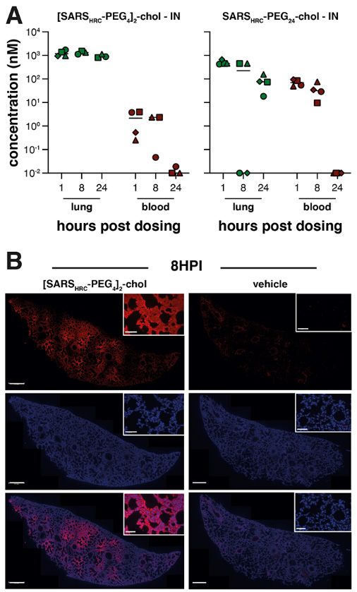
counterparts (Fig. 1D). A dimeric cholesterol-conjugated cells and human lung (19). The [SARSHRC-PEG4]2-chol peptide
lipopeptide based on the HPIV-3 F protein HRC domain, used dissolved in an aqueous buffer containing 2% dimethylsulfox-
as a negative control, did not inhibit fusion at any concentra- ide (DMSO) inhibited virus entry after 8 hours with an IC50
tion tested (black line in Fig. 1D, see fig. S1, B and C, for ad- ~300 nM in VeroE6 and ~5 nM in VeroE6-TMPRSS2 cells
ditional negative controls). Among the monomeric (Fig. 3A). To strengthen translational potential toward hu-
lipopeptides, the peptide bearing PEG24 was most potent. The man use, the lipopeptide was reformulated in sucrose instead
dimeric cholesterol-conjugated peptide ([SARSHRC-PEG4]2- of DMSO, resulting in equivalent in vitro potency (Fig. 3B). A
chol; red line in Fig. 1D) was the most potent lipopeptide control dimeric fusion-inhibitory lipopeptide directed
against SARS-CoV-2 among our panel. This peptide also ro- against HPIV-3 blocked infection by HPIV-3, but did not in-
bustly inhibited fusion mediated by the S proteins of several hibit SARS-CoV-2 infection. The in vitro efficacy data are
emerging SARS-CoV-2 variants [including D614G (15)], the summarized in table S1.
recent variants of concern B.1.1.7 and B.1.351 (16, 17) and the Ferrets are an ideal model for assessing respiratory virus
S protein of SARS-CoV and MERS-CoV (Fig. 1E). Proposed an- transmission, either by direct contact or by aerosol transmis-
choring of the dimeric lipopeptide in the host cell membrane sion (20, 21). Mustelids are highly susceptible to infection
Downloaded from http://science.sciencemag.org/ on February 17, 2021
and interactions with the viral S protein are shown in fig. S2 with SARS-CoV-2, as also illustrated by frequent COVID-19
and movie S1. Collectively, these data suggest that the outbreaks at mink farms. Direct contact transmission of
[SARSHRC-PEG4]2-chol lipopeptide is equipped to combat an SARS-CoV in ferrets was demonstrated in 2003 (22), and both
evolving pandemic. direct contact and airborne transmission have been shown in
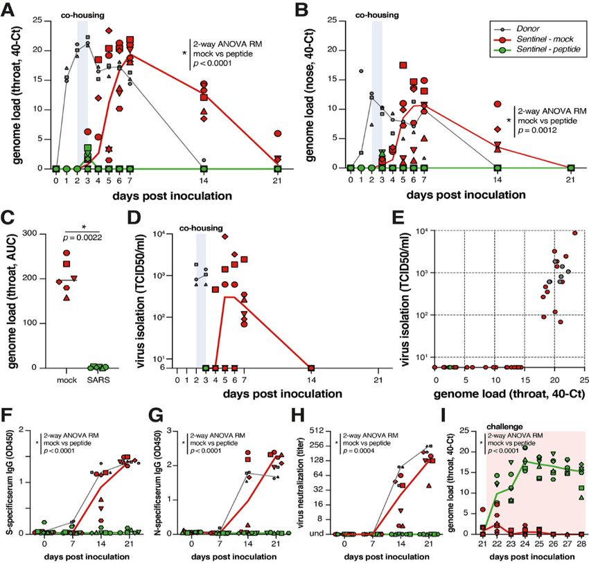
For other enveloped respiratory viruses, we previously ferrets for SARS-CoV-2 (20, 23). Direct contact transmission
showed that both ex vivo and in vivo dimeric lipopeptides in the ferret model is highly reproducible (100% transmission
(administered intranasally) displayed increased retention in from donor to acceptor animals), but ferrets display limited
the respiratory tract compared to monomeric compounds clinical signs. After infection via direct inoculation or trans-
(14). Here, we compared local and systemic biodistribution of mission, SARS-CoV-2 can readily be detected in and isolated
our most potent monomeric and dimeric lipopeptides from the throat and nose, and viral replication leads to sero-
(SARSHRC-PEG24-chol and [SARSHRC-PEG4]2-chol) at 1, 8, and conversion.
24 hours after intranasal inoculation or subcutaneous injec- To assess the efficacy of [SARSHRC-PEG4]2-chol in prevent-
tion in humanized K18 hACE2 mice (Fig. 2 and fig. S4). The ing SARS-CoV-2 transmission, naïve ferrets were dosed
two lipopeptides reached a similar lung concentration at 1 prophylactically with the lipopeptide before being co-housed
hour after intranasal administration (~1 to 2 μM). At 8 and with SARS-CoV-2 infected ferrets. In this setup, transmission
24 hours, the dimeric [SARSHRC-PEG4]2-chol lipopeptide re- via multiple routes can theoretically occur (aerosol, orofecal,
mained at high levels in the lung with minimal entry into the and scratching or biting), and ferrets are continuously ex-
blood, but the monomeric peptide entered the circulation posed to infectious virus during the period of co-housing,
and the lung concentration decreased (Fig. 2A). The dimeric providing a stringent test for antiviral efficacy. The study de-
[SARSHRC-PEG4]2-chol lipopeptide was distributed through- sign is shown in fig. S6. Three donor ferrets (gray in diagram)
out the lung after intranasal administration (Fig. 2B). A cel- were inoculated intranasally with 5 × 10⁵ TCID50 SARS-CoV-2
lular toxicity (MTT) assay in primary HAE cells showed on day 0. Twelve recipient ferrets housed separately were
minimal toxicity even after 6 days at the highest concentra- treated by nose drops with a mock preparation (red) or
tions tested (
ferrets were housed as separate groups. Additional [SARSHRC-      administration pre-exposure prophylaxis is promising, while
PEG4]2-chol peptide treatments were given to recipient ani-       the optimal formulation and dosing regimen is an area of on-
mals on 3 and 4 DPI.                                              going experimentation.
    The viral loads (detection of viral genomes via RT-qPCR)          The intranasal [SARSHRC-PEG4]2-chol peptide presented in
for directly inoculated donor animals (gray), mock-treated re-    this study is the first successful prophylaxis that prevents
cipient animals (red) and lipopeptide-treated recipient ani-      SARS-CoV-2 transmission in a relevant animal model, provid-
mals (green) are shown in Fig. 4, A and B. All directly           ing complete protection during a 24-hour period of intense
inoculated donor ferrets were productively infected, as           direct contact. Parallel approaches to prevent transmission
shown by SARS-CoV-2 genome detection in throat and nose           that target the interaction between S and ACE2 have shown
swabs, and efficiently and reproducibly transmitted the virus     promise in vitro [e.g., the “miniprotein” approach (24)]. The
to all mock-treated acceptor ferrets (Fig. 4, A and B, red        lipopeptide described here acts on the S2 domain after shed-
curves). Productive SARS-CoV-2 infection was not detected in      ding of S1 (fig. S2 and movie S1), and is complementary to
the throat or nose of any of the peptide-treated recipient an-    strategies that target S1’s functions or maintain S in its pre-
imals (Fig. 4, A and B, green curves). A slight rise in viral     fusion conformation, e.g., synthetic nanobodies (25, 26). Fu-

                                                                                                                                                                 Downloaded from http://science.sciencemag.org/ on February 17, 2021
loads in samples collected at 3 DPI was detected (at the end      sion-inhibitory lipopeptides could be used for pre- and post-
of the co-housing), confirming that peptide-treated animals       exposure prophylaxis in combination with these strategies,
were exposed to SARS-CoV-2. In Fig. 4C the area under the         and in conjunction with treatments [e.g., ribonucleoside an-
curve (AUC) shows the striking difference between the mock        alogs (27)] that reduce replication in a treated infected indi-
treated and the peptide treated animals. No infectious virus      vidual. A combination of drugs that target different aspects
was isolated from lipopeptide-treated ferrets, while infectious   of the viral life cycle is likely ideal for this rapidly-evolving
virus was detected in all mock-treated ferrets (Fig. 4D). Virus   virus. Of note, the [SARSHRC-PEG4]2-chol lipopeptide is
isolation data correlated with genome detection (Fig. 4E).        equally active against several emerging SARS-CoV-2 variants
    Seroconversion occurred in donor ferrets and 6/6 mock-        including the D614G as well as the recent variants of concerns
treated animals by 21 DPI, but in none of the peptide-treated     (B.1.1.7 and B.1.351). The [SARSHRC-PEG4]2-chol peptide has a
recipient animals, as shown by S- and N-specific IgG enzyme-      long shelf life, does not require refrigeration and can easily
linked immunosorbent assay (ELISA) and virus neutraliza-          be administered, making it particularly suited to treating
tion (Fig. 4, F to H). Successful challenge infection confirmed   hard-to-reach populations. This is key in the context of
that in-host virus replication had been completely blocked by     COVID-19, which has reached every community with the bur-
the [SARSHRC-PEG4]2-chol treatment (Fig. 4I and fig. S8) and      den falling disproportionately on low-income and otherwise
that none of the peptide-animals were protected, whereas the      marginalized communities. This HRC lipopeptide fusion in-
mock-treated animals (which had seroconverted) were all           hibitor is feasible for advancement to human use and should
protected. Collectively, these data show that intranasal          readily translate into a safe and effective nasal spray or inha-
prophylactic administration of the [SARSHRC-PEG4]2-chol           lation administered fusion inhibitor for SARS-CoV-2 prophy-
peptide had protected 6/6 ferrets from transmission and pro-      laxis, supporting containment of the ongoing COVID-19
ductive infection.                                                pandemic.
    In light of the persistence of the dimeric lipopeptide in     REFERENCES AND NOTES
the murine lung (Fig. 2 and fig. S4), we assessed the potential   1. F. Li, Structure, function, and evolution of coronavirus spike proteins. Annu. Rev.
for a single administration of sucrose-formulated lipopeptide          Virol. 3, 237–261 (2016). doi:10.1146/annurev-virology-110615-042301 Medline
in a ferret transmission experiment two hours before co-          2. M. Hoffmann, H. Kleine-Weber, S. Schroeder, N. Krüger, T. Herrler, S. Erichsen, T.
                                                                       S. Schiergens, G. Herrler, N.-H. Wu, A. Nitsche, M. A. Müller, C. Drosten, S.
housing to prevent or delay infection. In this experiment, we          Pöhlmann, SARS-CoV-2 cell entry depends on ACE2 and TMPRSS2 and is blocked
used a dimeric HPIV-3-specific lipopeptide as mock control             by a clinically proven protease inhibitor. Cell 181, 271–280.e8 (2020).
(fig. S9). Although sucrose formulation had resulted in prom-          doi:10.1016/j.cell.2020.02.052 Medline
ising results in vitro at small scale (Fig. 3B), formulation at   3. Y. Wan, J. Shang, R. Graham, R. S. Baric, F. Li, Receptor recognition by the novel
                                                                       coronavirus from Wuhan: an analysis based on decade-long structural studies of
larger scale resulted in incomplete dissolution. As a conse-           SARS coronavirus. J. Virol. 94, e00127-20 (2020). doi:10.1128/JVI.00127-20
quence, the sucrose-formulated [SARSHRC-PEG4]2-chol                    Medline
lipopeptide was administered at a substantially lower con-        4. V. K. Outlaw, F. T. Bovier, M. C. Mears, M. N. Cajimat, Y. Zhu, M. J. Lin, A. Addetia, N.
centration than in the experiment with the DMSO-                       A. P. Lieberman, V. Peddu, X. Xie, P.-Y. Shi, A. L. Greninger, S. H. Gellman, D. A.
                                                                       Bente, A. Moscona, M. Porotto, Inhibition of coronavirus entry in vitro and ex vivo
formulated lipopeptide (fig. S7, C and D). Nevertheless, the           by a lipid-conjugated peptide derived from the SARS-CoV-2 spike glycoprotein
SARS-CoV-2 lipopeptide provided a significant level of pro-            HRC domain. mBio 11, e01935-20 (2020). doi:10.1128/mBio.01935-20 Medline
tection as compared to the HPIV-3 control group, and four         5. S. Xia, L. Yan, W. Xu, A. S. Agrawal, A. Algaissi, C. K. Tseng, Q. Wang, L. Du, W. Tan,
out of six SARS-CoV-2 lipopeptide-treated animals were pro-            I. A. Wilson, S. Jiang, B. Yang, L. Lu, A pan-coronavirus fusion inhibitor targeting
                                                                       the HR1 domain of human coronavirus spike. Sci. Adv. 5, eaav4580 (2019).
tected against infection. This experiment suggests that single-        doi:10.1126/sciadv.aav4580 Medline

First release: 17 February 2021                       www.sciencemag.org             (Page numbers not final at time of first release)                      3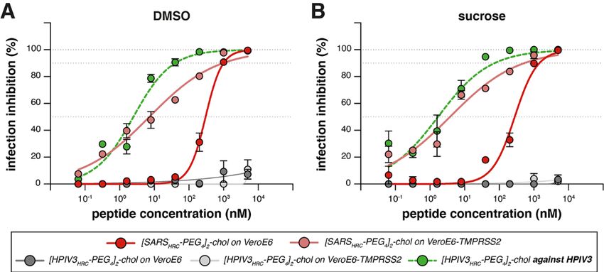
6. S. Xia, M. Liu, C. Wang, W. Xu, Q. Lan, S. Feng, F. Qi, L. Bao, L. Du, S. Liu, C. Qin, F.       between ferrets. Nat. Commun. 11, 3496 (2020). doi:10.1038/s41467-020-
     Sun, Z. Shi, Y. Zhu, S. Jiang, L. Lu, Inhibition of SARS-CoV-2 (previously 2019-              17367-2 Medline
     nCoV) infection by a highly potent pan-coronavirus fusion inhibitor targeting its         21. V. J. Munster, E. de Wit, J. M. A. van den Brand, S. Herfst, E. J. A. Schrauwen, T. M.
     spike protein that harbors a high capacity to mediate membrane fusion. Cell Res.              Bestebroer, D. van de Vijver, C. A. Boucher, M. Koopmans, G. F. Rimmelzwaan, T.
     30, 343–355 (2020). doi:10.1038/s41422-020-0305-x Medline                                     Kuiken, A. D. M. E. Osterhaus, R. A. M. Fouchier, Pathogenesis and transmission
7. Y. Zhu, D. Yu, H. Yan, H. Chong, Y. He, Design of potent membrane fusion inhibitors             of swine-origin 2009 A(H1N1) influenza virus in ferrets. Science 325, 481–483
     against SARS-CoV-2, an emerging coronavirus with high fusogenic activity. J.                  (2009). doi:10.1126/science.1177127 Medline
     Virol. 94, e00635-20 (2020). doi:10.1128/JVI.00635-20 Medline                             22. B. E. Martina, B. L. Haagmans, T. Kuiken, R. A. M. Fouchier, G. F. Rimmelzwaan, G.
8. X. Wang, S. Xia, Q. Wang, W. Xu, W. Li, L. Lu, S. Jiang, Broad-spectrum coronavirus             Van Amerongen, J. S. M. Peiris, W. Lim, A. D. M. E. Osterhaus, SARS virus infection
     fusion inhibitors to combat COVID-19 and other emerging coronavirus diseases.                 of cats and ferrets. Nature 425, 915 (2003). doi:10.1038/425915a Medline
     Int. J. Mol. Sci. 21, 3843 (2020). doi:10.3390/ijms21113843 Medline                       23. Y. I. Kim, S.-G. Kim, S.-M. Kim, E.-H. Kim, S.-J. Park, K.-M. Yu, J.-H. Chang, E. J.
9. M. Porotto, B. Rockx, C. C. Yokoyama, A. Talekar, I. Devito, L. M. Palermo, J. Liu, R.          Kim, S. Lee, M. A. B. Casel, J. Um, M.-S. Song, H. W. Jeong, V. D. Lai, Y. Kim, B. S.
     Cortese, M. Lu, H. Feldmann, A. Pessi, A. Moscona, Inhibition of Nipah virus                  Chin, J.-S. Park, K.-H. Chung, S.-S. Foo, H. Poo, I.-P. Mo, O.-J. Lee, R. J. Webby, J.
     infection in vivo: Targeting an early stage of paramyxovirus fusion activation                U. Jung, Y. K. Choi, Infection and rapid transmission of SARS-CoV-2 in ferrets. Cell
     during       viral     entry.    PLOS      Pathog.      6,      e1001168       (2010).        Host Microbe 27, 704–709.e2 (2020). doi:10.1016/j.chom.2020.03.023 Medline
     doi:10.1371/journal.ppat.1001168 Medline                                                  24. L. Cao, I. Goreshnik, B. Coventry, J. B. Case, L. Miller, L. Kozodoy, R. E. Chen, L.
10. A. Pessi, A. Langella, E. Capitò, S. Ghezzi, E. Vicenzi, G. Poli, T. Ketas, C. Mathieu,        Carter, A. C. Walls, Y.-J. Park, E.-M. Strauch, L. Stewart, M. S. Diamond, D. Veesler,
     R. Cortese, B. Horvat, A. Moscona, M. Porotto, A general strategy to endow natural            D. Baker, De novo design of picomolar SARS-CoV-2 miniprotein inhibitors. Science

                                                                                                                                                                                              Downloaded from http://science.sciencemag.org/ on February 17, 2021
     fusion-protein-derived peptides with potent antiviral activity. PLOS ONE 7,                   370, 426–431 (2020). doi:10.1126/science.abd9909 Medline
     e36833 (2012). doi:10.1371/journal.pone.0036833 Medline                                   25. M. Schoof, B. Faust, R. A. Saunders, S. Sangwan, V. Rezelj, N. Hoppe, M. Boone, C.
11. T. N. Figueira, D. A. Mendonça, D. Gaspar, M. N. Melo, A. Moscona, M. Porotto, M.              B. Billesbølle, C. Puchades, C. M. Azumaya, H. T. Kratochvil, M. Zimanyi, I.
     A. R. B. Castanho, A. S. Veiga, Structure-stability-function mechanistic links in the         Deshpande, J. Liang, S. Dickinson, H. C. Nguyen, C. M. Chio, G. E. Merz, M. C.
     anti-measles virus action of tocopherol-derivatized peptide nanoparticles. ACS                Thompson, D. Diwanji, K. Schaefer, A. A. Anand, N. Dobzinski, B. S. Zha, C. R.
     Nano 12, 9855–9865 (2018). doi:10.1021/acsnano.8b01422 Medline                                Simoneau, K. Leon, K. M. White, U. S. Chio, M. Gupta, M. Jin, F. Li, Y. Liu, K. Zhang,
12. T. N. Figueira, M. T. Augusto, K. Rybkina, D. Stelitano, M. G. Noval, O. E. Harder, A.         D. Bulkley, M. Sun, A. M. Smith, A. N. Rizo, F. Moss, A. F. Brilot, S. Pourmal, R.
     S. Veiga, D. Huey, C. A. Alabi, S. Biswas, S. Niewiesk, A. Moscona, N. C. Santos, M.          Trenker, T. Pospiech, S. Gupta, B. Barsi-Rhyne, V. Belyy, A. W. Barile-Hill, S. Nock,
     A. R. B. Castanho, M. Porotto, Effective in vivo targeting of influenza virus through         Y. Liu, N. J. Krogan, C. Y. Ralston, D. L. Swaney, A. García-Sastre, M. Ott, M.
     a cell-penetrating/fusion inhibitor tandem peptide anchored to the plasma                     Vignuzzi, QCRG Structural Biology Consortium, P. Walter, A. Manglik, An ultra-
     membrane.            Bioconjug.      Chem.        29,       3362–3376          (2018).        potent synthetic nanobody neutralizes SARS-CoV-2 by locking Spike into an
     doi:10.1021/acs.bioconjchem.8b00527 Medline                                                   inactive conformation. bioRxiv 2020.08.08.23846 [Preprint]. 17 August 2020.
13. C. Mathieu, M. T. Augusto, S. Niewiesk, B. Horvat, L. M. Palermo, G. Sanna, S.                 https://doi.org/10.1101/2020.08.08.238469.
     Madeddu, D. Huey, M. A. R. B. Castanho, M. Porotto, N. C. Santos, A. Moscona,             26. M. Schoof, B. Faust, R. A. Saunders, S. Sangwan, V. Rezelj, N. Hoppe, M. Boone, C.
     Broad spectrum antiviral activity for paramyxoviruses is modulated by                         B. Billesbølle, C. Puchades, C. M. Azumaya, H. T. Kratochvil, M. Zimanyi, I.
     biophysical properties of fusion inhibitory peptides. Sci. Rep. 7, 43610 (2017).              Deshpande, J. Liang, S. Dickinson, H. C. Nguyen, C. M. Chio, G. E. Merz, M. C.
     doi:10.1038/srep43610 Medline                                                                 Thompson, D. Diwanji, K. Schaefer, A. A. Anand, N. Dobzinski, B. S. Zha, C. R.
14. T. N. Figueira, L. M. Palermo, A. S. Veiga, D. Huey, C. A. Alabi, N. C. Santos, J. C.          Simoneau, K. Leon, K. M. White, U. S. Chio, M. Gupta, M. Jin, F. Li, Y. Liu, K. Zhang,
     Welsch, C. Mathieu, B. Horvat, S. Niewiesk, A. Moscona, M. A. R. B. Castanho, M.              D. Bulkley, M. Sun, A. M. Smith, A. N. Rizo, F. Moss, A. F. Brilot, S. Pourmal, R.
     Porotto, In vivo efficacy of measles virus fusion protein-derived peptides is                 Trenker, T. Pospiech, S. Gupta, B. Barsi-Rhyne, V. Belyy, A. W. Barile-Hill, S. Nock,
     modulated by the properties of self-assembly and membrane residence. J. Virol.                Y. Liu, N. J. Krogan, C. Y. Ralston, D. L. Swaney, A. García-Sastre, M. Ott, M.
     91, e01554-16 (2016). doi:10.1128/JVI.01554-16 Medline                                        Vignuzzi, P. Walter, A. Manglik; QCRG Structural Biology Consortium, An
15. L. Zhang, C. B. Jackson, H. Mou, A. Ojha, H. Peng, B. D. Quinlan, E. S. Rangarajan,            ultrapotent synthetic nanobody neutralizes SARS-CoV-2 by stabilizing inactive
     A. Pan, A. Vanderheiden, M. S. Suthar, W. Li, T. Izard, C. Rader, M. Farzan, H. Choe,         Spike. Science 370, 1473–1479 (2020). doi:10.1126/science.abe3255 Medline
     SARS-CoV-2 spike-protein D614G mutation increases virion spike density and                27. R. M. Cox, J. D. Wolf, R. K. Plemper, Therapeutically administered ribonucleoside
     infectivity. Nat. Commun. 11, 6013 (2020). doi:10.1038/s41467-020-19808-4                     analogue MK-4482/EIDD-2801 blocks SARS-CoV-2 transmission in ferrets. Nat.
     Medline                                                                                       Microbiol. 6, 11–18 (2021). doi:10.1038/s41564-020-00835-2 Medline
16. A. Muik, A.-K. Wallisch, B. Sänger, K. A. Swanson, J. Mühl, W. Chen, H. Cai, D.            28. T. A. Halgren, B. L. Bush, The Merck molecular force field (MMFF94). Extension
     Maurus, R. Sarkar, Ö. Türeci, P. R. Dormitzer, U. Şahin, Neutralization of SARS-              and application. Abstr. Pap. Am. Chem. Soc. 212, 2-Comp (1996).
     CoV-2 lineage B.1.1.7 pseudovirus by BNT162b2 vaccine-elicited human sera.                29. R. B. Best, X. Zhu, J. Shim, P. E. M. Lopes, J. Mittal, M. Feig, A. D. Mackerell Jr.,
     Science eabg6105 (2021). doi:10.1126/science.abg6105 Medline                                  Optimization of the additive CHARMM all-atom protein force field targeting
17. K. Wu, A. P. Werner, J. I. Moliva, M. Koch, A. Choi, G. B. E. Stewart-Jones, H.                improved sampling of the backbone φ, ψ and side-chain χ1 and χ2 dihedral angles.
     Bennett, S. Boyoglu-Barnum, W. Shi, B. S. Graham, A. Carfi, K. S. Corbett, R. A.              J. Chem. Theory Comput. 8, 3257–3273 (2012). doi:10.1021/ct300400x Medline
     Seder, D. K. Edwards, mRNA-1273 vaccine induces neutralizing antibodies against           30. S. J. Marrink, H. J. Risselada, S. Yefimov, D. P. Tieleman, A. H. de Vries, The
     spike mutants from global SARS-CoV-2 variants. bioRxiv 2021.01.25.427948                      MARTINI force field: Coarse grained model for biomolecular simulations. J. Phys.
     [Preprint]. 25 January 2021. https://doi.org/10.1101/2021.01.25.427948.                       Chem. B 111, 7812–7824 (2007). doi:10.1021/jp071097f Medline
18. A. Z. Mykytyn, T. I. Breugem, S. Riesebosch, D. Schipper, P. B. van den Doel, R. J.        31. Y. Cai, J. Zhang, T. Xiao, H. Peng, S. M. Sterling, R. M. Walsh Jr., S. Rawson, S. Rits-
     Rottier, M. M. Lamers, B. L. Haagmans, SARS-CoV-2 entry into human airway                     Volloch, B. Chen, Distinct conformational states of SARS-CoV-2 spike protein.
     organoids is serine protease-mediated and facilitated by the multibasic cleavage              Science 369, 1586–1592 (2020). doi:10.1126/science.abd4251 Medline
     site. eLife 10, e64508 (2021). doi:10.7554/eLife.64508 Medline                            32. S. Hakansson-McReynolds, S. Jiang, L. Rong, M. Caffrey, Solution structure of the
19. M. Hoffmann, K. Mösbauer, H. Hofmann-Winkler, A. Kaul, H. Kleine-Weber, N.                     severe acute respiratory syndrome-coronavirus heptad repeat 2 domain in the
     Krüger, N. C. Gassen, M. A. Müller, C. Drosten, S. Pöhlmann, Chloroquine does not             prefusion      state.      J.    Biol.   Chem.       281,    11965–11971       (2006).
     inhibit infection of human lung cells with SARS-CoV-2. Nature 585, 588–590                    doi:10.1074/jbc.M601174200 Medline
     (2020). doi:10.1038/s41586-020-2575-3 Medline                                             33. G. J. van Doornum, M. Schutten, J. Voermans, G. J. Guldemeester, H. G. Niesters,
20. M. Richard, A. Kok, D. de Meulder, T. M. Bestebroer, M. M. Lamers, N. M. A. Okba,              Development and implementation of real-time nucleic acid amplification for the
     M. Fentener van Vlissingen, B. Rockx, B. L. Haagmans, M. P. G. Koopmans, R. A.                detection of enterovirus infections in comparison to rapid culture of various
     M. Fouchier, S. Herfst, SARS-CoV-2 is transmitted via contact and via the air                 clinical specimens. J. Med. Virol. 79, 1868–1876 (2007). doi:10.1002/jmv.21031

First release: 17 February 2021                                                www.sciencemag.org                 (Page numbers not final at time of first release)                      4
Medline
34. V. M. Corman, O. Landt, M. Kaiser, R. Molenkamp, A. Meijer, D. K. W. Chu, T.
Bleicker, S. Brünink, J. Schneider, M. L. Schmidt, D. G. J. C. Mulders, B. L.
Haagmans, B. van der Veer, S. van den Brink, L. Wijsman, G. Goderski, J.-L.
Romette, J. Ellis, M. Zambon, M. Peiris, H. Goossens, C. Reusken, M. P. G.
Koopmans, C. Drosten, Detection of 2019 novel coronavirus (2019-nCoV) by real-
time RT-PCR. Euro Surveill. 25, 2000045 (2020). doi:10.2807/1560-
7917.ES.2020.25.3.2000045 Medline
ACKNOWLEDGMENTS
We thank J. S. Orange, S. G. Kernie, M. Lamers, E. Verveer, A. Mykytyn and
M. Koopmans for their contributions to this study. Funding: This work was
supported by funding from the National Institutes of Health (AI146980,
AI121349, and NS091263 to M.P., AI114736 to A.M., HHSN272201400008C to
S.H.), the Sharon Golub Fund at Columbia University Irving Medical Center
(CUIMC), the Children’s Health Innovation Nucleation Fund of the Pediatrics
Department at CUIMC and a Harrington Discovery Institute COVID-19 Award to
A.M. A.M. is the inaugural Sherie L. Morrison Professor of Immunology.
Downloaded from http://science.sciencemag.org/ on February 17, 2021
Author contributions: Conceptualization: R.D.d.V., S.H.G., C.A.A., R.L.d.S.,
A.M., M.P. Formal analysis: R.D.d.V., K.S.S., F.T.B., C.P., N.V.D., C.A.A.,
R.L.d.S., A.M., M.P. Funding acquisition: B.L.H., R.L.d.S., A.M., M.P.
Investigation: R.D.d.V., K.S.S., F.T.B., C.P., J.K., D.N., K.N.S., S.H., J.D.-B.,
S.B., G.M., N.V.D., S.H.G., C.A.A., R.L.d.S., A.M., M.P. Resources: B.L.H., B.R.,
N.V.D., C.A.A., R.L.d.S., A.M., M.P. Supervision: R.L.d.S., N.V.D., C.A.A.,
A.M., M.P. Visualization: R.D.d.V., K.S., F.T.B., J.K., G.M., N.V.D., S.H.G., C.A.A.,
A.M., M.P. Writing – original draft: R.D.d.V., R.L.d.S., A.M., M.P. Writing – final
version: all co-authors provided feedback to the final draft. Competing
interests: R.D.d.V., F.T.B., R.L.d.S., A.M. and M.P. are listed as inventors of
[SARSHRC-PEG4]2-chol on a provisional patent application covering findings
reported in this manuscript. Data and materials availability: All data are
available in the manuscript or the supplementary materials. Materials are
available by MTA with the Trustees of Columbia University, NYC. This work is
licensed under a Creative Commons Attribution 4.0 International (CC BY 4.0)
license, which permits unrestricted use, distribution, and reproduction in any
medium, provided the original work is properly cited. To view a copy of this
license, visit https://creativecommons.org/licenses/by/4.0/. This license does
not apply to figures/photos/artwork or other content included in the article that
is credited to a third party; obtain authorization from the rights holder before
using such material. Reagents are available from the corresponding authors
under a material agreement with Columbia University.
SUPPLEMENTARY MATERIALS
science.sciencemag.org/cgi/content/full/science.abf4896/DC1
Materials and Methods
Figs. S1 to S10
Table S1
References (28–34)
MDAR Reproducibility Checklist
Movie S1
29 October 2020; resubmitted 4 January 2021
Accepted 9 February 2021
Published online 17 February 2021
10.1126/science.abf4896
First release: 17 February 2021 www.sciencemag.org (Page numbers not final at time of first release) 5Downloaded from http://science.sciencemag.org/ on February 17, 2021
Fig. 1. Peptide-lipid conjugates that inhibit SARS-CoV-2 spike (S)–mediated fusion.
(A) The functional domains of SARS-CoV-2 S protein: receptor-binding domain (RBD)
and heptad repeats (HRN and HRC) are indicated. (B) Sequence of the peptides derived
from the HRC domain of SARS-CoV-2 S. (C) Monomeric and dimeric forms of lipid
tagged SARS-CoV-2 inhibitory peptides that were assessed in cell-cell fusion assays.
(D) Cell-cell fusion assays with different inhibitory peptides. The percentage inhibition
is shown for six different SARS-CoV-2-specific peptides and a control HPIV-3-specific
peptide at increasing concentrations. Percent inhibition was calculated as the ratio of
the relative luminescence units in the presence of a specific concentration of inhibitor
and the relative luminescence units in the absence of inhibitor, corrected for
background luminescence. % inhibition = 100 × [1 − (luminescence at X −
background)/(luminescence in absence of inhibitor − background)]. The difference
between the results for [SARSHRC-PEG4]2-chol and SARSHRC-PEG4-chol lipopeptides was
statistically significant (two-way ANOVA, P < 0.0001). (E) Fusion inhibitory activity of
[SARSHRC-PEG4]2-chol peptide against emerging SARS-CoV-2 S variants, MERS-CoV-2
S, and SARS-CoV S. Data in (D) and (E) are means ± standard error of the mean (SEM)
from three separate experiments with the curve representing a four-parameter dose-
response model.
First release: 17 February 2021 www.sciencemag.org (Page numbers not final at time of first release) 6Fig. 2. Biodistribution of [SARSHRC-PEG4]2-chol and
SARSHRC-PEG24 peptides after intranasal administration
to mice. (A) The concentration of lipopeptides (y axis) was
measured by ELISA in lung homogenates and plasma
samples (peptide-treated n = 3 to 4, mock n =1). Median is
indicated by horizontal bar. (B) Lung sections of [SARSHRC-
PEG4]2-chol-treated (or vehicle-treated) mice were stained
with anti-SARS-HRC antibody (red) confirming broad
distribution of [SARSHRC-PEG4]2-chol in the lung (8 hours
post-inoculation, 8HPI). Scale bar = 500 μm in lung tile scan,
50 μm in magnification, representative images and a full tile
scan are shown. Nuclei were counterstained with DAPI
(blue).
Downloaded from http://science.sciencemag.org/ on February 17, 2021
Fig. 3. Inhibition of infectious SARS-CoV-2 entry by [SARSHRC-PEG4]2-
chol and [HPIV-3HRC-PEG4]2-chol peptides. (A and B) The percentage
inhibition of infection is shown on VeroE6 and VeroE6-TMPRSS2 cells
with increasing concentrations of [SARSHRC-PEG4]2-chol (red lines) and
[HPIV-3HRC-PEG4]2-chol (gray lines). DMSO-formulated (A) and sucrose-
formulated stocks (B) were tested side-by-side. Mean ± SEM of
triplicates are shown, dotted lines show 50% and 90% inhibition.
Additionally, the potency of [HPIV-3HRC-PEG4]2-chol was confirmed by
inhibition of infectious HPIV-3 entry (dotted green lines, performed on
Vero cells).
First release: 17 February 2021 www.sciencemag.org (Page numbers not final at time of first release) 7Downloaded from http://science.sciencemag.org/ on February 17, 2021
Fig. 4. [SARSHRC-PEG4]2-chol prevents SARS-CoV-2 transmission in
vivo. (A and B) Viral loads detected in throat (A) and nose (B) swabs by
RT-qPCR. (C) Comparison of the area under the curve (AUC) from
genome loads reported in B for mock- and peptide-treated sentinels.
(D) Viral loads detected in throat swabs by virus isolation on VeroE6.
(E) Correlation between viral loads in the throat as detected via RT-qPCR
and virus isolation. Presence of anti-S (F) or anti-N (G) antibodies was
determined by IgG ELISA assay. Presence of neutralizing antibodies was
determined in a virus neutralization assay (H). Virus neutralizing
antibodies are displayed as the endpoint serum dilution factor that
blocks SARS-CoV-2 replication. Direct inoculation of peptide-treated or
mock-treated animals with SARS-CoV-2 led to productive infection in
only the previously peptide-treated animals (I), in the absence of S-
specific, N-specific and neutralizing antibodies. Donor animals shown in
gray, mock-treated animals in red, peptide-treated animals in green.
Symbols correspond to individual animals (defined in fig. S6). Line
graphs in (A), (B), (D), and (F) to (I) connect the median of individual
animals per time point. Mock- and peptide-treated groups were
compared via two-way ANOVA repeated measures [(A), (B), and (F) to
(I)] or Mann-Whitney test (C).
First release: 17 February 2021 www.sciencemag.org (Page numbers not final at time of first release) 8Intranasal fusion inhibitory lipopeptide prevents direct-contact SARS-CoV-2 transmission in
ferrets
Rory D. de Vries, Katharina S. Schmitz, Francesca T. Bovier, Camilla Predella, Jonathan Khao, Danny Noack, Bart L. Haagmans,
Sander Herfst, Kyle N. Stearns, Jennifer Drew-Bear, Sudipta Biswas, Barry Rockx, Gaël McGill, N. Valerio Dorrello, Samuel H.
Gellman, Christopher A. Alabi, Rik L. de Swart, Anne Moscona and Matteo Porotto
published online February 17, 2021
Downloaded from http://science.sciencemag.org/ on February 17, 2021
ARTICLE TOOLS http://science.sciencemag.org/content/early/2021/02/16/science.abf4896
SUPPLEMENTARY http://science.sciencemag.org/content/suppl/2021/02/16/science.abf4896.DC1
MATERIALS
REFERENCES This article cites 32 articles, 11 of which you can access for free
http://science.sciencemag.org/content/early/2021/02/16/science.abf4896#BIBL
PERMISSIONS http://www.sciencemag.org/help/reprints-and-permissions
Use of this article is subject to the Terms of Service
Science (print ISSN 0036-8075; online ISSN 1095-9203) is published by the American Association for the Advancement of
Science, 1200 New York Avenue NW, Washington, DC 20005. The title Science is a registered trademark of AAAS.
Copyright © 2021 The Authors, some rights reserved; exclusive licensee American Association for the Advancement of Science.
No claim to original U.S. Government Works. Distributed under a Creative Commons Attribution License 4.0 (CC BY).You can also read