Small-molecule polymerase inhibitor protects non-human primates from measles and reduces shedding - Nature
←
→
Page content transcription
If your browser does not render page correctly, please read the page content below
ARTICLE
                  https://doi.org/10.1038/s41467-021-25497-4                 OPEN

                  Small-molecule polymerase inhibitor protects non-
                  human primates from measles and reduces
                  shedding
                  Kevin Wittwer1, Danielle E. Anderson2, Kristin Pfeffermann1, Robert M. Cox 3, Josef D. Wolf3,
                  Sabine Santibanez4, Annette Mankertz4, Roland Plesker1, Zachary M. Sticher 5, Alexander A. Kolkykhalov5,
                  Michael G. Natchus5, Christian K. Pfaller 1 ✉, Richard K. Plemper 3 ✉ & Veronika von Messling1,6
1234567890():,;

                  Measles virus (MeV) is a highly contagious pathogen that enters the human host via the
                  respiratory route. Besides acute pathologies including fever, cough and the characteristic
                  measles rash, the infection of lymphocytes leads to substantial immunosuppression that can
                  exacerbate the outcome of infections with additional pathogens. Despite the availability of
                  effective vaccine prophylaxis, measles outbreaks continue to occur worldwide. We demon-
                  strate that prophylactic and post-exposure therapeutic treatment with an orally bioavailable
                  small-molecule polymerase inhibitor, ERDRP-0519, prevents measles disease in squirrel
                  monkeys (Saimiri sciureus). Treatment initiation at the onset of clinical signs reduced virus
                  shedding, which may support outbreak control. Results show that this clinical candidate has
                  the potential to alleviate clinical measles and augment measles virus eradication.

                  1 Veterinary Medicine Division, Paul-Ehrlich-Institute, Langen, Germany. 2 Programme in Emerging Infectious Diseases, Duke-NUS Medical School,

                  Singapore, Singapore. 3 Institute for Biomedical Sciences, Georgia State University, Atlanta, GA, USA. 4 WHO European Regional Reference Laboratory for
                  Measles and Rubella, Robert Koch-Institute, Berlin, Germany. 5 Emory Institute for Drug Development, Emory University, Atlanta, GA, USA. 6 Life Sciences
                  Unit, Federal Ministry of Education and Research, Berlin, Germany. ✉email: Christian.Pfaller@pei.de; rplemper@gsu.edu

                  NATURE COMMUNICATIONS | (2021)12:5233 | https://doi.org/10.1038/s41467-021-25497-4 | www.nature.com/naturecommunications                              1
ARTICLE                                                                 NATURE COMMUNICATIONS | https://doi.org/10.1038/s41467-021-25497-4

M
           easles virus (MeV) invades the body via the respiratory           conservatively estimated 1.5 µM ERDRP-0519 was nearly ster-
           route and infects immune cells in the upper respiratory           ilizing, resulting in a greater than three orders of magnitude
           tract through the signaling lymphocytic activation                reduction in progeny virus titer (Fig. 1e). Informed by the ex vivo
molecule receptor SLAM/CD150. During an incubation period of                 recapitulation of the antiviral effect of drug levels achievable after
~10–14 days, infected cells home to mediastinal lymph nodes,                 50 mg/kg oral ERDRP-0519, we selected this dose level, given in a
where the virus infects resident SLAM/CD150+T- and B-cells1,2.               b.i.d. regimen, for the efficacy study.
This event results in peripheral blood mononuclear cell (PBMC)-
associated viremia and is followed by viral spread to epithelial
                                                                             Clinical signs of measles virus infection in non-human pri-
cells, which coincides with the onset of clinical signs including
                                                                             mates are reduced under ERDRP-0519 treatment. To assess
fever, conjunctivitis, and the measles-typical rash2. While many
                                                                             antiviral efficacy in vivo, squirrel monkeys were infected intra-
patients fully recover, mortality increases substantially in the
                                                                             nasally with 106 TCID50 of the MeV field isolate MV/Frank-
presence of pre-existing conditions or malnutrition3, which led to
                                                                             furtMain.DEU/17.11 (genotype D8), which has recently been
over 200,000 measles virus-related deaths in 20194. Prolonged
                                                                             highly prevalent in Europe15 and responsible for major measles
immunosuppression in the aftermath of measles impairs memory
                                                                             outbreaks. A total of six animals per group were either left
responses to non-related infectious diseases5, compromising the
                                                                             untreated, treated with ERDRP-0519 12 h prior to infection
health prospect of recoverees. Humans are the sole MeV reservoir
                                                                             (prophylactic group), or treated 3 or 7 days after virus challenge
and infection or vaccination usually result in long-lasting
                                                                             (therapeutic groups), representing the onset of viremia and the
immunity, making the virus a candidate for eradication6.
                                                                             appearance of first clinical signs, respectively (Fig. 2a). In all cases,
Despite major concerted efforts for two decades, a recent eva-
                                                                             treatment was continued for 14 days b.i.d. Untreated animals
luation of global measles eradication concluded that the antici-
                                                                             developed characteristic measles rash with inflammation around
pated progress has not been made and previous gains on vaccine
                                                                             the mouth, nose and ears, and half of the monkeys in this control
coverage were lost4,7. Past achievements are further challenged by
                                                                             group presented a generalized rash (Fig. 2b, c, right panel).
the SARS-CoV-2 pandemic, which led to suspension of vacci-
                                                                             Prophylactically treated animals remained free of measles-typical
nation programs in developing countries, leaving 78 million
                                                                             clinical signs throughout the study (Fig. 2b, c, left panel). Four of
children susceptible to measles8. Due to the exceptionally high
                                                                             six animals in the day 3 therapeutic group and all animals of the
contagiousness of MeV (R0 = 13.7–18)9, measles is typically
                                                                             day 7 therapeutic group developed spots or mild rash in the face
among the first diseases to reemerge when vaccination coverage
                                                                             or inguinal region (Fig. 2b). However, none of the treated animals
drops in an area10. Since 2017, and thus before COVID-19, lapsed
                                                                             experienced severe or generalized rash, indicating a treatment
vaccination coverage has resulted in measles reappearing in
                                                                             benefit even when drug is administered late, at the time of onset
geographic regions that had previously been declared measles-
                                                                             of clinical signs.
free. These challenges create an urgent need for the development
of effective pharmacological countermeasures that can con-
solidate progress towards global measles control and support                 ERDRP-0519 treatment results in reduced viral replication and
vaccination-based eradication efforts11.                                     immunosuppression but efficient humoral response. Assess-
   Previous studies have established high in vitro efficacy of the            ment of trough serum concentration during multi-dose b.i.d.
small-molecule viral polymerase inhibitor ERDRP-0519 (Fig. 1a)               administration of ERDRP-0519 in 15 animals confirmed that
against MeV and related pathogens of the Morbillivirus genus,                repeat-dosing does not result in ERDRP-0519 accumulation
such as canine distemper virus (CDV)12–14. In vivo proof-of-                 (Fig. 3a). Mean drug plasma concentration of all animals over the
concept efficacy was established in a lethal CDV-ferret surrogate             full study period was ~1.6 µM, and mean trough concentrations
model of human measles, demonstrating unprecedented complete                 at individual time points exceeded the conservatively estimated
survival of all treated animals and nearly complete suppression of           1.5 µM in a 12-day window after treatment start used for ex vivo
clinical signs after post-exposure therapeutic dosing13. To assess           hPMBC recapitulation experiments (Fig. 1e). Rash coincided with
the clinical potential of the compound, we evaluated in this study           increases in body temperature in some of the animals but mean
the oral efficacy against a clinical MeV isolate in non-human                 temperatures in the distinct groups remained within the pre-
primates, which develop human measles-like disease.                          infection range of less than 40 °C (Fig. 3b). Body weight changes
                                                                             remained negligible in all animals throughout the study. One
                                                                             animal in the day 3 therapeutic group developed mild diarrhea
Results                                                                      and deceased on day 15 post-infection (illustrated in parenthesis).
Efficacy of ERDRP-0519 against MeV at concentrations                          Viremia titers and throat swab titers in this monkey were in the
achieved in vivo. A single-dose pharmacokinetic (PK) study was               lower range of animals in the group and leukocyte numbers were
performed in squirrel monkeys by oral (intragastric) delivery of             in the normal range. Drug serum concentrations of this animal
50 mg ERDRP-0519/kg bodyweight and blood sampling at seven                   were in the peak group of all treated animals (Fig. 1a), but none of
predefined time points after dosing (Fig. 1b). Serum concentra-               the other monkeys with similar drug serum concentrations
tions peaked ~2 h post-administration (Cmax = 3.27 µM), which                showed adverse effects. Nevertheless, to rule out toxic effects of
exceeded the in vitro EC50 values (0.07–0.3 µM, depending on the             the administered compound, we performed histological analysis
MeV-strain13) by 10- to more than 40-fold. Based on this PK                  of the liver of the deceased animal and found no pathological
profile, we recapitulated a once daily (q.d.) and twice daily (b.i.d.)        alternations when compared to an untreated control animal
treatment regimen ex vivo in human PBMCs infected with                       (Supplementary Fig. 1a–d). Furthermore, we examined the gut
reference MeV strain MV/New Jersey.USA/94/1 (genotype D6)                    histologically since a potential, underlying parasitic infections
over a 48-h period, initially assuming no significant drug accu-              may have worsen under stress of the study procedure. No mul-
mulation (Fig. 1c, d, Supplementary methods section, and Sup-                ticellular parasites were found in the lumen of the deceased
plementary Table 3). Resulting progeny MeV titers were                       animal and no infiltration of eosinophilic granulocytes was
significantly (p < 0.0001) lower than in untreated controls                   detected, indicating absence of a parasitic infection (Supple-
(Fig. 1e). The b.i.d. dosing regimen furthermore showed sig-                 mentary Fig. 1e–h). We concluded that the isolated death was due
nificantly (p = 0.0002) higher potency than q.d. administration.              to causes unrelated to ERDRP-0519 administration and/or MeV
Continuous exposure of MeV-infected hPBMCs to a                              infection and continued the study unchanged.

2                NATURE COMMUNICATIONS | (2021)12:5233 | https://doi.org/10.1038/s41467-021-25497-4 | www.nature.com/naturecommunications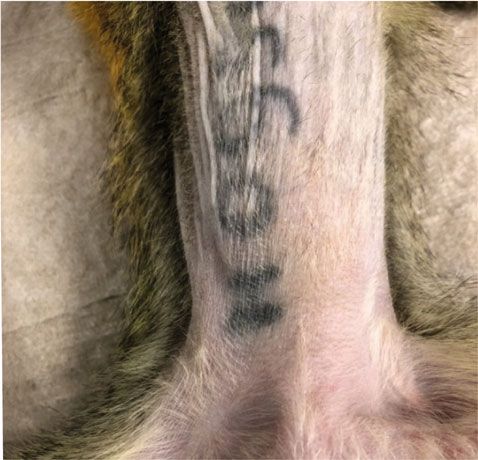
NATURE COMMUNICATIONS | https://doi.org/10.1038/s41467-021-25497-4                                                                                                                                                                                                 ARTICLE

    a                                                                                              c                              5       q.d. hPBMC ex vivo recapitulation
                                                        ERDRP-0519

                                                                                                       serum concentration [µM]
                          O                                                                                                       4

                                                     O                                                                                                                                                                               p
ARTICLE NATURE COMMUNICATIONS | https://doi.org/10.1038/s41467-021-25497-4
a infection Discussion
prophylactic
This study demonstrates the potential of ERDRP-0519 to improve
therapeutic (d3) management of severe measles and augment MeV eradication in
therapeutic (d7)
primates. Our results show that mean attained compound serum
titers in non-human primates after oral b.i.d. dosing were suffi-
-12 h 0 3 dpi 7 dpi 10 dpi 14 dpi 17 dpi 21 dpi cient to significantly reduce MeV replication in an ex vivo reca-
pitulation, anticipating that this dosing regimen inhibits viral
b replication in vivo. Indeed, we demonstrate a strong beneficial
no signs individual spots multiple spots mild rash general rash effect of ERDRP-0519 in MeV-infected squirrel monkeys leading
dpi 6 7 8 9 10 11 12 13 14 15 16 17 18 19 20 to reduced clinical signs. Treatment started as late as 7 days post-
infection prevented appearance of general rash. Earlier onsets of
untreated the treatment regimen further reduced disease severity.
ERDRP-0519 treatment reduced viral titers in PBMCs and throat
swabs. Notably, prophylactic treatment nearly completely prevented
prophylactic MeV viremia, and treatment started 3 days post-infection reduced
viremia markedly. In agreement with the intra-host life cycle of
therapeutic
MeV17,18, treatment starting 7 days post-infection was unable to
(d3) prevent PBMC-associated viremia, but still led to reduced viral titers
in the throat of these animals, indicating decreased viral spread into
and replication in epithelial tissues. The reduced viral load in airways
therapeutic
(d7)
may affect host-to-host transmission and thus have additional
benefits in breaking outbreak chains.
c Lymphocytopenia is a hallmark of MeV infection in humans,
leading to immunosuppression during and after acute infection5.
In our hands, animals treated with ERDRP-0519 trended to
exhibit reduced immunosuppression compared to untreated
animals, but this was not statistically significant. However, pre-
vious experiments of MeV-infected macaques had shown that
elevated numbers of monocytes and eosinophils can compensate
for MeV-induced lymphocytopenia19, indicating that a modest
reduction in total white blood cells may be associated with an
even more profound lymphocytopenia.
An important aspect of MeV infections in humans is the gen-
prophylactically treated untreated eration of life-long humoral immune responses that protect effi-
ciently against re-infection. We demonstrate here that ERDRP-0519
Fig. 2 Effect of ERDRP-0519 on manifestation of clinical signs in MeV- treatment does not prevent the generation of humoral immune
infected squirrel monkeys. a Schematic illustration of the experimental responses. All animals from the therapeutically treated groups
design. Each group was treated twice daily (b.i.d.) over a 14-day period, developed high levels of neutralizing antibodies. In the prophy-
illustrated by colored lines. Prophylactic treatment started 12 h prior to lactically treated group, two animals did not develop neutralizing
infection (green); therapeutic treatment started on day 3 post-infection antibodies until the end of the study. It is possible that ERDRP-0519
(therapeutic (d3), red), or on day 7 post-infection (therapeutic (d7), blue). treatment was so efficient in these individual animals that it pre-
Dpi, days post-infection. b Clinical scores of infected animals. Each row vented the minimal amount of viral replication required to mount
represents one animal of the respective group and color intensity indicates efficient immune responses. However, PBMC-associated viral titers
severity of clinical signs categorized as no signs (white) or individual spots, were similarly low in all animals of this group.
multiple spots, mild rash, and general rash (light to dark graduation). Gray, Absence of pathological alternations during necropsy and
crossed areas represent deceased animal. c Representative photographs of abnormalities that could be linked to a distinct group in histological
the abdomen of a prophylactically treated animal (left) and an untreated examination suggests that ERDRP-0519 does not evoke toxic side
animal (right), taken 13 days post-infection. effects at the administered dose, which is in line with previous
in vivo experiments of ERDRP-0519 treatment in ferrets13.
Safety of ERDRP-0519 treatment. All monkeys were sacrificed The evolution of drug-resistant mutant viruses is a potential
on day 21, followed by gross- and histopathological analyses. No problem for the safety of any antiviral drug. Escape mutant
histological signs of pharmacotoxicity were detected, but we hotspots occurring in the morbillivirus polymerase after ERDRP-
noted mild histopathological changes such as lymphatic lesions in 0519 treatment have been characterized in cell culture and
the lung (Fig. 4a, b), enlarged germinal centers in lymph nodes in vivo13,16. The relative fitness of these ERDRP-0519-resistant
(Fig. 4c, d), hematopoiesis in spleen (Fig. 4e, f), and mesangial viruses was reduced compared to that of the drug-sensitive parent
proliferation in glomeruli of the kidney (Fig. 4g, h), which are virus in all cases13, indicating that escape from the compound is
consistent with a recent virus infection. These and other mani- associated with a selection disadvantage. Viruses re-isolated from
festations emerged in a subset of animals of all treatment groups ERDRP-0519-treated animals did not evolve resistance muta-
and thus cannot be linked to distinct treatment regimens (Sup- tions, adding an additional positive aspect to the safety profile of
plementary Table 1). the drug. Although it is challenging to predict the impact of
Viral RNA was recovered from five animals per group at the emerging resistance on circulating MeV populations, the available
last time point of viremia or positive swab titers for Sanger datasets suggest that sustained circulation of ERDRP-0519-
sequencing of the polymerase L-gene. No mutations causing resistant MeV variants is unlikely.
amino acid changes were identified in the known clusters of the Consistent with the devastating measles burden found in pediatric
MeV polymerase protein associated with viral resistance to patients in many low- and middle-income countries, we believe that
ERDRP-0519 in vitro13,16. a viable therapeutic option must be amenable to cost-effective
4 NATURE COMMUNICATIONS | (2021)12:5233 | https://doi.org/10.1038/s41467-021-25497-4 | www.nature.com/naturecommunicationsNATURE COMMUNICATIONS | https://doi.org/10.1038/s41467-021-25497-4 ARTICLE
a ERDRP-0519 serum conc. in treated animals b rectal temperature
8 41
prophylactic
p=0.0217
( ) therapeutic (d3)
40
serum concentration [µM]
therapeutic (d7)
6
Temperature [°C]
mean 39 ( )
( ) ( )
( ) ( ) ( )
38 ( )
4
37
untreated
2 36 prophylactic
1,5 µM
therapeutic (d3)
35
therapeutic (d7)
0
0 3 7 10 14 0 3 7 10 14 17 21
days after onset of dosing days post-infection
c viral titers in PBMCs d viral titers in throat swabs
6 4 p=0.0042
p=0.0385 p=0.0146
p=0.0199
5
log10(T CID50/1 06 PBMCs)
p960
640
12
320
1 /dilution factor
10
160
1 03 cells/mm3
8 ( ) ( ) ( )
( ) 120
6 80
4 40
( )
( )
2 20
0ARTICLE NATURE COMMUNICATIONS | https://doi.org/10.1038/s41467-021-25497-4
pathological tissue inconspicuous tissue supplemented with 2% FBS and 1% L-Glutamine was used. Human peripheral
a blood mononuclear cells (iQ Biosciences; Donor 4327; Lot# P19L1400) were cul-
b tured in RPMI-1640 and stimulated with 0.2 μg/ml phytohaemagglutinin (PHA;
Sigma–Aldrich) for 24 h prior to use. The isolate MV/FrankfurtMain.DEU/17.11
was used for infections of animals, and the isolate MeV/NewJersey.USA/94/1 was
used for hPBMC infections. Each will be provided upon request.
lung
Compound synthesis and formulation. In total, 100 mg of ERDRP-051922 was
dissolved in 1 ml of poly(ethylene glycol)-200 (Sigma-Aldrich). On the days of
treatment, the drug stock was diluted 1:10 in 0.5% methylcellulose under vigorous
agitation to avoid precipitation. During all procedures, the drug was protected from
light to avoid degradation.
c d
Pharmacokinetics. Drug concentrations were determined using an internal stan-
* dard and a reversed phase isocratic HPLC method with positive ion electrospray
lymph node
ionization (ESI) mass spectrometry detection (LC/MS/MS) on an AB-SCIEX API
5500 MS/MS instrument (5 μl injection volume). Pharmacokinetic parameters were
* estimated using WinNonlin 5.3 (Pharsight).
PK and PD recapitulation in human blood mononuclear cells. Human periph-
eral blood mononuclear cells (hPBMCs) were infected (MOI = 0.1 TCID50 units
per cell) in a 24-well plate format with MeV/NewJersey.USA/94/1 (genotype D6) in
e the presence of ERDRP-0519 or DMSO (0.1%). The concentration of ERDRP-0519
f was maintained at a constant 1.5 µM (recapitulation of trough steady-state drug
* levels achieved during b.i.d. dosing regimen in squirrel monkeys) or adjusted at
various times post-infection to recapitulate ex vivo dynamic plasma drug con-
centrations for once (q.d.) or twice (b.i.d.) daily dosing regimens, based on single-
spleen
dose pharmacokinetics in squirrel monkeys. Cells were collected 48 h after infec-
tion and virus was subsequently harvested after two freeze thaw cycles. Released
virus titers were then determined by limited dilution method (TCID50) on Vero/
* hSLAM cells. Four biological repeats were used for each ex vivo PK recapitulation
and three biological repeats were used for DMSO-treated hPBMCs.
g h Virological and immunological sample analyses. White blood cell counts, viral
load quantification in PBMCs, and quantification of neutralizing antibody titers
were performed as follows: for white blood cell counts, blood was diluted 1:100 in
3% acetic acid and cells were counted using a Neubauer chamber. For viral load
glomeruli
quantification in PBMCs, blood was centrifuged at 3000 rpm for 15 min and serum
was frozen at −20 °C until further use. Red blood cell lysis was performed and the
remaining PBMCs were counted using a Neubauer chamber. Viral titers in PBMCs
were quantified in quadruplicates using limited dilution method and calculated as
TCID50/106 PBMCs. Neutralizing antibody titers were quantified by incubating
MeV with 2-fold serial dilutions of the respective serum for 20 min at room
temperature in quadruplicates. Vero/hSLAM cells were added and incubated for
3 days at 37 °C. Neutralizing antibody titers are expressed as the reciprocal of the
Fig. 4 Histological examination on day 21 post-infection. H&E-stained
highest serum dilution showing no signs of infection. For virus titration from
tissues from all animals of the study (n = 24) were examined and throat swabs, swabs were placed in 150 μl DMEM with 3% penicillin/streptomycin
representative pictures from individual animals are shown. a Perivascular and the titer was quantified by limited dilution method.
lesion due to lymphocytic infiltration in the lung. b Normal lung tissue. c
Lymph nodes with germinal centers (white asterisk) and histiocytosis Quantitative RT–PCR analysis of viral RNA in lymph nodes and PBMCs.
(white arrow). d Normal lymph node. e Spleen with germinal centers (white Organs (21 days post-infection) and PBMCs (twice a week) were frozen in
asterisk) and hematopoiesis (black asterisk). f Normal spleen tissue. g RNAlater (Qiagen) at −80 °C until further processing. For the deceased animal,
organs were frozen in RNAlater at the day of death. Lymph nodes were thawed on
Mesangial proliferation (black arrow) in glomeruli of the kidney. h Normal ice, transferred to TRIzol (ambion life technologies) and homogenized using a
glomerulus. Black bar represents 100 µm (a, b), 200 µm (c–f), 20 µm (g, h). tissue homogenizer. Total RNA of lymph nodes and PBMCs was extracted fol-
Representative pictures are shown from untreated group (c, g, h), lowing the TRIzol manufacturer’s protocol. RNA (1000 ng for lymph nodes; 110 ng
therapeutic day 3 (a, b, f), or therapeutic day 7 (d, e) group. for PBMCs) was then subjected to reverse transcription using random hexamer
primers and Superscript III reverse transcriptase in accordance with the manu-
facturer’s protocols. Subsequent quantitative PCR (qPCR) was carried out using
primer pairs specific for a fragment in the MeV N gene23 or GAPDH gene(Sup-
squirrel monkeys (Saimiri sciureus) were obtained from BioPRIM, 31450 Baziege, plementary Table 2) and the PowerUp SYBR Green Master Mix (Thermo Fisher
or CNRS 0846 Primatologie, 13790 Rousset, France. Each experimental group was Scientific) in an Applied Biosystems 7500 Real-Time PCR system. Samples were
housed in a separate room, and animals of the same sex were caged together within normalized for GAPDH. The comparative (ΔΔCt) Ct method was applied to
the respective groups. For the single-dose pharmacokinetic experiment, animals determine relative amounts of N-encoding RNA present in samples from treated
were anesthetized, and the drug was given through a stomach tube at a con- animals compared to those obtained from untreated control animals (lymph
centration of 50 mg/kg bodyweight. Blood samples were collected after 15, 30, and nodes) or compared to those obtained from untreated control animals on day 7
60 min, and 2, 3, 6, and 24 h. For efficacy assessment, groups of 6 animals were left post-infection (peak PBMC virus titer).
untreated or treated with 50 mg/kg of ERDRP-0519 twice daily for two weeks,
starting either 12 h before or 3 or 7 days after intranasal infection with 106 TCID50 Genetic analysis. Viruses isolated from PBMCs and throat swabs were grown on
of MV/FrankfurtMain.DEU/17.11. Clinical signs, rectal temperature, and weight Vero/hSLAM cells for one passage and RNA was isolated using the Direct-zol RNA
were recorded daily, and throat swabs and blood samples were collected before MiniPrep Kit (Zymo Research) according to the manufacturer’s instructions. RNA
infection and twice weekly thereafter. On day 21 post-infection, the animals were was reverse transcribed using random hexamer primers and Superscript III reverse
euthanized, necropsied, and samples were collected for histological analysis. transcriptase and cDNA sequence was assessed by Sanger sequencing for presence
of resistance mutations in clusters in the MeV L gene known to accumulate
resistance-conferring mutations in tissue culture in the presence of ERDRP-051910.
Cell culture and viruses. Vero cells stably expressing human SLAM (Vero/ Sanger sequencing was carried out using MeV L gene specific primers (Supple-
hSLAM) were maintained in Dulbecco’s modified Eagle’s medium (DMEM) with mentary Table 2) and obtained sequences were analyzed using Sequencher software
5% fetal bovine serum and 1% L-Glutamine. For infection experiments, DMEM (Gene Codes Corporation; Version 5.4.6).
6 NATURE COMMUNICATIONS | (2021)12:5233 | https://doi.org/10.1038/s41467-021-25497-4 | www.nature.com/naturecommunicationsNATURE COMMUNICATIONS | https://doi.org/10.1038/s41467-021-25497-4 ARTICLE
Necropsy and histology. After euthanasia, each animal was examined for gross 18. Ayasoufi, K. & Pfaller, C. K. Seek and hide: the manipulating interplay of
pathological changes, and samples were collected for histopathological examina- measles virus with the innate immune system. Curr. Opin. Virol. 41, 18–30
tion. After fixation, all tissue samples were embedded in paraffin. Histology slices (2020).
were mounted and stained using standard H&E staining techniques. 19. Auwaerter, P. G. et al. Measles virus infection in rhesus macaques: altered
immune responses and comparison of the virulence of six different virus
Statistical analysis. Statistical analysis was performed using GraphPad Prism 8. strains. J. Infect. Dis. 180, 950–958 (1999).
One-way analysis of variance (ANOVA) with Tukey’s multiple comparison post-hoc 20. Phadke, V. K., Bednarczyk, R. A., Salmon, D. A. & Omer, S. B. Association
test or two-way ANOVA, followed by Dunnett’s multiple comparisons post-hoc test between vaccine refusal and vaccine-preventable diseases in the United States:
were applied for statistical comparisons using the untreated group as reference if not a review of measles and pertussis. JAMA 315, 1149–1158 (2016).
stated otherwise. Different time points for each individual animal were paired. Bio- 21. Plemper, R. K. & Hammond, A. L. Synergizing vaccinations with therapeutics
logical repeat refers to measurements taken from distinct samples, and results obtained for measles eradication. Expert Opin. Drug Discov. 9, 201–214 (2014).
for each individual biological repeat are shown in the figures along with the exact size 22. Ndungu, J. M. et al. Non-nucleoside inhibitors of the measles virus RNA-
(n number) of biologically independent samples, animals, or independent experiments. dependent RNA polymerase: synthesis, structure-activity relationships, and
Measure of center (connecting lines and columns) are means throughout. Error bars pharmacokinetics. J. Med. Chem. 55, 4220–4230 (2012).
represent standard deviations (SD) throughout. For all experiments, the statistical 23. Hummel, K. B., Lowe, L., Bellini, W. J. & Rota, P. A. Development of
significance level α was set toYou can also read