Primary vulvar leiomyosarcoma localized in the Bartholin's gland area: A case report and review
←
→
Page content transcription
If your browser does not render page correctly, please read the page content below
MOLECULAR AND CLINICAL ONCOLOGY 14: 69, 2021

                  Primary vulvar leiomyosarcoma localized in the
                  Bartholin's gland area: A case report and review
  STELLA AKRIVI1, MICHAIL VARRAS2, ZOI ANASTASIADI3, CHRISTINA PAPPA3, AIKATERINI VLACHIOTI3,
    VIKTORIA‑KONSTANTINA VARRA4, FANI‑NIKI VARRA5, EUFEMIA BALASI6 and CHRISTOS AKRIVIS3

     1
       Department of Obstetrics and Gynecology, Royal Jubille Maternity Hospital, Belfast Trust, Belfast BT12 6BA, UK;
            2
              Fourth Department of Obstetrics and Gynecology, ‘Elena Venizelou’ General Hospital, Athens 11521;
            3
              Department of Obstetrics and Gynecology, ‘G. Chatzikosta’ General Hospital, Ioannina 45001, Epirus;
    4
      Department of Pharmacy, University of Patras, Patra 26504, Greece; 5Department of Pharmacy, Frederick University,
      Nicosia 1036, Cyprus; 6Pathology Department, ‘G. Chatzikosta’ General Hospital, Ioannina 45001, Epirus, Greece

                                      Received March 13, 2020; Accepted January 14, 2021

                                                    DOI: 10.3892/mco.2021.2231

Abstract. Vulvar sarcomas located in the Bartholin's gland           were observed 53 months after surgery. In conclusion, wide
area are extremely uncommon mesenchymal vulvar tumors.               local tumor excision with free surgical margins is a good option
These neoplasms can be mistaken as Bartholin' gland benign           of surgery for vulvar leiomyosarcomas. In recurrences, a new
lesions such as cysts or abscesses, leading to a delay in the        extensive surgical resection of the lesion and radiotherapy are
diagnosis of underlying malignancy. Currently, only a few            suggested. Ipsilateral lympadenectomy is indicated when there
cases of these aggressive cancers have been reported in the          is a pathologic lymph node. Chemotherapy is provided in cases
literature. A 42‑year‑old female patient without any previous        of distal metastases.
complaint presented to Obstetrics and Gynecology Department
of ‘G. Chaztikosta’ General Hospital due to a vulvar lump in         Introduction
the area of the left Bartholin's gland with a 6‑month history
of progressive swelling. Pelvic examination showed a solid           Sarcomas are tumors with mesenchymal origin and very poor
mass of 6.5‑cm in maximum diameter, localized in the left            prognosis compromising of 2% of all cancer deaths. These
Bartholin's gland. The patient underwent wide local excision         neoplasms are very rare and account for 1 to 2% of all malig‑
and histopathological examination of hematoxylin and                 nancies (1‑7). Sarcomas of the female genital tract have a low
eosin‑stained sections indicated intersecting fascicles of spindle   frequency of 3% (4,8,9). In particular, vulvar sarcomas account
cells, with moderate to severe atypia. The number of mitoses         for 1 to 2% of all the vulvar tumors, compared to the uterine
was up to 8 per 10 high power fields. The neoplasm to its greatest   sarcomas, which represent the vast majority of all sarcomas of
extent was circumscribed and in places had an invasive growth        the female reproductive system (90%) (2,5‑7).
pattern. Tumoral necrosis was not seen. Involved Bartholin'              Vulvar sarcomas comprise a highly heterogeneous
gland by the tumor was identified. The tumor extended                group of histological types, which include leiomyosar‑
focally to the surgical margin. The neoplastic cells showed          comas, epithelioid sarcomas, fibrosarcomas, liposarcomas,
positive staining for smooth muscle actin, desmin, HHF35,            hemangiosarcomas, rhabdomyosarcomas, angiosarcomas,
caldesmon, vimentin and estrogen and progesterone receptors.         malignant hemagiopericytomas, neurogenous sarcomas,
Immunohistochemistry was negative for S100, myoglobulin,             dermatofibrosarcomas protuberans, Ewing sarcomas,
keratin 116, CD117, CD34 and CD31. The patient denied                synovial sarcomas, clear cells carcinomas, malignant
further surgery or/and local radiotherapy, although the mass         fibrous histiocytomas and sometimes aggressive angiomyx‑
was >5‑cm and a focally infiltrative surgical margin was found.      omas (1,3,4,6,10‑12). The age of the described patients with
During the close follow‑up, no local recurrences or metastases       vulvar leiomyosarcomas at the time of clinical presentation
                                                                     ranged between 14 and 69 years of age, with an average age
                                                                     of approximately 30‑40 years (4,6,13,14). Vulvar embryonal
                                                                     rhabdomyosarcoma typically presents in girls less than
                                                                     8 years of age (15). The size of vulvar sarcomas varies
Correspondence to: Dr Michail Varras, Fourth Department of
                                                                     between 2 and 10‑cm (4). Vulvar sarcomas are usually
Obstetrics and Gynecology, ‘Elena Venizelou’ General Hospital,
Plateia Elenas Venizelou 2, Ampelokipoi, Athens 11521, Greece        asymptomatic or they are characterized by non‑specific
E‑mail: mnvarras@otenet.gr                                           clinical manifestations, such an enlarging vulvar mass with a
                                                                     local discomfort. Vulvar sarcomas located in the Bartholin's
Key words: Bartholin's gland, vulva, leiomyosarcoma, soft tissue,    gland area are often misdiagnosed as their clinical mani‑
vulvar sarcoma, vulvar neoplasm, vulvar diseases                     festations are very similar to benign lesions. These lesions
                                                                     are most commonly misdiagnosed as Bartholin's cysts or
                                                                     abscesses resulting in delaying diagnosis and worsening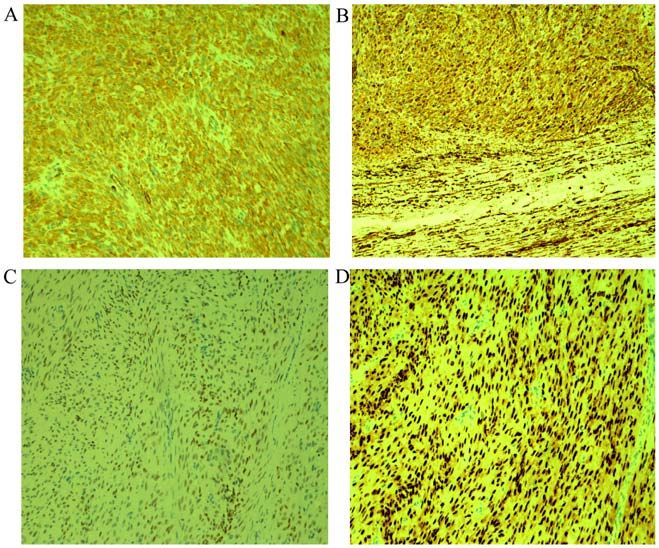
2 AKRIVI et al: PRIMARY VULVAR LEIOMYOSARCOMA LOCALIZED IN THE BARTHOLIN'S GLAND AREA
the prognosis (11,12,6). In these cases, the late symptoms Case report
include pain, ulceration, bleeding and voiding dysfunc‑
tion (3,6,11). Vulvar sarcomas are characterized by a high A 42‑year‑old patient presented to outpatient clinic because
chance of metastasis; it seems that chemotherapy achieves of a vulvar lump in the area of the left Bartholin's gland with
regression of lung metastasis (6,12,14,16). a 6‑month history of progressive swelling. In her gyneco‑
Leiomyosarcomas are the most common histological vari‑ logical history, the patient reported laparoscopic hysterectomy
ants of vulvar sarcomas (2,5,7,17‑22). Vulvar leiomyosarcomas without salpingo‑oophorectomy, two years ago, due to heavy
are considered to originate from the smooth muscles within metrorrhagia and the uterus was taken out through the vagina
erectile tissue, blood vessel walls, rough ligaments, dartos directly. The final histological exam of the hysterectomy
muscles, erector‑pili muscles and from stem cells localized showed benign leiomyomas within the uterus.
in Bartholin's gland (22‑24). Leiomyosarcomas occur most Pelvic examination showed a solid mass of 6.5‑cm in
frequently in the labia majora, followed in a decreasing maximum diameter, in the area of the left Bartholin's gland.
order by the Bartholin's gland area, the pericloitoral area Palpation of the inguinal lymph nodes in either groin was
and the labia minora (25,26). Localizations of leiomyosar‑ normal; there were no palpable nodes. The patient underwent
comas in the Bartholin's gland area are extremely rare and wide local excision and the mass was enucleated completely
only few such cases are reported in the international litera‑ from the bed. The surgical specimen was sent for pathological
ture (1,2,5,7,10,12,13,17,18). The biological behavior of the examination. We did not indicate a biopsy of the mass before
vulvar leiomyosarcomas is not fully understood and very little the excision because our initial clinical diagnosis was as a
information is available so far (1). Most of the described cases chronic left Bartholin's gland abscess.
of vulvar leiomyosarcomas in the international literature are Macroscopically, the tumor was 6.5x4.5x3.5‑cm in size
about patients from Western countries suggesting therefore with regular margins. Cut surface showed a whitish, homoge‑
genetic disposition and lifestyle connection (27). A possible neous lesion with fibroelastic consistency.
carcinogenetic precursor of these neoplasms is the chronic Microscopic examination of the hematoxylin and
inflammation. This hypothesis is supported by the findings eosin‑stained sections showed intersecting fascicles of
of coexistence between vulvar leiomyosarcomas and long‑ spindle cells (Fig. 1A and B), with moderate to severe
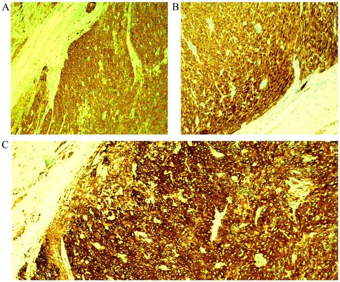
standing lichen sclerosus (28). Also, another predisposing atypia (Fig. 2A and B). The number of mitoses was up to 8
factor of vulvar leiomyosarcomas seems to be the pregnancy per 10 high power fields (Fig. 3A). The neoplasm to its greatest
because occurrence of them during pregnancy has been extent was circumscribed and in places had an invasive growth
reported (10,17,29). It seems that the positivity of estrogen pattern. Tumoral necrosis was not seen. Involved Bartholin'
and progesterone receptors is implicated in the develop‑ gland by the tumor was identified. The tumor extended focally
ment of vulvar leiomyosarcomas. The potential impact of to the surgical margin (Fig. 3B). The neoplastic cells showed
estrogens and progesterone signaling through their receptors positive staining for smooth muscle actin (SMA) (Fig. 4A),
may result in induction of proliferation and growth of DNA desmin (Fig. 4B), HHF35 (Fig. 4C), caldesmon (Fig. 5A),
damaged cells resulting finally in tumorogenesis of the vulvar vimentin (Fig. 5B) and estrogen (Fig. 5C) and progesterone
leiomyosarcomas (10,17,29). receptors (Fig. 5D). Immunohistochemistry was negative for
The histopathological examination of the excised mass S100, myoglobulin, keratin 116, CD117, CD34 and CD31.
confirms the diagnosis of the extremely rare entity of Thoracic and upper and lower abdominal CT scans were
vulvar leiomyosarcomas (2,5,7). Given the extreme rarity negative for malignancy. Also, pelvic MRI was non sugges‑
of vulvar leiomyosarcomas, an optimal treatment strategy tive for infiltrative pelvic lymph nodes. The diagnosis was
has not yet been elucidated. It seems that their primary primary vulvar leiomyosarcoma, localized in the Bartholin's
treatment of choice is the surgical excision, including wide gland area. The patient was discharged from the hospital and
local excision or radical hemivulvectomy. The usefulness during the follow‑up, every four months, no local recurrences
of ipsilateral lymphadenectomy in the treatment of vulvar or metastases of the disease were observed 53 months after
leiomyosarcomas is not clear because these neoplasms surgery and the patient is in excellent physical condition.
metastasize mostly by the bloodstream compared to Although the mass was greater than 5‑cm and a focally infil‑
lymphogenous metastasis. Locally recurrent tumors require trated surgical margin was found in the histological specimen,
adjuvant treatment in the form of radiotherapy. Adjuvant the patient denied further surgery or/and local radiotherapy.
chemotherapy is placed in the treatment of distal metas‑ During the close follow‑up the patient did not complain about
tases; however the exact role of adjuvant chemotherapy itching, burning, pain, numbness at the treatment site, body
and/or radiotherapy remains uncertain for vulvar leiomyo‑ image disturbance or sexual problems.
sarcomas, which display very aggressive behavior and rapid
progression (2,5,7). Discussion
Herein, we present a very rare case of vulvar leiomyosar‑
coma localized in the Bartholin's gland area in a 42‑year‑old Primary vulvar leiomyosarcomas are extremely rare tumors
female and describe its histopathological and immunohisto‑ and the localization of these tumors in the Bartholin's gland
chemical features. Also, the current literature is reviewed in area results in diagnostic delays (7). Clinical differential diag‑
terms of clinical signs and symptoms, diagnosis, biological nosis of vulvar leiomyosarcomas located in the Bartholin's
behavior, prognosis and treatment. Vulvar sarcomas located gland area include Bartholin's gland cyst or abscess, infectious
in the Bartholin's gland are often mistakenly considered as granuloma, syringeal, lipoma, fibroma, leiomyoma, hidrad‑
benign lesions resulting in delayed diagnosis. enoma, hidradentis suppurativa, hematoma, endometriosis,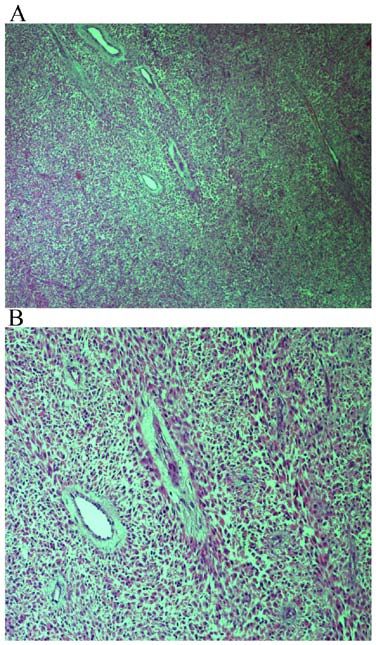
MOLECULAR AND CLINICAL ONCOLOGY 14: 69, 2021                                                              3

Figure 1. Spindle cell neoplasm with storiform pattern. Hematoxylin and       Figure 3. Presence of mitotic figures. (A) Hematoxylin and eosin staining
eosin staining of tumor tissues at a magnification of (A) x40 and (B) x100.   of tumor tissues at a magnification of x200. (B) Focally infiltrated margin,
                                                                              hematoxylin and eosin staining of tissue at a magnification of x40.

                                                                              accessory breast gland, warts, paraganglioma or squamous
                                                                              cell carcinoma, adenosquamous carcinoma, adenocarcinoma,
                                                                              adenoid cystic carcinoma, small cell carcinoma, transitional
                                                                              carcinoma, differential carcinoma, melanoma, spindle
                                                                              cell synovial sarcoma, embryonal rhabdomyosarcoma,
                                                                              dermatofibrosarcoma, malignant fibrous histiocytoma,
                                                                              extraskeletal Ewing's sarcoma, hibrosarcoma and epithelioid
                                                                              sarcoma (4,7,30‑38). Furthermore, when the tumor is located
                                                                              in the Bartholin's gland, leiomyosarcoma should be distin‑
                                                                              guished from adenosarcoma, which is composed by a mixture
                                                                              of benign glandular epithelium and a malignant sarcomatous
                                                                              stroma (39,40). Diagnosis is made by the histopathological
                                                                              examination of a biopsy of the lesion or of the complete exci‑
                                                                              sion of the neoplasm. In our case, non‑epithelial component
                                                                              was identified microscopically in the tumor and therefore we
                                                                              concluded the diagnosis of vulvar leiomyosarcoma. Metastatic
                                                                              dissemination of vulvar leiomyosarcomas is thought to be
                                                                              through the hematogenous routes. The most common sites
                                                                              of distant metastatic sites have been reported to be the liver
                                                                              and the lungs and occasionally the bones (1,37). The role of
                                                                              the lymphogenous routes for the metastatic dissemination of
                                                                              vulvar leiomyosarcomas is questionable (28).
                                                                                  Vulvar leiomyosarcomas can be distinguished from
                                                                              vulva r leiomyomas or vulva r atypical leiomyomas
                                                                              according to the suggestions of Nielsen et al (38). Three
                                                                              or more of the following histological and pathological
Figure 2. Moderate to severe cytological atypia. (A) Severe cytological
atypia; hematoxylin and eosin staining of tumor tissues at a magnification
                                                                              characteristics are important for the diagnosis of vulvar
of x100. (B) Moderate cytological atypia; hematoxylin and eosin staining of   leiomyosarcomas: i) The tumor has size greater than 5‑cm
tumor tissues at a magnification of x100.                                     in diameter; ii) infiltration is seen in the margins of the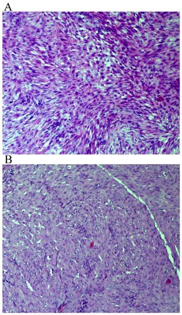
4                  AKRIVI et al: PRIMARY VULVAR LEIOMYOSARCOMA LOCALIZED IN THE BARTHOLIN'S GLAND AREA

Figure 4. Immunohistochemical expression of spindled tumor cells for (A) SMA at a magnification of x100; (B) desmin at a magnification of x100; and
(C) HHF35 at a magnification of x40.

Figure 5. Immunoreactivity of spindled tumor cells for (A) caldesmon at a magnification of x100; (B) vimentin at a magnification of x10; (C) estrogen receptors
at a magnification of x100; and (D) progesterone receptors at a magnification of x100.

tumor; iii) the tumoral background consists of more than                         actin and desmin and focal positivity for S‑100 and
five mitotic figures per 10 high‑power fields; and iv) the                       cytokeratin (10). In our case, the neoplastic cells showed
tumoral cells show moderate to severe atypia. Leiomyomas                         positive staining for smooth muscle actin (SMA), desmin,
exhibit only one of the above features; atypical leiomyomas                      HHF35, caldsmon, vimentin and estrogen and proges‑
exhibit only two of these features (38). Both leiomyomas                         terone receptors (Figs. 4 and 5).
and leiomyosarcomas exhibit immunopositivity for muscle                             A preoperative biopsy of vulvar masses located in the
markers including smooth muscle actin, muscle‑specific                           Bartholin's gland area and particularly in those with a compli‑
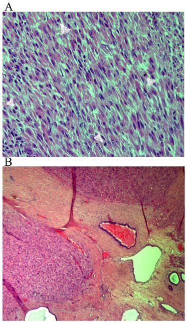
Table I. Histological and immunohistochemical features, treatment and clinical outcome of vulvar leiomyosarcoma reported cases.

Author‑               Age      Size				                                                 Initial		                      Local      Treatment of
Published year       (years)   (cm) MFI MTCN IHC                                      treatment Margins              recurrence    recurrence    Follow‑up   (Refs.)

Davos and Abell 41 NS   >10 NS      NS               Wide local         NS           NS           ‑                    28 months,                             (18)
(1976)						                                         excision				                                                      alive, NED
Davos and Abell 49 NS   >10 NS      NS               Local excision     NS           +            Wide local           9 years, alive,                        (18)
(1976)									                                                                                   excision             NED
Davos and Abell 84 NS   >10 NS      NS               Wide local         NS           +            No further treatment 6 months,                              (18)
(1976)						                                         excision				                                                      disseminated
										                                                                                                             metastases,
										                                                                                                             died of disease
Davos and Abell 54 NS   >10 NS      NS               Local excision     NS           +            Hemivulvectomy;      9 years,                               (18)
(1976)									                                                                                   hysterectomy and     disseminated
									                                                                                         excision of vaginal  metastases,
									                                                                                         mass                 died of disease
Davos and Abell 35 NS   >10 NS      NS               Local excision     NS           +            Re‑excision;         9 years, alive                         (18)
(1976)									                                                                                   radical vulvectomy
Møller et al    54  8    15  Yes‑   Desmin: Positive Radical            No           No           ‑                    30 months,                             (55)
(1990)				                   Focal		                 vulvectomy +				                                                  alive, NED
						                                               inguinofemoral
						                                               lymphadenectomy
Tawfik et al    52 15  25‑30 Yes‑   SMA, vimentin,   Local excision +   Infiltrative No           ‑                    14 months,                             (25)
(1994)				                   Focal  HHF‑35, ER,      radiotherapy				                                                  alive, NED
					p53: Positive
Aartsen and     15  7   NS   NS     NS               Radiotherapy		                  +; 2.5 years Wide Local           24 years,                               (1)
                                                                                                                                                                       MOLECULAR AND CLINICAL ONCOLOGY 14: 69, 2021

Albus‑Lutter						                                   40 Gy			                                     Excision +           alive, NED
(1994)									                                                                                   Radiotherapy 40 Gy
Aartsen and     41 4.5  NS   NS     NS               Wide local         ‑            ‑            ‑                    17 years,                               (1)
Albus‑Lutter						                                   excision				                                                      died of disease
(1994)
Aartsen and     75 10   NS   NS     NS               Radical vulvectomy NS           6 months & Local excision         4 months,                               (1)
Albus‑Lutter								                                                                 3 months     ‑Local excision      died of disease
(1994)
Aartsen and     67 10   NS   NS     NS               Radical vulvectomy NS           5 months     Radiotherapy         7 years,                                (1)
Albus‑Lutter									                                                                             49 Gy                died of disease
(1994)
                                                                                                                                                                       5
6
Table I. Continued.
Author‑Published Age Size Initial Local Treatment of
Year (years) (cm) MFI MTCN IHC treatment Margins Recurrence Recurrence Follow‑up (Refs.)
Fried‑Oginski et al 31 5 4 Yes‑ SMA, muscular Local Excision + NS 10 months Wide Local NS (56)
(1995) Focal actin, vimentin: 8 weeks later Wide Excision +
Positive Local Excision Radiotherapy
Nielsen et al 33 5.5 5 NS SMA, muscular Local Excision Infiltrative 4 months Wide local Excision 36 months, (38)
(1996) actin, vimentin, alive, NED
desmin, ER, PR:
Positive; keratin,
S‑100: Negative
Nielsen et al 46 10 5 NS SMA, muscular Modified Radical No ‑ ‑ 30 months, (38)
(1996) actin, vimentin, Vulvectomy alive, NED
desmin, ER, PR:
Positive; keratin,
S‑100: Negative
Nielsen et al 37 5 8 NS SMA, muscular Wide Local Infiltrative 3 months Wider Excision; 36 months, (38)
(1996) actin, vimentin, Excision positive margins alive, NED
ER, PR: Positive; again; modified
keratin, S‑100, radical vulvectomy
desmin: Negative
Nielsen et al 67 16 10 NS SMA, muscular NS NS ‑ Local, 7 months, (38)
(1996) actin, vimentin, radiotherapy metastases
ER, PR: Positive; chemotherapy to lungs and
keratin, S‑100, bones, died
desmin: Negative of disease
Nielsen et al 56 5.5 5 NS SMA, muscular NS Infiltrative NS NS NS (38)
(1996) actin, vimentin:
Positive; keratin,
AKRIVI et al: PRIMARY VULVAR LEIOMYOSARCOMA LOCALIZED IN THE BARTHOLIN'S GLAND AREA
desmin, S‑100,
ER, PR: NegativeTable I. Continued. Author‑Published Age Size Initial Local Treatment of Year (years) (cm) MFI MTCN IHC treatment Margins Recurrence Recurrence Follow‑up (Refs.) Torres et al 14 6 ‑ ‑ ‑ Local Excision ‑ +; Wide Local 22 Years, (13) (2000) 13 months Excision + alive, NED Chemotherapy; Wide Local Excision + Radiotherapy 50 Gy; Vulvectomy + Hysterectomy + Radiotherapy 65 Gy Rubin and Fletcher 26 1.2 6 Yes, SMA, desmin: Local Excision Infiltrative 8 months & NS 62 months, (57) (2000)
8
Table I. Continued.

Author‑                Age      Size				                               Initial		               Local       Treatment of
Published year        (years)   (cm) MFI MTCN IHC                    treatment Margins       recurrence     recurrence  Follow‑up     (Refs.)

González‑Bugatto  52      6    21  No SMA, Vimentin,   Hemivulvectomy + Yes               +;        Wide local           4 years,      (12)
et al					                            Desmin, ER,      ipsilateral		                      12 months excision             alive, NED
(2009)					                           PR: Positive;    lympadenectomy +
					                                 S‑100, CK, CD31, radiotherapy
					p53, c‑erbB‑2,                                    66.6 Gy +
					Leukocyte Common chemotherapy
					Antigen: Negative
Salehin et al     71      2    10  NS SMA, Ki‑67:      Hemivulvectomy + No                NS        NS                   NS             (4)
(2011)					                           Positive         hysterectomy +
						                                                 salpingo‑
						                                                 oophorectomy +
						                                                 inguinal
						                                                 lymphadenectomy
Mowers et al      48  4 & 10   NS  NS SMA, desmin,     Incision and        Infiltrative +; 5 years  Partial excision and 18 months,    (26)
(2014)		             (5 years			      ER, PR: Positive drainage			                                  marsupialization;    alive, NED
		                     later)							                                                                Left radical
									                                                                                           Hemivulvectomy +
									                                                                                           hysterectomy +
									                                                                                           bilateral salpingo‑
									                                                                                           oophorectomy +
									                                                                                           chemotherapy
Levy et al (2014) 57     2.5   16  NS SMA: Positive;   Local excision +    Infiltrative ‑ NS        NS                   NS            (16)
					Desmin: Positive                                  Radical excision    Then, no
							                                                                    residual
							                                                                    disease
Teramae et al     51    13.5  7‑10 NS Desmin, Ki‑67:   Local excision      No             ‑         ‑                    32 months,    (44)
(2014)					                           Positive					                                                                      alive, NED
Alnafisah and     37      5    NS  NS SMA, ER,         Wide local          Residual       15 months Left                 Lung          (14)
Alfieri (2016)					                   PR: Positive     excision +          disease		                hemivulvectomy +     metastatic
                                                                                                                                                AKRIVI et al: PRIMARY VULVAR LEIOMYOSARCOMA LOCALIZED IN THE BARTHOLIN'S GLAND AREA

						                                                 Chemotherapy;			                             ipsilateral iliac,   disease
						                                                 Left radical			                              inferior epigasric
						                                                 vulvectomy +			                              and obturator
						                                                 ipsilateral inguinal			                      lymphadenectomy
						                                                 lymphadenectomy
Table I. Continued.
Author‑ Age Size Initial Local Treatment of
Published year (years) (cm) MFI MTCN IHC treatment Margins recurrence recurrence Follow‑up (Refs.)
Korkmaz et al 65 6 20 Yes Calponin, Ki‑67: Local Excision NS NS ‑ 6 months, (7)
(2016) Positive; S‑100: alive, NED
Negative
a
Sayeed et al (2018) 72 11 8 Yes ‑ Wide local Infiltrative NS* NS* Died of (58)
excision disease
a
Sayeed et al (2018) 56 13.5 34 Yes ‑ Wide local Infiltrative NS* NS* Died of (58)
excision disease
a
Sayeed et al (2018) 68 5.5 23 Yes ‑ Wide local No NS* NS* Died of (58)
excision disease
Nath et al (2019) 38 8 4‑5 Yes SMA, vimentin: Wide local ‑ ‑ ‑ 21 months, (9)
Positive; CK, excision alive, NED
desmin, S‑100,
HMB‑45, CD34,
ER, PR: Negative
Sameeta et al, 63 2.1 25 No SMA, desmin, ER: Wide local Infiltrative NS NS NS (59)
(2019) Positive; S‑100, excision + (focal)
cytokeratin radiotherapy
AE1/AE3: Negative
Saquib et al 63 2.8 22 Yes‑ SMA, desmin, Local excision; Infiltrative ‑ ‑ NS (60)
(2020) Focal caldesmon, Ki‑67: ipsilateral (focal);
MOLECULAR AND CLINICAL ONCOLOGY 14: 69, 2021
Positive; CD117, lymphadenectomy + Negative:
CD4, vimentin, ipsilateral hemivul- After the
S100: Negative vectomy second
operation
Smith et al (2020) 46 3 NS NS SMA, desmin: Local excision; Yes; NS NS NS (61)
Positive; cytokeratin ipsilateral partial after 3
AE1/AE3, p40, radical vulvectomy; operations:
S100, myogenin, posterior radical Negative
myo‑D1, caldesmon, vulvectomy,
CD34, STAT‑6, bilateral
TLE1: Negative inguino‑femoral
lympadenectomy
910
Table I. Continued.
Author‑ Age Size Initial Local Treatment of
Published year (years) (cm) MFI MTCN IHC treatment Margins recurrence recurrence Follow‑up (Refs.)
Swanson et al 80 5.2 12 Yes NS Resection + No NS NS Liver (62)
(2020) chemotherapy metastasis at
the time of
resection
Swanson et al NS 9.7 36 No SMA: Diffusely Resection Infiltrative NS NS NS (62)
(2020) positive;
h‑Caldesmon:
Multifocally
positive; Desmine:
Negative
Swanson et al NS 2.5 12 No Desmin, Resection Infiltrative NS NS NS (62)
(2020) h‑Caldesmon and
SMA: Diffusely
positive; S100
protein: Negative
Akrivi et al 42 6.5 8‑10 No Desmin, Caldesmon, Wide local Focally No Wide local 53 months, ‑
(present case) Vimentin, SMA, excision infiltrative Excision alive, NED
ER, PR: Positive; margin
S100, myoglobulin,
Keratin‑116, CD117, CD34,
CD31: Negative
MFI, Mitotic figure index per ten high power fields; MTCN, Microscopic tumor cell necrosis; IHC, immunohistochemistry; NED, no evidence of disease; NS, not specified; aIn the study by Saveed et al
metastasis to lung occurred in 2 cases, at 1 and 36 months, respectively, and in the 3rd case occurred extensive local reccurence of the pelvis at 41 months. One patient was treated with adjuvant radiotherapy
after recurrence and 2 patients were treated with chemotherapy after lung metastasis. All of the three patients died of the disease at 4, 36 and 56 months.
AKRIVI et al: PRIMARY VULVAR LEIOMYOSARCOMA LOCALIZED IN THE BARTHOLIN'S GLAND AREAMOLECULAR AND CLINICAL ONCOLOGY 14: 69, 2021 11
cated appearance is highly recommended because early survival was positively correlated to high levels of interleukin
diagnosis is important for curative treatment without delays (IL)‑12 and mitochondrial pyruvate carrier 3 (MPC3) levels
of these extremely rare neoplasms (3,7,12,16,26). However, and negatively correlated with low levels of VEGF2 and high
in our case we did not indicate a biopsy of the mass before levels of placental growth factor (PGF) (48). In addition, in
operation because our preoperative diagnosis was as a chronic soft tissue sarcoma the activity of trabectedin was found
Bartholin's gland abscess. Superficial lymphadenopathy is to inversely correlate with the mRNA levels of BRCA1.
due to a cyst, inflammation, lymphoma or metastatic malig‑ Particularly, low levels of mRNA BRCA1 expression were
nant tumor (41,42). In the presence of a pathognomonic associated with improvement in outcome of patients (49).
inguinal node the use of fine‑needle aspiration biopsy is an In our case, the neoplastic cells showed positive staining
easy, reliable and safe office procedure, which immediately for estrogen and progesterone receptors (Fig. 5C and D),
provides an important tool of diagnosis (41). In the study by suggesting the potential role of estrogen and progesterone in
Altinboğa and Yüce (43) the lymph node fine‑needle aspiration the development and progression of vulvar leiomyosarcoma.
cytology (FNAC) showed sensitivity of 95.4%, specificity of However, in our case we did not examine the pre‑ and
94.1%, positive predictive value of 95.4% and negative predic‑ post‑operative serum estrogen levels of our patient.
tive value of 94.1% (43). Nethertheless, the predisposing role of estrogen and
The first‑line treatment involves surgical excision of the progesterone in vulvar leiomyosarcoma tumorogenesis is
lesion. Localized disease is usually managed with wide local supported by their occurrence during pregnancy (10,17,29).
excision with surgically free margins. According to Aartsen It is possible that the positivity of estrogen and progesterone
and Albus‑Lutter (1) the width of normal margin‑tissue receptors may initiate the proliferation and growth of DNA
proposed for the surgeon doing the excision is 2‑cm and for damaged cells resulting in development and progression of
the pathologist 1‑cm because of the shrinking effect. Poor vulvar leiomyosarcoma (10,17,29). The role of aromatase
prognostic factors are considered to be mitotic rate more than inhibitors (AIs), selective estrogen receptor modulators
10 mitotic figures per 10 HPF, high histological grade, tumor (SERMs), progestins and GnRH‑analogues for the treatment of
size >5‑cm, local recurrence and distal metastases (38,44). vulvar leiomyosarcoma and especially for their recurrences is
These neoplasms tend to metastasize by bloodstream and not clear due to the rarity of this entity and the lack of clinical
therefore ipsilateral lymphadenectomy is questionable (28). trials to determine the clinical efficacy and usefulness of the
The decision to treat with vulvectomy is made by the size of the above regiments for first‑ and second‑line therapies. However,
lesion (7,10,18). Inguinal lymphadenectomy does not improve we have some evidence in the case of uterine leiomyosarcoma.
treatment (1,22). The available data is limited and uncertain Particularly, patients with uterine leiomyosarcoma and a high
regarding radiosensitivity and chemosensitivity in the treat‑ expression rate of ER and PR show good response to hormonal
ment of the vulvar leiomyosarcomas. However, in the presence therapy (50). Yamaguchi et al suggested that Letrozole
of negative prognostic factors radiotherapy and chemotherapy as well as progestins could be the first choice for patients
should be given. Important favorite prognostic factors are the with recurrent or residual low‑grade endometrial stromal
lower grade of nuclear atypia, the smaller tumoral size, the sarcoma (51). It seems that letrozole is the first‑line hormone
pathological confirmation of at least 2‑cm negative surgical drug for postoperative adjuvant therapy in patients with stage I
margins and the absence of local recurrences (1,2,12,14,22,38). uterine leiomyosarcoma, while exemestane and anastrozole
Favor immunohistochemical factors include the overexpres‑ are for second‑line treatment in patients with recurrent,
sion of estrogen, progesterone and androgen receptors, the metastatic and unresectable uterine leiomyosarcoma (52). In a
moderate immunopositivity for Ki‑67 and the absence of p53 clinical trial by George et al (53) of 27 postmenopausal women
expression (4,22). It seems that the type of operation with with metastatic and/or unresectable uterine leiomyosarcoma
inadequately resected tumors is the most significant negative with positive estrogen receptors and/or postive progesteron
prognostic factor for the appearance of local recurrences and receptors it has been found that the rate of stable disease was
distal metastases, compared to tumoral size of more than 54% in all patients and the 12‑week rate of progression free
5‑cm (1). In addition to these factors, vulvar leiomyosarcomas survival 50%. These findings suggest that strong expression
with more aggressive behavior are defined on the basis of high of estrogen and progesterone expression in tumors is related
cellularity, anaplasia, mitotic activity more than ten per 10 with longer progression free survival (53). On the other
high‑power fields, high grade of nuclear atypia and presence hand, the use of progestins in uterine leiomyosarcoma
of tumoral necrosis (12,16). The indications for radiotherapy should be cautious because medroxyprogesterone acetate
includes: a) positive margin tumor involvement at the initial in higher doses (5 mg/day) is able to significantly increase
surgical treatment and b) tumor size >5‑cm (22,25,38,44,45). the growth of this neoplasm (52). In addition, it has been
Pharmacogenomics biomarkers for the prediction of response found that patients who receive tamoxifen treatment for
to chemotherapeutic regimens seem to be involved in the more than five years show an increase in the incidence of
improvement of molecular diagnostics in treatment of a specific uterine leiomyosarcoma to 17/100,000 women yearly (50,54).
subset of patients with soft tissue sarcomas. Actually, human Therefore, tamoxifen is contraindicated for the treatment of
equilibrative nucleotide transporter 1 (hENT1) is a molecular uterine leiomyosarcoma (52).
biomarker that could predict gemcitabine efficacy in leiomyo‑ In the present case, we chose to treat our patient with
sarcoma (46). Furthermore, TP53 mutations are significantly wide local excision because of the patients' age. Also, after
associated with longer progression‑free survival compared to the histopathological confirmation of the disease, in view of
TP53 wild‑type (47). In addition, after 12 weeks of treatment the absence of pelvic lymph node infiltration suggested by the
with pazopanib, it has been found that the progression‑free MRI examination, inguinal lymphadenectomy was decided12 AKRIVI et al: PRIMARY VULVAR LEIOMYOSARCOMA LOCALIZED IN THE BARTHOLIN'S GLAND AREA
not to be done. Our patient denied radiotherapy although Competing interests
the tumor extended focally to the surgical margin. In such
cases, we recommend radiotherapy because the risk of local The authors declare that they have no competing interests.
recurrence is related to the inadequate resection margins
and not to the size of the tumor (39). Furthermore, Aartsen References
and Albus‑Lutter suggested that the degree of differentiation
should not be determined for the case of vulvar leiomyosar‑ 1. Aartsen EJ and Albus‑Lutter CE: Vulvar sarcoma: Clinical
coma (1). Therefore, in our case we did not indicate the grade of implications. Eur J Obstet Gynecol Reprod Biol 56: 181‑189,
the vulvar leiomyosarcoma localized in the Bartholin's gland 1994.
2. Curtin JP, Saigo P, Slucher B, Venkatraman ES, Mychalczak B
area. In addition, in the present study we resumed reported and Hoskins WJ: Soft‑tissue sarcoma of the vagina and vulva: A
cases of vulvar leiomyosarcoma in a table according to their clinicopathologic study. Obstet Gynecol 86: 269‑272, 1995.
histological and immunohistochemical features, treatment and 3. Dewdney S, Kennedy CM and Galask RP: Leiomyosarcoma of
the vulva: A case report. J Reprod Med 50: 630‑632, 2005.
clinical outcome (Table I). 4. Salehin D, Haugk C, William M, Hemmerlein B, Thill M,
In conclusions, any vulvar lesion with unusual characteris‑ Diedrich K and Friedrich M: Leiomyosarcoma of the vulva. Eur
J Gynecol Oncol 33: 306‑308, 2012.
tics in the Bartholin's gland area should be carefully evaluated. 5. Duganzija T, Mikov MM, Salajic N, Nikolin B, Trifunovic J
Vulvar leiomyosarcomas localized in the Bartholin's gland and Ilic M: Increasing frequency of soft tissue sarcomas in
area could masquerade as chronic Bartholin's gland abscess Vojvodina‑comparison with the literature. Asian Pac J Cancer
Prev 15: 1011‑1014, 2014.
or as a benign lesion. For vulvar leiomyosarcomas, a wide 6. Chokoeva AA, Tchernev G, Cardoso JC, Tatterson JW, Dechev I,
local excision of the mass with free surgical margins should Valkanov S, Zanardelli M, Lotti T and Wollina U: Vulvar
be a good option of surgery. This is particularly important to sarcomas: Short guideline for histopathological recognition and
clinical management. Part I. Int J Immunopath Pharmacol 28:
perform an effective surgery in cases with these extremely 168‑177, 2015.
rare neoplasms to avoid recurrences and distal metastases. In 7. Korkmaz V, Kurdoğlu Z, Kardag B, Arslanca T, Caydere M
case of recurrence, a new extensive surgical resection of the and Ergun Y: A rare case of leiomyosarcoma localized in the
Bartholin's gland area and review of the literature. J Obstet
lesion with ipsilateral lympadenectomy and radiotherapy are Gynaecol Res 42: 589‑592, 2016.
suggested. Chemotherapy is given in cases of distal metastases. 8. Youssef A, Neji K, M'Barki M, Ben Amara F, Malek M and
Reziga H: Leimyoma of the vulva. Tunis Med 91: 78‑80, 2013.
9. Nath B, Gaikwad HS, Rajamani N, Chouhan M, Sharma M
Acknowledgements and Topden S: Vulvar smooth muscle tumours: Case series and
review of the literature. J Clin Diagn Res 13: QR01‑QR04, 2019.
Not applicable. 10. Di Gilio AR, Cormio G, Resta L, Carriero C, Loizzi V, Parisi AM
and Selvaggi L: Rapid growth of myxoid leiomyosarcoma of the
vulva during pregnancy: A case report. Int J Gynecol Cancer 14:
Funding 172‑175, 2004.
11. Shan ka r S, Todd PM, Rytina E and Crawford R A:
Leiomyosarcoma of the vulva. J Eur Acad Derm Venereol 20:
No funding was received. 116‑117, 2006.
12. González‑Bugatto F, Añόn‑Requena, Lόpez‑Guerrero MA,
Availability of data and materials Báez‑Perea JM, Bartha JL and Hervías‑Vivancos B: Vulvar
leiomyosarcoma in Bartholin's gland area: A case report and
literature review. Arch Gynecol Obstet 279: 171‑174, 2009.
The datasets used and analyzed during the current study 13. Torres Lobaton A, Cruz Ortiz H, Rojo Herrera G and Avila
are available from the corresponding author on reasonable Medrano L: Sarcomas of the vulva. Report of 2 cases. Ginecol
Obstet Mex 68: 429‑434, 2000 (In Spanish).
request. 14. Alnafisah F and Alfieri J: Lung Metastasis in a case of recurrent
poorly differentiated leiomyosarcoma of the Bartholin Gland: A
Authors' contributions case report and review of the literature. Cureus 8: e550, 2016.
15. Youngstrom EA and Bartkowski DP: Vulvar embyonal
rhabdomyosarcoma: A case report. J Pediatr Urol 9:e144‑e146,
CA, ZA, CP, AV were involved treated the patient. SA, MV, 2013.
ZA, CP, AV, EB, CA, conceived and designed this case report. 16. Levy RA, Winham WM, Bryant CS and Quick CM: Smooth
muscle neoplasms of the vulva masquerading as Bartholin gland
SA, MV, ZA, CP, AV, VKV, FNV, EB, CA, wrote the initial duct cysts. Proc (Bayl Univ Med Cent) 27: 25‑27, 2014.
draft of the report. SA, ZA, CP, AV, EB, CA, collected the 17. Kuller JA, Zucker PK and Peng TC: Vulvar leiomyosarcoma in
clinical data. EB performed histopathological and immuno‑ pregnancy. Am J Obstet Gynecol 162: 164‑166, 1990.
18. Davos I and Abell MR: Soft tissue sarcomas of vulva. Gynecol
histochemical analysis and provided the related images. CA, Oncol 4: 70‑86, 1976.
ZA and EB were responsible for confirming the assessment of 19. Tavassoli PA and Norris HJ: Smooth muscle tumors of the vulva.
the authenticity of all the raw data. All authors have read and Obstet Gynecol 53: 213‑217, 1979.
20. Newman PL and Fletcher CD: Smooth muscle tumors of the
approved the final version of the manuscript. extreme genitalia: Clinicopathological analysis of a series.
Histopathology 18: 523‑529, 1991.
Ethics approval and consent to participate 21. Grove A and Backman Nøhr S: Supperficial perineal leiomyo‑
sarcoma in an adolescent female and review of the literature
including vulvar leiomyosarcomas. APMIS 100: 1081‑1088,
Not applicable. 1992.
22. Lösch A, Joura EA, Stani J, Breitenecker G and Lahodny J:
Leiomyosarcoma of the vulva. A case report. J Reprod Med 46:
Patient consent for publication 609‑612, 2001.
23. Kaufman RH and Gardner HL: Benigh mesodermal tumors. Clin
Obstet Gynec 8: 953‑981, 1965.
The patient gave written informed consent for publication of 24. Reyad MM, Gazvani MR and Khine MM: A rare case of primary
the case details and associated images. leiomyoma of the vulva. J Obstet Gynecol 26: 73‑74, 2006.MOLECULAR AND CLINICAL ONCOLOGY 14: 69, 2021 13
25. Tawfik O, Huntrakoon M, Collins J, Owiety T, Seoud MA and 46. Vincenzi B, Stacchiotti S, Collini P, Pantano F, Rabitti C,
Weed J Jr: Leiomyosarcoma of the vulva: Report of a case. Perrone G, Iuliani M, Baldi A, Badalamenti G, Sanfilippo R, et al:
Gynaecol Oncol 54: 242‑249, 1994. Human equilibrative nucleoside transporter 1 gene expression
26. Mowers EL, Shank JJ, Frisch N and Reynolds RK: Myxoid is associated with gemcitabine efficacy in advanced leiomyo‑
leiomyosarcoma of the Bartholin Gland. Obstet Gynecol 124 sarcoma and angiosarcoma. Br J Cancer 117: 340‑346, 2017.
(Suppl 1): S433‑S435, 2014. 47. Koehler K, Liebner D and Chen JL: TP53 mutational status is
27. Mensch LS, Trask CE and Eltabbakh GH: Leiomyosarcoma of predictive of pazopanib response in advanced sarcomas. Ann
the vulva: A brief communication. Eur J Gynaecol Oncol 21: 61, Oncol 27: 539‑543, 2016.
2000. 48. Sleijfer S, Gorlia T, Lamers C, Burger H, Blay JY, Le Cesne A,
28. Rawal N, Saridogan E, Khan N and Weekes A: Leiomyosarcoma Scurr M, Collin F, Pandite L, Marreaud S and Hohenberger P:
of the vulva in association with lichen sclerosus. J Obstet Cytokine and angiogenic factors associated with efficacy and
Gynaecol 25: 87‑88, 2005. toxicity of pazopanib in advanced soft‑tissue sarcoma: An
29. Bakri YN, Akhtar M, El‑Senoussi M and Wierzbicki R: Vulvar EORTC‑STBSG study. Br J Cancer 107: 639‑645, 2012.
sarcoma: A report of four cases. Gynecol Oncol 46: 384‑390, 49. Schöffski P, Taron M, Jimeno J, Grosso F, Sanfilipio R, Casali PG,
1992. Le Cesne A, Jones RL, Blay JY, Poveda A, et al: Predictive impact
30. Tjalma WAA, Hauben EL, Deprez SME, Van Marck EA and of DNA repair functionality on clinical outcome of advanced
van Dam PA: Epitheliod sarcomaof the vulva. Gynecol Oncol 73: sarcoma patients treated with trabectedin: A retrospective multi‑
160‑164, 1999. centric study. Eur J Cancer 47: 1006‑1012, 2011.
31. Khoury‑Collado F, Elliott KS, Lee YC, Chen PC and Abulafia O: 50. Thanopoulou E, Thway K, Khabra K and Judson I: Treatment
Merkel cell carcinoma of the Bartholin's gland. Gynecol of hormone positive uterine leiomyosarcoma with aromatase
Oncol 97: 928‑931, 2005. inhibitors. Clin Sarcoma Res 4: 5, 2014.
32. Woida FM and Ribeiro‑Silva A: Adenoid cystic carcinoma of 51. Yamaguchi M, Erdenebaatar C, Saito F, Motohara T,
the Bartholin gland: An overview. Arch Pathol Lab Med 131: Miyahara Y, Tashiro H and Katabuchi H: Long‑term outcome
796‑798, 2007. of aromatase inhibitor therapy with letrozole in patients with
33. Kacerovska D, Nemcova J, Petrik R, Michal M and Kazakov DV: advanced low‑grade endometrial stromal sarcoma. Int J Gynecol
Lymphoepithelioma‑like carcinoma of the Bartholin gland. Am Cancer 25: 1645‑1651, 2015.
J Dermatopathol 30: 586‑589, 2008. 52. Zang Y, Dong M, Zhang K, Gao C, Guo F, Wang Y and Xue F:
34. Kyriazi MA, Carvounis EE, Kitsou M, Arkadopoulos N, Hormonal therapy in uterine sarcomas. Cancer Med 8: 1339‑1349,
Nicolaidou E, Fotiou S and Smyrniotis V: Myoepithelial 2019.
carcinoma of the vulva mimicking Bartholin gland abscess in 53. George S, Feng Y, Manola J, Nucci MR, Butrynski JE, Morgan JA,
a pregnant woman: Case report and review of literature. Inter Ramaiya N, Quek R, Penson RT, Wagner AJ, et al: Phase 2 trial
J Gynecol Pathol 29: 501‑504, 2010. of aromatase inhibition with letrozole in patients with uterine
35. Lee MY, Dalpiaz A, Schwamb R, Miao Y, Waltzer W and leiomyosarcomas expressing estrogen and/or progesterone
Khan A: Clinical pathology of Bartholin's glands: A review of receptors. Cancer 120: 738‑743, 2014.
the literature. Curr Urol 8: 22‑25, 2014. 54. Ricci S, Stone RL and Fader AN: Uterine leiomyosarcoma:
36. Morandeira C, Isusi M, Bárcena MV, Lecumberri G and Epidemiology, contemporary treatment strategies and the impact
Ibañez A: Vulvar Leiomyosarcoma in Bartholin's Gland. Eurorad of uterine morcellation. Gynecol Oncol 145: 208‑216, 2017.
Case: 14583, 2017. 55. Møller KL, Nygaard Nielsen M and Trolle C: Leiomyosarcoma
37. Vural B, Ozkan S, Yildiz K, Corakçi A and Gürbüz Y: Malignant vulvae. Acta Obstet Gynecol Scand 69: 187‑189, 1990.
fibrous histiocytoma of the vulva: A case report. Arch Gynecol 56. Fried‑Oginski W, Lovecchio JL, Farahani G and Smilari T:
Obstet 273: 122‑125, 2005. Malignant myxoid sarcoma of the Bartholin gland in pregnancy.
38. Nielsen GP, Rosenberg AE, Koerner FC, Young RH and Am J Obstet Gynecol 173: 1633‑1635, 1995.
Scully RE: Smooth‑muscle tumors of the vulva. A clinicopatho‑ 57. Rubin BP and Fletcher CD: Myxoid leiomyosarcoma of soft
logical study of 25 cases and review of the literature. Am J Surg tissue, an underrecognized variant. Am J Surg Pathol 24:
Pathol 20: 779‑793, 1996. 927‑936, 2000.
39. Ramos P, Ruiz A, Carabias E, Piñero I, Garzon A and Alvarez I: 58. Sayeed S, Xing D, Jenins SM, Weisman PS, Beuhler D,
Müllerian adenosarcoma of the cervix with heterogenous Warmke L, Uram‑Tuculescu C, Bakkum‑Gamez JN, Howitt BE,
elements: Report of a case and review of the literature. Gynecol Cortese C, et al: Criteria for risk stratification of vulvar and
Oncol 84: 161‑166, 2002. vaginal smooth muscle tumors: An evaluation of 71 cases
40. Gollard R, Kosty M, Bordin G, Wax A and Lancey C: Two comparing proposed classification systems. Am J Surg Pathol 42:
unusual presentations of Müllerian adenosarcoma: Case 84‑94, 2018.
reports, literature review and treatment considerations. Gynecol 59. Sameeta F, Haque M, Akbar S, Zotto VD and Kahn A:
Oncol 59: 412‑422, 1995. Leiomyosarcoma: A rare tumor of the vulva. Am J Clin
41. Kline TS, Kannan V and Kline IK: Lymphadenopathy and Pathol 152 (Suppl 1): S37‑S75, 2019.
aspiration biopsy cytology. Review of 376 superficial nodes. 60. Saquib S, Cherawala M, Abdel Rahman O and Keloth TE:
Cancer 54: 1076‑1081, 1984. Leiomyosarcoma of the vulva mimicking as chronic Bartholin
42. Choi AH, Bolaris M, Nguyen DK, Panosyan EH, Lasky JL III and cyst: A case report. Oman Med J 35: e153, 2020.
Duane GB: Clinicocytopathological correlation in an atypical 61. Smith S, Bou Zgheib N, Vallejos A and Cuda JD: Case report
presentation of lymphadenopathy with review of literature. Am of leiomyosarcoma of the vulva: A rare pathology. Marshall
J Clin Pathol 143: 749‑754, 2015. J Med 6: 6, 2020.
43. Altinboğa AA and Yüce G: Fine‑needle aspiration cytology in 62. Swanson AA, Howitt BE and Schoolmeester JK: Criteria for risk
the diagnosis of lymph nodes: Correlation with histopathological stratification of vulvar and vaginal smooth muscle tumors: A
diagnosis. South Clin Ist Euras 30: 52‑59, 2019. follow‑up study with application to leiomyoma variants, smooth
44. Teramae M, Fukuda T, Iami K, Yamauchi M, Hashiguchi Y, muscle tumors of uncertain malignant potential, and leiomyo‑
Ichimura T, Yasui T and Sumi T: Leiomyosarcoma of sarcomas. Hum Pathol 103: 83‑94, 2020.
the vulva: A case report. Int J Reprod Contracept Obstet
Gynecol 3: 225‑228, 2014.
45. Guidozzi F, Sadan O, Koller AB and Marks SR: Combined This work is licensed under a Creative Commons
chemotherapy and irradiation therapy after radical surgery for Attribution-NonCommercial-NoDerivatives 4.0
leiomyosarcoma of the vulva. A case report. S Afr Med J 71: International (CC BY-NC-ND 4.0) License.
327‑328, 1987.You can also read