ELEVATED CELLULAR PPIX POTENTIATES SONODYNAMIC THERAPY IN A MOUSE GLIOMA STEM CELL BEARING GLIOMA MODEL BY DOWNREGULATING THE AKT/ NF ΚB/MDR1 PATHWAY
←
→
Page content transcription
If your browser does not render page correctly, please read the page content below
www.nature.com/scientificreports
OPEN Elevated cellular PpIX potentiates
sonodynamic therapy in a mouse
glioma stem cell‑bearing glioma
model by downregulating the Akt/
NF‑κB/MDR1 pathway
Kenji Shono1, Yoshifumi Mizobuchi1*, Izumi Yamaguchi1, Kohei Nakajima1,
Yuri Fujiwara2, Toshitaka Fujihara1, Keiko Kitazato1, Kazuhito Matsuzaki1, Yoshihiro Uto2,
Oltea Sampetrean3, Hideyuki Saya3 & Yasushi Takagi1
Glioblastoma (GBM) has high mortality rates because of extreme therapeutic resistance. During
surgical resection for GBM, 5-aminolevulinic acid (5-ALA)-induced protoporphyrin IX (PpIX)
fluorescence is conventionally applied to distinguish GBM. However, surgical intervention is
insufficient for high invasive GBM. Sonodynamic therapy (SDT) combined with low-intensity
ultrasonication (US) and PpIX, as a sonosensitizer, is an emerging and promising approach, although
its efficacy is limited. Based on our previous study that down-regulation of multidrug resistant
protein (MDR1) in GBM augmented the anti-tumor effects of chemotherapy, we hypothesized that
elevation of cellular PpIX levels by down-regulation of MDR1 enhances anti-tumor effects by SDT. In
high invasive progeny cells from mouse glioma stem cells (GSCs) and a GSC-bearing mouse glioma
model, we assessed the anti-tumor effects of SDT with a COX-2 inhibitor, celecoxib. Down-regulation
of MDR1 by celecoxib increased cellular PpIX levels, as well as valspodar, an MDR1 inhibitor, and
augmented anti-tumor effects of SDT. MDR1 down-regulation via the Akt/NF-κB pathway by celecoxib
was confirmed, using an NF-κB inhibitor, CAPÉ. Thus, elevation of cellar PpIX by down-regulation of
MDR1 via the Akt/NF-κB pathway may be crucial to potentiate the efficacy of SDT in a site-directed
manner and provide a promising new therapeutic strategy for GBM.
Glioblastoma (GBM) is the most common primary malignant brain tumor and has high recurrence and mortality
rates1,2. Various current therapeutic approaches, including surgery, chemotherapy, radiotherapy, and immuno-
therapy, are clinically applied for treating GBMs3,4. However, surgical intervention is insufficient for eradicating
GBM because of its extremely high i nvasiveness3. Chemotherapy and radiotherapy not only damage tumor cells
but also partly harm normal cells. Furthermore, tumor cells acquire chemo-resistance during treatment, thus
deterring GBM treatment. Though various approaches including neoadjuvant anti-PD-1 immunotherapy have
been studied5,6, novel therapeutic strategies are required to improve the prognosis of GBM patients.
Sonodynamic therapy (SDT) is a promising and emerging noninvasive approach for cancer treatment. SDT
involves a combination of ultrasonication (US) via low-intensity ultrasound imaging and specialized chemical
agents known as sonosensitizers including 5-aminolevulinic acid (5-ALA)-induced protoporphyrin IX (PpIX)7,8.
After oral administration, 5-ALA can accumulate in GBM and surrounding infiltrating cancer cells such as
glioma stem cells (GSCs) outside of the tumor bulk. Once formed, PpIX emits fluorescence at a peak wavelength
of 635 nm after excitation with light near the Soret band peak around 410 nm9. Microbubbles generated during
US implode and release markedly high energy and initiate the emission of sonoluminescence light, subsequently
leading to the generation of reactive oxygen species (ROS). The anti-tumor effect of SDT is primarily attributed to
the generated ROS, suggesting that mitochondrial ROS generation is promoted under the presence of PpIX upon
1
Department of Neurosurgery, Tokushima University Graduate School of Biomedical Sciences, 3‑18‑15
Kuramoto‑cho, Tokushima 770‑8503, Japan. 2Graduate School of Technology, Industrial and Social Science,
Tokushima University, Tokushima, Japan. 3Division of Gene Regulation, Institute for Advanced Medical Research,
Keio University School of Medicine, Tokyo, Japan. *email: y.mizobuchi1203@gmail.com
Scientific Reports | (2021) 11:15105 | https://doi.org/10.1038/s41598-021-93896-0 1
Vol.:(0123456789)
www.nature.com/scientificreports/
Figure 1. Cell viability by sonodynamic therapy (SDT) in mouse glioma stem cells (GSCs). (a) Changes
of luminescence images and intensity in human glioblastoma U251 cells for 12 h after treatment with 1 μM
5-aminolevulinic acid (5-ALA) as a sonosensitizer. (b) Changes of luminescence images and intensity in mouse
GSCs for 24 h after treatment with 5-ALA. (c) Protocol and photos under ultrasonication (US) and changes
in cell viability. Time course of cell viability by 5-ALA, US, SDT; a combination of low-intensity US (1 MHz,
2 W/cm2, 2 min) after treatment with 1 μM 5-ALA. Each column data indicates mean ± SD (n = 6). *p < 0.05,
**p < 0.01 by Turkey–Kramer versus non-treated control. (d) Representative images of viable GFP-labeled GSCs
in control and treatment with 5-ALA or US alone and SDT.
SDT10, resulting in severe damage to tumor cells through hydrodynamic shear forces and selectively destroying
tumor cells10,11. Currently, 5-ALA is commonly administered to visualize GBM during resection surgery. It is
selectively taken up by tumor cells upon delivery and localizes to the mitochondria, being converted to P pIX10.
Because of its specificity and effectiveness towards highly invasive GBM c ells11, SDT might potentially be a
novel strategy for glioma therapy. However, SDT monotherapy cannot completely eradicate tumors. Since the
efficacy of SDT depends on cellular PpIX levels to induce intrinsic caspase-dependent a poptosis9–13, it may be
necessary to elevate the cellular PpIX level to promote the effects of SDT with new methods, including high-
intensity focused U S14.
P-Glycoprotein, referred to as multidrug resistance receptor (MDR1), is a transmembrane glycoprotein func-
tioning as an efflux pump and conferring multidrug resistance in brain tumors15,16. We have previously reported
high expression of MDR1 in GBM and demonstrated that down-regulation of MDR1 via Akt/NF-κB pathways
upon transfection of the Ad-DKK3 gene augmented the anti-tumor effects of temozolomide in GBM cells and
in a GBM-xenograft m odel17. We also reported that a selective cyclooxygenase-2 (COX-2) inhibitor, celecoxib
exerted anti-tumor effects associated with the down-regulation of Akt/NF-κB pathways in mouse glioma stem
cells (GSCs) and GSCs-bearing glioma m odel18,19. Based on these findings, we hypothesized that down-regulation
of MDR1 by celecoxib via Akt/NF-κB pathways may promote the uptake of 5-ALA into GBM, thereby elevating
cellular PpIX levels and enhance the anti-tumor effects of SDT.
This study shows that elevation of cellular PpIX through celecoxib-mediated MDR1 down-regulation potenti-
ates anti-tumor effects of SDT in a mouse GSC-bearing malignant glioma model which is highly invasive and
similar to GBM in many r espects20,21.
Results
Effects of SDT on GSC cell lines. Based on clinical usage in GBM patients, we first confirmed the time
course of PpIX fluorescence in a GBM cell line, U251 (Fig. 1a).and in GSCs (Fig. 1b). Since the luminescence
intensity peaked at 3 h after treatment with 5-ALA and was retained for 12 h in both cell lines (Fig. 1a,b), we
decided to perform US (1 MHz, 2 W/cm2 for 2 min) at 3 h after treatment with 1 μM 5-ALA, based on previous
study10,12. The effects of SDT in GSCs was assessed at 3, 6, 12, 24 and 48 h after SDT and compared with non-
treatment control, treatment with 5-ALA or US alone (Fig. 1c,d). A significant reduction of cell viability was
observed at 6–48 h after SDT.
Scientific Reports | (2021) 11:15105 | https://doi.org/10.1038/s41598-021-93896-0 2
Vol:.(1234567890)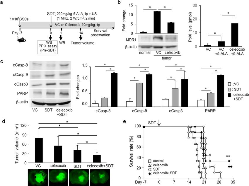
www.nature.com/scientificreports/
Figure 2. Anti-tumor effects of SDT in the mouse GSC-bearing glioma model. (a) Protocol in mouse GSC-
bearing glioma model; SDT performed 7 days after injection of GSCs. The mice subjected to US 1 MHz, 2 W/
cm2 2 min) at 3 h after injection of 200 mg/kg 5-ALA. Photographs indicate mice subjected to SDT, GSC
injection area, and glioma. (b) Representative western blot analysis on day 1 after SDT compared to non-
treatment control in a GSC-bearing mouse glioma model. Each column indicates mean ± SD (n = 4). *p < 0.05 by
Student’s t-test. (c) DAB immunohistochemistry of apoptosis-related molecules on day 1 after SDT in a glioma-
bearing mouse. Each specimen was co-stained with hematoxylin. Arrows point to positive cells. (d) GFP-labeled
GSCs and tumor volume in brain on days 3–14 after SDT. Each column data indicates mean ± SD (n = 6).
*p < 0.05 by Student’s t-test. (e) Changes in body weight before and after SDT. (f) Kaplan–Meier survival estimate
(%), no significant difference by log-rank test (n = 10).
Anti‑tumor effects of SDT in the mouse GSC‑bearing glioma model. We further examined the
effects of SDT in the mouse GSC-bearing glioma model (Fig. 2a). SDT was performed 7 d after GSC injection.
One day after SDT, we observed up-regulation of cCasp-9, -3 and PARP but not cCasp-8 by western blot analysis
(Fig. 2b) and immunohistochemistry (Fig. 2c), compared to non-treatment control, indicating the induction of
intrinsic apoptosis by SDT. SDT significantly decreased the tumor size (Fig. 2d) without affecting body weights
14 d after GSC injection (Fig. 2e) but did not significantly extend the survival period compared with the values
in other groups (Fig. 2f). The anti-tumor effects of single SDT may be limited in this model.
An increase in cellular PpIX via celecoxib‑mediated MDR1 down‑regulation augmented the
anti‑tumor effect of SDT. To enhance the anti-tumor effects of SDT, we examined whether MDR1 down-
regulation can increase the cellular PpIX levels after 5-ALA injection. Based on our previous findings in human
glioma cells17, we first observed that the expression levels of COX-2 and MDR1 in human glioblastoma and the
mouse GSC-bearing glioma model were higher than those in normal brain tissue (Fig. 3a). Next, using a COX-2
inhibitor, celecoxib, we assessed the effects on the MDR1 expression in GSCs.
Treatment with celecoxib 60 μM (IC50 dose) decreased MDR1 expression levels compared to diluted DMSO
as vehicle control and the PpIX levels were significantly higher upon combination treatment with 1 μM 5-ALA
and celecoxib than with 5-ALA monotherapy (Fig. 3b). To confirm the effects of MDR1 down-regulation on
PpIX levels, we examined the effects of an MDR1 inhibitor, valspodar (Fig. 3c). MDR1 was down-regulated by
1 μM valspodar (Fig. 3c) and cellular PpIX levels increased as expected, resulting in the enhanced anti-tumor
effects of SDT by the combination of valspodar and SDT in GSCs (Fig. 3d). The increase in cellular PpIX levels
through MDR1 decreased by celecoxib may be at least partly attributable to the enhancement of the anti-tumor
effects of SDT.
Combination therapy with SDT and celecoxib enhanced apoptosis induction, thereby enhanc‑
ing anti‑tumor effects in the mouse GSC‑bearing glioma model. To further confirm whether SDT
with down-regulation of MDR1 by celecoxib augments anti-tumor effects, we assessed the efficacy in the mouse
Scientific Reports | (2021) 11:15105 | https://doi.org/10.1038/s41598-021-93896-0 3
Vol.:(0123456789)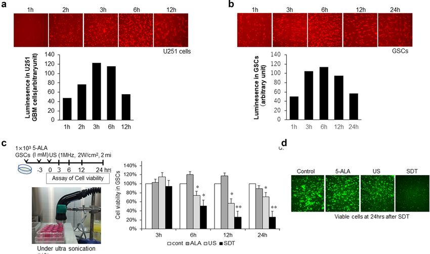
www.nature.com/scientificreports/

                                            Figure 3.  Elevation of PpXI through down-regulation of MDR1 leading to low cell viability in GSCs. (a)
                                            Representative expression of COX-2 and MDR1 by DAB stain in human GBM and GSCs-bearing mouse
                                            glioma model and each normal tissue. (b) Changes in MDR1 expression and PpIX level treated with celecoxib
                                            and 5-ALA. Western blot analysis performed at 24 h after treatment with 60 μM celecoxib and compared with
                                            DMSO as a vehicle control (VC). PpXI levels were determined at 3 h after 1 μM 5-ALA. Each column indicates
                                            mean ± SD values (n = 4). (c) Changes in MDR expression and PpIX levels upon treatment with valspodar as a
                                            MDR1 inhibitor and 5-ALA. Western blot analysis performed at 3 h after valspodar and PpXI level determined
                                            at 3 h after 5-ALA. (d) Cell viability of GSCs treated with 1 μM 5-ALA at 3 h after treatment with 1 μM
                                            valspodar. US was performed at 3 h after 5-ALA. Each column indicates mean ± SD values (n = 8). *p < 0.05 by
                                            Student’s t-test versus VC (2 groups) or Turkey–Kramer’s test versus others (> 3 groups).

                                            GSC-bearing glioma model (Fig. 4a). Compared to normal tissue, the expression of MDR1 was elevated in brain
                                            tumor tissue after GSCs injection, which was reduced upon treatment with 10 mg/kg celecoxib for 7 days before
                                            SDT (Fig. 4b). Concurrent with this finding, cellular PpIX levels in brain tumor after 5-ALA injection were
                                            significantly increased (Fig. 4b). The elevation of cellular PpIX levels upon combination therapy with SDT and
                                            celecoxib escalated the induction of intrinsic and extrinsic apoptosis 1 day after SDT (Fig. 4c), thus decreasing
                                            the tumor volume (Fig. 4d) and prolonging survival (Fig. 4e). The combination therapy with SDT and celecoxib
                                            may be a promising means for GBM treatment.

                                            Enhancement of anti‑tumor effects by SDT was attributed to celecoxib‑mediated MDR1
                                            down‑regulation through the AKT/NF‑κB pathway. To clarify the mechanisms underlying celecoxib-
                                            mediated MDR1 down-regulation, we treated (i.p.) the mouse GSC-bearing glioma model with 10 mg/kg
                                            celecoxib for 7 days (Fig. 5a). AKT2, pAkt, pNF-κB, and MDR1 were significantly up-regulated in the glioma
                                            model than in normal tissue and were attenuated through celecoxib treatment (Fig. 5a). Furthermore, to confirm
                                            the transcriptional regulation of MDR1, we used a proteasome inhibitor, caffeic acid phenethyl ester (CAPE),
                                            which inhibits the phosphorylation of inhibitor of κB (I-κB), thus facilitating the phosphorylation of NF-κB
                                            (pNF-κB) and regulating the transcription of target genes. In GSCs, up-regulation of MDR1 mRNA and protein
                                            was significantly attenuated upon treatment with 1 μM CAPE, accompanied by the reduction of pI-κB and
                                            pNF-κB (Fig. 5b), suggesting transcriptional regulation of MDR1 through NF-κB. Together, celecoxib-mediated
                                            MDR1 down-regulation through the Akt/NF-κB pathway may increase cellular PpIX levels by increasing the
                                            intratumoral uptake of 5-ALA, thus promoting apoptosis induction via SDT (Fig. 5c). MDR1 down-regulation
                                            may be attributable to the improvement of not only chemo-resistance, but also the therapeutic efficacy of SDT
                                            in GBM.

          Scientific Reports |   (2021) 11:15105 |              https://doi.org/10.1038/s41598-021-93896-0                                                 4

Vol:.(1234567890)
www.nature.com/scientificreports/
Figure 4. Elevation of cellular PpXI levels via MDR1 down-regulation by celecoxib augmented anti-tumor
effects by SDT in the mouse glioma model. (a) Protocol: celecoxib was continuously injected during the survival
period after GSC injection. (b) Expression of MDR1 by western blot analysis and PpIX level 3 h after SDT at
7 days after GSCs injection. The MDR1 expression was compared with normal- and tumor brain tissue treated
with celecoxib or a vehicle control (VC) included DMSO/HBC. Each data indicates mean ± SD (n = 4). *p < 0.05
by Turkey–Kramer’s test versus others (> n = 3). (c) Expression analysis of apoptosis-related molecules by
western blotting at day 1 after SDT (each, n = 4). (d) Tumor volume 14 days after SDT. (each, n = 6). (e) Kaplan–
Meier survival estimate (%) and survival period prolonged by the combination therapy with SDT and celecoxib.
**p < 0.01 versus other groups by the log-rank test (n = 9–10).
Discussion
In this study, we initially show that combination of SDT and celecoxib to down-regulate MDR1 augmented
the anti-tumor effects in GSCs and the mouse GSC-bearing malignant glioma model indicating invasive and
therapeutic resistance. First, we confirmed that SDT monotherapy exerted anti-tumor effects in GSCs and GSCs-
bearing glioma model; however, it displayed transient and limited efficacy. Since we have demonstrated the anti-
tumor effects by a COX2 inhibitor, c elecoxib18,19, we assessed the efficacy of SDT combined with pre-treatment
by celecoxib. Expectedly, the pre-treatment by celecoxib decreased the expression of MDR-1 and elevated the
cellular PpIX levels induced by 5-ALA, resulting in the enhanced anti-tumor effects of the combination therapy
in the GSC-bearing glioma model. Next, to confirm the mechanisms underlying the downregulation of MDR-1
by celecoxib, we used valspodar as an MDR-1 inhibitor. It decreased the expression of MDR-1 and elevated the
cellular PpIX levels induced by 5-ALA, thereby reducing cell viability. Finally, we verified the down-regulation of
MDR-1 by celecoxib via Akt/NF-κB pathway, using CAPE, a NF-κB inhibitor. The combination therapy of SDT
and celecoxib enhanced the induction of both intrinsic and extrinsic apoptosis pathways in the GSCs-bearing
malignant glioma model. Taken together, combination therapy with SDT and celecoxib may be a promising
strategy to retard GBM recurrence.
SDT has been developed as a novel promising noninvasive approach derived from photodynamic therapy
(PDT)22,23. Because PDT is tightly focused with a short penetration depth of light in soft tissue up to several tens
of centimeters, PDT is not effective for the treatment of deep-seated tumors24,25. In contrast, SDT is anticipated
to overcome the major limitation of PDT. It may be a promising treatment method for brain tumors and other
tumors26. PpIX, converted from 5-ALA in mitochondria, is used in heme formation in a reaction catalyzed by
Scientific Reports | (2021) 11:15105 | https://doi.org/10.1038/s41598-021-93896-0 5
Vol.:(0123456789)www.nature.com/scientificreports/
Figure 5. Anti-tumor effects of SDT combined with the inhibition of the AKT/NF-κB/MDR1 pathway by
celecoxib in the GSCs-bearing mouse glioma model. (a) AKT/NF-κB signaling pathways and MDR1 expression
upon western blotting after consecutive treatment with celecoxib for 7 days in the GSC-bearing mouse glioma
model. Each expression level was compared between normal brain and tumor brain tissue treated with vehicle
control (VC) or celecoxib. Each data indicates mean ± SD (n = 6). (b) Protein and mRNA levels of MDR1 in
GSCs treated with or without 1 μM CAPE, a potent and a specific inhibitor of NF-κB activation. (each, n = 6). (c)
Schematic representation of the anti-tumor effects upon combination therapy with SDT and celecoxib.
the enzyme ferrochelatase. In GBM, the decrease of ferrochelatase may contribute to the accumulation of PpIX
within gliomas and GSCs and lead to its preferential localization in t umor9. Although we did not assess the
effects of ferrochelatase, we suggested that the increased cellular PpIX level may be at least partly attributable
to the uptake of 5-ALA into brain or GSCs through down-regulation of MDR-1 by celecoxib, thereby enhanc-
ing anti-tumor effects. Because GBM occurs in the brain parenchyma surrounded by the skull and differs from
other epithelial cancers, thus far, SDT has been performed for GBM patients only during surgical i ntervention27.
Although the ultrasound instrument allows the repeated application of STD, this technique is limited to only a
few institutes. Therefore, to enhance the repeatability of SDT, the development of feasible and safe equipment
with new s onosensitizers28 are urgently required.
MDR1 (ABCB1) gene encodes P-glycoprotein, a drug transporter that is a critical component of the blood-
brain barrier, which prevents entry of many potentially toxic compounds into the central nervous system29.
We previously reported that the reduction of MDR1 upon transfection of DKK3 gene in human glioma cells
enhanced the anti-tumor effects of temozolomide17. Concurrent with a previous report indicating increased
mRNA levels of MDR1 in COX-2-overexpressing cells, the present study showed that MDR1 was highly up-
regulated and COX-2 was overexpressed in GSC-derived glioma tissue and in human glioma tissue. COX-2,
an inducible form of the enzyme catalyzing the first step in prostanoid synthesis, has been reported to be over-
expressed in various tumors and possesses proangiogenic and anti-apoptotic properties. MDR1 is expressed
in normal liver and kidney tissue, where it functions to actively transport lipophilic xenobiotic compounds
and serves as an efflux pump for chemotherapeutic agents30. Thus, up-regulation of MDR1 and COX-2 may be
associated with not only chemotherapeutic resistance but also with the limited uptake of 5-ALA in GSCs and
the mouse GSC-bearing glioma model. To confirm the enhanced efficacy of SDT through the cellular up-take of
5-ALA upon MDR1 down-regulation in GSCs, we treated GSCs with a COX-2 inhibitor, celecoxib, or an MDR1
inhibitor, valspodar. As expected, cellular PpIX levels increased upon MDR1 down-regulation by each chemi-
cal compound in GSCs. Whereas celecoxib is applicable in a clinical setting, valspodar has not shown evidence
Scientific Reports | (2021) 11:15105 | https://doi.org/10.1038/s41598-021-93896-0 6
Vol:.(1234567890)www.nature.com/scientificreports/
of clinical efficacy. Therefore, we used celecoxib but not valspodar to reduce MDR1 in our in-vivo study and
confirmed the efficacy of SDT enhanced by celecoxib.
Although glioma-associated macrophages may take up large amounts of 5-ALA, we did not directly assess the
effects of celecoxib on macrophages. However, we previously verified the reduction on chemokines/its receptors
(CCL2/CCR2 and CXCL10/CXCR3) by celecoxib18. The reduction of these molecules may inhibit the recruitment
of macrophages into the tumor, resulting in anti-tumor effects in the GSC-bearing glioma model. Therefore, the
ability to take up 5-ALA into macrophages may be limited in the presence of celecoxib. Taken together, treatment
with celecoxib before SDT may contribute to increase the uptake of 5-ALA into the tumor, thereby enhancing
the anti-tumor effects.
In addition, MDR1 expression is associated with several cellular signaling pathways and protein kinases,
chaperons, ubiquitin-related enzymes, and transcription f actors31. As the COX-2 inhibitor induced apoptosis by
inhibiting the AKT pathway in low-grade glioma cells in a previous study18, we observed MDR1 down-regulation
through the Akt/NF-κB pathway upon celecoxib treatment. Furthermore, we observed the down-regulation of
MDR1 by a NF-κB inhibitor, CAPE. Thus, celecoxib may affect both apoptosis induction and MDR1 down-
regulation by inhibiting the Akt/NF-κB pathway, resulting in an enhanced anti-tumor effects of not only SDT
but also other anti-tumor agents.
This study has some limitations. Within a short period after SDT, apoptosis induction was enhanced in com-
bination with celecoxib, and the cell viability was decreased. Therefore, we need the assessment of anti-tumor
effects through repeated SDT upon combination during a longer period treatment with celecoxib. We previously
confirmed that on combination treatment with 5-ALA and US, ROS generation and anti-tumor effects were
greater than those upon monotherapy10. Unfortunately, in the current study we did not assess ROS genera-
tion, although the elevated cellular PpIX level was confirmed. Further investigation is necessary to determine
whether elevated PpIX in combination therapy with SDT and celecoxib is associated with the enhanced ROS
generation, thereby leading to the augment of the anti-tumor effects. SDT activates the mitochondrial caspase
pathway and down-regulates ATP-binding cassette transporters such as MDR1, thus selectively improving the
uptake of chemotherapeutic drugs into tumor cells and reducing the toxic effects on normal cells and t issues31,32.
Therefore, sequential treatment with celecoxib after SDT may improve the uptake of celecoxib by itself into
glioma cells, contributing to the prolonged survival in our glioma model. Although combination therapy with
SDT and celecoxib may exert various synergistic therapeutic effects on GBM, we cannot exclude the favorable
effects of celecoxib after SDT beyond MDR1 down-regulation33. Further studies are required to develop clinically
applicable, handy, repeatable, and feasible devices for SDT.
In conclusion, the combination therapy with SDT and celecoxib resulted in enhanced anti-tumor efficacy
among GSCs and a mouse GSC-bearing glioma model. MDR1 down-regulation via the Akt/NF-κB pathway
may be a promising mean for treatment of GBM patients. Our results warrant for further verification of the
anti-tumor effects of combination therapy with SDT and other chemotherapeutic agents and the development
of new US systems capable of penetrating the skull.
Method
Study approval and informed consent. This study was approved by the ethical review board of Tokush-
ima University Hospital for human study and the ethics committee of Tokushima University Graduate School of
Biomedical Sciences, Tokushima, Japan. Human tissue samples were obtained during routine clinical procedures
after informed consent including the use of tissue sample from patients with brain tumors at the Department of
Neurosurgery, Tokushima University Hospital. For the use of samples obtained from patients, we have obtained
a statement attesting to informed consent for all patients with or without neurosurgery in our department. Each
sample was fixed in 4% formalin in phosphate-buffered saline (PBS) and processed for paraffin embedding. The
samples were classified by neuropathologists in accordance with the WHO classification of brain tumors. Sec-
tions from non-neoplastic regions (NNRs) were purchased from BioChain Institute (Newark, NJ, USA). The
study was performed in accordance with the tenets of the Declaration of Helsinki. All animal experiments were
approved and performed in accordance with the animal care guidelines of Tokushima University.
Cell lines. Human GBM cell line U251MG were purchased from American Type Culture Collection (Manas-
sas, VA, USA) and cultured in RPMI-1640 medium (Invitrogen, NJ, USA) with 10% fetal bovine serum (GIBCO-
BRL, NY, USA) at 37 °C in an atmosphere of 5% C O2 and 95% humidified air. Mice GSCs were established
and provided by OS and HS, Keio University19,20. GSCs were cultured in Dulbecco’s Modified Eagle’s medium/
nutrient mixture F-12 Ham (Sigma-Aldrich, St. Louis, MO, USA) supplemented with 20 ng/ml recombinant
human epidermal growth factor (PeproTech, Rocky Hill, NJ, USA), 20 ng/ml recombinant human basic fibro-
blast growth factor (PeproTech), B-27 supplement without vitamin A (Life Technologies, Carlsbad, CA, USA),
200 ng/ml heparin sulfate, 100 U/ml penicillin, and 100 μg/ml streptomycin (Nacalai Tesque, Kyoto, Japan).
Cell viability assay. GSCs (1 × 10³ cells/well) were plated in 96-well tissue culture plates. To enumerate via-
ble cells, the conversion of WST-8 to formazan by metabolically active cells was quantified using WST-8 reagent
(Dojindo, Osaka, Japan) on a microplate reader (Infinit F200 PRO, TECAN) at 450 nm. We used PBS-treated
cells as the control to represent 100% viability and the percent viability was determined in each treatment.
Establishment of the animal model and assessment of anti‑tumor effects. All experimental
protocol was approved by Tokushima University institutional committee (No. T27-2) and carried out in com-
pliance with the animal care guidelines of Tokushima University and the ARRIVE guidelines (PLoS Bio 8(6),
e1000412, 2010).
Scientific Reports | (2021) 11:15105 | https://doi.org/10.1038/s41598-021-93896-0 7
Vol.:(0123456789)www.nature.com/scientificreports/
Six-week-old male C57BL/6 mice were subjected to inhalation anesthesia with isoflurane and a stereotactic
apparatus was placed in the right brain. With a dental drill, a small hole was bored into the skull 2.0 mm lateral to
the bregma. In a malignant glioma model with mouse GSCs, established by Sampetrean and S aya20, and Shibao,
et al.21, GSC progeny cells (1 × 10³) in 2 μl of Hank’s balanced salt solution (Sigma-Aldrich) were injected into
the right cerebral hemisphere 3 mm below the brain surface, using a 10-μl Hamilton syringe. To examine the
anti-tumor effects of SDT, the mice were randomized and treated with 5-ALA, US, or SDT and compared to the
non-treated control (Fig. 2). For SDT, a 0.2-ml solution of 5-ALA in PBS was intraperitoneally injected at a dose
of 200 mg/kg body weight. Three hours later, the mouse right brain was placed on the stereotactic apparatus and
subjected to US imaging (1 MHz, 2 W/cm2 for 2 min) under inhalation anesthesia. On day 1 after SDT, apoptosis
induction by SDT was confirmed and tumor volume on days 3, 7, and 14 and the survival rate were analyzed.
Mice were euthanized and their brains were sliced on a brain slicer matrix at 1.0-mm intervals and the tumor vol-
ume, represented by the GFP-positive area, was microscopically determined (Keyence BZ-X710, Osaka, Japan).
In addition, to assess the effect of combination therapy of SDT and celecoxib, another set of mice were rand-
omized and treated with vehicle, celecoxib, SDT, or a combination of celecoxib and SDT (Fig. 4). Celecoxib, lysed
with dimethyl sulfoxide (DMSO) and hydroxypropyl-β-cyclodextrin (HBC), was injected (i.p.) at a dose of 10 mg/
kg consecutively after mouse GSC implantation. Vehicle controls received equivalent doses of DMSO/HBC and
normal saline at the same dosing schedule. To validate the efficacy of celecoxib during SDT, tumor volume on day
14 and the survival rate during the observation period were assessed in each group (Fig. 4) as described above.
Measurement of cellular PpIX levels and SDT. Luminescence was measured 1, 2, 3, 6, 12 or 24 h after
treatment of human GBM U251 cells with 1 μM 5-ALA in human GBM U251 cells and GSCs, using the image
analyzer in the BZ-X710 microscope (KEYENCE). Thereafter, the viability assessed for 3-48h of GSCs upon
combination therapy with SDT and US (1 MHz, 2 W/cm2 for 2 min) at 3 h after 5-ALA treatment was assessed
after 24 h.
Celecoxib (Sigma-Aldrich, PHR1683) was dissolved in DMSO and supplemented in the culture medium at a
final concentration of 60 μM. After 3-h incubation of cells in medium supplemented with 1 μM 5-ALA (SBI ALA
promo, Tokyo, Japan), the medium was replaced with fresh complete medium, and the 96-well plate was exposed
to LED irradiation (630 nm, 80 mW/cm2) for 5 min. The LED light spot was an equally illuminated rectangular
spot encompassing the entire culture plate. SDT was performed using the ultrasonic generator UST-770 (ITO Co.
Ltd., Tokyo, Japan). In the mouse GSC-bearing glioma model, the tumor area was extracted 3 h after treatment
with 200 mg/kg 5-ALA and PpIX levels were analyzed as previously d escribed20.
Quantitative real‑time PCR (qRT‑PCR). Total RNA was isolated and extracted using the MagNA Pure
RNA isolation kit (Roche, Tokyo, Japan) and the MagNa lyser (Roche), in accordance with the manufacturer’s
instructions. We used Transcriptor Universal cDNA Master (Roche) to reverse-transcribe total RNA to cDNA
and a LightCycler 2.0 (Roche Diagnostics, Tokyo, Japan) for qRT-PCR. The following primers were used: mouse
mdr1, 5′-primer, GGC ATT GCC TAC CTG TTG G-3′; 3′-primer GCT TTC TGT GGA CAC TTC TG, and
mouse glyceraldehyde-3-phosphate dehydrogenase (Gapdh), 5′-CAG AAC ATC ATC CCT GCA TC-3′ and
5′-CTG CTT CAC CAC CTT CTT GA-3′. The mRNA levels were normalized to those of Gapdh. The PCR con-
ditions were as follows: 95 °C for 10 min, followed by 40 cycles at 95 °C for 10 s, 60 °C for 10 s, and 72 °C for 8 s.
We subjected 4 samples in each group to the qRT-PCR assay to determine the gene expression levels.
Western blot analysis. According to our previous study6, cells or tissue samples were homogenized in
RIPA buffer containing a protease/phosphatase inhibitor cocktail (Cell Signaling Technology, CST, 5872). After
10-min centrifugation at 12,000 rpm, 4 °C, the protein concentration in the supernatants was determined using
BCA kit (Thermo Fisher Scientific, USA). Protein (20 or 50 μg) was separated by SDS-PAGE and transferred to
polyvinylidene fluoride membranes (immune-blot PVDF membrane, BIO-RAD, Hercules, CA, USA) by elec-
troblotting. Based on the molecular weight marker, each membrane was cut before hybridization. The mem-
branes were immersed in blocking buffer (5% skim milk or 2% BSA in tris-buffered saline, TBS) for 1 h and
incubated with primary antibodies: anti-MDR1 (BD Biosciences, NJ, USA, 1:1,000), anti-cCaspase-8, -9, -3
and anti-PARP (Cell Signaling Technology, MA, USA, 1:1,000), anti-AKT2 (Abcam, Cambridge, UK, rabbit,
1:1,000), pAkt (Santa Cruz Biotechnology, CA, USA, rabbit, 1:500), anti-pNF-κB (CST, 1:1000), anti-pI-κB (CST,
1:1000) and β-actin (Sigma-Aldrich, mouse, 1:5000) were diluted in Can Get Signal Solution 1 (Toyobo). After
washing in Tween-TBS (T-TBS), the membranes were incubated for 1 h with horseradish peroxidase-conjugated
secondary antibodies in Can Get Signal Solution 2 (dilution 1:3000). After washing, the protein-antibody com-
plexes were detected with Amersham ECL prime Western blotting detection reagents (GE Healthcare, UK) using
a Lumino image analyzer (Image Quant LAS-4000 mini, GE Healthcare Japan, Tokyo, Japan) and ImageJ 1.52
software (NIH, Bethesda, MD, USA) was used to analyze the protein expression levels. Each experiment was
repeated four times.
Immunohistochemistry. Murine tissue samples were fixed with 4% paraformaldehyde and 5-μm-thick
frozen sections were mounted on Matsunami adhesive saline-coated glass slides (Matsunami Glass, Tokyo,
Japan). As previously reported6, human glioma tissue sections from the paraffin-embedded block were dewaxed,
rehydrated, and subjected to antigen retrieval. The sections were blocked for 30 min with 1–3% hydrogen per-
oxide solution, and stained overnight at 4 °C with the following antibodies: anti-MDR1 (D-11) (Santa Cruz
Biotechnology, Inc., Dallas, TX, USA, 1:100), anti-COX-2 (Abcam, ab15191), rabbit monoclonal anti-MDR1
(ab170904; 1:100), anti-cleaved caspase-8 (cCasp-8), anti-cCasp-9, anti-cCasp-3, and anti-PARP (CST, 1:1,000).
Thereafter, they were incubated with biotinylated secondary antibody (30 min, 30 °C), visualized using DAB
Scientific Reports | (2021) 11:15105 | https://doi.org/10.1038/s41598-021-93896-0 8
Vol:.(1234567890)www.nature.com/scientificreports/
buffer tablets, and counterstained with hematoxylin. Photographs were obtained under a light microscope, using
KEYENCE BZ-X710.
Statistical analysis. Survival estimates and median survivals were determined using Kaplan–Meier sur-
vival curves. A log-rank (Mantel-Cox) test was performed to determine the p values derived from Kaplan–Meier
survival curves. To determine statistical significance, between-group comparisons were performed using Stu-
dent’s t-test. For multiple comparisons, one-way ANOVA, followed by the Tukey–Kramer tests. Error bars indi-
cate the standard deviation values. All statistical analyses were performed using JMP 13.2 (SAS Institute Inc.)
and the differences with p < 0.05 were considered significant.
Received: 20 November 2020; Accepted: 1 July 2021
References
1. Ostrom, Q. T. et al. The epidemiology of glioma in adults: A “state of the science” review. Neuro Oncol. 17, 624–626 (2015).
2. Lu, V. M., Jue, T. R., McDonald, K. L. & Rovin, R. A. The survival, effect of repeat surgery at glioblastoma recurrence and its trend:
A systematic review and meta-analysis. World Neurosurg. 115, 453–459 (2018).
3. Hervey-Jumper, S. L. & Berger, M. S. Maximizing safe resection of low- and high-grade glioma. J. Neurooncol. 130, 269–282 (2016).
4. Bush, N. A., Chang, S. M. & Berger, M. S. Current and future strategies for treatment of glioma. Neurosurg. Rev. 40, 1–14 (2017).
5. Adhikaree, J., Moreno-Vicente, J., Kaur, A. P., Jackson, A. M. & Patel, P. M. Resistance mechanisms and barriers to successful
immunotherapy for treating glioblastoma. Cells 21, 263 (2020).
6. Yamaguchi, I. et al. Downregulation of PD-L1 via FKBP5 by celecoxib augments antitumor effects of PD-1 blockade in a malignant
glioma model. Neuro-Oncol. Adv. 2, vdz058 (2020).
7. Wu, S. K., Santos, M. A., Marcus, S. L. & Hynynen, K. MR-guided focused ultrasound facilitates sonodynamic therapy with 5-ami-
nolevulinic acid in a rat glioma model. Sci. Rep. 9, 1–10 (2019).
8. Mason, T. J. Therapeutic ultrasound an overview. Ultrason. Sonochem 18, 847–852 (2011).
9. Mahmoudi, K. et al. 5-aminolevulinic acid photodynamic therapy for the treatment of high-grade gliomas. J. Neurooncol. 141,
595–607 (2019).
10. Shimamura, Y. et al. 5-Aminolevulinic acid enhances ultrasound-mediated antitumor activity via mitochondrial oxidative damage
in breast cancer. Anticancer Res. 36, 3607–3612 (2016).
11. Prada, F. et al. Applications of focused ultrasound in cerebrovascular diseases and brain tumors. Neurotherapeutics 16, 67–87
(2019).
12. Song, D. et al. Study of the mechanism of sonodynamic therapy in a rat glioma model. OncoTargets Ther. 7, 1801–1810 (2014).
13. Chen, Z., Li, J., Song, X., Wang, Z. & Yue, W. Use of a novel sonosensitizer in sonodynamic therapy of U251 glioma cells in vitro.
Exp. Ther. Med. 3, 273–278 (2012).
14. Suehiro, S. et al. Enhancement of antitumor activity by using 5-ALA-mediated sonodynamic therapy to induce apoptosis in
malignant gliomas: Significance of high-intensity focused ultrasound on 5-ALA-SDT in a mouse glioma model. J. Neurosurg. 129,
1416–1428 (2018).
15. Chen, L., Shi, L., Wang, W. & Zhou, Y. ABCG2 downregulation in glioma stem cells enhances the therapeutic efficacy of demeth-
oxycurcumin. Oncotarget 8, 43237–43247 (2017).
16. de Trizio, I., Errede, M., d’Amati, A., Girolamo, F. & Virgintino, D. Expression of P-gp in glioblastoma: What we can learn from
brain development. Curr. Pharm. Des. 26, 1428–1437 (2020).
17. Fujihara, T. et al. Down-regulation of MDR1 by Ad-DKK3 via Akt/NFκB pathways augments the anti-tumor effect of temozolomide
in glioblastoma cells and a murine xenograft model. J. Neurooncol. 139, 323–332 (2018).
18. Shono, K. et al. Downregulation of the CCL2/CCR2 and CXCL10/CXCR3 axes contributes to antitumor effects in a mouse model
of malignant glioma. Sci. Rep. 10, 15286 (2020).
19. Sato, A. et al. Blocking COX-2 induces apoptosis and inhibits cell proliferation via the Akt/survivin- and Akt/ID3 pathway in
low-grade-glioma. J. Neurooncol. 132, 231–238 (2017).
20. Sampetrean, O. & Saya, H. Characteristics of glioma stem cells. Brain Tumor Pathol. 30, 209–214 (2013).
21. Shibao, S. et al. Metabolic heterogeneity and plasticity of glioma stem cells in a mouse glioblastoma model. Neuro Oncol. 20,
343–354 (2018).
22. Shinohara, Y. et al. Development of a novel Schiff base derivative for enhancing the anticancer potential of 5-aminolevulinic acid-
based photodynamic therapy. Photodiagnosis Photodyn. Ther. 20, 182–188 (2017).
23. Bilmin, K., Kujawska, T. & Grieb, P. Sonodynamic therapy for gliomas. Perspectives and prospects of selective sonosensitization
of glioma cells. Cells 8, 1428 (2019).
24. Stepp, H. & Stummer, W. 5-ALA in the management of malignant glioma. Lasers Surg. Med. 50, 399–419 (2018).
25. Dolmans, D. E., Fukumura, D. & Jain, R. K. Photodynamic therapy for cancer. Nat. Rev. Cancer 3, 380–387 (2003).
26. Yuan, S. X. et al. Underlying mechanism of the photodynamic activity of hematoporphyrin-induced apoptosis in U87 glioma cells.
Int. J. Mol. Med. 41, 2288–2296 (2018).
27. Sun, Y. et al. Tumor targeting DVDMS-nanoliposomes for an enhanced sonodynamic therapy of gliomas. Biomater. Sci. 26, 985–994
(2019).
28. Bilmin, K., Kujawska, T. & Grieb, P. Sonodynamic therapy for gliomas. Perspectives and prospects of selective sonosensitization
of glioma cells. Cells 13, 1428 (2019).
29. Munoz, J. L., Walker, N. D., Scotto, K. W. & Rameshwar, P. Temozolomide competes for P-glycoprotein and contributes to chem-
oresistance in glioblastoma cells. Cancer Lett. 10, 69–75 (2015).
30. Patel, V. A., Dunn, M. J. & Sorokin, A. Regulation of MDR-1 (P-glycoprotein) by cyclooxygenase-2. J. Biol. Chem. 277, 38915–38920
(2002).
31. Robey, R. W. et al. Revisiting the role of ABC transporters in multidrug-resistant cancer. Nat. Rev. Cancer 18, 452–464 (2018).
32. Xi, G. et al. CD133 and DNA-PK regulate MDR1 via the PI3K- or Akt-NF-κB pathway in multidrug-resistant glioblastoma cells
in vitro. Oncogene 35, 241–250 (2016).
33. Lin, F. et al. PI3K-mTOR pathway inhibition exhibits efficacy against high-grade glioma in clinically relevant mouse models. Clin.
Cancer Res. 23, 1286–1298 (2017).
Acknowledgements
We thank Emiko Nishikawa and Akiko Sumi for excellent technical support.
Scientific Reports | (2021) 11:15105 | https://doi.org/10.1038/s41598-021-93896-0 9
Vol.:(0123456789)www.nature.com/scientificreports/
Author contributions
K.S. and I.Y. performed the in vivo and in vitro experiments; T.F., K.N., Y.F. and K.M. performed the in vitro
experiments; O.U. and H.S. provided transgenic cell; Y.U. provided ultrasonic generator; Y.M. and K.T.K.
designed the work and contributed at the revision stage of the paper; Y.T. designed and organized the work; all
authors contributed to the final version of the paper.
Funding
Funding for this work was provided by a Grant-in-Aid for Scientific Research (No. 18K16586 to K.S.) and (No.
25462264 to Y. M.) from the Ministry of Education, Culture, Sports, Science, and Technology of Japan.
Competing interests
The authors declare no competing interests.
Additional information
Supplementary Information The online version contains supplementary material available at https://doi.org/
10.1038/s41598-021-93896-0.
Correspondence and requests for materials should be addressed to Y.M.
Reprints and permissions information is available at www.nature.com/reprints.
Publisher’s note Springer Nature remains neutral with regard to jurisdictional claims in published maps and
institutional affiliations.
Open Access This article is licensed under a Creative Commons Attribution 4.0 International
License, which permits use, sharing, adaptation, distribution and reproduction in any medium or
format, as long as you give appropriate credit to the original author(s) and the source, provide a link to the
Creative Commons licence, and indicate if changes were made. The images or other third party material in this
article are included in the article’s Creative Commons licence, unless indicated otherwise in a credit line to the
material. If material is not included in the article’s Creative Commons licence and your intended use is not
permitted by statutory regulation or exceeds the permitted use, you will need to obtain permission directly from
the copyright holder. To view a copy of this licence, visit http://creativecommons.org/licenses/by/4.0/.
© The Author(s) 2021
Scientific Reports | (2021) 11:15105 | https://doi.org/10.1038/s41598-021-93896-0 10
Vol:.(1234567890)You can also read