Cancer Association of South Africa (CANSA)
←
→
Page content transcription
If your browser does not render page correctly, please read the page content below
Cancer Association of South Africa (CANSA)
Fact Sheet
on
Von Hippel-Lindau Disease
Introduction
Two eye doctors, Dr Eugen von Hippel in Germany and Dr Arvid Lindau in Sweden, were the
first to publish descriptions of tumours in patients’ eyes and brains, hallmarks of von Hippel-
Lindau disease. In the 1960s the disease was name VHL (von Hippel-Lindau) to recognise
their contribution in describing the disease (Stanford Cancer Institute).
Dr Eugen von Hippel (1867 – 1938) Dr Arvid Lindau (1892 – 1958)
[Picture Credit: VHL Alliance] [Picture Credit: VHL Alliance]
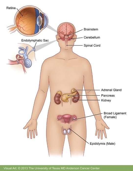
Von Hippel-Lindau Disease (VHL)
Von Hippel-Lindau (VHL) disease is an inherited disorder causing multiple tumours, both
benign and malignant, in the central nervous system (CNS) and viscera (the internal organs
in the main cavities of the body, especially those in the abdomen, e.g. the intestines). The
most common tumours are retinal and CNS haemangioblastomas, renal cell
carcinoma (RCC), and phaeochromocytoma. Tumours also arise in the pancreas, epididymis
or broad ligament of the uterus, and the inner ear (endolymphatic sac). The age at which the
tumours present ranges from early childhood to the seventh decade of life. Early diagnosis,
screening of family members and lifelong surveillance of VHL patients for tumours is
recommended (Patient.co.uk).
Tumours called haemangioblastomas are characteristic of VHL. These growths are made of
newly formed blood vessels. Although they are typically noncancerous, they can cause
serious or life-threatening complications. Haemangioblastomas that develop in the brain and
spinal cord can cause headaches, vomiting, weakness, and a loss of muscle coordination
(ataxia). Haemangioblastomas can also occur in the light-sensitive tissue that lines the back
Researched and Authored by Prof Michael C Herbst
[D Litt et Phil (Health Studies); D N Ed; M Art et Scien; B A Cur; Dip Occupational Health; Dip Genetic
Counselling; Diagnostic Radiographer; Dip Audiometry and Noise Measurement]
Approved by Ms Elize Joubert, Chief Executive Officer [BA Social Work (cum laude); MA Social Work]
December 2017 Page 1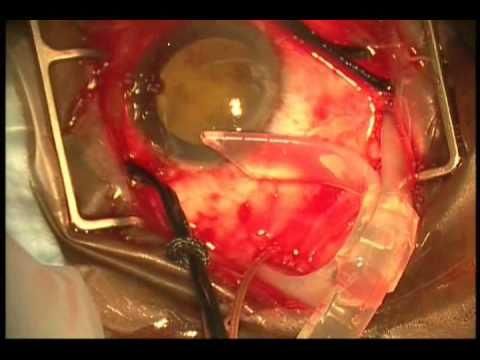
of the eye (the retina). These tumours, which are also called retinal angiomas, may cause
vision loss.
                [Picture Credit: VHL Disease]

People with VHL commonly develop
cysts in the kidneys, pancreas, and
genital tract. They are also at an
increased risk of developing a type of
kidney cancer called clear cell renal
cell carcinoma and a type of
pancreatic cancer called a pancreatic
neuroendocrine tumour.

VHL is associated with a type of
tumour called a pheochromocytoma,
which most commonly occurs in the
adrenal glands (small hormone-
producing glands located on top of
each kidney). Pheochromocytomas
are usually noncancerous. They may
cause no symptoms, but in some
cases they are associated with
headaches, panic attacks, excess
sweating, or dangerously high blood
pressure that may not respond to
medication. Pheochromocytomas are
particularly dangerous if they develop
during pregnancy.

About 10 percent of people with VHL develop endolymphatic sac tumours, which are
noncancerous tumours in the inner ear. These growths can cause hearing loss in one or
both ears, as well as ringing in the ears (tinnitus) and problems with balance. Without
treatment, these tumours can cause sudden profound deafness.
(Genetics Home Reference).

The VHL Gene
DNA (deoxyribonucleic acid) is the biochemical basis of life and of heredity. All of an
individual’s characteristics are written in DNA in a kind of code. DNA is assembled into
microscopic structures called chromosomes. In the human species there are 46
chromosomes, 23 from the mother and 23 from the father. There are 22 autosomes,
numbered 1 to 22, of which each person has a pair (two copies of chromosome 1, two of
chromosome 2, etc.) and one pair of the ‘sex’ chromosomes, XX for females and XY for
males. On each chromosome are the genes that contain the specific information necessary
for the manufacture of proteins. Each gene has two copies, one inherited from the father,
and one from the mother. The condition called VHL is caused by a dominant gene, since
only one altered copy of the VHL gene will cause the condition. VHL occurs in both men and
women. Each child of a person with VHL is at 50% risk of inheriting the altered copy of the
gene.

Researched and Authored by Prof Michael C Herbst
[D Litt et Phil (Health Studies); D N Ed; M Art et Scien; B A Cur; Dip Occupational Health; Dip Genetic
Counselling; Diagnostic Radiographer; Dip Audiometry and Noise Measurement]
Approved by Ms Elize Joubert, Chief Executive Officer [BA Social Work (cum laude); MA Social Work]
December 2017                                                                                      Page 2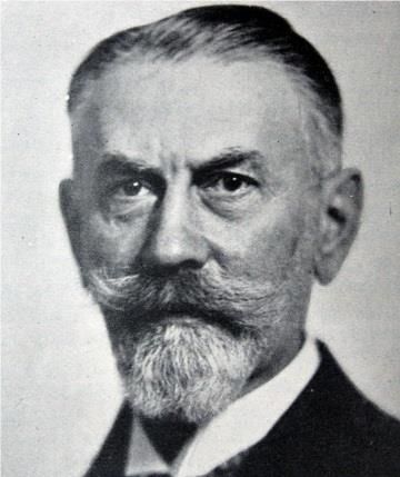
VHL gene location. The VHL gene is located on
the short arm of chromosome 3 at a site called
3p25-p26. An international team of scientists
identified the precise structure of this gene in
1993. Alterations in the normal structure of this
gene are known to result in the condition called
VHL.

The VHL gene encodes the formula for a protein whose function seems extremely important
in the fundamental process called ‘transcription’ which permits DNA to be transformed into a
more simple molecule, RNA, which is used to create the protein.

The normal VHL gene acts as a ‘tumour-suppressor gene’, whose normal function is to
suppress the formation of tumours. In order for a tumour to form, both copies of the VHL
gene (the one from the father and the one from the mother) must become inactivated. In an
individual who does not have the inherited alteration in the VHL gene, it is necessary for
each of these two normal copies of the VHL gene to undergo some change that inactivates
the VHL protein and allows a tumour to form. This may take some time, and multiple
damaging ‘hits’ to the genes in this cell, before the tumour will form. This explains why when
these tumours occur in the general population they are usually single occurrences in a single
organ, and the average age of onset of symptomatic kidney cancer in the general population
is age 62.

In the case of people who have inherited one copy of the gene that does not work correctly
in the beginning, it is only necessary to deactivate the one remaining copy before a tumour
may form. This is a much more likely occurrence, which means that tumours develop more
often, at younger ages, and in more organs than in people in the general population. Without
preventive action, the average age of onset of symptomatic kidney cancer in people with
VHL is age 42.

These alterations (or ‘mutations’) of the VHL gene can now be identified in most people with
VHL. The alteration is always the same in members of a single family. Conversely, the
precise alteration in the gene will be different from one VHL family to another. There is a
significant relationship between certain kinds of mutations and the likelihood of
pheochromocytomas, or the aggressiveness of Pancreatic NETs. Researchers are studying
other specific mutations which may be responsible for different aspects of VHL.

In most cases, the alteration in the VHL gene occurred a very long time ago, and the original
mutation has been passed down through several generations in a family. VHL in the Black
Forest Family in Germany and Pennsylvania has been documented back to the early 1600’s.
There are certain people, though, perhaps as many as 20%, who are the first in their family
to have an alteration in the VHL gene. Neither parent is affected, and these people have a
case of VHL, occurring ‘de novo’, for the first time. This ‘new mutation’ is caused by a
change in the gene in one sperm from the father, or in one egg from the mother, or in the
copying of the gene in one of the first stages of division of the embryo. This alteration in the
VHL gene can now be passed to future children of this affected person, and necessitates
medical screening of these children as well. There are no reliable statistics yet on the rate of
new VHL mutations.
(VHL Alliance).

Researched and Authored by Prof Michael C Herbst
[D Litt et Phil (Health Studies); D N Ed; M Art et Scien; B A Cur; Dip Occupational Health; Dip Genetic
Counselling; Diagnostic Radiographer; Dip Audiometry and Noise Measurement]
Approved by Ms Elize Joubert, Chief Executive Officer [BA Social Work (cum laude); MA Social Work]
December 2017                                                                                      Page 3
Neoplastic Risks of Von Hippel-Lindau Disease (VHL)
The neoplastic risks (risk of being able to form new growths) of VHL include:

    o   Central nervous system (CNS) haemangioblastomas may cause life-threatening
        complications in spite of their benign nature and classic slow-growing course and
        remain a major cause of morbidity and mortality in VHL disease
    o Retinal haemangioblastomas may cause retinal detachment, haemorrhage,
        glaucoma and cataract, leading to blindness, in absence of early detection and
        treatment
    o Renal cell carcinomas is becoming the main cause of death in the disease, because
        of secondary dissemination mainly due to delay in diagnosis
    o Pheochromocytomas are malignant in about 5-10% of cases
    o Neuroendocrine pancreatic tumours tend to be slow growing but have the potential of
        a truly malignant course with locoregional dissemination
    o Endolymphatic sac tumours is a low grade papillary adenocarcinoma resulting in
        progressive hearing loss. It can grow to the pontocerebelline angle and/or the middle
        ear, then destroying the temporal bone
    o Epididymal cysts and cystadenomas of the broad ligament are benign tumours
(Atlas of Genetics and Cytogenetics in Oncology and Haematology).

Incidence and Survival Rate for Von Hippel-Lindau Disease (VHL)
Because VHL itself is not a type of cancer, the National Cancer Registry (2013) does not
provide information on the incidence of VHL.

It is not a rare condition in the United States of America where the incidence is roughly 1:36
000 live births and has over 90% penetrance by 65 years of age. Before comprehensive
screening surveys became routine, median survival of patients with the disease was less
than 50 years of age. The main causes of death were complications linked to renal cell
carcinomas (RCC) and CNS haemangioblastomas.
(Lonser, et al., 2003).

Signs & Symptoms of Von Hippel-Lindau Disease (VHL)
Sometimes von Hippel Lindau disease has no symptoms. When it does have signs, they
vary from person to person and depend on the problems caused by the disease. These
symptoms usually do not mean one has VHL. However, it is important to discuss any
symptoms with a doctor, since they may signal other health problems.

Pheochromocytoma Symptoms - pheochromocytomas may cause symptoms that are like
what one feels in an emergency (‘fight or flight’) situation. These include:
   o High blood pressure, either all the time or just sometimes
   o Sweating
   o Headache
   o Rapid or irregular heartbeats
   o Feelings of anxiety, panic and fear
   o Pale skin
   o Dizziness or lightheadedness upon standing
   o Tremor
   o Weight loss

Researched and Authored by Prof Michael C Herbst
[D Litt et Phil (Health Studies); D N Ed; M Art et Scien; B A Cur; Dip Occupational Health; Dip Genetic
Counselling; Diagnostic Radiographer; Dip Audiometry and Noise Measurement]
Approved by Ms Elize Joubert, Chief Executive Officer [BA Social Work (cum laude); MA Social Work]
December 2017                                                                                      Page 4
Hemangioblastoma Symptoms - symptoms of hemangioblastomas vary depending on their
location.

Cerebellum
   o Difficulty walking and with muscle coordination
   o Dizziness
   o Headaches
   o Double vision
   o Vomiting

Spinal Cord
   o Decreased feeling in the arms, legs and body
   o Weakness
   o Difficulty walking
   o Difficulties with bowel and bladder function

Brain Stem
   o Decreased feeling in the arms, legs and body
   o Walking difficulties
   o Swallowing difficulties
   o Headaches
   o Poor coordination
(MD Anderson Cancer Center).

Diagnosis of Von Hippel-Lindau Disease (VHL)
The doctor will ask about symptoms and medical history. A physical examination will be
done. If the patient has any VHL symptoms, he/she should consider being tested for the VHL
gene. This is advised even if the patient has no known family history of the disease. This
could be the first person in the family to have VHL. Or could be the first one to have it
properly diagnosed since many people are not aware they have it.

A blood test that analyses DNA may be done to determine if the patient has the VHL gene.
Not all families with VHL have an identifiable VHL mutation. If members of a family are
positive for the gene and the patient is not, the patient does not need any further testing.

However, if other family members have been diagnosed with VHL despite a negative genetic
test, or if the person tests positive for the VHL gene, he/she needs to have regular medical
examinations and tests to uncover early signs. Even in the absence of symptoms, screening
should begin in childhood and continue periodically throughout life.

Screening for VHL complications includes a physical examination with special attention to
the eyes and nervous system.

Bodily fluids may be tested. This can be done with:
   o Blood tests
   o Urine tests

Images may be taken of bodily structures. This can be done with:

Researched and Authored by Prof Michael C Herbst
[D Litt et Phil (Health Studies); D N Ed; M Art et Scien; B A Cur; Dip Occupational Health; Dip Genetic
Counselling; Diagnostic Radiographer; Dip Audiometry and Noise Measurement]
Approved by Ms Elize Joubert, Chief Executive Officer [BA Social Work (cum laude); MA Social Work]
December 2017                                                                                      Page 5
o MRI scan
   o CT scan
   o Ultrasound
   o Angiography
(NYU Langone Medical Center).

Treatment of Von Hippel-Lindau Disease (VHL)
Von Hippel-Lindau disease (VHL) is usually a progressive disease. Surgical treatment may
consist of argon laser photocoagulation, cryotherapy, fluid drainage, scleral buckling,
penetrating diathermy, vitreous surgery, or endodiathermy. VHL varies according to the
location and size of the tumour and its associated cyst. In general, the objective of treatment
is to treat the growths when they are causing symptoms, but while they are still small so that
they do not cause permanent problems by putting pressure on the brain or spine, blocking
the flow of cerebrospinal fluid in the nervous system, or impairing vision. Certain tumours
can be treated with Stereotactic Radiation and Radio Frequency Ablation.
(Minneapolis Clinic of Neurology).

Argon laser photocoagulation - Argon plasma
coagulation (APC) is a non-contact thermal method of
haemostasis that has generated much attention and
excitement in recent years. It was introduced as an
alternative to contact thermal coagulation (heater probe
and bipolar cautery) and to existing non-contact
technologies (primarily laser). The theoretical
advantages of APC include its ease of application,
speedy treatment of multiple lesions in the case of angiodysplasias or wide areas (the base
of resected polyps or tumour bleeding), safety due to reduced depth of penetration, and
lower cost compared to laser (Shroff Eye Hospital; UpToDate).

Cryotherapy - is the local or general use of low temperatures in
medical therapy. Cryotherapy is used to treat a variety of benign
and malignant tissue damage, medically called lesions. The term
‘cryotherapy’ comes from the Greek cryo (κρύο) meaning cold,
and therapy (θεραπεία) meaning cure. Cryotherapy has been
used as early as the seventeenth century (Retina Macula
Institute; Wikipedia).

Scleral buckling - Scleral buckling surgery is a common way to
treat retinal detachment. It is a method of closing breaks and
flattening the retina of the eye. A scleral buckle is a piece of
silicone sponge, rubber, or semi-hard plastic that the
ophthalmologist places on the outside of the eye (the sclera, or
the white of the eye). The material is sewn to the eye to keep it in
place. The buckling element is usually left in place permanently
(Scleral buckling; WebMD).

Researched and Authored by Prof Michael C Herbst
[D Litt et Phil (Health Studies); D N Ed; M Art et Scien; B A Cur; Dip Occupational Health; Dip Genetic
Counselling; Diagnostic Radiographer; Dip Audiometry and Noise Measurement]
Approved by Ms Elize Joubert, Chief Executive Officer [BA Social Work (cum laude); MA Social Work]
December 2017                                                                                      Page 6
Penetrating diathermy - penetrating diathermy is used to fix the retina and to create retinal
microincarceration while air is simultaneously injected into the eye (Unbound Medicine).
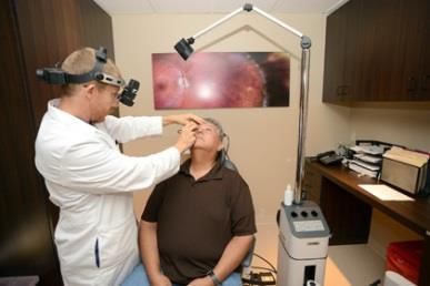
Vitreous surgery - Vitreous surgery is a relatively new type of microsurgery developed in
recent years which now enables eye surgeons to treat patients
with diseases of the retina and vitreous. Patients who, until the
advent of this type of surgery, might have been considered
hopelessly blind. This pamphlet has been prepared to provide a
basic understanding of vitreous surgery, the indications for
surgery, and the kinds of things a patient undergoing this
treatment can expect (Community Eye Health Journal; Florida
Retina Institute).
[Picture Credit: Vitreous Surgery]
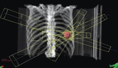
Stereotactic radiation – uses focused radiation beams targeting a
well-defined tumour, relying on detailed imaging, computerised
three-dimensional treatment planning and precise treatment set-
up to deliver the radiation dose with extreme accuracy
(Stereotactic Radiation).
[Picture Credit: Stereotactic Radiation]
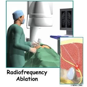
Radio Frequency Ablation – a medical procedure in which part of the
electrical conduction system of the heart, tumour, or other dysfunctional
tissue is ablated using the heat generated from high frequency
alternating current in the range of 350-500kHz |(Radio Frequency
Ablation; Wikipedia).
[Picture Credit: Radio Frequency Ablation]
About Clinical Trials
Clinical trials are research studies that involve people. These studies test new ways to
prevent, detect, diagnose, or treat diseases. People who take part in cancer clinical trials
have an opportunity to contribute to scientists’ knowledge about cancer and to help in the
development of improved cancer treatments. They also receive state-of-the-art care from
cancer experts.
Types of Clinical Trials
Cancer clinical trials differ according to their primary purpose. They include the following
types:
Treatment - these trials test the effectiveness of new treatments or new ways of using
current treatments in people who have cancer. The treatments tested may include new
drugs or new combinations of currently used drugs, new surgery or radiation therapy
techniques, and vaccines or other treatments that stimulate a person’s immune system to
fight cancer. Combinations of different treatment types may also be tested in these trials.
Researched and Authored by Prof Michael C Herbst
[D Litt et Phil (Health Studies); D N Ed; M Art et Scien; B A Cur; Dip Occupational Health; Dip Genetic
Counselling; Diagnostic Radiographer; Dip Audiometry and Noise Measurement]
Approved by Ms Elize Joubert, Chief Executive Officer [BA Social Work (cum laude); MA Social Work]
December 2017 Page 7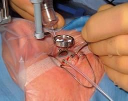
Prevention - these trials test new interventions that may lower the risk of developing certain
types of cancer. Most cancer prevention trials involve healthy people who have not had
cancer; however, they often only include people who have a higher than average risk of
developing a specific type of cancer. Some cancer prevention trials involve people who have
had cancer in the past; these trials test interventions that may help prevent the return
(recurrence) of the original cancer or reduce the chance of developing a new type of cancer

Screening - these trials test new ways of finding cancer early. When cancer is found early, it
may be easier to treat and there may be a better chance of long-term survival. Cancer
screening trials usually involve people who do not have any signs or symptoms of cancer.
However, participation in these trials is often limited to people who have a higher than
average risk of developing a certain type of cancer because they have a family history of that
type of cancer or they have a history of exposure to cancer-causing substances (e.g.,
cigarette smoke).

Diagnostic - these trials study new tests or procedures that may help identify, or diagnose,
cancer more accurately. Diagnostic trials usually involve people who have some signs or
symptoms of cancer.

Quality of life or supportive care - these trials focus on the comfort and quality of life of
cancer patients and cancer survivors. New ways to decrease the number or severity of side
effects of cancer or its treatment are often studied in these trials. How a specific type of
cancer or its treatment affects a person’s everyday life may also be studied.

Where Clinical Trials are Conducted
Cancer clinical trials take place in cities and towns in doctors’ offices, cancer centres and
other medical centres, community hospitals and clinics. A single trial may take place at one
or two specialised medical centres only or at hundreds of offices, hospitals, and centres.

Each clinical trial is managed by a research team that can include doctors, nurses, research
assistants, data analysts, and other specialists. The research team works closely with other
health professionals, including other doctors and nurses, laboratory technicians,
pharmacists, dieticians, and social workers, to provide medical and supportive care to
people who take part in a clinical trial.

Research Team
The research team closely monitors the health of people taking part in the clinical trial and
gives them specific instructions when necessary. To ensure the reliability of the trial’s
results, it is important for the participants to follow the research team’s instructions. The
instructions may include keeping logs or answering questionnaires. The research team may
also seek to contact the participants regularly after the trial ends to get updates on their
health.

Clinical Trial Protocol
Every clinical trial has a protocol, or action plan, that describes what will be done in the trial,
how the trial will be conducted, and why each part of the trial is necessary. The protocol also
includes guidelines for who can and cannot participate in the trial. These guidelines, called
eligibility criteria, describe the characteristics that all interested people must have before
they can take part in the trial. Eligibility criteria can include age, sex, medical history, and

Researched and Authored by Prof Michael C Herbst
[D Litt et Phil (Health Studies); D N Ed; M Art et Scien; B A Cur; Dip Occupational Health; Dip Genetic
Counselling; Diagnostic Radiographer; Dip Audiometry and Noise Measurement]
Approved by Ms Elize Joubert, Chief Executive Officer [BA Social Work (cum laude); MA Social Work]
December 2017                                                                                      Page 8
current health status. Eligibility criteria for cancer treatment trials often include the type and
stage of cancer, as well as the type(s) of cancer treatment already received.

Enrolling people who have similar characteristics helps ensure that the outcome of a trial is
due to the intervention being tested and not to other factors. In this way, eligibility criteria
help researchers obtain the most accurate and meaningful results possible.

National and International Regulations
National and international regulations and policies have been developed to help ensure that
research involving people is conducted according to strict scientific and ethical principles. In
these regulations and policies, people who participate in research are usually referred to as
“human subjects.”

Informed Consent
Informed consent is a process through which people learn the important facts about a clinical
trial to help them decide whether or not to take part in it, and continue to learn new
information about the trial that helps them decide whether or not to continue participating in
it.

During the first part of the informed consent process, people are given detailed information
about a trial, including information about the purpose of the trial, the tests and other
procedures that will be required, and the possible benefits and harms of taking part in the
trial. Besides talking with a doctor or nurse, potential trial participants are given a form,
called an informed consent form, that provides information about the trial in writing. People
who agree to take part in the trial are asked to sign the form. However, signing this form
does not mean that a person must remain in the trial. Anyone can choose to leave a trial at
any time—either before it starts or at any time during the trial or during the follow-up period.
It is important for people who decide to leave a trial to get information from the research
team about how to leave the trial safely.

The informed consent process continues throughout a trial. If new benefits, risks, or side
effects are discovered during the course of a trial, the researchers must inform the
participants so they can decide whether or not they want to continue to take part in the trial.
In some cases, participants who want to continue to take part in a trial may be asked to sign
a new informed consent form.

New interventions are often studied in a stepwise fashion, with each step representing a
different “phase” in the clinical research process. The following phases are used for cancer
treatment trials:

Phases of a Clinical Trial
Phase 0. These trials represent the earliest step in testing new treatments in humans. In a
phase 0 trial, a very small dose of a chemical or biologic agent is given to a small number of
people (approximately 10-15) to gather preliminary information about how the agent is
processed by the body (pharmacokinetics) and how the agent affects the body
(pharmacodynamics). Because the agents are given in such small amounts, no information
is obtained about their safety or effectiveness in treating cancer. Phase 0 trials are also
called micro-dosing studies, exploratory Investigational New Drug (IND) trials, or early phase

Researched and Authored by Prof Michael C Herbst
[D Litt et Phil (Health Studies); D N Ed; M Art et Scien; B A Cur; Dip Occupational Health; Dip Genetic
Counselling; Diagnostic Radiographer; Dip Audiometry and Noise Measurement]
Approved by Ms Elize Joubert, Chief Executive Officer [BA Social Work (cum laude); MA Social Work]
December 2017                                                                                      Page 9
I trials. The people who take part in these trials usually have advanced disease, and no
known, effective treatment options are available to them.

Phase I (also called phase 1). These trials are conducted mainly to evaluate the safety of
chemical or biologic agents or other types of interventions (e.g., a new radiation therapy
technique). They help determine the maximum dose that can be given safely (also known as
the maximum tolerated dose) and whether an intervention causes harmful side effects.
Phase I trials enrol small numbers of people (20 or more) who have advanced cancer that
cannot be treated effectively with standard (usual) treatments or for which no standard
treatment exists. Although evaluating the effectiveness of interventions is not a primary goal
of these trials, doctors do look for evidence that the interventions might be useful as
treatments.

Phase II (also called phase 2). These trials test the effectiveness of interventions in people
who have a specific type of cancer or related cancers. They also continue to look at the
safety of interventions. Phase II trials usually enrol fewer than 100 people but may include as
many as 300. The people who participate in phase II trials may or may not have been
treated previously with standard therapy for their type of cancer. If a person has been treated
previously, their eligibility to participate in a specific trial may depend on the type and amount
of prior treatment they received. Although phase II trials can give some indication of whether
or not an intervention works, they are almost never designed to show whether an
intervention is better than standard therapy.

Phase III (also called phase 3). These trials compare the effectiveness of a new intervention,
or new use of an existing intervention, with the current standard of care (usual treatment) for
a particular type of cancer. Phase III trials also examine how the side effects of the new
intervention compare with those of the usual treatment. If the new intervention is more
effective than the usual treatment and/or is easier to tolerate, it may become the new
standard of care.

Phase III trials usually involve large groups of people (100 to several thousand), who are
randomly assigned to one of two treatment groups, or “trial arms”: (1) a control group, in
which everyone in the group receives usual treatment for their type of cancer, or 2) an
investigational or experimental group, in which everyone in the group receives the new
intervention or new use of an existing intervention. The trial participants are assigned to their
individual groups by random assignment, or randomisation. Randomisation helps ensure
that the groups have similar characteristics. This balance is necessary so the researchers
can have confidence that any differences they observe in how the two groups respond to the
treatments they receive are due to the treatments and not to other differences between the
groups.

Randomisation is usually done by a computer program to ensure that human choices do not
influence the assignment to groups. The trial participants cannot request to be in a particular
group, and the researchers cannot influence how people are assigned to the groups.
Usually, neither the participants nor their doctors know what treatment the participants are
receiving.

Researched and Authored by Prof Michael C Herbst
[D Litt et Phil (Health Studies); D N Ed; M Art et Scien; B A Cur; Dip Occupational Health; Dip Genetic
Counselling; Diagnostic Radiographer; Dip Audiometry and Noise Measurement]
Approved by Ms Elize Joubert, Chief Executive Officer [BA Social Work (cum laude); MA Social Work]
December 2017                                                                                      Page 10
People who participate in phase III trials may or may not have been treated previously. If
they have been treated previously, their eligibility to participate in a specific trial may depend
on the type and the amount of prior treatment they received.
In most cases, an intervention will move into phase III testing only after it has shown promise
in phase I and phase II trials.
Phase IV (also called phase 4). These trials further evaluate the effectiveness and long-term
safety of drugs or other interventions. They usually take place after a drug or intervention
has been approved by the medicine regulatory office for standard use. Several hundred to
several thousand people may take part in a phase IV trial. These trials are also known as
post-marketing surveillance trials. They are generally sponsored by drug companies.
Sometimes clinical trial phases may be combined (e.g., phase I/II or phase II/III trials) to
minimize the risks to participants and/or to allow faster development of a new intervention.
Although treatment trials are always assigned a phase, other clinical trials (e.g., screening,
prevention, diagnostic, and quality-of-life trials) may not be labelled this way.
Use of Placebos
The use of placebos as comparison or “control” interventions in cancer treatment trials is
rare. If a placebo is used by itself, it is because no standard treatment exists. In this case, a
trial would compare the effects of a new treatment with the effects of a placebo. More often,
however, placebos are given along with a standard treatment. For example, a trial might
compare the effects of a standard treatment plus a new treatment with the effects of the
same standard treatment plus a placebo.
Possible benefits of taking part in a clinical trial
The benefits of participating in a clinical trial include the following:
Trial participants have access to promising new interventions that are generally not
available outside of a clinical trial.
The intervention being studied may be more effective than standard therapy. If it is
more effective, trial participants may be the first to benefit from it.
Trial participants receive regular and careful medical attention from a research team
that includes doctors, nurses, and other health professionals.
The results of the trial may help other people who need cancer treatment in the
future.
Trial participants are helping scientists learn more about cancer (e.g., how it grows,
how it acts, and what influences its growth and spread).
Potential harms associated with taking part in a clinical trial
The potential harms of participating in a clinical trial include the following:
The new intervention being studied may not be better than standard therapy, or it
may have harmful side effects that doctors do not expect or that are worse than
those associated with standard therapy.
Researched and Authored by Prof Michael C Herbst
[D Litt et Phil (Health Studies); D N Ed; M Art et Scien; B A Cur; Dip Occupational Health; Dip Genetic
Counselling; Diagnostic Radiographer; Dip Audiometry and Noise Measurement]
Approved by Ms Elize Joubert, Chief Executive Officer [BA Social Work (cum laude); MA Social Work]
December 2017 Page 11 Trial participants may be required to make more visits to the doctor than they would if
they were not in a clinical trial and/or may need to travel farther for those visits.
Correlative research studies, and how they are related to clinical trials
In addition to answering questions about the effectiveness of new interventions, clinical trials
provide the opportunity for additional research. These additional research studies, called
correlative or ancillary studies, may use blood, tumour, or other tissue specimens (also
known as ‘biospecimens’) obtained from trial participants before, during, or after treatment.
For example, the molecular characteristics of tumour specimens collected during a trial
might be analysed to see if there is a relationship between the presence of a certain gene
mutation or the amount of a specific protein and how trial participants responded to the
treatment they received. Information obtained from these types of studies could lead to more
accurate predictions about how individual patients will respond to certain cancer treatments,
improved ways of finding cancer earlier, new methods of identifying people who have an
increased risk of cancer, and new approaches to try to prevent cancer.
Clinical trial participants must give their permission before biospecimens obtained from them
can be used for research purposes.
When a clinical trial is over
After a clinical trial is completed, the researchers look carefully at the data collected during
the trial to understand the meaning of the findings and to plan further research. After a phase
I or phase II trial, the researchers decide whether or not to move on to the next phase or
stop testing the intervention because it was not safe or effective. When a phase III trial is
completed, the researchers analyse the data to determine whether the results have medical
importance and, if so, whether the tested intervention could become the new standard of
care.
The results of clinical trials are often published in peer-reviewed scientific journals. Peer
review is a process by which cancer research experts not associated with a trial review the
study report before it is published to make sure that the data are sound, the data analysis
was performed correctly, and the conclusions are appropriate. If the results are particularly
important, they may be reported by the media and discussed at a scientific meeting and by
patient advocacy groups before they are published in a journal. Once a new intervention has
proven safe and effective in a clinical trial, it may become a new standard of care.
(National Cancer Institute).
Medical Disclaimer
This Fact Sheet is intended to provide general information only and, as such, should not be
considered as a substitute for advice, medically or otherwise, covering any specific situation.
Users should seek appropriate advice before taking or refraining from taking any action in
reliance on any information contained in this Fact Sheet. So far as permissible by law, the
Cancer Association of South Africa (CANSA) does not accept any liability to any person (or
his/her dependants/estate/heirs) relating to the use of any information contained in this Fact
Sheet.
Whilst the Cancer Association of South Africa (CANSA) has taken every precaution in
compiling this Fact Sheet, neither it, nor any contributor(s) to this Fact Sheet can be held
Researched and Authored by Prof Michael C Herbst
[D Litt et Phil (Health Studies); D N Ed; M Art et Scien; B A Cur; Dip Occupational Health; Dip Genetic
Counselling; Diagnostic Radiographer; Dip Audiometry and Noise Measurement]
Approved by Ms Elize Joubert, Chief Executive Officer [BA Social Work (cum laude); MA Social Work]
December 2017 Page 12responsible for any action (or the lack thereof) taken by any person or organisation wherever they shall be based, as a result, direct or otherwise, of information contained in, or accessed through, this Fact Sheet. Researched and Authored by Prof Michael C Herbst [D Litt et Phil (Health Studies); D N Ed; M Art et Scien; B A Cur; Dip Occupational Health; Dip Genetic Counselling; Diagnostic Radiographer; Dip Audiometry and Noise Measurement] Approved by Ms Elize Joubert, Chief Executive Officer [BA Social Work (cum laude); MA Social Work] December 2017 Page 13
Sources and References

Atlas of Genetics and Cytogenetics in Oncology and Haematology
http://atlasgeneticsoncology.org/Kprones/VHLKpr10010.html

Community Eye Health Journal
http://www.cehjournal.org/article/management-of-capsular-rupture-and-vitreous-loss-in-
cataract-surgery/

Florida Retina Institute
http://www.floridaretinainstitute.com/services_vitreous.html

Genetics Home Reference
http://ghr.nlm.nih.gov/condition/von-hippel-lindau-syndrome
http://ghr.nlm.nih.gov/gene/VHL

Lonser, R.R., Glenn, G.M., Walther, M., Chew, E.Y., Libutti, S.K., Linehan, W.M. &
Oldfield, E.H. 2003. Von Hippel-Lindau disease. The Lancet, 361:2059 – 2067.

MD Anderson Cancer Center
http://www.mdanderson.org/patient-and-cancer-information/cancer-information/cancer-
types/von-hippel-lindau-disease/symptoms/index.html

Minneapolis Clinic of Neurology
http://www.minneapolisclinic.com/medical-services/127-von-hippel-lindau-disease.html

National Cancer Institute
http://www.cancer.gov/cancertopics/factsheet/clinicaltrials/clinical-trials

NYU Langone Medical Center
http://www.med.nyu.edu/content?ChunkIID=22504

Patient.co.uk
http://www.patient.co.uk/doctor/von-Hippel-Lindau-Disease.htm

Radio Frequency Ablation
http://www.smpt.biz/Pain-Care/Radiofrequency-Ablation/a~6789/article.html

Retina Macula Institute
http://www.retinamaculainstitute.com/treatments/cryotherapy/

Scleral Buckling
https://www.google.co.za/search?q=cryotherapy+VHL&source=lnms&tbm=isch&sa=X&ei=E
0iZU6XoCqSV7AaB04CwCg&ved=0CAYQ_AUoAQ&biw=1517&bih=714&dpr=0.9#q=scleral
+buckling+procedure&tbm=isch&facrc=_&imgdii=_&imgrc=YbtUQs4aiKZyRM%253A%3Bwn
Ay2IC4Uewg_M%3Bhttp%253A%252F%252Fi1.ytimg.com%252Fvi%252Fdep0-
iEZexM%252F0.jpg%3Bhttp%253A%252F%252Fshelf3d.com%252FSearch%252FAugen%
25252BOP's%25252BPlayListIDPL020A07169A6AF04C%3B480%3B360

Shroff Eye Hospital
http://www.shroffeye.org/services-view/argon-laser/

Researched and Authored by Prof Michael C Herbst
[D Litt et Phil (Health Studies); D N Ed; M Art et Scien; B A Cur; Dip Occupational Health; Dip Genetic
Counselling; Diagnostic Radiographer; Dip Audiometry and Noise Measurement]
Approved by Ms Elize Joubert, Chief Executive Officer [BA Social Work (cum laude); MA Social Work]
December 2017                                                                                      Page 14
Stanford Cancer Institute
http://cancer.stanford.edu/information/geneticsAndCancer/types/vhl.html

Stereotactic Radiation
http://www.rsna.org/NewsDetail.aspx?id=9297

Unbound Medicine
http://www.unboundmedicine.com/medline/citation/6367726/Retinal_microincarceration_with
_penetrating_diathermy_in_the_management_of_giant_retinal_tears_

UpToDate
http://www.uptodate.com/contents/argon-plasma-coagulation-in-the-management-of-
gastrointestinal-hemorrhage

VHL Alliance
http://www.vhl.org/wordpress/patients-caregivers/basic-facts-about-vhl/the-history-of-vhl/dr-
eugen-von-hippel/
http://www.vhl.org/wordpress/patients-caregivers/basic-facts-about-vhl/the-history-of-vhl/dr-
arvid-lindau/
http://www.vhl.org/wordpress/patients-caregivers/progress-toward-a-cure/genetic-research-
and-vhl/

VHL Disease
https://www.google.co.za/search?q=von+hippel-
lindau+syndrome+(vhl)&source=lnms&tbm=isch&sa=X&ei=YrWWU4O0EszB7Aaex4HoBQ&
ved=0CAYQ_AUoAQ&biw=1517&bih=714&dpr=0.9#facrc=_&imgdii=_&imgrc=0McloZgj8Pic
vM%253A%3BH1J4eorvcAycyM%3Bhttp%253A%252F%252Fwww.mdanderson.org%252F
patient-and-cancer-information%252Fcancer-information%252Fcancer-types%252Fvon-
hippel-lindau-disease%252Fvhl-
full.jpg%3Bhttp%253A%252F%252Fwww.mdanderson.org%252Fpatient-and-cancer-
information%252Fcancer-information%252Fcancer-types%252Fvon-hippel-lindau-
disease%252Findex.html%3B750%3B963

Vitreous Surgery
http://www.cehjournal.org/article/management-of-capsular-rupture-and-vitreous-loss-in-
cataract-surgery/

WebMD
http://www.webmd.com/eye-health/scleral-buckling-surgery-for-retinal-detachment

Wikipedia
http://en.wikipedia.org/wiki/Radiofrequency_ablation
http://en.wikipedia.org/wiki/Cryotherapy

Researched and Authored by Prof Michael C Herbst
[D Litt et Phil (Health Studies); D N Ed; M Art et Scien; B A Cur; Dip Occupational Health; Dip Genetic
Counselling; Diagnostic Radiographer; Dip Audiometry and Noise Measurement]
Approved by Ms Elize Joubert, Chief Executive Officer [BA Social Work (cum laude); MA Social Work]
December 2017                                                                                      Page 15
You can also read