Pretreatment with LCK inhibitors chemosensitizes cisplatin resistant endometrioid ovarian tumors
←
→
Page content transcription
If your browser does not render page correctly, please read the page content below
Crean-Tate et al. Journal of Ovarian Research          (2021) 14:55
https://doi.org/10.1186/s13048-021-00797-x

 RESEARCH                                                                                                                                          Open Access

Pretreatment with LCK inhibitors
chemosensitizes cisplatin‐resistant
endometrioid ovarian tumors
Katie K. Crean-Tate1,2*, Chad Braley3, Goutam Dey3, Emily Esakov3, Caner Saygin4, Alexandria Trestan3,
Daniel J. Silver3, Soumya M. Turaga3, Elizabeth V. Connor5, Robert DeBernardo1, Chad M. Michener1, Peter G. Rose1,
Justin Lathia3 and Ofer Reizes3*

  Abstract
  Background: Ovarian cancer is the most fatal gynecologic malignancy in the United States. While chemotherapy is
  effective in the vast majority of ovarian cancer patients, recurrence and resistance to standard systemic therapy is
  nearly inevitable. We discovered that activation of the non-receptor tyrosine kinase Lymphocyte Cell-Specific
  Protein-Tyrosine Kinase (LCK) promoted cisplatin resistance. Here, we hypothesized that treating high grade,
  platinum resistant endometrioid cancer cells with an LCK inhibitor (LCKi) followed by co-treatment with cisplatin
  would lead to increased cisplatin efficacy. Our objective was to assess clinical outcomes associated with increased
  LCK expression, test our hypothesis of utilizing LCKi as pre-treatment followed by co-treatment with cisplatin in
  platinum resistant ovarian cancer in vitro, and evaluate our findings in vivo to assess LCKi applicability as a
  therapeutic agent.
  Results: Kaplan-Meier (KM) plotter data indicated LCK expression is associated with significantly worse median
  progression-free survival (HR 3.19, p = 0.02), and a trend toward decreased overall survival in endometrioid ovarian
  tumors with elevated LCK expression (HR 2.45, p = 0.41). In vitro, cisplatin resistant ovarian endometrioid cells
  treated first with LCKi followed by combination LCKi-cisplatin treatment showed decreased cell viability and
  increased apoptosis. Immunoblot studies revealed LCKi led to increased expression of phosphorylated H2A histone
  family X (γ-H2AX), a marker for DNA damage. In vivo results demonstrate treatment with LCKi followed by LCKi-
  cisplatin led to significantly slowed tumor growth.
  Conclusions: We identified a strategy to therapeutically target cisplatin resistant endometrioid ovarian cancer
  leading to chemosensitization to platinum chemotherapy via treatment with LCKi followed by co-treatment with
  LCKi-cisplatin.
  Keywords: Ovarian cancer, Platinum resistance, LCK inhibitor, Chemosensitization

* Correspondence: kkcrean@gmail.com; reizeso@ccf.org
1
 Department of Gynecologic Oncology, Cleveland Clinic Foundation,
Women’s Health Institute, OH, Cleveland, USA
3
 Department of Cardiovascular and Metabolic Sciences, Lerner Research
Institute, Case Comprehensive Cancer Center, The Laura J. Fogarty Endowed
Chair in Uterine Cancer Research, 9500 Euclid Avenue, NC10, OH 44195
Cleveland, USA
Full list of author information is available at the end of the article

                                       © The Author(s). 2021 Open Access This article is licensed under a Creative Commons Attribution 4.0 International License,
                                       which permits use, sharing, adaptation, distribution and reproduction in any medium or format, as long as you give
                                       appropriate credit to the original author(s) and the source, provide a link to the Creative Commons licence, and indicate if
                                       changes were made. The images or other third party material in this article are included in the article's Creative Commons
                                       licence, unless indicated otherwise in a credit line to the material. If material is not included in the article's Creative Commons
                                       licence and your intended use is not permitted by statutory regulation or exceeds the permitted use, you will need to obtain
                                       permission directly from the copyright holder. To view a copy of this licence, visit http://creativecommons.org/licenses/by/4.0/.
                                       The Creative Commons Public Domain Dedication waiver (http://creativecommons.org/publicdomain/zero/1.0/) applies to the
                                       data made available in this article, unless otherwise stated in a credit line to the data.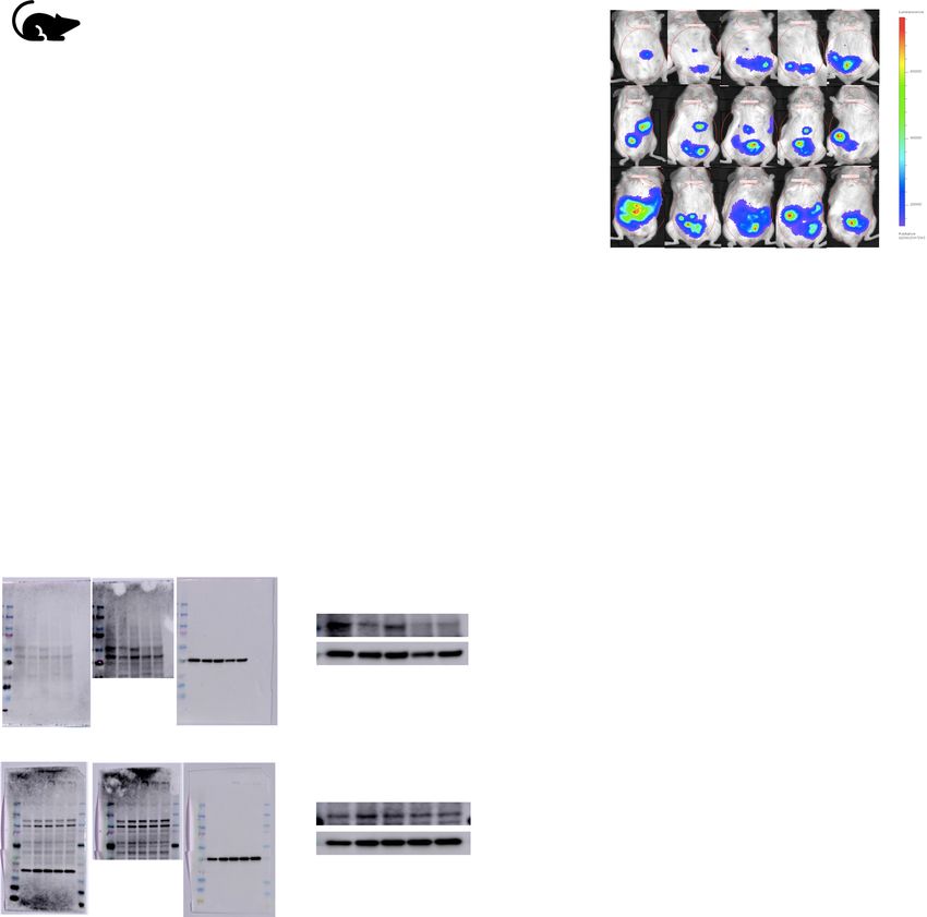
Crean-Tate et al. Journal of Ovarian Research   (2021) 14:55                                                    Page 2 of 10

Background                                                     With our initial results and proposed mechanism of
Gynecologic malignancy is common, with endometrial             action of chemosensitization, we hypothesized that treat-
and ovarian cancers being the most common incident             ing cancer cells first with an LCK inhibitor, followed by
types in the United States. Ovarian cancer is the most         co-treatment with an LCK inhibitor and cisplatin, would
fatal gynecologic malignancy in the United States, with        enhance the chemosensitization effect. Our primary
only a 48 % survival at 5 years after diagnosis [1]. Typic-    objective was to test this hypothesis in vitro in a cisplatin
ally, advanced disease in ovarian cancer is treated with       resistant endometrioid cancer model, followed by in vivo
cytoreductive surgery and platinum-based chemother-            as a proof of concept.
apy. Up to 15 % of ovarian cancers have endometrioid
subtype histologically [2]. Unfortunately, high-grade          Results
endometrioid cancers prove difficult to treat due to re-       LCK expression is associated with poor patient survival
currence and chemoresistance [3]. In ovarian cancer,           Given the previously described mechanism of cisplatin
while up to 85 % of patients will enter remission with         resistance via the LCK pathway, we hypothesized that in-
standard treatment of debulking surgery and platinum-          creased LCK expression would be associated with worse
taxane chemotherapy, most of these patients will recur         clinical outcomes. We assessed survival outcomes with
[4]. In the 15 % of patients failing standard therapy, dis-    increased LCK expression for endometrioid ovarian can-
ease persists or progresses within the first six months        cer using Kaplan-Meier Plotter database (KM Plotter:
after chemotherapy, indicating platinum-resistant dis-         http://kmplot.com/analysis/). The database was queried
ease. For those who enter remission, progression to            for patients of all stages and grades with endometrioid
platinum-resistant disease is pervasive [5]. The prognosis     ovarian cancer. In endometrioid ovarian cancer, LCK ex-
is particularly poor in those with platinum-resistant dis-     pression is associated with significantly worse median
ease, with response rates below 20 % for subsequent            progression-free survival (HR 3.19, p = 0.02, Fig. 1a).
lines of chemotherapy and continued decrease in disease        Overall survival is not significantly different between
free interval with each subsequent therapy [6]. Recurrent      groups, though a non-significant trend toward decreased
ovarian cancer is considered incurable, with goals of care     survival was seen with HR 2.45 (p = 0.41, Fig. 1b). This is
aimed at symptom management with alternative regi-             also supported by gene expression profiling data from
mens of chemotherapy [7]. Given the poor prognosis in          Tothill et al. that found an over 2-fold increased expres-
patients with platinum-resistant disease, identification of    sion in LCK in ovarian cancer subtypes containing high
chemoresistance pathways is necessary for development          grade endometrioid cells and associated significantly de-
of therapies to sensitize resistant ovarian cancer [8].        creased PFS (p < 0.001) and OS (p < 0.001) in this sub-
   Endometrioid tumors have previously been shown to           type [16]. This compares with half the gene expression
exhibit a self-renewing population of cells termed cancer      of LCK in the predominantly low grade endometrioid
stem cells (CSC) [9, 10]. CSCs are associated with both        ovarian cancer subtype, which showed the most favor-
tumor recurrence and chemoresistance in multiple               able PFS and OS [16]. These data indicate increased
tumor types [9, 11–13]. Previously in the Reizes lab, Say-     LCK expression in endometrioid ovarian cancer corre-
gin et al. identified a novel pathway in CSCs leading to       lates with poorer clinical outcomes.
chemoresistance in endometrioid tumors via activation
of the non-receptor tyrosine kinase LCK [10]. Saygin           Pretreatment with LCK inhibitors chemosensitize cisplatin
et al. utilized pharmacologic inhibition of LCK to incur       resistant endometrioid cells and increase apoptosis
increased sensitization to cisplatin in endometrioid           In our prior studies, LCK inhibition in the ovarian endo-
CSCs. This finding was verified with LCK-silenced CSCs         metrioid CSC population led to increased cisplatin sensi-
via shRNA (short hairpin RNA) constructs. Additionally,        tivity [10]. Given that CSCs are known to be a
LCK overexpression in non-CSCs indicated higher sur-           chemoresistant population closely associated with dis-
vival rates and lower apoptosis rates compared to an           ease recurrence, we hypothesized that inhibition of the
empty vector control [10]. In vitro studies using saracati-    LCK pathway would lead to sensitization of platinum re-
nib, an investigational drug that inhibits LCK, showed         sistant endometrioid cells. We performed an in vitro cel-
sensitization of cisplatin resistant endometrioid cells to     lular proliferation assay in cisplatin resistant ovarian
cisplatin in a CSC population. With inhibition of LCK,         endometrioid cells (CP70) treated with vehicle or the
DNA repair genes were attenuated and cells showed in-          LCK inhibitor (LCKi) saracatinib followed by assessing
creased sensitivity to cisplatin [10]. Phase I studies of      chemosensitivity to cisplatin via dose response. There
saracatinib utilized in multiple cancer types have indi-       was no significant difference with co-treatment of LCKi
cated an appropriate safety profile, though follow up          and cisplatin alone, so we hypothesized that pretreat-
randomized human trials in ovarian cancer have fallen          ment with LCKi is required to effectively sensitize these
short in translating the effect into clinical use [14, 15].    cells to cisplatin. CP70 cells pretreated with saracatinib
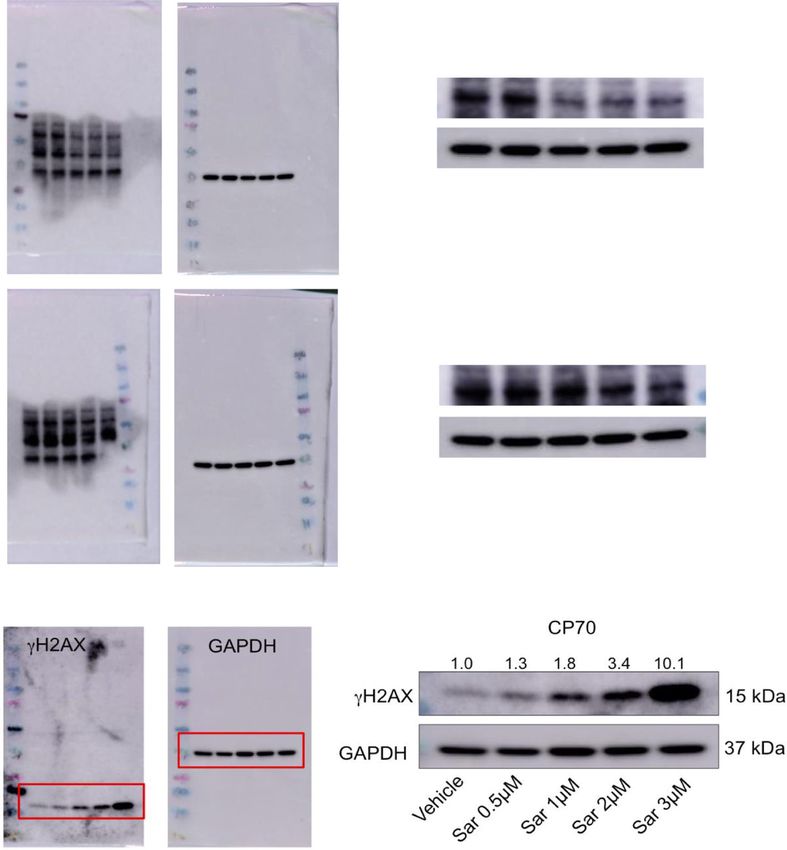
Crean-Tate et al. Journal of Ovarian Research      (2021) 14:55                                                                        Page 3 of 10

 Fig. 1 LCK expression is associated with poorer patient survival. Kaplan-Meier progression-free and overall survival curves were obtained from
 Kaplan-Meier Plotter (KM Plotter: http://kmplot.com/analysis/) for endometrioid ovarian cancer patients who had high versus low tumor mRNA
 expression of LCK (a, b) prior to therapy

and then treated with combination saracatinib-cisplatin                     Cisplatin resistant endometrioid cells treated with LCK
exhibited significantly reduced proliferation (Fig. 2a). In                 inhibitors lead to increased DNA double strand breaks
parallel, we tested the effect of saracatinib-cisplatin                     To investigate the mechanism by which LCK inhibitors
treatment on apoptosis using Caspase Glo. We deter-                         decrease cisplatin resistance, we tested whether LCK
mined that CP70 cells pretreated with saracatinib                           inhibitors decrease phosphorylation of LCK in CP70
followed by cisplatin plus saracatinib led to increased                     cells by immunoblot. We found that Y394 phosphory-
apoptosis compared to no pretreatment (Fig. 2b). These                      lated LCK (P-LCK) was significantly reduced when cells
findings were replicated in HEC1a, a cisplatin resistant                    are exposed to LCK inhibitors saracatinib or PP2. Y394
endometrioid endometrial cancer model. HEC1a pre-                           P-LCK is the autophosphorylation site on LCK necessary
treated with saracatinib followed by cisplatin co-                          for kinase activation and function [17]. Total LCK (T-
treatment with saracatinib exhibited inhibition of cell                     LCK) was unchanged in vehicle and saracatinib-treated
viability (Fig. 2c) and increased apoptosis (Fig. 2d) com-                  cells, whereas there was a reduction in PP2-treated cells.
pared to vehicle treated cells.                                             GAPDH was used as a loading control (Fig. 3a). We
  To validate our hypothesis that treatment with                            tested for DNA damage by blotting for          H2AX, a
LCKi followed by co-treatment with cisplatin leads to                       marker of DNA double strand breaks based on
decreased proliferation specifically in cisplatin resist-                   phosphorylation of histones [18, 19]. We found an
ant endometrioid cells, we performed a proliferation                        increase in      H2AX in LCKi treated CP70 cells
assay in A2780 cells, the chemo-naive parent cell line                      (Fig. 3b).These data indicate that endometrioid ovarian
of CP70. We found that pretreatment with saracati-                          cancer cells exposed to LCKi demonstrate a decrease in
nib, as well as simultaneous treatment with saracati-                       phosphorylated LCK and an increase in DNA double
nib and cisplatin, did not alter cell viability (Fig. 2e).                  strand breaks.
To validate our hypothesis that saracatinib chemosen-
sitizes these cells via LCK inhibition, we pretreated                       Treatment with LCK inhibitor followed by co‐treatment
CP70 cells with a more selective LCKi, PP2, followed                        with cisplatin decreases tumor growth in vivo
by cisplatin co-treatment, and found a similar left                         Given the in vitro findings, we next tested our hypoth-
shift in dose response compared to control groups                           esis in an in vivo model. We injected NOD severe
treated with cisplatin alone (Fig. 2f). These data indi-                    combined immunodeficient (SCID) IL2R gamma (NSG)
cate that platinum resistant endometrioid cells pre-                        mice with CP70 cells virally transduced with luciferase
treated with LCKi followed by co-treatment with                             and utilized the in vivo imaging system (IVIS) to detect
LCKi and cisplatin show decreased cell proliferation                        tumor growth on a weekly frequency. After injection of
and increased apoptosis.                                                    tumor cells (day 0), mice were placed in one of two
Crean-Tate et al. Journal of Ovarian Research      (2021) 14:55                                                                         Page 4 of 10

    A.                                                                  B.

    C.                                                                  D.

    E.                                                                  F.

 Fig. 2 LCK inhibitors chemosensitize cisplatin resistant endometrioid cells and increase apoptosis. Cisplatin resistant ovarian endometrioid cells
 (CP70) were cultured and pretreated with an LCK inhibitor (saracatinib) or vehicle, followed by vehicle or combination LCKi-cisplatin, followed by
 cell viability assay performed with the CellTiterGlo Assay (a). Caspase 3/7 Assay was then performed to assess apoptosis (b). A second cisplatin
 resistant endometrioid cell line (HEC1a) was similarly treated and tested with subsequent proliferation and apoptosis assays performed (c, d).
 Cisplatin sensitive ovarian endometrioid cells (A2780) were cultured and treated according to the aforementioned paradigm (e). An alternative
 LCK inhibitor (PP2) was utilized for pretreatment in CP70 cells followed by co-treatment with PP2-cisplatin (f). All data represent at minimum
 three independent experiments with three technical replicates

arms: pre-treatment with saracatinib or vehicle via oral                     cisplatin and saracatinib all showed a steady increase in
gavage three days per week, initiated on day 3. On day                       tumor burden over time. However, in mice first treated
16 when all mice were confirmed to have detectable                           with saracatinib followed by combination cisplatin and
tumor on IVIS, mice pretreated with saracatinib were                         saracatinib therapy, tumor growth appeared stable or
injected with cisplatin three times weekly, and mice pre-                    attenuated (Fig. 4b, c). At the experimental endpoint
treated with vehicle were randomly assigned to one of                        (Day 30), tumor growth in the vehicle group was similar
four arms: cisplatin, saracatinib, combination cisplatin                     to cisplatin, saracatinib, and combination cisplatin and
and saracatinib, or vehicle alone, given three times per                     saracatinib arms. In contrast, pretreatment with saracati-
week. Mice were then euthanized on day 30 (Fig. 4a).                         nib followed by combination saracatinib-cisplatin
Images presented for each subgroup were taken from                           treatment exhibited a significant reduction in tumor
the same mouse over time. Mice treated first with ve-                        growth compared to vehicle as well as combination
hicle followed by cisplatin, saracatinib, or combination                     alone (Fig. 4d). These findings were also reflected in the
Crean-Tate et al. Journal of Ovarian Research (2021) 14:55 Page 5 of 10 Fig. 3 Cisplatin resistant endometrioid cells treated with LCK inhibitors indicate decreased P-LCK and ovarian endometrioid cells treated with LCK inhibitor indicate increased DNA double strand breaks. Cisplatin resistant ovarian endometrioid cancer cells (CP70) were treated with DMSO, LCK inhibitor saracatinib (Sar) or PP2 at indicated doses for 48 h. Protein lysates were then immunoblotted for phosphorylated LCK (P-LCK Y394) and total LCK (T-LCK). Fold changes of protein expression are shown in the figure, with values normalized to vehicle control. GAPDH was used as loading control (a). CP70 cells treated with the indicated varied doses of saracatinib (Sar) were immunoblotted for H2AX. Fold changes of protein expression are shown in the figure, with values normalized to vehicle control. GAPDH was used as a loading control (b). Each experiment was performed with at least three technical replicates mouse body weight, with greatest weight increase ob- Treatment with LCK inhibitor in vivo leads to decreased served in vehicle and saracatinib arms, and the largest P-LCK in cisplatin‐resistant endometrioid tumors reduction in weight, though nonsignificant (p = 0.059), To validate the efficacy of LCKi, we assessed Y394 observed in the pretreatment saracatinib arm (Fig. 4f). P-LCK expression in tumors extracted from our Of note, this data represents tumor growth corrected in vivo study. We found that P-LCK in tumors from to baseline, not absolute tumor size, indicating that mice treated with saracatinib or a combination of the rate at which tumor growth is occurring is signifi- saracatinib and cisplatin expressed lower P-LCK cantly reduced relative to other treatment groups. than mice treated only with vehicle or cisplatin. T- These data demonstrate that pretreatment with LCKi LCK did not exhibit a significant reduction in ex- followed by LCKi-cisplatin co-treatment leads to de- pression. GAPDH was used as a loading control creased tumor burden in cisplatin resistant endome- (Fig. 4e). Thus, LCKi was efficacious at inhibiting trioid ovarian cancer in vivo. LCK activation.
Crean-Tate et al. Journal of Ovarian Research     (2021) 14:55                                                                       Page 6 of 10

  A.                                                                                B.

  C.                                                                                D.

  E.                                                                                F.

 Fig. 4 Pretreatment with LCK inhibitor followed by LCKi-cisplatin treatment attenuates tumor burden, and treated tumors indicate decreased P-
 LCK. NSG mice were injected with CP70-luciferase transfected cells followed by pretreatment with LCKi (6 mice) or vehicle (24 mice) for 14 days.
 LCKi mice were then co-treated with LCKi and cisplatin, and vehicle mice were randomized to further treatment with vehicle, cisplatin,
 saracatinib, or combination (6 mice per arm) (a). IVIS imaging was obtained on a weekly basis to assess tumor growth (b). IVIS luminescence was
 corrected to baseline for each arm and assessed over time (c) and at the experimental endpoint (d). Tumors from NSG mice were extracted, and
 protein lysates were prepared and immunoblotted for protein expression of P-LCK (Y394) and T-LCK. Fold changes of protein expression are
 shown in the figure, with values normalized to vehicle control. GAPDH was used as a loading control (e). Two tumors per condition were probed,
 with at least three technical replicates performed. Mouse body weight at day 7, 21, and 28 were obtained for each arm, with weights shown as
 fold change from baseline (f)

Discussion                                                                 cause for the poor survival rates seen in ovarian cancer
Despite most ovarian cancers displaying an excellent ini-                  today [3, 5, 9]. Studies have focused on identifying a tar-
tial response to standard chemotherapy, the majority of                    getable pathway promoting chemoresistance in order to
advanced stage patients recur, with eventual resistance                    reduce recurrence [9, 11, 12]. Previous studies by Saygin
to our most effective chemotherapy agents. This pattern                    et al [10] identified a novel pathway leading to chemore-
of pervasive relapse and ensuing chemoresistance is the                    sistance in endometrioid tumors in which CD55
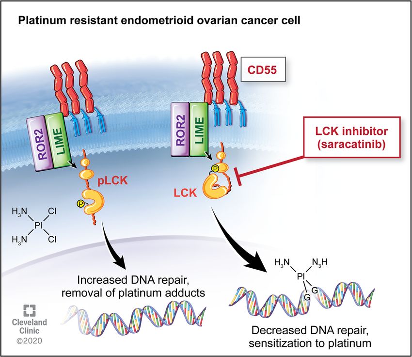
Crean-Tate et al. Journal of Ovarian Research    (2021) 14:55                                                                      Page 7 of 10

mediated DNA repair via phosphorylation of LCK. We                       formation with LCK inhibition in immunoblot studies
assessed clinical outcomes associated with LCK expres-                   (Fig. 3), an indication that targeting this pathway allows
sion. We found that high LCK expression predicted a                      platinum therapy to function in a previously cisplatin
significant effect on PFS with a three-fold increase in                  resistant cell population.
survival from 13 months to 34 months in low versus                         A common challenge in translational research is that
high LCK expressing tumors (Fig. 1). This data suggests                  while in vitro studies may prove promising, translating
a clinical benefit to addressing tumors with increased                   this to effective in vivo studies and clinical trials can
LCK expression, and thus a potential targetable pathway                  prove difficult. Saracatinib, an investigational LCKi, has
in recurrent ovarian endometrioid tumors.                                been studied for several cancer types, with mixed results.
  Standard chemotherapy in ovarian cancer includes a                     Studies on safety found appropriate dosing for saracati-
platinum and taxane agent, and survival decreases as                     nib in humans for effective pharmacodynamics while
response to platinum therapy diminishes. Prior studies                   limiting toxicity, indicating this drug would be tolerable
have found that cisplatin resistance is seen with multiple               in clinical trials [14]. While utilizing saracatinib as
pathways, including increased DNA repair enzyme                          monotherapy has not proven efficacious, combination
expression and associated reduction in DNA adducts [9,                   therapy has yielded more promising results. In a study
10, 20]. Through LCK inhibition, DNA repair enzyme                       combining saracatinib with carboplatin and/or paclitaxel
expression is attenuated, one of the known pathways to                   in solid tumors, objective responses were seen in
cisplatin resistance [10]. Given this anticipated initial                ovarian, breast and skin cancers, with longest response
chemosensitization step, we pursued pretreatment with                    durations seen in patients with ovarian cancer [14].
LCK inhibitors followed by co-treatment with cisplatin                   However, a randomized trial further assessed treatment
and found that this technique was effective in decreasing                with saracatinib in combination with weekly paclitaxel in
cancer cell populations and increasing apoptosis in vitro                platinum-resistant ovarian cancer, and found that co-
(Fig. 2). We verified the effects of inhibition of LCK on                treatment of saracatinib with weekly paclitaxel did not
DNA damage and found an increase in DNA adduct                           improve outcomes [15]. Of note, the majority of these

 Fig. 5 LCK pathway regulates cisplatin resistance in endometrioid tumors. Downstream of CD55, LCK stimulates expression of DNA repair genes,
 leading to cisplatin resistance. This targetable pathway identifies LCK inhibitors as adjunctive therapy for platinum resistant ovarian
 endometrioid cancer
Crean-Tate et al. Journal of Ovarian Research   (2021) 14:55                                                   Page 8 of 10

tumors were serous histology, and patients received only       conditions. Cell lines were obtained from the Cleveland
weekly Taxol without platinum in addition to saracati-         Clinic centralized research core facility, through which
nib. There is no clinical randomized data assessing sara-      cell lines were previously obtained from the American
catinib with cisplatin use in platinum resistant patients.     Type Culture Collection (ATCC) and authenticated. At
We see from this clinical data that saracatinib is well tol-   approximately 80 % confluence, trypsin (0.25 %)/EDTA
erated and may have a role in combination therapy in           solution or Accutase was used to lift cells for passaging
platinum resistant disease. We tested this hypothesis          as needed for continued experiments until passage 10, at
with a novel administration of saracatinib followed by         which point a fresh allotment of cells was plated. Cis-
co-treatment with cisplatin and found a decreased rate         platin was obtained from Cleveland Clinic Hospital
of tumor growth in vivo (Fig. 4), identifying a targetable     pharmacy, with 1 mg/mL stock solutions stored at room
pathway (Fig. 5) and providing a novel therapeutic regi-       temperature protected from light given its photosensitiv-
men for platinum resistant ovarian endometrioid                ity. Saracatinib (AZD0530) was purchased from Selleck
carcinoma.                                                     Chemicals, dissolved in DMSO (Sigma Cat#D2650), and
  This study’s strength lies in the proof of concept find-     10 μM stock solutions were aliquoted and stored at
ings using both in vitro and in vivo models. Additionally,     -20 °C. PP2 (AG1879) was purchased from Selleck Che-
whereas prior studies focused on a specific cell popula-       micals, dissolved in DMSO (Sigma Cat#D2650), and 10
tion, cancer stem cells, this study utilized a more hetero-    μM stock solutions aliquoted and stored at -20 °C.
geneous cell population, more closely simulating a             Mycoplasma testing was performed and negative.
typical tumor microenvironment. Further investigation
should also be performed in additional histologic types        Proliferation assays and caspase 3/7 assays
such as serous and clear cell, as well as substitution of      The appropriate cancer cells for each experiment were
cisplatin for co-treatment with carboplatin, a commonly        pre-treated with saracatinib (1μM), PP2 (10–50 μM), or
used platinum agent. Patients with platinum resistant          vehicle (DMSO at similar concentration to drug of inter-
disease have often received multiple lines of chemother-       est) for 4 days in T75 flasks. Cells were then plated in
apy previously and would benefit from further treatment        96-well plates at 5,000 cells/well on seeding Day 0,
options. Given our promising findings, further studies         manually counted by hemocytometer using Trypan blue
are indicated to pursue LCK inhibitors as an adjunctive        dye exclusion as live cell marker. Cisplatin was then ap-
therapy to platinum resistant disease in clinical trials.      plied the next day at doses of 0–10 μM, with/without
                                                               saracatinib, PP2 or vehicle, and treatment was ongoing
Conclusions                                                    for 4 to 6 days. Measured proliferation was assessed by
In summary, we identified a targetable pathway for che-        CellTiter-Glo (Promega, Southampton, UK) as per man-
mosensitization of platinum resistant ovarian endome-          ufacturer’s instructions. Percentage survival was normal-
trioid cancer. We found that pretreatment with LCK             ized to the untreated control for each group.
inhibitors followed by co-treatment with cisplatin leads          Caspase 3/7 Assay kit (Promega, Southampton, UK)
to decreased cell viability and increased apoptosis            was utilized to assess apoptosis as per manufacturer’s in-
in vitro. This is associated with increased DNA adduct         structions. This was performed alongside CellTiter-Glo
formation and significantly reduced tumor growth               to correct for viable cell density. Relative Caspase activ-
in vivo. Further studies are needed to assess the mecha-       ities were normalized to untreated controls in each
nisms behind the enhanced efficacy of pretreatment, as         group, with activity assessed from 30 to 120 min. Three
well as further investigation of LCK inhibitors as ad-         independent experiments were performed at minimum,
junctive therapy for platinum resistant endometrioid           each with three technical replicates.
ovarian carcinoma, including other histological subtypes.
                                                               Immunoblotting
Methods                                                        Protein lysates were obtained with cell lysis in 20mM
Cell culture                                                   Tris-HCl (pH 7.5), 150mM NaCl, 1 mM Na2EDTA, 1 %
Ovarian endometrioid adenocarcinoma cell lines A2780           NP-40, 1 mM EGTA, 1 % sodium pyrophosphate, 1 mM
(cisplatin sensitive) and its cisplatin resistant daughter     β-glycerophosphate, 1mM sodium orthovanadate, 1 μg/
cell line CP70 were cultured in Dulbecco’s Modified            mL leupeptin, 20 mM NaF and 1 mM PMSF. Protein
Eagle Medium (DMEM) supplemented with 10 % heat-               concentrations were measured with BCA Protein Assay
inactivated fetal bovine serum at 37 °C in a humidified        Kit (ThermoFisher Scientific). Protein concentrations of
atmosphere in 5 % CO2. Cisplatin resistant endometrioid        40μg of total protein were resolved in 10–12 % SDS-
endometrial cancer cell line HEC1a was cultured in             PAGE and transferred to PVDF membrane. Membranes
modified McCoy’s 5a medium supplemented with 10 %              were incubated overnight at 4 °C with primary anti-
heat-inactivated fetal bovine serum, also at similar           bodies against P-LCK (Y394) (1:1000) (R&D Systems,
Crean-Tate et al. Journal of Ovarian Research (2021) 14:55 Page 9 of 10
Catalog#MAB7500, Clone#755,103), T-LCK (1:1000) Statistical analysis
(Proteintech, Catalog#12477-1-AP), GAPDH (1:1000) Statistical analysis was calculated by one-way ANOVA and
(Proteintech, Catalog#HRP-60,004), and γ-H2AX (1: two sample t-test, with p-values included. Statistical signifi-
1000) (Cell Signaling, Catalog#2257). Secondary anti- cance is denoted via * to represent p-value of < 0.05 but >
mouse or anti-rabbit IgG antibodies conjugated to 0.01, ** representing p-value of < 0.01 but > 0.001, and **
horseradish peroxidase (HRP) (1:3000) (Cell Signaling, representing p-value < 0.001. For proliferation assays, IC50
Catalog#7076) or (1:25,000) (ProMega, Catalog#W4011) was calculated using nonparametric values set to nonlinear
were used. ECl (Pierce) was then used to visualize im- fit curve as per statistical analysis performed with GraphPad
munoreactive bands. Prism. Survival data was obtained from Kaplan-Meier Plotter
(KM Plotter: http://kmplot.com/analysis/) for endometrioid
In vivo study ovarian cancer based on CD55 and LCK mRNA expression.
All animal procedures were evaluated and approved KM Plotter survival data is obtained from an online database
prior to initiation by the Institutional Animal Care and collected from The Cancer Genome Atlas (TCGA), Gene
Use Committee (IACUC) of the Cleveland Clinic Lerner Expression Omnibus (GEO) and European Genome-
Research Institute. NOD severe combined immunodefi- phenome Archive (EGA).
cient (SCID) IL2R gamma (NSG) mice were purchased
from the Biological Response Unit (BRU) at the Acknowledgements
We acknowledge our gynecologic oncology patients, who continue to be
Cleveland Clinic and housed in microisolator units the motivation of our research.
under IACUC protocol #2018 − 1940. Thirty mice were
injected intraperitoneally with 1 million CP70-luciferase Authors’ contributions
virally transduced cells. At the time of injection (day 0), The first author KKCT generated the hypothesis and research question,
created study design, executed experiments, analyzed data, and performed
mice were placed in one of two arms, which started day primary manuscript authorship. CB and GD contributed to experiment
3: six mice began receiving pre-treatment with saracati- execution and data collection. EE contributed to data organization, analysis,
nib (Selleck), 25 mg/kg dissolved in 0.5 % hydroxypropyl and manuscript editing. CS and EC contributed to hypothesis generation,
experiment execution and data organization. AT contributed to experiment
methylcellulose (Sigma-Aldrich), 0.1 % Tween 80 execution and data collection. DS contributed to data organization and
(Sigma-Aldrich) via oral gavage three days per week, and manuscript editing. ST contributed to experiment execution. RD, CM, and
24 mice received vehicle via oral gavage on the same PGR all contributed to data review and manuscript review. JL contributed to
hypothesis generation and study design. OR performed significant
schedule. contributions to hypothesis generation, study design, data review and
Bioluminescence images to detect tumor burden were analysis, and manuscript editing. All authors read and approved the final
taken with Xenogen in vivo imaging system (IVIS, Perki- manuscript.
nElmer) using D-luciferin as previously described [21].
Funding
Mice received an IP injection of D-luciferin (Goldbio Research in the Reizes laboratory is supported by the VeloSano Bike to Cure
LUCK-1G, 150 mg/kg in 150μL) under inhaled isoflur- Impact Award, the Laura J. Fogarty Endowed Chair for Uterine Cancer
ane anesthesia. Images were analyzed (Living Image Research and the Cleveland Clinic Foundation Lerner Institute.
Software) and bioluminescence plots of photon flux
Availability of data and materials
(photons/second/cm2/steradian) over time were com- The KM plotter dataset is available at https://kmplot.com/analysis/index.
puted for each mouse, with normalization against day 0 php?p=service&cancer=ovar. The datasets used and analyzed for the current
signal values. Non-tumor and black backgrounds were study are available from the corresponding author upon reasonable request.
also subtracted from each tumor burden region of inter-
est. All images were obtained with a 15 s exposure. On Declarations
day 16 when all mice were confirmed to have tumor by Author’s Information
IVIS, mice pretreated with saracatinib were also treated OR and PGR are co-directors of the Center for Research Excellence in Gyne-
cologic Cancer at the Cleveland Clinic.
with cisplatin (2.5 mg/kg, 3 times per week) injected in-
traperitoneally, as previously described [10]. On day 16,
Ethics approval and consent to participate
mice pretreated with vehicle were randomly assigned to All mouse experiments were performed with adherence to protocols
one of four arms (6 mice per arm), whereby they were approved by the Institutional Animal Care and Use Committee at the Lerner
treated with cisplatin, saracatinib, combination cisplatin Research Institute at the Cleveland Clinic (IACUC protocol #2018 − 1940).
and saracatinib, or vehicle alone. Mice were sacrificed
Consent for publication
on day 30 and all visible tumor was collected for future Not applicable.
studies. There were no significant observable treatment
toxicities. All mouse procedures were performed under Competing interests
adherence to protocols approved by the Institute Animal OR has a patent for CD55 as a therapeutic target in cisplatin resistant
endometrial cancer pending. CM receives personal fees from Clovis
Care and Use Committee at the Lerner Research Oncology. The remaining authors have no relevant financial or conflicts of
Institute, Cleveland Clinic. interest to disclose for this work.Crean-Tate et al. Journal of Ovarian Research (2021) 14:55 Page 10 of 10
Author details 20. Damia G, Broggini M. Platinum Resistance in Ovarian Cancer: Role of DNA
1
Department of Gynecologic Oncology, Cleveland Clinic Foundation, Repair. Cancers (Basel). 2019 Jan 20;11(1).
Women’s Health Institute, OH, Cleveland, USA. 2Department of Gynecologic 21. Toyoshima M, Tanaka Y, Matumoto M, Yamazaki M, Nagase S, Sugamura K,
Oncology, Sutter Cancer Center, 2800 L Street, Suite 300, CA 95816 et al. Generation of a syngeneic mouse model to study the intraperitoneal
Sacramento, USA. 3Department of Cardiovascular and Metabolic Sciences, dissemination of ovarian cancer with in vivo luciferase imaging.
Lerner Research Institute, Case Comprehensive Cancer Center, The Laura J. Luminescence. 2009;24(5):324–31.
Fogarty Endowed Chair in Uterine Cancer Research, 9500 Euclid Avenue,
NC10, OH 44195 Cleveland, USA. 4Department of Internal Medicine, The Ohio
State University, OH, Columbus, USA. 5Department of Gynecologic Oncology, Publisher’s Note
Billings Clinic Cancer Center, MT, Billings, USA. Springer Nature remains neutral with regard to jurisdictional claims in
published maps and institutional affiliations.
Received: 6 November 2020 Accepted: 15 March 2021
References
1. Cancer of the Ovary -. Cancer Stat Facts, SEER (Surveillance, Epidemiology,
and End Results Program) Database. https://seer.cancer.gov/statfacts/html/
ovary.html. Accessed 2020 May 8.
2. Disaia PJ, Creasman WT, Mannel RS, McMeekin DS, Mutch DG. Clinical
Gynecologic Oncology. 9th ed. Philadelphia: Elsevier; 2012.
3. Ozols RF. Treatment goals in ovarian cancer. International Journal of
Gynecologic Cancer. 2005;15(Suppl 1):3–11.
4. Corrado G, Salutari V, Palluzzi E, Distefano MG, Scambia G, Ferrandina G.
Optimizing treatment in recurrent epithelial ovarian cancer. Expert Rev
Anticancer Ther. 2017;17(12):1147–58.
5. Pujade-Lauraine E, Combe P. Recurrent ovarian cancer. Ann Oncol. 2016;
27(suppl_1):i63–5.
6. Hanker LC, Loibl S, Burchardi N, Pfisterer J, Meier W, Pujade-Lauraine E, et al.
The impact of second to sixth line therapy on survival of relapsed ovarian
cancer after primary taxane/platinum-based therapy. Annals of oncology.
2012;23(10):2605–12.
7. Fung-Kee-Fung M, Oliver T, Elit L, Oza A, Hirte HW, Bryson P. Optimal
chemotherapy treatment for women with recurrent ovarian cancer. Curr
Oncol. 2007;14(5):195–208.
8. Monk J, Herzog BJ, Tewari TS. K. Evolution of chemosensitivity and
resistance assays as predictors of clinical outcomes in epithelial ovarian
cancer patients. Curr Pharm Design. 2016;22(30):4717–28.
9. Wiechert A, Saygin C, Thiagarajan PS, Rao VS, Hale JS, Gupta N, et al.
Cisplatin induces stemness in ovarian cancer. Oncotarget. 2016 Apr 20;7(21):
30511–22.
10. Saygin C, Wiechert A, Rao VS, Alluri R, Connor E, Thiagarajan PS, et al. CD55
regulates self-renewal and cisplatin resistance in endometrioid tumors. J
Exp Med. 2017;214(9):2715–32.
11. Kyo S, Maida Y, Inoue M. Stem cells in endometrium and endometrial
cancer: accumulating evidence and unresolved questions. Cancer letters.
2011;308(2):123–33.
12. Reya T, Morrison SJ, Clarke MF, Weissman IL. Stem cells, cancer, and cancer
stem cells. nature. 2001;414(6859):105–11.
13. Xiang D, Shigdar S, Bean AG, Bruce M, Yang W, Mathesh M, et al.
Transforming doxorubicin into a cancer stem cell killer via EpCAM aptamer-
mediated delivery. Theranostics. 2017 Sep 20;7(17):4071–86.
14. Kaye S, Aamdal S, Jones R, Freyer G, Pujade-Lauraine E, de Vries EGE, et al.
Phase I study of saracatinib (AZD0530) in combination with paclitaxel and/
or carboplatin in patients with solid tumours. Br J Cancer. 2012 May 22;
106(11):1728–34.
15. McNeish IA, Ledermann JA, Webber L, James L, Kaye SB, Hall M, et al. A
randomised, placebo-controlled trial of weekly paclitaxel and saracatinib
(AZD0530) in platinum-resistant ovarian, fallopian tube or primary peritoneal
cancer†. Ann Oncol. 2014;25(10):1988–95.
16. Tothill RW, Tinker AV, George J, Brown R, Fox SB, Lade S, et al. Novel
molecular subtypes of serous and endometrioid ovarian cancer linked to
clinical outcome. Clin Cancer Res. 2008 Aug 15;14(16):5198–208.
17. Chapman NM, Connolly SF, Reinl EL, Houtman JCD. Focal adhesion kinase
negatively regulates Lck function downstream of the T cell antigen
receptor. J Immunol. 2013;15(12):6208–21.
18. Kuo LJ, Yang L-X. Gamma-H2AX - a novel biomarker for DNA double-strand
breaks. In Vivo. 2008 Jun;22(3):305–9.
19. Kinner A, Wu W, Staudt C, Iliakis G. γ-H2AX in recognition and signaling of
DNA double-strand breaks in the context of chromatin. Nucleic acids
research. 2008;36(17):5678–94.You can also read