PUMA is critical for neonatal cardiomyocyte apoptosis induced by endoplasmic reticulum stress
←
→
Page content transcription
If your browser does not render page correctly, please read the page content below
Cardiovascular Research 73 (2007) 48 – 56
                                                                                                                        www.elsevier.com/locate/cardiores

         PUMA is critical for neonatal cardiomyocyte apoptosis induced
                        by endoplasmic reticulum stress
                                       Philip Nickson, Ambrus Toth, Peter Erhardt ⁎
                                  Boston Biomedical Research Institute, 64 Grove Street, Watertown, MA 02472, USA
                            Received 10 May 2006; received in revised form 12 September 2006; accepted 2 October 2006
                                                         Available online 6 October 2006
                                                        Time for primary review 36 days

Abstract

Objective: Puma (p53-upregulated modulator of apoptosis), a proapoptotic BH3-only member of the Bcl-2 protein family, has been
implicated in the pathomechanism of several diseases, including cancer, AIDS, and ischemic brain disease. We have recently shown that
Puma is required for cardiac cell death upon ischemia/reperfusion of mouse hearts. Since ischemia/reperfusion is also associated with

                                                                                                                                                            Downloaded from by guest on July 24, 2015
endoplasmic reticulum (ER) stress, in the present study we investigated whether Puma contributes to the ER stress-dependent component of
cardiomyocyte apoptosis.
Methods: Primary cultures of rat and mouse neonatal cardiomyocytes were treated with 3 μM thapsigargin or 100 ng mL− 1 tunicamycin.
Puma levels were suppressed by adenoviral delivery of shRNA or targeted deletion of the puma gene. Puma expression was detected by RT-
PCR and Western blotting. Apoptosis was assessed by TUNEL assay, caspase-3 cleavage, and cytochrome c release.
Results: We have shown that in rat neonatal cardiac myocytes, thapsigargin or tunicamycin treatment led to ER-stress, transcriptional
upregulation of Puma, and apoptosis. Most importantly, cardiac myocytes acquired resistance to ER stress-induced apoptosis if Puma
expression was downregulated by adenoviral delivery of shRNA or eliminated by targeted deletion in knockout mice.
Conclusion: Taken together, our data indicate that Puma is a critical component of ER stress-induced apoptosis in cardiac myocytes, and
inhibition of Puma activity may be used to treat cardiac infarcts or prevent heart failure by blocking ER stress-induced apoptosis.
© 2006 European Society of Cardiology. Published by Elsevier B.V. All rights reserved.

Keywords: Myocytes; Apoptosis; SR function; Signal transduction

  This article is referred to in the Editorial by A.A.                            Nascent secretory proteins are synthesized on ER ribo-
Knowlton (pages 1–2) in this issue.                                            somes and posttranslationally modified within the ER lumen
                                                                               to adopt their native conformation. In a normally functioning
1. Introduction                                                                ER, there is a balance between the translocation of newly
                                                                               synthesized unfolded proteins into the ER and the secretion
    Over recent years the endoplasmic reticulum (ER) has                       of mature, correctly folded proteins [3]. If this balance is
become increasingly recognized as a highly dynamic and                         disturbed, the ER responds by attempting to accommodate to
multifunctional signaling organelle [1]. The ER is a principal                 the changes in its environment [3]. The adaptive mechanisms
site for secretory protein synthesis and folding, calcium (Ca2+)               – collectively referred to as the Unfolded Protein Response
storage and signaling, and is also involved in the regulation of               (UPR) – include the activation of a highly conserved tran-
several other fundamental cellular processes, including                        scriptional program to increase the protein folding capacity
programmed cell death (apoptosis) [1,2].                                       of the ER, the degradation of misfolded proteins, and the
                                                                               inhibition of global protein synthesis [4].
                                                                                  The signaling pathways involved in the UPR are relatively
 ⁎ Corresponding author. Tel.: +1 617 658 7853; fax: +1 617 972 1761.          well characterized [3–5]. In a normally functioning cell, the
   E-mail address: erhardt@bbri.org (P. Erhardt).                              ER-resident chaperones, for example BiP (Grp78), bind to
0008-6363/$ - see front matter © 2006 European Society of Cardiology. Published by Elsevier B.V. All rights reserved.
doi:10.1016/j.cardiores.2006.10.001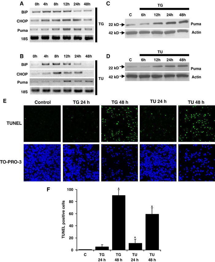
P. Nickson et al. / Cardiovascular Research 73 (2007) 48–56                                   49

the ER sensory proteins PKR-like endoplasmic reticulum                   during ischemia/reperfusion, and that the induction of ER
kinase (PERK), activating transcription factor 6 (ATF6) and              stress genes by ATF6 protected against ischemia/reperfusion
endoribonuclease inositol-requiring enzyme-1 (Ire1) [3–5].               injury [15,16]. These studies indicate that ER stress-induced
When unfolded proteins accumulate in the ER, however, they               apoptosis may be involved in the pathogenesis of a variety of
rapidly occupy the chaperones, and consequently, the ER                  different cardiac diseases, in particular ischemic heart dis-
sensory proteins are released, become activated and launch               ease and heart failure. The role of Bcl-2 family proteins in
the UPR [3–5]. Upon removal of the chaperones, the protein               ER stress-induced apoptosis in cardiomyocytes, however,
kinase PERK oligomerizes, and phosphorylates the eukary-                 has not yet been elucidated.
otic initiation factor 2-alpha (eIF2α), leading to transient                 In a recent paper we described that the proapoptotic Bcl-2
suppression of protein synthesis and decreased ER workload               family protein Puma is critical for ischemia/reperfusion-
[4]. Liberated ATF6 translocates to the Golgi apparatus and is           induced cell death in cardiomyocytes [17]. Since cardiac
cleaved by proteases, resulting in a highly potent transcrip-            ischemia/reperfusion has been demonstrated to induce
tion factor that promotes transcription of ER-resident chap-             ER stress [13,15,16], in the current paper we investigated
erons and folding enzymes [4]. Oligomerized Ire1 results in              whether ER stress requires Puma to promote the apoptotic
production of the active form of X box-binding protein 1                 response. To our knowledge, our experiments demonstrate
(XBP-1), which is another transcription factor involved in               for the first time that Puma is critical for ER stress-dependent
induction of genes responsible for protein folding [5].                  apoptosis in cardiomyocytes. Puma, therefore, may provide a
   The ER also regulates Ca2+ homeostasis by governing the               potential target to block apoptotic cell death in the ischemic
movement of Ca2+ between the ER lumen and the cytosol.                   heart.
Ca2+ exits the lumen through the inositol 1,4,5-triphosphate
receptors and the ryanodine receptors and is actively pumped             2. Methods
back into the ER by the sarcoplasmic/endoplasmic reticulum
Ca2+ ATPases (SERCAs) [2,6]. Since the function of ER-                   2.1. Isolation and culture of primary rat cardiomyocytes
resident chaperones is Ca2+ -dependent, in addition to

                                                                                                                                            Downloaded from by guest on July 24, 2015
accumulation of unfolded proteins, aberrant Ca2+ regulation                 Neonatal cardiac myocytes were prepared using the Percoll
in the ER also leads to activation of the UPR [2,7].                     gradient method, as described earlier [17,18]. Cardiomyocytes
   By coordinating the suppression of global protein synthesis           were cultured for 24 h in a serum containing medium (4:1
and the upregulation of ER-resident chaperones and other                 Dulbecco's modified Eagle's medium (DMEM):medium 199
relevant proteins, the UPR is often able to restore homeostasis,         (M199), 10% heat-inactivated horse serum (HS), 5% heat-
and, therefore, is cytoprotective [4,5,7]. Intense or persistent         inactivated fetal bovine serum (FBS), 100 units/ml penicillin,
ER stress, however, triggers cell death, usually in the form of          100 μg/ml streptomycin, 10 mM glutamine, and 0.2 mM
apoptosis [4,5,7]. The mechanism of ER stress-induced                    bromodeoxyuridine (BrdU)). Following a 24 h incubation
apoptosis has already been extensively investigated and                  period, serum containing medium was replaced with a low
well-known general regulators of the central apoptotic ma-               serum medium (4:1 DMEM:M199, 5% heat inactivated HS,
chinery, including the Bcl-2 family, have been identified to             100 units/ml penicillin, 100 μg/ml streptomycin, 10 mM
play a role in UPR [5,7,8]. While the antiapoptotic Bcl-2-like           glutamine, and 0.2mM BrdU) to prevent hypertrophy.
proteins blocked ER stress-induced apoptosis, both BH3-only
(Bim, BIK and Puma) and multidomain (Bax and Bak)                        2.2. Isolation, culture, and genotyping of primary mouse
proapoptotic Bcl-2 family members were induced or activated              cardiomyocytes
by ER stress [5,7,8]. Importantly, Puma appeared to be critical
for ER stress-induced apoptosis in neuronal cells, mouse                    Cardiomyocytes were isolated from hearts of 1-day-old
embryonic fibroblasts and MCF7 breast carcinoma cells [9–                pups of wild-type and Puma knockout mice, as described
11]. Interestingly, C/EBP homologous protein (CHOP), a                   earlier for rats [17,18], with minor modifications. Briefly,
proapoptotic transcription factor induced by ER stress was also          non-myocyte contaminants were removed by 2 rounds of
suggested to be a regulator of Bcl-2-like proteins [4,5].                pre-plating for 2 h on 35-mm plastic tissue culture dishes in
   Several recent studies have highlighted the importance of             a humidified incubator at 37 °C with 5% CO2. Cardio-
ER stress in cardiac diseases. It was shown that pressure                myocytes from each animal were plated separately into 2
overload by transverse aortic constriction induced prolonged             wells of a 24-well tissue culture plate and cultured in
ER stress during progression from cardiac hypertrophy to                 serum-containing medium. Following a 24 h incubation
heart failure [12]. It was also demonstrated that AMP-ac-                period, serum-containing medium was replaced with low-
tivated protein kinase protected cardiac myocytes against                serum medium to prevent hypertrophy. Following seeding,
hypoxic injury by attenuating ER stress [13]. Consistent with            mice were genotyped using tail DNA as described earlier
these observations, aberrant ER quality control in transgenic            [17,18].
mice with mutant Lys–Asp–Glu–Leu (KDEL) receptor (a                         This investigation conforms with the Guide for the Care and
receptor for ER chaperons) caused dilated cardiomyopathy                 Use of Laboratory Animals published by the US National
[14]. Lastly, it was described that the UPR was activated                Institutes of Health (NIH Publication No. 85-23, revised 1996).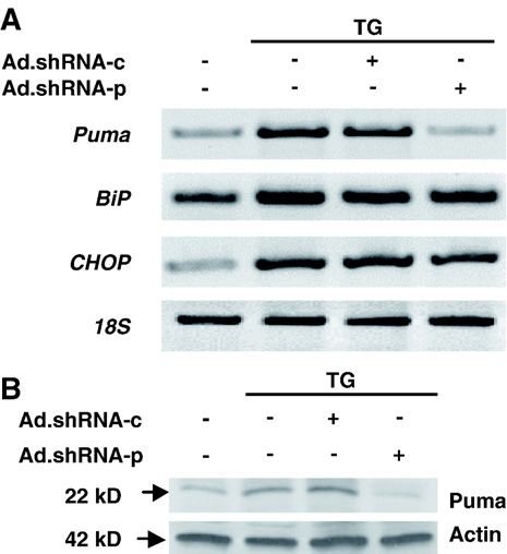
50                                              P. Nickson et al. / Cardiovascular Research 73 (2007) 48–56

2.3. Preparation of adenoviral shRNA (Ad.shRNA)                                     first–designated Ad.shRNA-p–led to a significant decrease
                                                                                    in Puma levels, whereas the second – designated Ad.
   Using the previously described pSuppressorAdeno gene                             shRNA-c – was inactive. The sequence of the inserts in these
suppression system [19], we generated several Ad.shRNA                              recombinant adenoviruses corresponds to two distinct 21
constructs and selected two for further experiments. The                            base pair regions within the Rattus norvegicus Puma cDNA

                                                                                                                                                                    Downloaded from by guest on July 24, 2015

Fig. 1. Thapsigargin and tunicamycin-induced ER stress promotes PUMA expression and apoptosis. A, B, C, and D, thapsigargin (TG) and tunicamycin (TU)-
induced ER stress expression profiles in cardiac myocytes. Cardiomyocytes were grown in tissue culture dishes and exposed to 3 μM thapsigargin or 100 ng
mL− 1 tunicamycin for up to 48 h. A and B, samples were collected at 4, 8, 12, 24, and 48 h time points following the addition of thapsigargin or tunicamycin and
semi-quantitative RT-PCR was performed using oligonucleotides directed against BiP, CHOP, Puma, and 18S as a loading control. C and D, samples were
collected at 6, 12, 24, and 48 h time points after the addition of thapsigargin or tunicamycin. Immunoblot analysis was performed using anti-Puma antibodies.
Actin is shown as a loading control. E, identification of apoptotic cardiac myocytes. Cardiomyocytes were grown on plastic chamber slides and treated with 3 μM
thapsigargin or 100 ng mL− 1 tunicamycin. At 24 and 48 h time points, cells were stained for apoptosis by TUNEL assay (green) and nuclei were identified by
staining with TO-PRO-3 (blue). Images were taken using a confocal fluorescence microscope at 100× magnification. F, quantification of cardiomyocyte
apoptosis. Apoptotic nuclei were scored on the basis of TUNEL positivity as described in E. Data are averaged from three experiments in 10 randomly selected
fields (at least 200 cells) from each group. Error bars represent the S.E. of the mean. ⁎, significant difference from the control ( p b 0.01).
P. Nickson et al. / Cardiovascular Research 73 (2007) 48–56 51
(accession number AY157758.1). Recombinant adeno- 2.7. Semi-quantitative RT-PCR
viruses were generated according to the manufacturer's
instructions (Imgenex Corporation). Briefly, complementary RNA was isolated from cardiomyocytes using Trizol
oligonucleotides were synthesized representing 21-mer reagent (Invitrogen) and processed according to the manu-
sense and anti-sense PUMA sequences with Xho I and facturer's instructions. Reverse transcription was performed
Xba I overhangs, respectively. Sense and anti-sense with the iScript cDNA synthesis kit (BioRad) using 1 μg total
sequences were separated by a short hairpin sequence (5′- RNA, and semi-quantitative PCR was carried out using the
ATCGAT-3′), which encodes a Cla I restriction endonuclease following primers: PUMA 5′-TGGGTGCACTGATGGA
site. shRNA-c encodes a 21-mer sequence complementary to GATA-3′ (sense), 5′-AACCTATGCAATGGGATGGA-3′
R. norvegicus Puma nucleotides 1324–1345 (5′-GAGCA- (anti-sense), BiP 5′-GCCACGGGATGGTTCCTTGCC-3′
TATGAGCCAAACCTGA-3′). shRNA-p encodes a 21-mer (sense), 5′-GCGGATCCAGGTCGACGCCGG-3′ (anti-
sequence complementary to nucleotides 1560–1581 (5′- sense), CHOP 5′-CGGAACCTGAGGAGAGAGTG-3′
CGTGTGACCACTGGCATTCAT-3′). Sense and anti-sense (sense), 5′-CGTTTCCTGGGGATGAGATA-3′ (anti-sense),
oligonucleotides were annealed and the resulting hairpins 18S 5′-CGGCGACGACCCATTCGAAC-3′ (sense), 5′-
cloned into Xho I and Xba I of the shuttle plasmid IMG- GAATCGAACCCTGATTCCCCGTC-3′ (anti-sense).
1200-1. Cloning was confirmed using Cla I digestion.
Shuttle plasmids were then cotransfected into HEK293 cells 2.8. Immunocytochemistry
along with adenovirus backbones for generation of adeno-
viral genomes. Adenoviruses were then amplified in Immunocytochemical analysis was performed as previ-
HEK293 cells and purified as described earlier [17,18,20]. ously described [17,18], with minor modifications. Briefly,
cardiac myocytes were cultured on gelatin-coated 8-well
2.4. Adenovirus infections and induction of ER stress plastic chamber slides and fixed in 3.7% formaldehyde (in
1 × PBS) for 10 min. Cells were then permeabilized with 0.5%
Downloaded from by guest on July 24, 2015
Cardiac myocytes were infected with adenoviruses Triton-X-100 in 1 × PBS for 5 min, incubated in blocking
(multiplicity of infection of 25–50 plaque-forming units/ buffer (5% goat serum and 1% BSA in 1 × PBS) and then
cell) for 2 h, after which the virus-containing medium was probed with primary and secondary antibodies in blocking
replaced with a virus-free medium and cells were buffer. Sections were analyzed by confocal fluorescence
incubated for up to 72 h. ER stress was then induced microscopy (Bio-Rad). Apoptotic cells were detected using
by treating the cells with 3 μM thapsigargin or 100 ng the terminal deoxynucleotidyltransferase-mediated UTP in
mL − 1 tunicamycin for the indicated times, or left situ nick end labeling (TUNEL) method (Roche). Sections
untreated. were costained with anti-sarcomeric actinin (Sigma).
2.5. Cell culture and transfection 2.9. Cell viability assays
MCF7 cells were cultured in DMEM supplemented Cell viability assays were performed using the CellTiter
with 10% heat-inactivated FBS, 100 units/ml penicillin, 96 Aqueous One Solution Cell Proliferation assay, according
100 μg/ml streptomycin, and 10 mM glutamine. Transfec- to the manufacturer's instruction (Promega). Briefly, cells
tion with pCDNA3.0 PUMA (rat) was performed using were cultured on gelatin-coated 96-well plates at a density of
FuGENE 6 (Roche) according to the manufacturer's 8.0 × 10⁴ cells/ml. Following treatment, cells were incubated
instructions. in the presence of the assay reagent at 37 °C for 4 h. Optical
densities were recorded at 490 nm.
2.6. Immunoblot analysis
2.10. Detection of cytosolic cytochrome c
Immunoblot analysis was performed as described earlier
[17,21]. Briefly, cells were lysed in radioimmunoprecipita- Isolation of the cytosolic fraction for the detection of
tion assay (RIPA) buffer complemented with protease in- cytochrome c was performed as previously described [21].
hibitors. Protein samples (10–20 μg) were electrophoresed
in 15% denaturing polyacrylamide gels (BioRad) and then 3. Results
transferred onto nitrocellulose membranes. Membranes
were incubated with primary antibodies specific for 3.1. Induction of ER stress promotes Puma expression and
PUMAα NT (Imgenex), actin (Sigma), cleaved caspase 3 apoptosis in neonatal cardiac myocytes
(Cell Signaling Technology), and cytochrome c (BD
Biosciences), followed by incubation with horseradish To determine whether ER stress-induced apoptosis is
peroxidase-conjugated secondary antibodies. Proteins were mediated by expression of the Puma protein in cardio-
identified using the SuperSignal chemiluminescence system myocytes, isolated cells from neonatal rat hearts were
(Pierce). treated with the SERCA inhibitor thapsigargin or with52 P. Nickson et al. / Cardiovascular Research 73 (2007) 48–56
of apoptotic cell death was recorded for tunicamycin at 48 h
(Fig. 1E–F).
These experiments demonstrate that thapsigargin-induced
Ca+ release and tunicamycin-induced inhibition of ER
protein glycosylation trigger Puma expression and cardio-
myocyte apoptosis in parallel with the induction of ER-
stress-specific chaperones and transcription factors.
3.2. Development of an adenoviral shRNA for knocking
down Puma
Fig. 2. Ad.shRNA-p attenuates both exogenous and endogenous Puma To further investigate the importance of Puma in ER
expression. A, validation of shRNA by transient transfection. MCF7 cells stress-induced apoptosis of cardiac myocytes, we developed
plated at a density of 1.5 × 10⁶ cells were infected with either the pSup-
recombinant adenoviruses encoding anti-Puma short hairpin
pressorAdeno (non-recombinant virus), Ad.shRNA-c, or Ad.shRNA-p and
incubated for 48 h. Cells were then transfected with pCMS-Puma and RNA (shRNA) to suppress Puma mRNA levels. Several
incubated for an additional 24 h—a total of 72 h. Immunoblot analysis was adenoviral constructs were generated and tested for anti-
performed using anti-Puma antibody. The position of exogenous FLAG- Puma activity. Two of these adenoviral shRNAs were se-
tagged Puma band is indicated by an arrow. Actin is shown as a loading lected and designated as Ad.shRNA-p and Ad.shRNA-c,
control. Results are representative of three independent experiments. B,
based on their ability to knock down Puma. The efficacy of
validation of shRNA in cardiac myocytes. Cells were grown in tissue culture
dishes and infected with either Ad.shRNA-c or Ad.shRNA-p for 72 h. these constructs was validated by analyzing exogenous and
Immunoblot analysis was performed using anti-Puma antibody. Actin is endogenous Puma expression.
shown as a loading control. Results are representative of three independent Ad.shRNAs were initially tested against exogenous Puma
experiments. using MCF7 cells transfected with plasmids carrying the rat
Downloaded from by guest on July 24, 2015
puma cDNA (pCMS-puma). While Ad.shRNA-p efficiently
decreased the rat Puma protein levels, Ad.shRNA-c did not
tunicamycin, which blocks ER protein glycosylation. Both evoke any significant effect (Fig. 2A). To analyze the efficacy
thapsigargin and tunicamycin were earlier shown to induce of the Ad.shRNA in cardiomyocytes, we infected isolated rat
ER stress in cardiac cells [12]. We then followed the ER neonatal cardiac myocytes with different concentrations of
stress response by analyzing the mRNA levels of two ER shRNA adenoviruses and analyzed endogenous Puma
stress-specific proteins, the chaperone BiP and the tran- protein expression levels. Consistent with the data obtained
scription factor CHOP, using RT-PCR. Following treat- from MCF7 cells, endogenous Puma expression levels were
ment with thapsigargin and tunicamycin, both BiP and
CHOP were induced after 4 h of treatment, reached maximal
levels at 12 h, and receded back to basal levels by 24 or 48 h
(Fig. 1A–B).
Following the induction of the ER-stress response, Puma
mRNA was also upregulated, albeit delayed in comparison
with BiP and CHOP, with the first signs of induction
observed at 8 h and maximal induction at 12 h (Fig. 1A–B).
By 48 h, Puma mRNA levels almost returned to the control
level (Fig. 1A–B). This transcriptional Puma induction was
accompanied by a corresponding increase in Puma protein
levels (Fig. 1C–D). However, while Puma mRNA levels
were barely detectable at 48 h, Puma protein was still
highly elevated (Fig. 1C–D). Interestingly, the kinetics of
Puma expression were comparable for both ER stress
activators.
Since Puma is a proapoptotic member of the Bcl-2 family,
we next determined whether Puma induction was followed
by concomitant increase in apoptosis. As expected, elevated Fig. 3. Ad.shRNA-p inhibits Puma expression during thapsigargin-induced
Puma expression was accompanied by apoptosis, as ER stress in cardiac myocytes. Cardiomyocytes were grown in tissue culture
discerned using TUNEL assay. After 24 h of thapsigargin dishes and infected with either Ad.shRNA-c or Ad.shRNA-p for 72 h. Cells
treatment, less than 10% of the cells were TUNEL positive. were then exposed to 3 μM thapsigargin for an additional 12 h. A, semi-
quantitative RT-PCR was performed using oligonucleotides directed against
At the 48 h time-point, however, the majority of cells were BiP, CHOP, Puma, and 18S as a loading control. B, immunoblot analysis
apoptotic. A similar pattern was observed following was performed using anti-Puma antibodies. Actin is shown as a loading
tunicamycin treatment, however, overall, a lower percentage control.P. Nickson et al. / Cardiovascular Research 73 (2007) 48–56 53
reduced in a concentration-dependent manner when cardio- These data indicate that Ad.shRNA-p efficiently down-
myocytes were infected with Ad.shRNA-p (Fig. 2B). In regulates Puma levels whereas Ad.shRNA-c is ineffective. In
contrast, Ad.shRNA-c had no effect on endogenous rat Puma later experiments these two viruses were used either to knock
expression (Fig. 2B). down Puma levels or as a negative control virus, respectively.
Downloaded from by guest on July 24, 2015
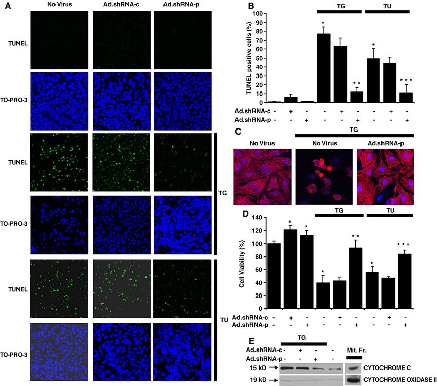
Fig. 4. Suppression of Puma attenuates thapsigargin and tunicamycin-induced apoptosis in cardiac myocytes. A, Ad.shRNA-p reduces thapsigargin and
tunicamycin-induced apoptosis. Cardiomyocytes were grown in tissue culture dishes and infected with either Ad.shRNA-c or Ad.shRNA-p for 48 h. Cells were
then exposed to 3 μM thapsigargin or 100 ng mL− 1 tunicamycin for an additional 48 h. Cells were stained for apoptosis by TUNEL assay (green) and nuclei were
identified by staining with TO-PRO-3 (blue). Images were taken using a confocal fluorescence microscope at 100× magnification. B, quantification of
cardiomyocyte apoptosis. Apoptotic nuclei were scored on the basis of TUNEL positivity as described in A. Data are averaged from three experiments in 10
randomly selected fields (at least 200 cells) from each group. Error bars represent the S.E. of the mean. ⁎, significant difference from the control ( p b 0.01). ⁎⁎,
significant difference from the TG group ( p b 0.01). ⁎⁎⁎, significant difference from the TU group ( p b 0.01). C. High magnification micrographs of
cardiomyocytes subjected to treatment with TG, TG plus Ad.shRNA-p, or left untreated, as described in A. Cells were stained with anti-sarcomeric actinin (red)
and TO-PRO-3 (blue). Images were taken using a confocal fluorescence microscope at 400× magnification. D, quantification of cardiomyocyte viability.
Cardiomyocytes were grown in 96-well tissue culture plates and were infected with either Ad.shRNA-c or Ad.shRNA-p for 48 h. Cells were then exposed to
3 μM thapsigargin or 100 ng mL− 1 tunicamycin for an additional 48 h. Cell viability was then assessed using the CellTiter 96 Aqueous One solution. Optical
densities were recorded at 490 nm. Data are averaged from 3 independent experiments, each comprising 5 independent replicates. Error bars represent the S.E of
the mean. ⁎, significant difference from the control ( p b 0.01). ⁎⁎, significant difference from the TG group ( p b 0.01). ⁎⁎, significant difference from the TU
group ( p b 0.01). E. Ad.shRNA-p prevents thapsigargin-induced cytochrome c release into the cytosol. Cardiomyocytes were grown and treated as described in
A. Equal amounts of cytosolic proteins were immunoblotted for cytochrome c and cytochrome oxidase subunit 2 (as control), along with similar amounts of
mitochondrial fractions of non-treated cells (Mit.Fr.).54 P. Nickson et al. / Cardiovascular Research 73 (2007) 48–56
3.3. Inhibition of thapsigargin-induced Puma expression by These data were corroborated by analyzing the amount of
adenoviral delivery of shRNA in cardiac myocytes cytochrome c in the cytosol. We detected a small amount
of cytochrome c in the cytosol, probably due to partial
We next examined whether shRNA-p is able to effectively disruption of mitochondria during cell fractionation
suppress the induction of Puma mRNA and protein by (Fig. 4E). Cytochrome c release was significantly induced
thapsigargin. Cardiac myocytes were infected with shRNA upon thapsigargin treatment or if thapsigargin was
adenoviruses for 72 h prior to thapsigargin treatment to allow combined with infection with Ad.shRNA-c (Fig. 4E). In
preloading of cytoplasmic shRNA. Cells were then exposed contrast, Ad.shRNA-p effectively decreased cytosolic
to thapsigargin for 12 h and collected for mRNA and protein cytochrome c to a level comparable to the control
analysis. Using RT-PCR we found that Ad.shRNA-p, but not (Fig. 4E). Collectively, these data provide evidence that
Ad.shRNA-c, inhibited puma mRNA accumulation induced Puma is a critical factor for ER stress-induced apoptosis in
by thapsigargin (Fig. 3A). This effect was specific, because cardiomyocytes, because downregulation of Puma blocks
neither BiP, nor CHOP mRNA levels were affected in cardiomyocyte apoptosis.
identical samples (Fig. 3A). Consistent with the mRNA
changes, induction of the Puma protein by thapsigargin was 3.5. ER stress-induced apoptosis is inhibited in Puma−/−
blocked only by shRNA-p, whereas shRNA-c did not have neonatal mouse cardiac myocytes
any effect (Fig. 3B).
In summary, the shRNA-p virus specifically blocks To corroborate that Puma is a critical factor for ER stress-
expression of the Puma mRNA and protein upon thapsi- induced apoptosis in cardiac myocytes, we compared the
gargin induction. sensitivity of isolated mouse wild-type, puma+/− and puma−/−
cardiomyocytes to thapsigargin treatment. Based on data of
3.4. Inhibition of thapsigargin-induced apoptosis in cardiac Fig. 1, isolated cells were exposed to 3 μM thapsigargin for
myocytes 48 h and then assayed for caspase-3 cleavage and Puma
Downloaded from by guest on July 24, 2015
expression by immunoblotting. Following the induction of ER
To determine if Puma is essential for ER-stress-induced stress, we observed a significant increase in the proteolytic
cardiomyocyte apoptosis, we treated control and Ad.shRNA cleavage of caspase-3 in samples obtained from both wild-type
infected cells with thapsigargin and tunicamycin for 48 h, at and heterozygous animals (Fig. 5). In contrast, in puma−/−
which time-point we identified widespread apoptosis in cardiomyocytes, thapsigargin treatment did not affect the
earlier experiments (Fig. 1E–F). Subsequently, the rate of cleavage of caspase-3 (Fig. 5). Consistent with the genotypes,
apoptosis was measured with TUNEL assay. In control cells the basal level of Puma was detected only in puma+/+
or Ad.shRNA infected cells we observed less than 5% cardiomyocytes, where Puma was strongly induced by
apoptosis (Fig. 4A–B). In contrast, more than 70% of the thapsigargin (Fig. 5). Basal Puma expression was not detected
cells exposed to thapsigargin underwent apoptosis, and this in heterozygous cells, but was increased following thapsigar-
was not significantly inhibited by Ad.shRNA-c (Fig. 4A–B). gin treatment, albeit at a lower level than in puma+/+ cells
Importantly, Ad.shRNA-p, which was shown to decrease (Fig. 5). Finally, in puma−/− cardiomyocytes, Puma was not
Puma expression under several different conditions detected, independently of the thapsigargin treatment (Fig. 5).
(Figs. 2 and 3), reduced apoptosis to approximately 10% These data together confirm that Puma is a critical factor
(Fig. 4A–B). Similarly, close to 50% of the cells exposed for ER stress-induced cardiomyocyte apoptosis. They also
to tunicamycin underwent apoptosis, and again, this re- suggest that reaching a threshold in Puma levels is sufficient
sponse was inhibited by Ad.shRNA-p to approximately to induce apoptosis, resulting in similar sensitivity of wild-
10% (Fig. 4A–B). Ad.shRNA-c had no effect. Of note, type and heterozygote cardiomyocytes to ER stress-induced
representative high magnification micrographs of healthy apoptosis. Importantly, this phenomenon was also seen by
(no virus), dead (TG) and rescued (Ad.shRNA-p) cardiac
myocytes stained by alpha-actinin and TO-PRO3 are
shown in Fig. 4C.
To ascertain the viability of the TUNEL negative cells, we
determined the number of viable cells following the various
treatments, using a Promega kit (see Methods). In thapsi-
gargin treated cells, the number of viable cells decreased to
less than half of the control and this was unaffected by the
presence of Ad.shRNA-c, whereas the viability of Ad.
shRNA-p treated cells remained close to 100% (Fig. 4D). Fig. 5. Targeted deletion of Puma inhibits thapsigargin-induced apoptosis.
Importantly, a similar pattern of viability changes was Cardiomyocytes isolated from wild-type (n = 5), puma+/− (n = 4), and Puma−/−
(n = 3) mice were grown in tissue culture dishes and exposed to 3 μM
observed upon tunicamycin treatment (Fig. 4D). In summa- thapsigargin for 48 h, or left untreated. Immunoblot analysis was performed
ry, the viability data shown in Fig. 4D are consistent with the using cleaved capase 3 and Puma antibodies. Actin is shown as a loading
TUNEL assay measurements presented in Fig. 4 A–B. control. Data are representative of at least three independent experiments.P. Nickson et al. / Cardiovascular Research 73 (2007) 48–56 55
other investigators earlier in other cell types using different [4,5]. The hypothesis that CHOP activates the Puma gene
apoptosis inducers [27]. in cardiomyocytes exposed to ER stress is now under
investigation in our laboratory.
4. Discussion ER stress-induced cell death appears to be especially
important in the heart where increased protein synthesis
In a recent study, we demonstrated that cardiomyocytes triggered by hypertrophy of cardiomyocytes may overload
with Puma deletion acquire resistance to ischemia/reperfu- the ER chaperone system, leading to cell death. This may
sion-induced cell death [17]. Since ischemia/reperfusion further aggravate cardiac remodeling, eventually culminat-
causes protein misfolding and consequent ER stress [15,16], ing in dilatation of the heart and heart failure. In fact,
in our current study we aimed to determine whether the ER recent papers have described that ER stress occurs in the
stress component of ischemia/reperfusion injury is also heart upon ischemia/reperfusion, which is suggested to
dependent on Puma activity in cardiac myocytes. There are contribute to the pathogenesis of heart failure [12–16]. Our
three well-characterized experimental ways to provoke ER current paper provides evidence that Puma is an essential
stress by pharmacological inducers. These include agents component of the ER stress pathway in cardiac myocytes,
that interfere with the function of ER-resident glycosylases, because suppressed Puma expression leads to inhibition of
such as tunicamycin, that inhibit the formation of disulfide experimentally induced ER stress. Similarly, we showed
bonds, such as dithiothreitol, and that lower calcium levels in earlier that ischemia/reperfusion-induced cell death also
the ER, such as thapsigargin [3]. In these studies, we used the required Puma [17]. Together these studies strongly
SERCA inhibitor thapsigargin, which promotes leakage of suggest that Puma provides a novel therapeutic target to
Ca2+ from the ER [22] and, thereby, blocks the function of block apoptosis associated with ischemic heart disease and
Ca 2+ -dependent ER-resident chaperons, including BiP heart failure. In addition, inhibition of Puma may improve
(Grp78), Grp94 and calreticulin [4]. We have also applied the viability of transplanted cells used for cardiac cell
tunicamycin, which induces accumulation of misfolded therapy.
Downloaded from by guest on July 24, 2015
proteins and ER stress without directly causing cytoplasmic
Ca2+ overload. Acknowledgements
We have demonstrated with multiple assays that Puma is a
critical component of ER stress-induced apoptosis in The authors are grateful to Dr. Noriaki Ikemoto (Boston
cardiomyocytes. Our data provide evidence that Puma is Biomedical Research Institute) for critically reading the
transcriptionally activated during ER stress and that its manuscript, to Dr. Subhas Biswas (Columbia University)
expression is clearly necessary for the development of for the rat puma plasmid, and to Drs. John Jeffers and
apoptosis. However, our studies do not identify the tran- Gerald Zambetti (St. Jude Children's Hospital) for the
scription factor responsible for ER stress-related induction of Puma knockout mice. This work was supported by grants
Puma. Puma was originally described as a p53-inducible from NIH (HL-68126 to P.E.), from NSF (MCB-9982789 to
target gene [23,24]. Despite this, it is less likely that in- P.E.), and by an American Heart Association fellowship
duction of Puma during ER stress of cardiac myocytes is (to A.T.).
p53-dependent for several reasons. Firstly, in cardiac
myocytes hypoxia/reoxygenation-induced apoptosis
References
requires Puma [17], but not p53 [25,26]. Secondly, Puma
is known to be activated in a p53-independent manner by [1] Berridgem M. The endoplasmic reticulum: a multifunctional signaling
several inducers, including cytokine removal, dexametha- organelle. Cell Calcium 2002;32:235–49.
sone and serum starvation [27–29]. Finally, in neuronal cells [2] Wehrens X, Lehnart S, Marks A. Intracellular calcium release and
and in embryonic mouse fibroblasts, the induction of Puma cardiac disease. Annu Rev Physiol 2005;67:69–98.
[3] Stavridi E, Halazonetis T. p53 and stress in the ER. Genes Dev
by ER stress does not depend on p53 [9–11]. Moreover, in
2004;18:241–4.
recent studies, p53 was inhibited, not activated by ER stress, [4] Xu C, Bailly-Maitre B, Reed J. Endoplasmic reticulum stress: cell life
probably through a glycogen synthase kinase-3-dependent and death decisions. J Clin Invest 2005;115:2656–64.
mechanism [30,31]. Our experiments rather suggest that [5] Boyce M, Yuan J. Cellular response to endoplasmic reticulum stress: a
CHOP could be responsible for ER stress-induced Puma matter of life or death. Cell Death Differ 2006;13:363–73.
[6] Diaz M, Graham H, O neill S, Trafford A, Eisner D. The control of
activation, because CHOP activation precedes Puma activa-
sarcoplasmic reticulum Ca content in cardiac muscle. Cell Calcium
tion in cardiac myocytes (Fig. 1). In addition, upon CHOP 2005;38:391–6.
overexpression, induction of apoptosis can be inhibited by [7] Crow M, Mani K, Nam Y, Kitsis R. The mitochondrial death pathway
Bcl-2 [4,5], suggesting that CHOP activates a proapoptotic and cardiac myocyte apoptosis. Circ Res 2004;95:957–70.
Bcl-2 family member, potentially Puma. Consistent with this [8] Foo R, Mani K, Kitsis R. Death begets failure in the heart. J Clin Invest
2005;115:565–71.
hypothesis, the CHOP promoter contains binding sites for all
[9] Rao R, Ellerby H, Bredesen D. Coupling endoplasmic reticulum stress
major inducers of the ER stress response, such as ATF4, to the cell death program. Cell Death Differ 2004;11:372–80.
ATF6 and XBP-1. Finally, CHOP is activated by two well- [10] Reimertz C, Kgel D, Rami A, Chittenden T, Prehn J. Gene expression
known proapoptotic proteins, p38-MAP kinase and JNK during ER stress-induced apoptosis in neurons: induction of the BH3-56 P. Nickson et al. / Cardiovascular Research 73 (2007) 48–56
only protein Bbc3/PUMA and activation of the mitochondrial [20] Anderson R, Haskell R, Xia H, Roessler B, Davidson B. A simple
apoptosis pathway. J Cell Biol 2003;162:587–97. method for the rapid generation of recombinant adenovirus vectors.
[11] Li J, Lee B, Lee A. Endoplasmic reticulum stress-induced apoptosis: Gene Ther 2000;7:1034–8.
multiple pathways and activation of p53-up-regulated modulator of [21] Erhardt P, Schremser E, Cooper G. B-Raf inhibits programmed cell
apoptosis (PUMA) and NOXA by p53. J Biol Chem 2006;281:7260–70. death downstream of cytochrome c release from mitochondria by
[12] Okada K, Minamino T, Tsukamoto Y, Liao Y, Tsukamoto O, Takashima activating the MEK/Erk pathway. Mol Cell Biol 1999;19:5308–15.
S, et al. Prolonged endoplasmic reticulum stress in hypertrophic and [22] Inesi G, Sagara Y. Specific inhibitors of intracellular Ca2+ transport
failing heart after aortic constriction: possible contribution of ATPases. J Membr Biol 1994;141:1–6.
endoplasmic reticulum stress to cardiac myocyte apoptosis. Circulation [23] Yu J, Zhang L, Hwang P, Kinzler K, Vogelstein B. PUMA induces the
2004;110:705–12. rapid apoptosis of colorectal cancer cells. Mol Cell 2001;7:673–82.
[13] Terai K, Hiramoto Y, Masaki M, Sugiyama S, Kuroda T, Hori M, et al. [24] Nakano K, Vousden K. PUMA, a novel proapoptotic gene, is induced
AMP-activated protein kinase protects cardiomyocytes against hyp- by p53. Mol Cell 2001;7:683–94.
oxic injury through attenuation of endoplasmic reticulum stress. Mol [25] Bialik S, Geenen D, Sasson I, Cheng R, Horner J, Evans S, et al.
Cell Biol 2005;25:9554–75. Myocyte apoptosis during acute myocardial infarction in the mouse
[14] Hamada H, Suzuki M, Yuasa S, Mimura N, Shinozuka N, Takada Y, localizes to hypoxic regions but occurs independently of p53. J Clin
et al. Dilated cardiomyopathy caused by aberrant endoplasmic Invest 1997;100:1363–72.
reticulum quality control in mutant KDEL receptor transgenic mice. [26] Webster K, Discher D, Kaiser S, Hernandez O, Sato B, Bishopric N.
Mol Cell Biol 2004;24:8007–17. Hypoxia-activated apoptosis of cardiac myocytes requires reoxygenation
[15] Martindale J, Fernandez R, Thuerauf D, Whittaker R, Gude N, or a pH shift and is independent of p53. J Clin Invest 1999;104:239–52.
Sussman M, et al. Endoplasmic reticulum stress gene induction and [27] Jeffers J, Parganas E, Lee Y, Yang C, Wang J, Brennan J, et al. Puma is
protection from ischemia/reperfusion injury in the hearts of an essential mediator of p53-dependent and -independent apoptotic
transgenic mice with a tamoxifen-regulated form of ATF6. Circ pathways. Cancer Cells 2003;4:321–8.
Res 2006;98:1186–93. [28] Han J, Flemington C, Houghton A, Gu Z, Zambetti G, Lutz R, et al.
[16] Azfer A, Niu J, Rogers L, Adamski F, Kolattukudy P. Activation of Expression of bbc3, a pro-apoptotic BH3-only gene, is regulated by
endoplasmic reticulum stress response during the development diverse cell death and survival signals. Proc Natl Acad Sci U S A
of ischemic heart disease. Am J Physiol Heart Circ Physiol 2001;98:11318–23.
2006;291:1411–20. [29] Villunger A, Michalak E, Coultas L, Mllauer F, Bock G, Ausserlechner
[17] Toth A, Jeffers J, Nickson P, Min J, Morgan J, Zambetti G, et al. M, et al. p53-and drug-induced apoptotic responses mediated by BH3-
Downloaded from by guest on July 24, 2015
Targeted deletion of puma attenuates cardiomyocyte death and only proteins puma and noxa. Science 2003;302:1036–8.
improves cardiac function during ischemia/reperfusion. Am J Physiol [30] Pluquet O, Qu L, Baltzis D, Koromilas A. Endoplasmic reticulum
Heart Circ Physiol 2006;291:52–60. stress accelerates p53 degradation by the cooperative actions of Hdm2
[18] Toth A, Nickson P, Qin L, Erhardt P. Differential regulation of and glycogen synthase kinase 3beta. Mol Cell Biol 2005;25:9392–405.
cardiomyocyte survival and hypertrophy by MDM2, an E3 ubiquitin [31] Qu L, Koromilas A. Control of tumor suppressor p53 function by
ligase. J Biol Chem 2006;281:3679–89. endoplasmic reticulum stress. Cell Cycle 2004;3:567–70.
[19] Xia H, Mao Q, Paulson H, Davidson B. siRNA-mediated gene
silencing in vitro and in vivo. Nat Biotechnol 2002;20:1006–10.You can also read