TRANSFORMING GROWTH FACTOR BETA HAS DUAL EFFECTS - SciELO Colombia
←
→
Page content transcription
If your browser does not render page correctly, please read the page content below
Facultad de Ciencias
ACTA BIOLÓGICA COLOMBIANA Departamento de Biología
http://www.revistas.unal.edu.co/index.php/actabiol Sede Bogotá
ARTÍCULO DE INVESTIGACIÓN / RESEARCH ARTICLE BIOQUÍMICA
TRANSFORMING GROWTH FACTOR BETA HAS DUAL EFFECTS
ON MMP9 AND uPA EXPRESSION IN HTR-8/SVneo HUMAN
TROPHOBLASTIC CELL LINE
El factor de crecimiento transformante beta tiene efecto dual en
la expresión de MMP9 y uPA en la línea celular de trofoblasto
HTR-8/SVneo
Sandra Susana NOVOA HERRÁN1, Mariela CASTELBLANCO1, Myriam SÁNCHEZ -GÓMEZ1, Adriana UMAÑA PÉREZ1*
1
Universidad Nacional de Colombia–Sede Bogotá, Facultad de Ciencias, Departamento de Química, Grupo de investigación en
Hormonas. Cra 30 n°. 45-03 Ed. 451 Of. 464, Bogotá D.C., Colombia.
*For correspondence. yaumanap@unal.edu.co
Received: 20th December 2017, Returned for revision: 25th August 2018, Accepted: 17th October 2018.
Associate Editor: Argel Aguilar Valles.
Citation/Citar este artículo como: Novoa Herran SS, Castelblanco M, Sanchez-Gomez M, Umaña-Perez A. Transforming Growth Factor Beta Has
Dual Effects on MMP9 and uPA Expression in HTR-8/SVneo Human Trophoblastic Cell Line. Acta biol. Colomb. 2019;24(1):26-37. DOI: http://dx.doi.
org/10.15446/abc.v24n1.69527
ABSTRACT
Invasion of trophoblast into endometrium is vital for successful pregnancy development. MMP9 and uPA are key proteases in this
process, but it is still not clear the regulation of its expression by Transforming Growth Factor Beta (TGF-), a known negative
regulator of trophoblast invasion. We evaluated the effect of TGF- on the transcriptional expression of uPA and MMP9 over time,
in HTR-8/SVneo trophoblast cells cultured with or without 0.5 % fetal bovine serum, via RT qPCR. The involved transcription factors
and signaling pathways were analyzed in silico, using Proscan, Enrich, PCViz and WikiPathway. Results showed that TGF- temporarily
regulates the expression of uPA and MMP9. Serum modified the nature of TGF-’s effects on uPA expression, from negative without
serum to positive with it, showing opposite effects on MMP9 expression. In silico analysis evidenced different transcription factors
for each protease, some belonging to TGF- signaling pathway, and crosstalk with MAPK and Wnt/-catenin pathways. The TGF-
dual role is discussed proposing that serum affects the cellular context. Transcriptional regulation of MMP9 and uPA by TGF- is
differential and depends on serum presence and evaluation time.
Keywords: Computational biology, matrix metalloproteinase, placenta, plasminogen, polymerase chain reaction.
RESUMEN
La invasión del trofoblasto al endometrio es vital para el correcto desarrollo del embarazo. Las proteasas MMP9 y uPA son claves en
este proceso, pero aún no es clara la regulación de su expresión por parte del Factor de Crecimiento Transformante beta (TGF-),
conocido por sus acciones no invasivas sobre el trofoblasto. En este trabajo evaluamos el efecto del TGF- sobre la expresión
transcripcional de uPA y MMP9 en células de la línea de trofoblasto HTR-8/SVneo cultivadas con o sin suero fetal bovino al 0,5 %,
mediante RT qPCR. Se analizaron in sillico los potenciales factores de transcripción y vías de señalización involucradas empleando
Proscan, Enrich, PCViz y WikiPathway. Los resultados muestran que el TGF- regula temporalmente la expresión de uPA y MMP9. El
suero modificó la naturaleza del efecto del TGF- sobre la expresión de uPA, de negativo en ausencia de suero a positivo en presencia
de este, presentando efectos opuestos para la expresión de MMP9. El análisis in sillico evidenció diferentes factores de transcripción
para cada proteasa, algunos pertenecientes a la vía de señalización del TGF-, y un entrecruzamiento con la vía MAPK y Wnt/-
catenina. Los resultados sugieren que la regulación transcripcional de MMP9 y uPA por parte del TGF- es diferencial y depende de
la presencia de suero y tiempo de evaluación.
Palabras clave: Biología computacional, metaloproteinasa de matrix, placenta, plasminógeno, reacción en cadena polimerasa.
26 - Acta biol. Colomb., 24(1):26-37, Enero- Abril 2019
DOI: http://dx.doi.org/10.15446/abc.v24n1.69527TGF- Dual Effects on MMP-9 and UPA Expression in HTR-8/SVneo
INTRODUCTION more recent works show that TGF- promotes invasion of
Extravillous cytotrophoblast (EVT) is an integral tissue of the HTR-8/SVneo cells in advanced culture passages, involving
placenta which invades the uterine endometrium and the expression changes in members of its signaling pathway
maternal spiral arteries, remodeling them (Lunghi et al., (Yanzhen et al., 2014). Furthermore, TGF-1 upregulated
2007; He et al., 2017). An adequate EVT invasion is vital cadherin-11 expression in HTR-8/SVneo through activating
for blastocyst implantation and successful pregnancy SMAD2 and SMAD3 signaling pathways inducing epithelial
development. When this process is modified, some to mesenchymal transition (Cheng et al., 2018). This
pathologies occur such as preeclampsia, intrauterine apparent contradiction in the effect of TGF- on protease
growth restriction, and gestational trophoblastic expression and cellular invasion of the trophoblast requires
diseases. This invasive process requires the expression additional studies that consider the expression over time
and activation of extracellular proteases, highlighting and in vitro culture conditions, specifically the treatment in
matrix metalloproteinases (MMPs) and urokinase-type presence or absence of fetal bovine serum (FBS).
plasminogen activator (uPA). An increase in the expression In previous studies with HTR-8/SVneo cells cultured
of MMPs and uPA is a uniform phenomenon in diverse for 24 hours with or without fetal bovine serum (0 %, 5 %,
cancer types, and it is correlated with tumor progression and 10 %), we have seen a change in protein profiles which
and metastasis (Friedl and Wolf, 2003; Turunen et al., 2017; suggests a metabolic adaptation due to serum absence
Mahmood et al., 2018). Amongst MMPs, MMP2 and nine (Novoa-Herran et al., 2016).
have been the most characterized enzymes in cancer cases, Additionally, it has been seen that the effects of IGF-I and
and MMP9 plays a relevant role in invasive trophoblast insulin on the invasion of cellular trophoblast models are
processes (Staun-Ram et al., 2004; Cohen et al., 2006; Zhang serum-dependant (Mandl et al., 2002).
et al., 2018). On the other side, it has been demonstrated Considering the above, it is relevant to study the effects
that the activator system of plasminogen uPA plays an
of TGF- on the expression of MMP9 and uPA in different
important role in the regulation of invasion and migration
culture conditions and over time, since expression of these
of trophoblast (Lala and Chakraborty, 2003; Zheng et
proteases is an inflection point in the regulation of EVT
al., 2018). Even though EVT cells behave as metastatic
invasion. In this work, transcriptional expression of uPA
(Ferretti et al., 2007), their invasion in vivo is transitory,
and MM9, as a response to different doses of TGF-, was
spatially confined and highly regulated by a plethora of
evaluated by means of quantitative PCR in HRT-8/SVneo
factors (Cohen et al., 2006; Menkhorst et al., 2016; Li and
cell line, in presence or absence of 0.5 % FBS and over
Shao, 2017), including the transforming growth factor
time; possible transcription factors and involved signaling
beta (TGF-), which has negative effects on trophoblast
pathways as well, were identified by a bioinformatic analysis.
proliferation, migration and invasion (Chakraborty et al.,
2002; Lunghi et al., 2007; Yi et al., 2018) and promote its
MATERIALS AND METHODS
differentiation (Cheng et al., 2018).
TGF- causes a significant reduction in secretion
Cell line and culture conditions
and activity of uPA, and induces the expression and
secretion of its inhibitor PAI-1 and the tissue inhibitor The immortalized cell line of human extravillous trophoblast
of metalloproteases TIMP-1 and -2, which inhibit the HTR-8/SVneo was donated by Dr. Ángela Cadavid
extracellular matrix degradation performed by the MMPs (Universidad de Antioquia, Colombia). This cell line was
(Lala et al., 1998). It has been seen that TGF- reduces the developed from a culture of first-trimester human placenta
expression and secretion of MMP9 in primary cultures of explant and was immortalized by transfection with a cDNA
trophoblast isolated from first-trimester placenta (Meisser that codes for the simian virus 40 (SV40) large T-antigen
et al., 1999), in explants of first-trimester placenta (Lash et (Graham et al., 1993). These cells exhibit a high proliferation
al., 2005), in the choriocarcinoma cell line JEG-3 (Karmakar index and share several phenotypic similarities with parental
and Das, 2002) and in the trophoblast cell line NPC (Zhao HTR-8 cells, including invasive abilities in vitro. Cells were
et al., 2006). Nevertheless, other studies in primary cultures manipulated from passage 15 to 20 (own records) and
of the first-trimester cytotrophoblast have demonstrated cultured at 37 °C in humidified atmosphere with 5 % CO2
that TGF- stimulates expression and secretion of MMP9 in RPMI 1640 medium (Sigma Chemical Co.) supplemented
(Shimonovitz et al., 1996). The immortalized first-trimester with 10 % FBS (Gibco, Invitrogen), 2 mM L-glutamine
EVT cell line HTR-8/SVneo was established and described (Gibco, Invitrogen), and 40 µg/mL gentamicin (Genfar).
as a model that partially responds to the regulatory actions Cells were seeded in 60 mm culture dishes and cultured in
of TGF-, with a decrease in secretion and activity of uPA supplemented medium for one to two days until 60-80 %
without affecting the invasion level (Graham et al., 1993), confluency was achieved, were starved overnight in serum-
being proposed as a model for the study of human placental free medium, after that stimulus was performed in the
function and tumor progression (Lala et al., 2002). However, mentioned doses and times.
Acta biol. Colomb., 24(1):26-37, Enero- Abril 2019 - 27Sandra Susana Novoa Herrán, Mariela Castelblanco, Myriam Sánchez-Gómez, Adriana Umaña Pérez
Treatment CTCACACTTACACTCACAGC; Hrs-18S: D: ATGTGGTGTTGA
Cells were stimulated with transforming growth factor GGAAAGC, R: TACTGGCGTGGATTCTGC; -actin: D: GCG TGA
isoform 1 (hBA-112, Santa Cruz Biotechnology Inc., Receptor CATTAAGGAGAAG,R:GAAGGAAGGCTGGAAGAG. Forty cycles
grade) in a concentration of 10 ng/mL and for 0, 6, 12, 18 were performed with 10 s at 95 °C, 20 s at 56 °C and 20 s
and 24 hours. Additionally, TGF-1 in concentrations of 0.1, at 72 °C. The fluorescence threshold (Ct) was calculated with
1, 5, 10 and 20 ng/mL was used. Different concentrations the software CFX Manager version 1.6 (BioRad Laboratories
up to 20 ng/mL as indicated for each experiment. These Inc) and a melting curve was made once the last amplification
treatments were performed in serum-free RPMI medium or cycle finished as a criterion to detect unspecificity and dimer
concomitant with 0. 5% FBS using serum-free RPMI medium formation. Each determination was performed by triplicate,
or with 0.5 % FBS as control, respectively. obtaining the relative expression normalized to genes 18S and
-actin used as reference genes, accordingly to the data treatment
RNA extraction and error propagation suggested by Hellemans et al., (2007).
Each culture dish was treated with 1 mL of TRIzol reagent Finally, the normalized relative expression (NRQ) and the
(Invitrogen, USA) according to the instructions of the obtained standard deviation (SD) was re-scaled, normalizing the
manufacturer. The cells were lysed with the reagent, data to each biological control and following the propagation of
generating a monophasic solution composed of phenol- error formulas.
guanidium isothiocyanate, and extracting total RNA in the
aqueous phase by phase-partitioning with chloroform. RNA Statistical analysis
was precipitated with isopropanol, washed with 75 % ethanol For simple comparisons between treatments and controls,
in RNase-free water and solubilized in 30 µL of RNase-free unpaired Two-tailed T-test was used. For comparison
water. The concentration and purity of the obtained RNA between treatments and times or stimuli doses a two-
were determined by spectrophotometry (Pharmacia Biotech way analysis of variance (ANOVA) was performed, and
Ultraspec, 2000) and its integrity was verified by agarose gel Bonferroni test for subsequent analysis (biological replicate,
electrophoresis. n=3, pTGF- Dual Effects on MMP-9 and UPA Expression in HTR-8/SVneo
order to detect intermediaries that could mediate functional (Fig. 1 C; 10 and 20 ng/mL at 24 h) and with serum (Fig. 1 D;
relationships between the cytokine and proteases. 1 and 20 ng/mL at 12 h). Specifically, the treatment of
TFG- with 0.5 % FBS caused a decrease between two and
RESULTS four times the expression of MMP9 at 12 hours, depending
on the doses (0 vs 0.1 ng/mL 3.57-fold p=0.0192; vs 1 ng/ml
The presence of serum differentially affects the expression 4.17-fold p=0.0151; vs 5 ng/mL 3.23-fold p=0.0221; vs 10
of MMP9 and uPA as a response to different doses of TGF- ng/mL 2.63-fold n.s.), while at 24 hours a decrease higher
The effect of TGF- on the expression of uPA and MMP9 than five times with the dose 0.1 ng/mL (0 vs 0.1 ng/mL 5.56-
proteases was evaluated in cells of the immortalized fold p=0.0490) was recorded. In the case of uPA we found
trophoblast-derived cell line HTR-8/SVneo, by RT-PCR in a two to three times higher increase as a response to TGF-
real time, normalizing the results against the 18S ribosomal with FBS at 12 hours (0 vs 1 ng/mL 3.15-fold p=0.0306; vs
unit and the -actin genes, before the specific normalizations 20 ng/mL 2.74-fold p=0.0323). When the treatment was
of each analysis. performed in the absence of serum, the expression of uPA
In general, the nature and magnitude of the effect decreased as a response to TGF-, being significant at 24
of TGF- on gene expression change with the dose, the hours (0 vs. 10 ng/mL 2.78-fold p=0.0005; vs. 20 ng/mL
presence of serum and with the evaluation time (Fig. 1). No 1.79-fold p=0.0062).
significant differences were observed among the different When evaluating the influence of serum on the effect of
TGF- doses, although this factor affects the expression of the treatment, the expression of these genes as a response to
each protease, being significant for the expression of MMP9 this cytokine differed significantly between with or without
with serum (Fig. 1B; 0.1 ng/mL at 12 and 24 h, 1 and 5 FBS (Fig. 2), specially for uPA which presented an expression
ng/mL at 12 h) and the expression of uPA without serum profile in response to TGF- that was clearly distinctive
Figure 1. Effect of TGF- on the expression of MMP9 and uPA. We evaluated the response to TGF- doses until 20 ng/mL at 12 and 24 hours,
without serum (A and C) or with 0.5% FBS (B and D) on the expression of MMP9 and uPA respectively. Relative expression to 18S and -actin genes,
normalized against control (TGF- 0 ng/mL). Unpaired Two-tailed T-test compared to control (n=3 p>0.05, ** pSandra Susana Novoa Herrán, Mariela Castelblanco, Myriam Sánchez-Gómez, Adriana Umaña Pérez between with or without serum treatment and independent to TGF-, HTR-8/SVneo cells were cultured in RPMI medium on the dose or the evaluated time. Noticeably we observed with 0.5 % FBS, with or without 10 ng/mL TGF- and mRNA that presence or absence of serum modified in a significant was extracted every 6 hours leading to analysis times of 0, manner the nature of the effect of TGF-, changing from 6, 12, 18 and 24 hours. The employed dose was 10 ng/mL, decreasing the expression of uPA in absence of serum to an widely used in the literature. Cells cultured for the same time increase in presence of 0.5 % FBS both at 12 hours (Fig. in medium with 0.5 % FBS were used as a control. Initially, 2 C, 1 ng/mL t=3761 p
TGF- Dual Effects on MMP-9 and UPA Expression in HTR-8/SVneo
Additionally, when normalizing the relative expression and JunB are reported as regulators of this gene but not of
over time against expression at zero time, a dual effect on PLAU (Fig.4A), suggesting they only follow the TGF-/SMADs
the proteases expression over time was detected, following canonic pathway (Fig. 5).
a sinusoidal pattern, with out-of-phase peaks –coordinated Using the Pathway Commons an analysis was performed
but opposed between the genes. In this way, while at 6 hours to evaluate the interaction network between TGF- and
TGF- increased the expression of MMP9 (2.5-fold) and MMP9 or uPA, as well as the network between the cytokine
decreased the expression of uPA (1.74-fold), at 18 hours and both proteases (Fig. 4B). The role of Jun as a common
TGF- increased the expression of uPA (6.61-fold) and transcription factor for both proteases was confirmed. We
decreased the expression of MMP9 (18.34-fold) (Fig. 3). observed in the network from TGF- to MMP9, that SMAD7
– that mediates the signaling from the ligand connects to
Analysis of promoters and regulation of transcription CTNNB1 or -catenin, a key downstream component of
factors the canonic Wnt pathway. In the case of uPA network, a
The observed differences on the TGF- transcriptional remarkable interaction was observed between TGF- and
regulation of the genes under study, could be the result of a members of the MAPK pathway as MAPK1 and MMAPK3,
differential signaling pathway activation, and therefore, on the reinforcing the results obtained, when crossing the
corresponding transcription factors. A bioinformatics analysis transcription factors that regulate uPA in an exclusive form,
of the promoters and transcription factors of MMP9 and PLAU with the TGF- pathway from WikiPathways.
(uPA) genes was performed using the Proscan and Enrich
tools. MMP9 and PLAU genes are in different chromosomes DISCUSSION
(ch20q13.12 and chr10q22.2, respectively) and despite a In the present work, we found that the presence of fetal
common group of transcription factors was found, most of bovine serum (0.5 % FBS) affects in a significant way the
them were specific for each gene. Figure 4A contains a final nature of the effects of TGF- on the expression of uPA in
representation, indicating the transcription factors that bind, cells of the HTR-8/SVneo trophoblast-derived cell line.
or probably bind, to the promoter of each gene. Treatment with TGF- in conjunction with 0.5 % FBS
Linking these transcription factors with the TGF- pathway increased the expression of uPA at 12 and 24 hours, while
reported by Wikipathway (Fig. 5), some common elements as in the absence of serum, the expression was decreased. In
Fos/Jun were observed. Fos/Jun form a multimeric complex the case of MMP9, we also observed a serum-dependent
with Smad2-3/Smad4, as well as Myc, EP300, Sp1, and response at 24 hours, but in an inversely way: in the absence
JunD, which coordinate the expression of both proteases. of serum TGF- increased the expression of MMP9, whereas
Among the reported transcription factors for PLAU but in the presence of serum a variable response was observed.
not for MMP9, are included CREBBP, Ets1, and E2F4, On the other hand, at 12 hours a decrease was observed
highlighting ATF2, atf3 and MEF2A, which are activated by in MMP-9 expression, without an apparent effect of serum.
phosphorylation by MAPK14. As for MMP9, SMAD4, TP53, This finding suggests that the transcriptional regulation of
Figure 3. Kinetics of the effect of TGF- over the expression of MMP9 and uPA. We evaluated the expression of MMP9 and uPA at 0, 6, 12,
18 and 24 hours after treatment with 10 ng/mL TGF- with 0.5% FBS. Statistical analysis: Two-way ANOVA, comparing the NRQ of the treatment
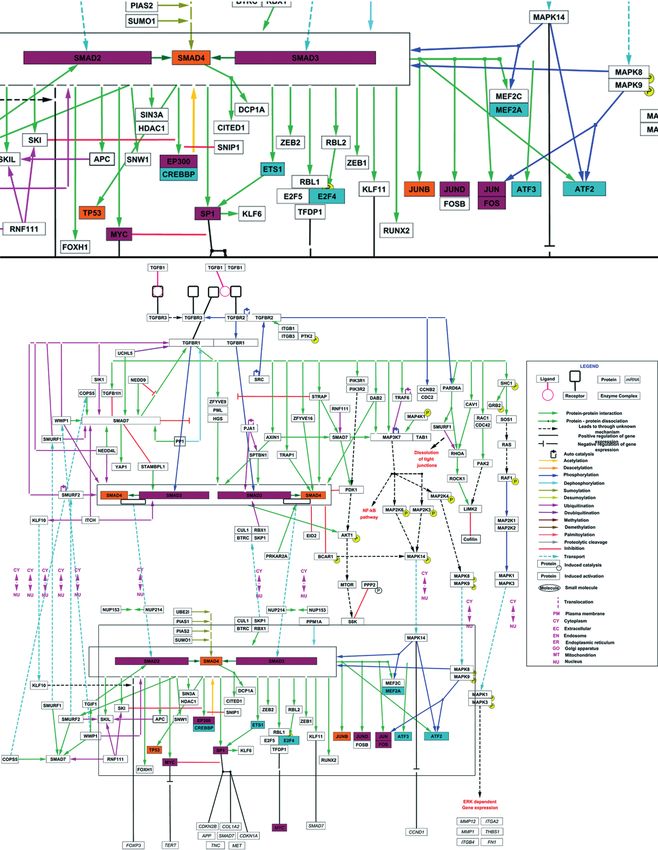
(T = TGF-) against control (C = TGF- 0 ng/mL, 0.5% FBS) at each time for each gene: (Txh/Cxh) (n=3, *p0.01, *** pSandra Susana Novoa Herrán, Mariela Castelblanco, Myriam Sánchez-Gómez, Adriana Umaña Pérez Figure 4. A. Transcription factors with binding sites in of MMP9 and PLAU (uPA) genes promoters. Consensus list created from Proscan v. 1.7 and Enrich analysis (ChIP ChEA 2015, ENCODE TF ChIP-seq 2015, TRANSFAC and JASPAR PWMs and Genome Browser PMWs). B. Analysis of Pathways commons pathways. Obtained network between the TGF-, MMP9 and PLAU genes, obtained with PCViz and addition of 34 nodes. Blue connections: state changes, green connections: expression. Dotted line frames: connector nodes shared between TGF- and both proteases. Continuous line frames: connector nodes exclusive to MMP9 or PLAU. 32 - Acta biol. Colomb., 24(1):26-37, Enero- Abril 2019
TGF- Dual Effects on MMP-9 and UPA Expression in HTR-8/SVneo
Figure 5. TGF- signalling pathway and transcription factors involved in the expression of MMP9 and PLAU (uPA). Inferred transcription
factors that regulates the expression of MMP9 and PLAU (purple nodes), only MMP9 (orange nodes) or only PLAU (blue nodes), belonging to the
TGF- signalling pathway reported by WikiPathways.
uPA can be more dependent on the presence of serum, and In addition to the influence of serum, our results also
therefore on the environmental conditions, in comparison showed that the effects of TGF- on the expression of
with MMP9 expression. MMP9 and uPA changed with time, evidencing a dual effect
Acta biol. Colomb., 24(1):26-37, Enero- Abril 2019 - 33Sandra Susana Novoa Herrán, Mariela Castelblanco, Myriam Sánchez-Gómez, Adriana Umaña Pérez
of TGF- with sinusoidal kinetics. A coordinated regulatory on the expression of MMP9 and uPA, key proteases in the
pattern for both genes was obtained, but with out-of-phase invasive process of both trophoblast (Lala and Chakraborty,
peaks, due to the opposed effects of TGF- : while at 6 2003; Staun-Ram et al., 2004; Cohen et al., 2006; Zheng et
hours post-stimuli, TGF- inhibits the expression of uPA and al., 2018) and metastatic tumors (Friedl and Wolf, 2003;
increases the expression of MMP9, at 12 hours the effect Mahmood et al., 2018). Even though the presence of 0.5 %
is the opposite. This phenomenon could be explained in FBS can modify the cellular status and therefore the response
part, by the activation of different signaling pathways and capacity to exogenous factors, based on the performed
their corresponding transcriptional mediators operating study, it is not possible to discern the role that FBS plays.
at their kinetics. Another factor may arise from a possible Until now, no systematic studies about the expression of
modification of the culture conditions, due to nutrient uptake these two genes in response to TGF- over time, or about
and conditioning of the medium with autocrine factors that the influence of serum during the treatment had been
altogether modify the characteristics of the culture medium. performed. In this work, we found a differential response to
These changes could influence the response to TGF-, TGF- as a function of time, for both uPA and MMP9 genes,
generating other input signals, which explain the changes in exhibiting dual kinetics, which partially agrees with the
response to TGF- due to the presence of serum and over literature. In general, it has been seen that TGF- decreases
time, highlighting the influence of microenvironment or the secretion and activity of uPA in the HTR-8/SVneo cell
cellular context. line (48 h, serum-free ExCell medium) (Graham et al., 1993)
Studies on the effects of insulin and IGF-I, acting separately and in first-trimester placenta explants (24h, serum-free
or in synergy with the serum, on the secretion of chorionic ExCell 300 medium) (Lash et al., 2005), simultaneously
gonadotropin beta (hCG), proliferation and invasion of decreasing the secretion and activity of MMP9 in primary
BeWo, JAR and JEG-3 choriocarcinoma cell lines, a strong first-trimester cytotrophobast cultures (4 days, serum-free
synergistic effect with serum was revealed (Mandl et al., DMEM medium) (Meisser et al., 1999), and in placenta
2002). Other studies have demonstrated that a high glucose explants (24 h, serum-free ExCell 300 medium) (Lash et al.,
concentration inhibits the activity of uPA and in vitro invasion 2005), and confirming a reduction in the expression and
of HTR-8/SVneo cells, with no observed effects on the levels activity of uPA and MMP9 in the choriocarcinoma cell line
of MMP2, MMP9, PAI-1 or uPAR (Belkacemi et al., 2005). JEG-3 (10 ng/mL, 24 h) (Karmakar and Das, 2002).
The observed dependence of the signaling of TGF- on the Nevertheless, in primary first-trimester cytotrophobast
culture conditions and its versatility, can be explained based cultures we observed an increase in the expression of
on its dual behavior, as tumor suppressor in certain cell types MMP9 at 8 hours and its activity at 12 hours, as a response
and as pro-metastatic in many invasive tumors (Pardali and to TGF-1 (10 and 20 ng/mL, serum-free RPMI 1640
Moustakas, 2007), and whose effects greatly depend on the medium) (Shimonovitz et al., 1996); similarly, an increase in
cellular context (Ikushima and Miyazono, 2010). MMP9 and 2 as a response to TGF- was observed in early
The trophoblastic tissue has been considered as “pseudo- cytotrophobast cultures and the choriocarcinoma cell line
malignant” or “physiologically metastatic” because its BeWo (Yudate et al., 1996). This agrees with an increase in
cellular abilities (Ferretti et al., 2007; Li and Shao, 2017) the expression of MMP9 at 6 hours in 0.5 % FBS medium or
and similar to solid tumors, the placenta has several at 24 hours in serum-free medium, highlighting the duality
trophoblast subtypes, which vary with the gestational of TGF- and its context-dependent actions.
stage (Liu et al., 2018); even more, the HTR-8/SVneo cell The disparity in the previous results can be due not only
line after immortalization by transfection with SV40 virus to the specific culture conditions –such as cellular model,
large T-antigen (Graham et al., 1993) can present additional medium type, the presence of serum, dose and stimulus
features, being proposed as a tumor progression model time– but also to the type of technique used for transcript
(Lala et al., 1998). In spheroid formation assay, HTR-8/ measuring. In the present study, the genic expression was
SVneo spheroids showed trophoblast progenitor cell-like evaluated in a time range from 6 to 24 hours by qPCR, which
characteristics and are suggested that these spheroids contain despite being implemented as a relative comparison, has a
the progenitor cell characteristic of repopulating activity much higher sensibility and allows to detect small changes
in a near in vivo model (Weber et al., 2013). Nevertheless, in transcript abundance, making possible to determine
TGF- has been studied only as a tumor suppressor in these that TGF- has a different effect at short times (6 hours).
models, based on its classic anti-proliferative, anti-invasive At longer culture times, the medium is conditioned by
and anti-migratory actions (Chakraborty et al., 2002; Lala et trophoblast-secreted factors that could have an autocrine
al., 2002), without considering its dual effect according to effect, increasing the complexity in both the signaling and
the biological context in which it performs its actions. Taking the results interpretation.
into account the HTR-8/SVneo cell line characteristics and The difference in the TGF- transcriptional regulation on
its origin, as well as the dual role or dichotomy of TGF-, it each gene may be explained in part by the different sets of
is not surprising to observe the opposed effects of this factor transcription factors found for each gene, especially those
34 - Acta biol. Colomb., 24(1):26-37, Enero- Abril 2019TGF- Dual Effects on MMP-9 and UPA Expression in HTR-8/SVneo
belonging to the TGF- signaling pathway, as Smad4, serum, being the culture conditions and stimulation relevant
TP53 and JUNB for MMP9, and CREBBP, Ets1, E2F4, when analyzing the effect of this cytokine.
ATF2, ATF3 and MEF2A for PLAU. This difference can
also be due to a differential signaling activation, as the ACKNOWLEDGMENT
bioinformatic analysis suggests. It is necessary to explore This project was funded by the Research Direction (DIB) of
possible crossed signaling events, including co-activation Universidad Nacional de Colombia, Sede Bogotá (Project
of the MAPK pathway. Previously it was seen that TGF- code 20697). S Novoa-Herrán thanks Colciencias for her
promotes invasion of HTR-8/SVneo cells in advanced credit-grant for PhD studies “Generación del Bicentenario”.
culture passages, involving a reduction in the expression of
TRI, Smad3, and Smad4, increase of Smad7 (Yanzhen et CONFLICT OF INTEREST
al., 2014), and induction of expression of MMP2 through The authors declare that there is no conflict of interest.
Smad2, with no Smad 4 involvement (Lin et al., 2006), and
more recently TGF- was reported to induce invasion in JEG- REFERENCES
3 cells and up-regulates the MMP-9 expression (Huang et Belkacemi L, Lash GE, Macdonald-Goodfellow SK, Caldwell JD,
al., 2017). Cellular context-specific factors whose action Graham CH. Inhibition of human trophoblast invasiveness
depends on environmental conditions can generate signaling by high glucose concentrations. J Clin Endocrinol Metab.
dynamics and spatial-temporal regulations (Kholodenko et 2005;90(8):4846-51. Doi:http://dx.doi.org/10.1210/jc.200
al., 2010). Further studies besides the activation of MPK 4-2242
and TGF-/SMADS pathways can explain the phenomena Cerami EG, Gross BE, Demir E, Rodchenkov I, Babur Ö,
observed here, confirming the formulated hypotheses and Anwar N, et al. Pathway Commons, a web resource for
bioinformatic inferences. Additionally, it is necessary to biological pathway data. Nucleic Acids Res. 2011;39(suppl
verify the expression at the protein level and furthermore, 1):D685-D690. Doi:http://dx.doi.org/10.1093/nar/gkq1039
verify the secretion and extracellular activity of these enzymes Cohen M, Meisser A, Bischof P. Metalloproteinases and Human
to corroborate a possible temporal regulation, like the one Placental Invasiveness. Placenta. 2006;27(8):783-793.
we propose. Doi:http://dx.doi.org/10.1016/j.placenta.2005.08.006
The invasion of the maternal endometrium by EVTs is a Chakraborty C, Gleeson LM, McKinnon T, Lala PK. Regulation
key process (He et al., 2017) whose deregulation, seen as of human trophoblast migration and invasiveness. Can.J.
hypoinvasiveness or hyperinvasiveness, lead to preeclampsia Physiol. Pharmacol 2002;80(2):116-124. Doi:http://dx.doi.
or trophoblastic neoplasms, respectively (Chakraborty et al., org/10.1139/Y02-016
2002). Being TGF- a main regulatory factor in trophoblast Chen EY, Tan CM, Kou Y, Duan Q, Wang Z, Meirelles GV, et
physiology, including invasion (Pollheimer and Knöfler, al. Enrichr: interactive and collaborative HTML5 gene list
2005), and taking into account the over-expression of this enrichment analysis tool. BMC bioinformatics. 2013;14:128.
factor in preeclampsia and hydatidiform mole (Pang and Doi:http://dx.doi.org/10.1186/1471-2105-14-128
Xing, 2003); understanding the molecular mechanism Cheng J-C, Yi Y, Chang H-M, Leung PCK. TGF-1 up-regulates
underlying this regulation is important. The possible cadherin-11 expression through Snail: A potential mechanism
temporal regulation and the observed dual effects of TGF- for human trophoblast cell differentiation. Cell Signal.
on the expression of MMP9 and uPA, key proteases in the 2018;43:55-61. Doi:https://doi.org/10.1016/j.cellsig.2017.
invasive phenotype, are of remarkable interest and they 12.004
confer an important role to these enzymes in the invasive Ferretti C, Bruni L, Dangles-Marie V, Pecking AP, Bellet D.
process of trophoblastic cells, which is highly regulated in a Molecular circuits shared by placental and cancer cells,
spatial-temporal way (Menkhorst et al., 2016). These results and their implications in the proliferative, invasive and
are in agreement with previous reports about the effects migratory capacities of trophoblasts. Hum Reprod Update.
of TGF- in trophoblast and are compatible with the dual 2007;13(2):121-141. Doi:http://dx.doi.org/10.1093/humu
and context-dependent TGF- role in cancer (Ikushima and pd/dml048
Miyazono, 2010). Additionally, novel results were obtained, Friedl P, Wolf K. Tumour-cell invasion and migration: diversity
as the temporal regulation of the expression of MMP9 and and escape mechanisms. Nat Rev Cancer. 2003;3(5):362-
uPA, involving opposed effects of TGF-; and remarkably, 374. Doi:http://dx.doi.org/10.1038/nrc1075
the influence of serum on the nature and effect of TGF- Graham CH, Hawley TS, Hawley RC, MacDougall JR, Kerbel
on the expression of uPA, a concomitance not previously RS, Khoo N, et al. Establishment and Characterization of
studied in this model. First Trimester Human Trophoblast Cells with Extended
Lifespan. Exp Cell Res. 1993;206(2):204-211. Doi:http://
CONCLUSIONS dx.doi.org/10.1006/excr.1993.1139
The nature and magnitude of the effects of TGF- on the He N, van Iperen L, de Jong D, Szuhai K, Helmerhorst FM, van
expression of uPA and MMP9 in HTR-8/SVneo human der Westerlaken LAJ, et al. Human Extravillous Trophoblasts
immortalized trophoblastic cells depend on the presence of Penetrate Decidual Veins and Lymphatics before Remodeling
Acta biol. Colomb., 24(1):26-37, Enero- Abril 2019 - 35Sandra Susana Novoa Herrán, Mariela Castelblanco, Myriam Sánchez-Gómez, Adriana Umaña Pérez Spiral Arteries during Early Pregnancy. PLOS ONE. growth factor-beta1-induced trophoblast expression of 2017;12(1):e0169849. Doi:http://dx.doi.org/10.1371/journal. matrix metalloproteinase-2. Front Biosci . 2006;11:637- pone.0169849 46. Doi:http://dx.doi.org/10.2741/1823 Hellemans J, Mortier G, De Paepe A, Speleman F, Liu Y, Fan X, Wang R, Lu X, Dang Y-L, Wang H, et al. Single-cell Vandesompele J. qBase relative quantification framework RNA-seq reveals the diversity of trophoblast subtypes and and software for management and automated analysis patterns of differentiation in the human placenta. Cell Res. of real-time quantitative PCR data. Genome Biol. 2018;28(8):819-832. Doi:10.1038/s41422-018-0066-y 2007;8(2):R19. Doi:http://dx.doi.org/10.1186/gb-2007- Lunghi L, Ferretti M, Medici S, Biondi C, Vesce F. Control 8-2-r19 of human trophoblast function. Reprod Biol Endocrinol. Huang Z, Li S, Fan W, Ma Q. Transforming growth factor 2007;5(1):6. Doi:http://dx.doi.org/10.1186/1477-7827- beta1 promotes invasion of human JEG-3 trophoblast 5-6 cells via TGF-beta/Smad3 signaling pathway. Oncotarget. Mahmood N, Mihalcioiu C, Rabbani SA. Multifaceted Role 2017;8(20):33560-33570. Doi:http://dx.doi.org/10.18632/ of the Urokinase-Type Plasminogen Activator (uPA) and Its oncotarget.16826 Receptor (uPAR): Diagnostic, Prognostic, and Therapeutic Ikushima H, Miyazono K. TGF signalling: a complex web Applications. Front Oncol. 2018;8(24). Doi:http://dx.doi. in cancer progression. Nat Rev Cancer. 2010;10(6):415- org/10.3389/fonc.2018.00024 424. Doi:http://dx.doi.org/10.1038/nrc2853F Mandl M, Haas J, Bischof P, Nohammer G, Desoye G. Serum- Karmakar S, Das C. Regulation of Trophoblast Invasion by IL-1 dependent effects of IGF-I and insulin on proliferation and and TGF- 1. Am J Of Reprod Immunol. 2002;48(4):210-219. invasion of human first trimester trophoblast cell models. Doi:http://dx.doi.org/10.1034/j.1600-0897.2002.01151.x Histochem Cell Biol. 2002;117(5):391-9. Doi:http:// Kholodenko BN, Hancock JF, Kolch W. Signalling ballet in dx.doi.org/10.1007/s00418-002-0403-5 space and time. Nat Rev Mol Cell Biol. 2010;11(6):414- Meisser A, Chardonnens D, Campana A, Bischof P. Effects of 426. Doi:http://dx.doi.org/10.1038/nrm2901 tumour necrosis factor-, interleukin-1 , macrophage colony Kutmon M, Lotia S, Evelo CT, Pico AR. WikiPathways App stimulating factor and transforming growth factor on for Cytoscape: Making biological pathways amenable to trophoblastic matrix metalloproteinases. Mol Hum Reprod. network analysis and visualization. F1000Res. 2014;3:152. 1999;5(3):252-260. Doi:http://dx.doi.org/10.1093/molehr Doi:http://dx.doi.org/10.12688/f1000research.4254.2 /5.3.252 Lala PK, Graham CH, Lysiak JJ, Khoo NKS, Hamilton GS. Menkhorst E, Winship A, Van Sinderen M, Dimitriadis E. TGF regulation of trophoblast function. Placenta. Human extravillous trophoblast invasion: intrinsic and 1998;19:149-157. Doi:http://dx.doi.org/10.1016/S0143- extrinsic regulation. Reprod Fertil Dev. 2016;28(4):406- 4004(98)80012-X 415. Doi:https://doi.org/10.1071/RD14208 Lala PK, Lee BP, Xu G, Chakraborty C. Human placental Novoa-Herran S, Umaña-Perez A, Canals F, Sanchez-Gomez trophoblast as an in vitro model for tumor progression. M. Serum depletion induces changes in protein expression Can.J. Physiol. Pharmacol. 2002;80(2):142-149. Doi:http:// in the trophoblast-derived cell line HTR-8/SVneo. Cell Mol dx.doi.org/10.1139/y02-006 Biol Lett. 2016;21(1):22. Doi:http://dx.doi.org/10.1186/ Lala PK, Chakraborty C. Factors Regulating Trophoblast s11658-016-0018-9 Migration and Invasiveness: Possible Derangements Pang Z-J, Xing F-Q. Expression of transforming growth factor- Contributing to Pre-eclampsia and Fetal Injury. Placenta. and insulin-like growth factor in molar and placental tissues. 2003;24(6):575-587. Doi:http://dx.doi.org/10.1016/S014 Arch Gynecol Obstet. 2003;269(1):1-4. Doi:10.1007/ 3-4004(03)00063-8 s00404-002-0379-3 Lash GE, Otun HA, Innes BA, Bulmer JN, Searle RF, Robson Pardali K, Moustakas A. Actions of TGF-[beta] as tumor SC. Inhibition of Trophoblast Cell Invasion by TGFB1, 2, suppressor and pro-metastatic factor in human cancer. and 3 Is Associated with a Decrease in Active Proteases. Biochim Biophys Acta Rev Cancer. 2007;1775(1):21-62. Biol Reprod. 2005;73(2):374-381. Doi:http://dx.doi. Doi:http://dx.doi.org/10.1016/j.bbcan.2006.06.004 org/10.1095/biolreprod.105.040337 Pollheimer J, Knöfler M. Signalling pathways regulating the Li DQ, Shao ZM. Chapter 19 - Emerging Therapeutic Targets invasive differentiation of human trophoblasts: a review. for Cancer Metastasis: From the Perspective of Embryo Placenta. 2005;26:21-30. Doi:http://dx.doi.org/10.1016/j. Implantation. In: Ahmad A, editor. Introduction to Cancer placenta.2004.11.013 Metastasis. Ciudad: Academic Press; 2017. p. 353-373. Shannon P, Markiel A, Ozier O, Baliga NS, Wang JT, Ramage Doi:https://doi.org/10.1016/B978-0-12-804003-4.00 D, et al. Cytoscape: A Software Environment for Integrated 019-0 Models of Biomolecular Interaction Networks. Genome Res. Lin HY, Yang Q, Wang HM, Qi JG, Zhang H, Wang HX, et al. 2003;13(11):2498-2504. Doi:http://dx.doi.org/10.1101/ Involvement of SMAD4, but not of SMAD2, in transforming gr.1239303 36 - Acta biol. Colomb., 24(1):26-37, Enero- Abril 2019
TGF- Dual Effects on MMP-9 and UPA Expression in HTR-8/SVneo
Shimonovitz S, Hurwitz A, Barak V, Dushnik M, Adashi EY, Anteby on the proliferation and invasion of the HTR-8/SVneo cell
E, et al. Cytokine-mediated regulation of type IV collagenase line. Oncol Lett. 2014;8(5):2187-2192. Doi:http://dx.doi.
expression and production in human trophoblast cells. J org/10.3892/ol.2014.2451
Clin Endocrinol Metab. 1996;81(8):3091-6. Doi:http:// Yi Y, Cheng J-C, Klausen C, Leung PCK. TGF-beta 1 inhibits human
dx.doi.org/10.1210/jc.81.8.3091 trophoblast cell invasion by upregulating cyclooxygenase-2.
Staun-Ram E, Goldman S, Gabarin D, Shalev E. Expression and Placenta. 2018;68:44-51. Doi:http://dx.doi.org/10.1016/j.
importance of matrix metalloproteinase 2 and 9 (MMP-2 and placenta.2018.06.313
-9) in human trophoblast invasion. Reprod Biol Endocrinol. Yudate T, Isaka K, Kosugi Y, Koshiishi M, Shiraishi K, Hosaka M,
2004;2(1):59. Doi:http://dx.doi.org/10.1186/1477-7827- et al. Analysis of the mechanism of trophoblast infiltration.
2-59 Nihon Sanka Fujinka Gakkai Zasshi. 1996;48(3):191-8.
Turunen SP, Tatti-Bugaeva O, Lehti K. Membrane-type matrix Zhang S-M, Tian F-J, Zeng W-H, Ma X-L, Ren J-B, Lin Y. XCL1-
metalloproteases as diverse effectors of cancer progression. XCR1 pathway promotes trophoblast invasion at maternal-
Biochim Biophys Acta Mol Cell Res. 2017;1864(11, Part fetal interface by inducing MMP-2/MMP-9 activity. Am J Of
A):1974-1988. Doi:https://doi.org/10.1016/j.bbamcr.20 Reprod Immunol. . 2018;80(3):e12990. Doi:https://doi.
17.04.002 org/10.1111/aji.12990
Weber M, Knoefler I, Schleussner E, Markert UR, Fitzgerald JS. Zhao M-r, Qiu W, Li Y-x, Zhang Z-b, Li D, Wang Y-l. Dual effect
HTR8/SVneo Cells Display Trophoblast Progenitor Cell-Like of transforming growth factor 1 on cell adhesion and
Characteristics Indicative of Self-Renewal, Repopulation invasion in human placenta trophoblast cells. Reproduction.
Activity, and Expression of “Stemness-”; Associated 2006;132(2):333-341. Doi:http://dx.doi.org/10.1530/rep.
Transcription Factors. BioMed Res Int. 2013;2013:10. 1.01112
Doi:10.1155/2013/243649 Zheng Q, Yang Y, Cui X, Zhang D, Liu S, Yan Q. AP1 mediates uPA/
Yanzhen ZUO, Zhihua FU, Yatao HU, Yuhong LI, Qian XU, uPAR induced FUT4 expression and trophoblast invasion. J
Dayong SUN, et al. Effects of transforming growth factor-1 Cell Biochem. 2018;119(8):6442-6451. Doi:http://dx.doi.
org/10.1002/jcb.26648
Acta biol. Colomb., 24(1):26-37, Enero- Abril 2019 - 37You can also read