Hair follicle differentiation and regulation
←
→
Page content transcription
If your browser does not render page correctly, please read the page content below
Int. J. Dev. Biol. 48: 163-170 (2004)

                          Hair follicle differentiation and regulation
                                                           GEORGE E. ROGERS*
                              School of Molecular and Biomedical Sciences, University of Adelaide, South Australia

                   ABSTRACT Ten years ago, Hardy (1992) wrote a timely review on the major features of hair follicle
                   development and hair growth which she referred to as a secret life. Many of these secrets are now
                   being revealed. The information discussed in this brief review comprises the structure of the hair
                   and hair follicle, the continuing characterisation of the genes for keratin and keratin associated
                   proteins, the determination of the location of their expression in the different cell layers of the hair
                   follicle, molecular signals which control keratin gene expression and post-translational events in
                   the terminal stages of hair formation.

                   KEY WORDS: hair, follicle layer, keratinocyte, keratin gene, gene expression

A hair arises from the integrated activities of several                     both follicle induction and hair growth. The molecular factors
keratinocyte layers in the hair follicle                                    and receptors that are expressed during follicle induction and in
                                                                            the anagen growth phase are becoming increasingly well-
    Keratinocytes differentiate into several distinct cellular lay-         defined and the fluctuations of their activity are discussed in
ers of the follicle during the growth phase (anagen) of the hair            reviews (Botchkarev and Kishimoto, 2003, Fuchs et al., 2001).
cycle (Figs. 1,2). From the outermost aspect of the follicle the            Prominent regulatory proteins in both developing and anagen
histological structures are: the outer root sheath (ORS) con-               follicles include the BMPs, Sonic hedgehog and several WNT
sisting of several cell layers and the innermost adjacent to the            proteins and the receptors, BMPR1A, EGFR, FGFR and TGFR.
inner root sheath (IRS) is called the companion layer (Orwin,                   A daughter cell from a mitotic event might move out of the
1971). Adjoining the ORS on the dermal side is a basket-like                matrix zone of the follicle and differentiate or could remain in the
arrangement of two orthogonally arrayed layers of collagen                  zone and continue dividing. Whether a cell moves or stays
fibres, the glassy layer (Rogers, 1957) now known as the                    within the critical region might be controlled by the level of β1-
dermal sheath.The Henle, Huxley and cuticle layers of the IRS;              integrin expression since the expression of β1-integrin is greater
the IRS cuticle layer adjoins the cuticle of the presumptive hair           in epidermal stem cells and is reduced when they enter a
fibre.The presumptive hair shaft comprises an outer layer of                differentiation phase (Zhu et al., 1999). The importance of β1–
overlapping cuticle cells surrounding a cellular cortex and                 integrin for the maintenance of proliferating cells in the hair bulb
sometimes a central medulla.                                                perhaps as a stem cell population was also evident from
    The primary activities in anagen hair follicle to produce a             observations made on the hair follicles of β1-integrin null mice
hair involve proliferation of the germinative epithelial cells in           (Brakebusch et al., 2000). The importance of BMPs in restrict-
the bulb region, the determination of cell lineages for all the             ing a normal proliferating population to the bulb region was
follicle layers (Fig. 2)and terminal differentiation (keratinisation).      demonstrated by the over expression of the BMP inhibitor
The differentiation products of the temporally regulated pro-               Noggin, in transgenic mice. In such mice, dividing cells were
cesses consist of structural proteins within the cells and adhe-            then observed in the hair shaft distal to the bulb (Kulessa et al.,
sion proteins between the cells that hold them tightly packed               2000).
together within the cylindrical-shaped hair.                                    The synthesis and intracellular deposition of keratin struc-
    The region in the bulb where keratinocytes proliferate rap-             tural proteins in the spindle-shaped keratinocytes of the cortex
idly is called the critical region or hair matrix zone (Auber,              leads to assembly of a composite of intermediate filaments (IFs)
1950, Orwin, 1979) (see Fig. 2); it surrounds the dermal papilla
separated by a basement membrane. Since the seminal ex-
periments of (Cohen, 1961) and (Oliver, 1966) it has been                   Abbreviations used in this paper: IF, intermediate filament; IRS, inner root
known that the dermal papilla provides essential stimuli for                sheath; KAP, keratin associated protein; ORS, outer root sheath.

*Address correspondence to: Dr. George Rogers. School of Molecular and Biomedical Sciences, University of Adelaide, South Australia 5005, Australia.
Fax: +61-8-8303-4262. email: george.rogers@adelaide.edu.au

0214-6282/2004/$25.00
© UBC Press
Printed in Spain
www.ijdb.ehu.es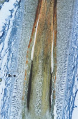
164 G.E. Rogers
and a matrix of keratin associated proteins (called either IFAPs or were divided into a high-sulphur KAP group with less than 30 mol%
KAPs) (Powell and Rogers, 1997), often referred to as the IF-matrix of cysteine and an ultra-high sulphur group with cysteine contents
complex. During keratinisation this composite is finally stabilised above that value (Gillespie, 1991). Sequence comparisons of the
mainly by the formation of inter- and intra-molecular disulfide genes for wool proteins showed that there are at least eight families
bonds. Presumptive cuticle cells that surround the cortex undergo of cysteine–rich proteins and two main groups with more than
flattening as they emerge from the bulb region and are interlocked twenty glycine/tyrosine-rich proteins (Powell and Rogers, 1997).
with the IRS during their passage up the follicle anchoring the hair Studies of the human genome for equivalent genes encoding
in the follicle. glycine-tyrosine rich proteins identified a domain of 17 genes on
chromosome 21q22.1 (Rogers et al., 2002). Furthermore, on the
The keratin and keratin-associated proteins expressed same domain seven KAP genes for high-sulphur proteins were
in the cortex and cuticle located which extends an earlier study that revealed a cluster of 37
genes for the sulphur-rich KAP group (Rogers et al., 2001) on
The term “keratin” is now generally restricted to designating the chromosome 17q12-2 interestingly within a domain of Type I IF
intermediate filament proteins of the fibre cortex. The proteins of genes and could be grouped into seven gene families.
two large families, Type I and Type II, form the IFs of the “hard”
keratins of hair (also the “soft” keratins of the epidermis). The two The formation of IF and KAP proteins in hair follicles
keratin families are distinguished by their isoelectric points (Type
I, acidic; Type II, basic/neutral). Equimolar amounts of the two There is a high rate of protein synthesis in the hair follicle. For
types are required to form the IFs (Steinert and Roop, 1988). a hair fibre of diameter 100 µm and length growth rate of about 20
Studies of the keratins of sheep (Powell and Rogers, 1997) µm per h, 5-10µg of protein are produced in a single follicle every
indicated that the families of chains consist of four Type I chains 24 h. The large families of IF and IFAP genes may be necessary
and four Type II chains. However, investigation of human hair for the high rate of synthesis in enabling transcription of mRNAs at
keratin gene families have strikingly revealed that the Type I family a level commensurate with demand.
has nine members and the Type II has six members (Langbein et Since the zone of synthesis and maturation in the approximately
al., 1999, Langbein et al., 2001). The genes are respectively on lower third of the follicle (called the keratinisation zone) through
human chromosomes 17q12-21 and 12q13 (Rogers et al., 1995). which a cortical cell passes is about 1000 µm long, it follows that
The keratin-associated (KAP) proteins that constitute the matrix the cells are completely filled with the keratin complex and cross-
of the keratin composite of wool are a large group of possibly up to linked over a period of 48 h as they pass through the zone.
100 different proteins. Originally, they were divided into three main A notable feature is the orderly expression of the IF and KAP
families according to their amino acid composition and molecular proteins in the developing hair. The temporal sequence with which
size. The proteins containing 35-60 mol% of glycine and tyrosine they are laid down has been determined by in situ hybridisation
(now KAPs 6, 7 & 8, (Powell and Rogers, 1997)) were originally detection of specific mRNAs for the different gene families using
referred to as the high-glycine/tyrosine group whereas the sulphur- cRNA probes (Powell et al., 1992, Powell and Rogers, 1997).
rich (cysteine-rich) proteins (KAPs 1-5 (Powell and Rogers, 1997)) These studies (Fig. 3) and a more recent and detailed investigation
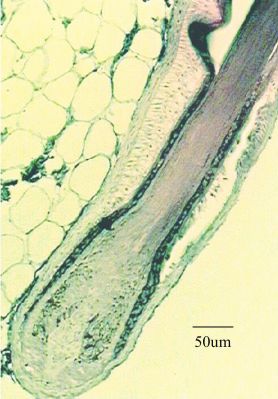
Fig. 1 (Left). Light micrograph of the different layers of the hair follicle. In the
bulb region, a proliferating epithelial matrix surrounds the mesenchymal dermal
papilla. The hair shaft of cortex, medulla and cuticle layers enclosed by the inner
root sheath move outwards within the outer root sheath which is continuous with
the epidermis. (From Millar, 1999; reproduced with permission from Elsevier).
Fig. 2 (Right). Cartoon of the follicle bulb. The diagram illustrates the several
lineages of cells which are determined in their differentiation pathway and leave
the follicle forming the different layers of the hair and surrounding follicle.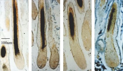
Hair keratinocyte differentiation and regulation                 165

                                                                                 different members specifically pair to form the keratin IFs remains
                                                                                 unclear. In vitro experiments, similar in kind to those conducted
                                                                                 with epidermal IFs (Coulombe et al., 1990, Coulombe and Fuchs,
                                                                                 1990) to determine the relative affinities of different combinations
                                                                                 of IF proteins would appear to be a major direction for elucidating
                                                                                 the most likely combinations of Type I and Type II chains for hair
                                                                                 keratin IF formation. Such investigations are in progress but so far
                                                                                 inconclusive (Herrling and Sparrow, 1991, Thomas et al., 1986,
                                                                                 Wang et al., 2000) and an important improvement is the utilisation
                                                                                 of recombinant IF proteins in such recombination experiments
                                                                                 (Hofmann et al., 2002).
                                                                                     Ultra-high sulphur KAP families including those of the cuticle are
 A                   B                   C                  D                    the last KAPs to be expressed. It should be noted that the
                                                                                 expression of the different gene families are not distinct but merge
Fig. 3. Localisation of expression of mRNAs encoding the keratin IF              one into the other and the translation of their mRNAs continue until
and KAP proteins of wool fibre cortex by in situ hybridisation. Specific         the last stages of hair formation. There is no precise evidence so
3’-cRNA probes were used to localise the sequential expression of (A)            far of down-regulation of one set of genes and up-regulation of
keratin IF; (B) Glycine-tyrosine rich KAPs; (C) Cysteine –rich KAPS (< 30        another compared to the transcription of epidermal genes (Fuchs
mol%) and (D) Cysteine-rich KAPs ( >30 mol%), the last to be expressed.          et al., 1989). Biochemical events are reflected in structural changes
                                                                                 visualised by transmission electron microscopy (TEM) of the
(Langbein et al., 2001) have conclusively shown that IF genes are                keratinised hair fibre cortex at high resolution. When the cortex is
the first to be expressed followed by the high glycine/tyrosine                  forming the IFs are seen to be aggregated into fibrils and in
genes and then the cysteine-rich protein families.                               conjunction with the in situ expression data it can be concluded that
                                                                                 as the matrix proteins are expressed they migrate into the spaces
Hair cortex                                                                      within the IF aggregates. The synthesis and insertion of matrix
                                                                                 proteins become coincident processes especially in the late phases
    In situ hybridisation experiments with cRNA probes (Powell et                of cortical cell differentiation and aggregation of the IFs. Evidence
al., 1992) revealed that IF genes K2.12, K2.9, K2.10 and K2.11 for               for this is that in the keratinising zone the matrix proteins appear to
wool keratins are expressed in that order in the cortical cells                  aggregate as “blocks” (Fig. 4) that subsequently disperse between
beginning with K2.12 just above the critical zone of the follicle bulb           the filaments as differentiation advances (Fig. 5). The matrix
and the later expression of K2.11 in the upper bulb region. The                  proteins and IFs interact further to produce either the typical quasi-
expression is coincident with Type I gene expression. Extensive                  hexagonal or cylindrical packing of the keratinised hair cortex (Fig.
studies of human genes (Langbein et al., 1999, Langbein et al.,                  6) (Fraser et al., 1972, Rogers, 1959b). The relative abundance of
2001) have shown that the pattern of IF gene expression is also                  the two major KAP groups and possibly the IFs as well may be
complex and as to which Type I and II proteins out of some twelve                structural factors responsible for these organisational patterns.

Fig. 4 (Left). Electron microscopic image (TEM) of loose bundles of
keratin IFs in a longitudinal section of a wool follicle. The TEM shows
the presence of dense aggregates (arrow) within bundles of IFs (macrofibrils)
and are assumed to be KAP proteins. They are also readily observed in
cross sections at a later stage of follicles, in which it can be seen that the
granules begin to disperse and interact with the IFs (see Fig. 5).

Fig. 5 (Right). TEM of part of a cortical cell in an osmium-fixed follicle. The aggregates seen as “blocks” in Fig. 4 appear as electron-dense matrix
incompletely distributed between the IFs which constitute macrofibrils. Dense cytoplasmic material is abundant between the macrofibrils. This
differentiation process occurs in cortical cells at mid level of the keratinisation zone of a hair follicle.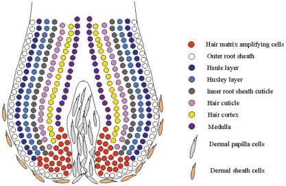
166       G.E. Rogers

                                                                                                        Fig. 6 (Left). Cross-section of
                                                                                                        macrofibrils of the developing cortex at
                                                                                                        a late stage of differentiation. The kera-
                                                                                                        tin IF in the macrofibrils are completely
                                                                                                        separated by the electron-dense matrix
                                                                                                        consisting of the KAP proteins which have
                                                                                                        dispersed between them. The IFs display
                                                                                                        the cylindrical mode of packing. The cyto-
                                                                                                        plasmic material between the macrofibrils
                                                                                                        has markedly decreased in abundance.

                                                                                                        Fig. 7 (Right). Localisation of the ex-
                                                                                                        pression of KAP 5 mRNA by in situ
                                                                                                        hybridisation. The expression of the KAP
                                                                                                        5 gene was detected using a 3’- cRNA
                                                                                                        probe. Strong expression signal appears in
                                                                                                        the cuticle at a late stage of hair formation
                                                                                                        in a human follicle.

   From X-ray diffraction studies and TEM studies (Jones et al.,         more than 50% of the fatty acids is 18-methyleicosanoic acid
1997, Strelkov et al., 2003) it has been deduced that the                (MEA) and a genetic defect affecting the synthesis of MEA pro-
organisation of keratin chains within the IFs consists of a regular      duces structural defects in the intercellular layers of the cuticle. The
array of dimer units and an average of 32 chains in the IF cross-        details of the site and synthesis of these cuticular proteolipids are
section. However the precise location of covalent links between          unknown. A summary of the structure of the cuticle of hair in shown
IFs and especially between the IFs and matrix proteins have yet          in Fig. 8.
to be mapped in a detail that would more precisely explain the
physical properties of hair.                                             Inner root sheath

Hair cuticle                                                                 The IRS is adjacent and adherent to the growing hair and is
                                                                         responsible for the surface topography of the fibre. It is degraded
    The amorphous cystine-rich contents of scale cells (Bradbury,        and sloughed as the hair emerges from the follicle. The establish-
1973, Fraser et al., 1972) consist of at least two unique families of    ment of the IRS cell lineage is dependent on the activities of several
proteins (KAP5 and KAP10) of the cuticle (Fig. 7) (Jenkins and           factors and recently the transcription factor GATA-3 has been
Powell, 1994, MacKinnon et al., 1990, Rogers et al., 2004). The          identified as playing a central role. The IRS is not formed in GATA-
evidence for the expression of specific IF proteins, Type I (hHa2        3 null mice, the normal coat is absent and aberrant hairs are
and hHa5) and Type II (hHb2 and hHb5) in the developing cells was        produced from embryonic skin grafted onto nude mice (Kaufman
unexpected (Langbein et al., 2001, Rogers et al., 1996). These           et al., 2003, Kobielak et al., 2003).
chemical findings appear to be in conflict with the microscopic              The IFs in the IRS cells are morphologically indistinguishable
evidence from TEM that IFs are not prevalent in developing cuticle       from IFs of the cortex but markedly different in protein composition.
cells. Some “tufts” of IFs can be visualised in the developing scale     The expressed genes for a Type I in sheep have been identified
cells in the bulb region but the prominent structures that are           and localised in the IRS (Fig. 9), one Type II in mice (Aoki et al.,
produced are globular masses that fuse to form the exocuticle            2001) and four in human (follicles) (Langbein et al., 2003). It is not
(Orwin, 1979, Powell and Rogers, 1997, Rogers, 1959a, Rogers,            known whether they specifically pair but the deficiency of one IF in
1959b). The KAP 5 and KAP10 proteins are major components of             the IRS through a spontaneous mutation in mice, causes collapse
the exocuticle (unpublished observations). An explanation might          of the IF network and interrupts normal IRS formation (Peters et al.,
be that the IF proteins are degraded in the later phases of              2003). It would seem likely that there are more members of the IRS
differentiation. Other proteins yet to be confirmed in scale cells are   intermediate filament family to be found.
those related to keratinocyte cell envelopes such as involucrin and          Differentiation of the IRS is characterised by the synthesis of
loricrin (Kalinin et al., 2002, Steinert and Marekov, 1995) that are     trichohyalin a specific precursor matrix material that finally binds
cross-linked by isopeptide bonds (Rice et al., 1994). An additional      the filaments into a cross-linked composite analogous to that of the
feature of the hair cuticle is the presence of a group of long-chain     cortex. As the cells move upward with the hair the trichohyalin that
fatty acids that is responsible for the hydrophobicity of the hair       is present as cytoplasmic aggregates, undergoes a post-transla-
surface. Chemical evidence indicates that the fatty acids are linked     tional modification of arginine residues to citrulline causing the
by thioester bonds to protein(s) that are probably components of         protein to disperse between the filaments (Rogers et al., 1997,
the scale cell envelope (Jones and Rivett, 1997). Unexpectedly           Rothnagel and Rogers, 1986). The final IF-matrix composite does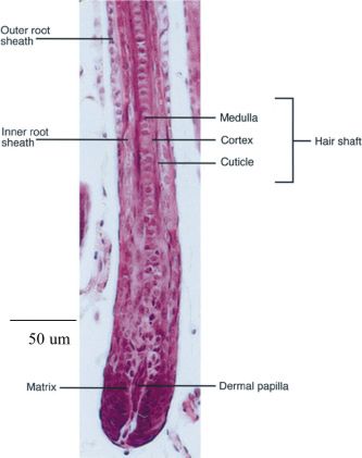
Hair keratinocyte differentiation and regulation              167

not have the quasi-crystalline packing seen in cortical keratinocytes             1995, Winter et al., 1998) are expressed in the companion layer
and instead of disulphide bonds it is extensively cross-linked by                 and presumably they correspond to the keratin IFs seen in the cells.
isopeptide bonds produced by transglutaminase activity present in
the follicle.                                                                     Signals which regulate cell specificity and gene expres-
    Other differentiation products reported in the cells of the IRS               sion
include µ-crystallin, a protein primarily found in the eye. It pos-
sesses both enzymic and structural properties (Aoki et al., 2000).                    The regulatory molecules and their networks that control differ-
Several proteins are calcium-binding proteins, namely trichohyalin                entiation of hair keratinocytes are becoming increasingly defined
itself and the enzymes, peptidylarginine deiminase and                            and indeed complex. Notch has been identified as a factor in the
transglutaminase                                                                  determination of cell type (Kopan and Weintraub, 1993, Lin et al.,
    Why are the proteins of the IRS different from those of the hair              2000). BMP signalling inhibits follicle development (Botchkarev,
shaft? The answer surely lies in the requirement for IRS cells to be              2003) and when the abundance of BMPs was reduced by over-
finally sloughed as the anagen hair emerges from the skin surface                 expressing Noggin the differentiation of keratinocytes into mature
and is released from the supporting layers of the IRS. This change                cortical and cuticle cells was severely impaired demonstrating the
is achieved by the proteins of the IRS being readily degradable by                key role of BMPs in the formation of the hair layers (Kulessa et al.,
proteases (Rogers, 1964a) whereas the molecular organization of                   2000). The central role of BMP and its linkage to the WNT pathway
hair keratin makes it resistant to proteolysis. Proteolytic activity has          has been strikingly substantiated (Kobielak et al., 2003) by knock-
been observed to be a central feature of epithelial desquamation                  ing out the gene for the BMP receptor BMPRIA resulting in hairless
and is present in the hair follicle distal to the opening of the                  mice with malformed follicles. Follicle growth was inhibited and the
sebaceous duct (Ekholm and Egelrud, 1998).                                        follicles lacked an IRS although those features are not necessarily
                                                                                  causally related.
Outer root sheath                                                                     The regulation of gene activity in the anagen hair follicle shares
                                                                                  a significant degree of commonality with the keratinocytes of the
    The outer root sheath (ORS) is continuous with the epidermis                  epidermis for which a large number of transcription factors for
but the layer of cells immediately adjacent to the IRS Henle layer                both positive and negative regulation have been recognised
has some features that differentiate it as a distinct entity (Rogers,             (Eckert et al., 1997, Fuchs et al., 2001, Nakamura et al., 2001).
1964b)]. Notable are tufts of intermediate filaments located at the               The involvement of LEF1 in hair keratin gene expression indi-
ORS/IRS junction complex oriented so that they encircle the                       cated the participation of this factor in hair follicle development
follicle. This layer was later named the companion layer (Orwin,                  through the WNT regulatory pathway (DasGupta and Fuchs,
1971) but it’s role in the dynamics of hair follicle function is unclear.         1999). The importance of that pathway has been demonstrated by
Certainly the junction adjoining the Henle layer is different in                  numerous findings that when it is dysfunctional through natural or
lacking desmosomes and that could indicate that the IRS moves                     experimental mutations, hair follicle development is affected and
relative to the companion layer during outward growth of the hair.                produces a variety of hair and follicle phenotypes. It is also
The alternative is for the layer to move with the IRS as that layer               essential for the maintenance of inducing activity of the dermal
differentiates with the growing hair. Families of K6 proteins to-                 papilla (Kishimoto et al., 2000; Shimizu and Morgan, 2004). The
gether with K16 (Rothnagel and Roop, 1995, Takahashi et al.,                      central molecule in the WNT pathway is β-catenin which has dual

Fig. 8 (Left). Diagrammatic representation of a scale cell of the hair cuticle. The major
intracellular layers, hydrophobic layer, A layer, exocuticle and endocuticleand protein components
of a scale cell are indicated in cross-section.

Fig. 9 (Right). Localisation by in situ hybridisation of the expression of the mRNA encoding
a Type I IF protein. The mRNA detected by a 3’- probe is highly expressed in the IRS of a hair follicle.
The dark material in the bulb region is melanin.
168 G.E. Rogers
roles of being part of the cadherin complexes of cell junctions or In the differentiation of both hair and the IRS keratinocytes, the
acting within the nuclei of cells after transportation from the junctions are replaced with a new cell membrane complex (CMC)
cytoplasm (DasGupta and Fuchs, 1999, Merrill et al., 2001) and that gradually develops as a continuous layer between the cells.
forming a transcription complex with the LEF1/TCF DNA binding This complex consists of an electron-dense central (δ) layer about
family of proteins. This complex activates genes involved in hair 15nm thick surrounded by β-layers that are approximately 5nm
follicle development and presumably, hair keratin genes as well, wide (Jones and Rivett, 1997, Rogers, 1959a, Rogers, 1959b).
given that the LEF1 consensus is present in the proximal promot- Once the growing hair has passed through the keratinisation zone
ers referred to earlier. in the lower third of the follicle, morphological changes occur in the
An early survey for control sequences in the 5’ promoter region nuclei and cytoplasm of all the cells. Although nuclei remain in the
of several hair keratin genes (Powell et al., 1992, Powell et al., 1991) cells the chromatin is degraded and mostly resorbed. Several
revealed several binding sites that are commonly active in the control markers have shown that apoptosis participates in the mecha-
of gene expression. A sequence CTTTGAAGA was found to be nisms that occur during morphogenesis of the hair follicle and in
common to some 15 hair keratin genes and located between 180 and catagen and anagen of the hair cycle (Magerl et al., 2001, Müller-
240 bp upstream of the transcription start sites (Powell et al., 1991). Röver et al., 1998). A low degree of apoptosis continues in the outer
This sequence was later recognised as the site for LEF1 binding root sheath of the anagen follicle but not in the keratinising hair
(Zhou et al., 1995) An investigation using a K2.10 –lacZ transgene shaft above the bulb region. Instead the nuclear membrane be-
expressing in transgenic mice, demonstrated that all the regulatory comes insoluble and remains in the keratinised cell as an elon-
elements for expression appear to be located in 400 bp of the gated structure in the cortex. The changes that make it insoluble
promoter of the K2.10 gene (Dunn et al., 1998). Reduction of the are probably isopeptide links (Rice et al., 1994).
promoter to 200 bp including deletion of the LEF1 site resulted in no The aqueous milieu that supports the biochemical processes of
expression of the transgene. Site-directed mutagenesis of the LEF1 differentiation disappears in the late stages of hardening. The loss of
binding site in the 400 bp transgene allowed patchy expression and water is probably aided by the rapid disulfide cross-linking of sulfhy-
indicates that a different factor(s) is required for follicle cell specificity. dryl groups in the newly synthesised keratin proteins (1650 µmoles
The presence of trans-acting regulatory factors that bind to the cysteine/g reduces to about 30 µmoles/g; see Gillespie (1991). This
promoter of the K2.10 keratin gene was demonstrated in hair follicle event occurs at the upper region of the keratinisation zone over a
extracts by DNAse-1 foot printing. distance approximating the length (100 µm) of a cortical cell. How
It has been suggested (Powell and Beltrame, 1994) that the these events are catalysed and regulated is not known although
coordination of keratin gene expression could be under the control copper in some biochemical form has been implicated in wool growth
of locus control regions (LCRs) that open up chromatin domains (Gillespie, 1991, Marston, 1946, Marston, 1949).
and thereby direct the activation of keratin genes as found for the
globin gene loci (Trimborn et al., 1999). The regions for control of Acknowledgements
expression would include Type I and Type II gene families of hair Dr. Lesley Jones kindly provided the TEMs for Figs. 5 & 6 and I am
and epidermis keratin IF linked into separate large clusters (Powell grateful to Dr. Barry Powell for helpful discussions.
and Rogers, 1997) and KAP genes that are linked to the Type I
keratin IF domain an organisation that has been more recently References
extensively revealed (Langbein et al., 1999, Langbein et al., 2001,
AOKI, N., ITO, K.and ITO, M. (2000). µ−Crystallin, thyroid hormone-binding protein is
Rogers et al., 2004). At the present time it is presumed that the expressed abundantly in the murine inner root sheath cells. J. Invest. Dermatol.
establishment of loci of different families of homologous genes 115: 402-405.
arose by gene duplication but the elements controlling the activity AOKI, N., SAWADA, S., ROGERS, M. A., SCHWEIZER, J., SHIMOMURA, Y.,
of these loci have yet to be elucidated. TSUJIMOTO, T., ITO, K.and ITO, M. (2001). A novel Type II keratin mK6irs is
expressed in the Huxley and Henle layers of the mouse inner rooot sheath. J.
Invest. Dermatol. 116: 359-365.
The final events of keratinisation
AUBER, L. (1950). The anatomy of follicles producing wool fibres with special
reference to keratinization. Transac. Roy. Soc. Edinb. 52 (part I): 191-254.
As the regulated expression of the keratin genes progresses to
BOTCHKAREV, V. A. (2003). Bone morphogenetic proteins and their antagonists in
the last stages and the IF and KAP proteins are laid down, the skin and hair follicle biology. J. Invest. Dermatol. 120: 36-47.
keratinocytes of the differentiating hair and the accompanying IRS
BOTCHKAREV, V. A.and KISHIMOTO, J. (2003). Molecular control of epithelial-
cells undergo other structural changes. mesenchymal interactions during hair follicle cycling. J. Invest. Dermatol. Symp.
Desmosomes, gap junctions and tight junctions are established Proc. 8: 46-55.
between differentiating keratinocytes of the hair fibre and the IRS BRADBURY, J. H. (1973). The structure and chemistry of keratin fibres. Adv. Protein
to varying extents on their upward journey in the follicle. According Chem. 27: 111-211.
to electron microscopic studies (Orwin et al., 1973a, Orwin et al., BRAKEBUSCH, C., GROSE, R., QUONDAMATTEO, F., RAMIREZ, A., JORCANO,
1973b, Orwin et al., 1973c) gap junctions and desmosomes cover J. L., PIRRO, A., SVENSSON, M., HERKEN, R., SASAKI, T., TIMPL, R.,
about 10% of the plasma membrane surface of cortical cells in the WERNER, S.and FÄSSLER, R. (2000). Skin and hair follicle integrity is crucially
dependent on B1 interin expression on keratinocytes. EMBO J. 19: 3990-4003.
bulb region and then gradually degenerate. Desmosomes are
COHEN, J. (1961). The transplantation of individual rat and guinea pig whisker
more abundant in the IRS compared to the developing cortex. Tight
papillae. J. Embryol. Exp. Morph. 9: 117-127.
junctions (zonula occludens) are also established between Henle
COULOMBE, P. A., CHAN, Y.-M., ALBERS, K.and FUCHS, E. (1990). Deletions in
and Huxley layers and between Henle cells and those of the epidermal keratins leading to alterations in filament organization in vivo and in
apposed companion layer of the outer root sheath (ORS). These intermediate filament assembly in vitro. J. Cell Biol. 111: 3049-3064.
junctions probably alter the movement of small molecules (signal- COULOMBE, P. A.and FUCHS, E. (1990). Elucidating the early stages of keratin
ling molecules and metabolites) between the cells. filament assembly. J. Cell Biol. 111: 153-169.Hair keratinocyte differentiation and regulation 169
DASGUPTA, R.and FUCHS, E. (1999). Multiple roles for activated LEF/TCF tran- MACKINNON, P. J., POWELL, B. C.and ROGERS, G. E. (1990). Structure and
scription complexes during the hair folicle development and differentiation. Devel- expression of genes for a class of cysteine-rich proteins of the cuticle layers of
opment 126: 4557-4568. differentiating wool and hair follicles. J. Cell Biol. 111: 2587-2600.
DUNN, S. M., KEOUGH, R. A., ROGERS, G. E.and POWELL, B. C. (1998). MAGERL, M., TOBIN, D. J., MULLER-ROVER, S., HAGEN, E., LINDNER, G.,
Regulation of a hair follicle intermediate filament gene promoter. J. Cell Sci. 111: MCKAY, I. A.and PAUS, R. (2001). Patterns of proliferation and apoptosis during
3487-3496. murine hair follicle morphogenesis. J. Invest. Dermatol. 116: 947-955.
ECKERT, R. L., CRISH, J. F., BANKS, E. B.and WELTER, J. F. (1997). The epidermis: MARSTON, H. R. (1946). Nutrition and wool production. Pages 207-214. Symposium
genes on and off. J. Invest. Dermatol. 109: 501-509. on Fibrous Proteins. Society of Dyers and Colourists, Leeds.
EKHOLM, E.and EGELRUD, T. (1998). Stratum corneum chymotryptic enzyme may MARSTON, H. R. (1949). The organisation and work of the Division of Biochemistry
be involved in desquamation also in terminal hair follicles. Br. J. Dermatol. 139: and General Nutrition of C.S.I.R. Proc. Roy. Soc., London, A 149: 273-294.
585-5909. MERRILL, B. J., GAT, U., DASGUPTA, R.and FUCHS, E. (2001). TCF3 and LEF1
FRASER, R. D. B., MACRAE, T. P.and ROGERS, G. E. (1972). Keratins. Their regulate lineage differentiation of multipotent stem cells in skin. Genes Dev. 15:
Composition, Structure and Biosynthesis. Charles C. Thomas, Springfield, Illinois. 1688-1705.
FUCHS, E., MERRILL, B. J., JAMORA, C.and DASGUPTA, R. (2001). At the roots of MÜLLER-RÖVER, S., ROSSITER, H., LINDNER, G., PETERS, E. M., KUPPER, T.
a never-ending cycle. Dev. Cell 1: 13-25. S.and PAUS, R. (1998). Hair follicle apoptosis and Bcl-2, Proceedings of the
FUCHS, E., STOLER, A., KOPAN, R.and ROSENBERG, M. (1989). The differential Second Intercontinental Meeting of Hair Research Societies, Washington, DC. J.
expression of keratin genes in human epidermal cells. In The Biology of Wool and Invest. Dermatol. Symp. Proc. 4: 272-277.
Hair. (Ed. G. E. Rogers, P. J. Reis, K. A. Wardand R. C. Marshall.). Chapman and NAKAMURA, M., SUNDBERG, J. P.and PAUS, R. (2001). Mutant laboratory mice
Hall, London, pp.287-309. with abnormalities in hair follicle morphogenesis, cyclingand/or structure:annotated
GILLESPIE, J. M. (1991). The structural proteins of hair: isolation, characterization tables. Exptl. Dermatol. 10: 369-390.
and regulation of biosynthesis. In Physiology, Biochemistry and Molecular Biology OLIVER, R. F. (1966). Whisker growth after removal of the dermal papilla and lengths
of the Skin. (Ed. L. A. Goldsmith.). Oxford University Press, Oxford, pp.625-659. of the follicle in the hooded rat. J. Embryol. Exp. Morph. 15: 331-347.
HARDY, M. H. (1992). The secret life of the hair follicle. Trends. Genet. 8: 55-61. ORWIN, D. F. G. (1971). Cell differentiation in the lower outer root sheath: a
HERRLING, J.and SPARROW, L. G. (1991). Interactions of intermediate filament companion layer. Australian Journal of Biological Science 24: 989-999.
proteins from wool. Internat. J. Biol. Macromol. 13: 115-119. ORWIN, D. F. G. (1979). The cytology and cytochemistry of the wool follicle. Internat.
HOFMANN, I., WINTER, H., MUCKE, N., LANGOWSKI, J.and SCHWEIZER, J. Rev. Cytol. 60: 331-374.
(2002). The in vitro assembly of hair follicle keratins: comparison of cortex and ORWIN, D. F. G., THOMSON, R. W.and FLOWER, N. E. (1973a). Plasma membrane
companion layer keratins. Biol. Chem. 383: 1373-1381. differentiations of keratinising cells of the wool follicle I. Gap junctions. J. Ultrastruct.
JENKINS, B. J.and POWELL, B. C. (1994). Differential expression of genes encoding Res. 45: 1-14.
a cysteine-rich keratin family in the hair cuticle. J. Invest. Dermatol. 103: 310-317. ORWIN, D. F. G., THOMSON, R. W.and FLOWER, N. E. (1973b). Plasma membrane
JONES, L. N.and RIVETT, D. E. (1997). The role of 18-methyleicosanoic acid in the differentiations of keratinising cells of the wool follicle II. Desmosomes. J. Ultrastruct.
structure and formation of mammalian hair fibres. Micron 28: 469-485. Res. 45: 15-29.
JONES, L. N., SIMON, M., WATTS, N. R., BOOY, F. P., STEVEN, A. C.and PARRY, ORWIN, D. F. G., THOMSON, R. W.and FLOWER, N. E. (1973c). Plasma membrane
D. A. (1997). Intermediate filament structure: hard alpha-keratin. Biophys. Chem. differentiations of keratinising cells of the wool follicle III. Tight junctions. J.
68: 83-93. Ultrastruct. Res. 45: 30-40.
KALININ, A. E., KAJAVA, A. V.and STEINERT, P. M. (2002). Epithelial barrier PETERS, T., SEDLMEIER, R., BUSSOW, H., RUNKEL, F., LUERS, G. H., KORTHAUS,
function: assembly and structural features of the cornified cell envelope. Bioessays D., FUCHS, H., HRABE DE ANGELIS, M., STUMM, G., RUSS, A. P., PORTER,
24: 789-800. R. M., AUGUSTIN, M.and FRANZ, T. (2003). Alopecia in a novel mouse model
RCO3 is caused by mK6irs1 deficiency. J Invest Dermatol 121: 674-680.
KAUFMAN, C. K., ZHOU, P., PASOLLI, H. A., RENDL, M., BOLOTIN, D., LIM, K. C.,
DAI, X., ALEGRE, M. L.and FUCHS, E. (2003). GATA-3: an unexpected regulator POWELL, B., CROCKER, L. A.and ROGERS, G. E. (1992). Hair follicle differentiation:
of cell lineage determination in skin. Genes Dev. 17: 2108-2122. expression, structure and evolutionary conservation of the hair type II keratin
intermediate filament gene family. Development 114: 417-434.
KISHIMOTO, J., BURGESON, R. E., and MORGAN, B. A. (2000). Wnt signalling
maintains the hair inducing activity of the dermal papilla. Genes Dev. 14: 1181- POWELL, B. C.and BELTRAME, J. S. (1994). Characterisation of a hair (wool) keratin
1185. intermediate filament gene domain. J. Invest. Dermatol. 102: 171-177.
POWELL, B. C., NESCI, A.and ROGERS, G. E. (1991). Regulation of keratin gene
KOBIELAK, K., PASOLLI, H. A., ALONSO, L., POLAK, L.and FUCHS, E. (2003).
expression in hair follicle differentiation. Annals NY Acad. Sci. 642: 1-20.
Defining BMP functions in the hair follicle by conditional ablation of BMP receptor
IA. J. Cell Biol. 163: 609-623. POWELL, B. C.and ROGERS, G. E. (1997). The role of keratin proteins and their
genes in the growth, structure and properties of hair. In Formation and structure
KOPAN, R.and WEINTRAUB, H. (1993). Mouse notch: expression in hair follicle
of hair. (Ed. P. Jolles, H. Zahnand H. Hocker). Birkhauser Verlag, Basel, pp.59-
correlates with cell fate determination. J. Cell Biol. 121: 631-641.
148.
KULESSA, H., TURK, G.and HOGAN, B. L. (2000). Inhibition of Bmp signaling affects
RICE, R. H., WONG, V. J.and PINKERTON, K. E. (1994). Ultrastructural visualization
growth and differentiation in the anagen hair follicle. EMBO J. 19: 6664-6674.
of cross-linked protein features in epidermal appendages. J. Cell Sci.: 1985-1992.
LANGBEIN, L., ROGERS, M. A., PRAETZEL, S., WINTER, H.and SCHWEIZER, J.
ROGERS, G., WINTER, B., MCLAUGHLAN, C.and POWELL, B. (1997).
(2003). K6irs1, K6irs2, K6irs3and K6irs4 represent the inner-root-sheath-specific
Peptidylarginine deiminase of the hair follicle: characterisation, localisation and
type II epithelial keratins of the human hair follicle. J Invest Dermatol 120: 512-522.
function in keratinising tissues. J. Invest. Dermatol. 108: 700-707.
LANGBEIN, L., ROGERS, M. A., WINTER, H., PRAETZEL, S., BECKHAUS, U.,
ROGERS, G. E. (1957). Electron microscope observations on the glassy layer of the
RACKWITZ, H.-R.and SCHWEITZER, J. (1999). The catalog of human hair
hair follicle. Exptl. Cell. Res. 13: 521-528.
keratins I. Expression of the nine Type I members in the hair follicle. J. Biol. Chem.
274: 19874-19884. ROGERS, G. E. (1959a). Electron microscope studies of hair and wool. Ann. N.Y.
Acad. Sci. 83: 378-399.
LANGBEIN, L., ROGERS, M. A., WINTER, H., PRAETZEL, S.and SCHWEITZER, J.
(2001). The catalog of human hair keratins II. Expression of the six Type II ROGERS, G. E. (1959b). Electron microscopy of wool. J. Ultrastruct. Res. 2: 309-330.
members in the hair follicle and the combined catalog of human TypeI and II ROGERS, G. E. (1964a). Isolation and properties of inner root sheath cells of hair
keratins. J. Biol. Chem. 276: 35123-35132. follicles. Exptl. Cell. Res. 33: 264-276.
LIN, M.-H., LEIMEISTER, C., GEISSLER, M.and KOPAN, R. (2000). Activation of the ROGERS, G. E. (1964b). Structural and biochemical features of the hair follicle. In The
Notch pathway in the hair cortex leads to aberrant differentiation of the adjacent Epidermis. (Ed. W. Montagna and W. C. Lobitz.). Academic Press, New York,
hair shaft layers. Development 127: 2421-2432. pp.179-236.170 G.E. Rogers
ROGERS, M. A., LANGBEIN, L., WINTER, H., BECKMANN, I., PRAETZEL, S.and isodipeptide cross-linked compnents of the human epidermal cornified cell
SCHWEIZER, J. (2004). Hair keratin associated proteins: characterization of a envelope. J. Biol. Chem. 270: 17702-17711.
second high sulfur KAP gene domain on human chromosome 21. J Invest STEINERT, P. M.and ROOP, D. R. (1988). Molecular and cellular biology of
Dermatol 122: 147-158. intermediate filaments. Ann. Rev. Biochem. 57: 593-625.
ROGERS, M. A., LANGBEIN, L., WINTER, H., EHMANN, C., PRAETZEL, S., STRELKOV, S. V., HERRMANN, H.and AEBI, U. (2003). Molecular architecture of
KORN, B.and SCHWEIZER, J. (2001). Characterisation of a cluster of humanhigh/ intermediate filaments. Bioessays 25: 243-251.
ultrahigh sulfur keratin -associated protein genes embedded in the Type I
keratin gene domain on chromosome 17q12-21. J. Biol. Chem. 276: 19440- TAKAHASHI, K., PALADINI, R. D.and COULOMBE, P. A. (1995). Cloning and
19451. characterisation of multiple human genes and cDNAs encoding highly related
typeII keratin 6 isoforms. J. Biol. Chem. 270: 18581-18592.
ROGERS, M. A., LANGBEIN, L., WINTER, H., EHMANN, C., PRAETZEL, S.and
SCHWEIZER, J. (2002). Characterization of a first domain of human high THOMAS, H., CONRADS, A., PHAN, K.-H., LOCHT, M. V. D.and ZAHN, H. (1986).
glycine-tyrosine and high sulfur keratin-associated protein (KAP) genes on In vitro reconstitution of wool intermediate filaments. Internat. J. Biol. Macromol.
chromosome 21q22.1. J Biol Chem 277: 48993-49002. 8: 258-265.
ROGERS, M. A., NISCHT, R., KORGE, B., KRIEG, T., FINK, T. M., LICHTER, P., TRIMBORN, T., GRIBNAU, J., GROSVELD, F.and FRASER, P. (1999). Mecha-
WINTER, H.and SCHWEIZER, J. (1995). Sequence data and chromosomal nisms of developmental control of transcription in the murine a- and b-loci.
localization of human Type I and Type II hair keratin genes. Exptl. Cell. Res. 220: Genes Dev. 13: 112-124.
357-362. WANG, H., PARRY, D. A. D., JONES, L. N., IDLER, W. I., MAREKOV, L. N.and
ROGERS, M. A., WINTER, H., LANGBEIN, L., KRIEG, T.and SCHWEIZER, J. STEINERT, P. M. (2000). In vitro assembly and structure of trichocyte keratin
(1996). Genomic characterization of the human type I cuticular hair keratin hHa2 intermediate filaments: a novel role for stabilisation by disulfide bonding. J. Cell
and identification of an adjacent novel type I hair keratin gene hHa5. J Invest Biol. 151: 1459-1468.
Dermatol 107: 633-638. WINTER, H. L., LANGBEIN, S., PRAETZEL, S., JACOBS, M. A., ROGERS, M. A.,
ROTHNAGEL, J. A.and ROGERS, G. E. (1986). Trichohyalin, an intermediate LEIGH, I. M., TIDMAN, N.and SCHWEITZER, J. (1998). A novel human type II
filament-associated protein of the hair follicle. J. Cell Biol. 102: 1419-1429. cytokeratin, K6hf, specifically expresed in the companion layer of the hair
follicle. J. Invest. Dermatol. 111: 955-962.
ROTHNAGEL, J. A.and ROOP, D. R. (1995). The hair follicle companion layer:
reacquainting an old friend. J. Invest. Dermatol. 104: 42S-43S. ZHOU, P., BYRNE, C., JACOBS, J.and FUCHS, E. (1995). Lymphoid enhancer
factor 1 directs hair follicle patterning and epithelial cell fate. Genes Dev. 9: 700-
SHIMIZU, H.and MORGAN, B. A. (2004). Wnt signaling through the beta-catenin 713.
pathway is sufficient to maintain, but not restore, anagen-phase characteristics
of dermal papilla cells. J Invest Dermatol 122: 239-245. ZHU, A. J., HAASE, I.and WATT, F. M. (1999). Signalling via b1 integrins and
mitogen-activated protein kinase determines human epidermal stem cell fate in
STEINERT, P. M.and MAREKOV, L. N. (1995). The proteins elafin, filaggrin, keratin vitro. Proc. Natl. Acad. Sci. USA 96: 6728-6733.
intermediate filaments, loricrin and small proline-rich proteins 1 and 2 areYou can also read