Multi gene signature of microcalcification and risk prediction among Taiwanese breast cancer
←
→
Page content transcription
If your browser does not render page correctly, please read the page content below
www.nature.com/scientificreports
OPEN Multi‑gene signature
of microcalcification and risk
prediction among Taiwanese breast
cancer
Hsin‑Tien Tsai1,15, Ching‑Shui Huang1,2,15, Chao‑Chiang Tu3,4, Chih‑Yi Liu5, Chi‑Jung Huang6,7,
Yuan‑Soon Ho8,9,10,11, Shih‑Hsin Tu2,9,12, Ling‑Ming Tseng13,14* & Chi‑Cheng Huang13*
Microcalcification is one of the most common radiological and pathological features of breast ductal
carcinoma in situ (DCIS), and to a lesser extent, invasive ductal carcinoma. We evaluated messenger
RNA (mRNA) transcriptional profiles associated with ectopic mammary mineralization. A total of 109
breast cancers were assayed with oligonucleotide microarrays. The associations of mRNA abundance
with microcalcifications and relevant clinical features were evaluated. Microcalcifications were present
in 86 (79%) patients by pathological examination, and 81 (94%) were with coexistent DCIS, while
only 13 (57%) of 23 patients without microcalcification, the invasive diseases were accompanied with
DCIS (χ2-test, P < 0.001). There were 69 genes with differential mRNA abundance between breast
cancers with and without microcalcifications, and 11 were associated with high-grade (comedo) type
DCIS. Enriched Gene Ontology categories included glycosaminoglycan and aminoglycan metabolic
processes and protein ubiquitination, indicating an active secretory process. The intersection (18
genes) of microcalcificaion-associated and DCIS-associated genes provided the best predictive
accuracy of 82% with Bayesian compound covariate predictor. Ten genes were further selected for
prognostic index score construction, and five-year relapse free survival was 91% for low-risk and 83%
for high-risk group (log-rank test, P = 0.10). Our study suggested that microcalcification is not only
the earliest detectable radiological sign for mammography screening but the phenomenon itself
may reflect the underling events during mammary carcinogenesis. Future studies to evaluate the
prognostic significance of microcalcifications are warranted.
Major efforts have been made for the prevention and management of breast cancer in the last century. Risk factors
of breast cancer are rarely modified; therefore, the task of breast cancer prevention should emphasize early detec-
tion and appropriate treatment. The results of early detection are summarized among population-based t rials1;
early detection of breast cancer by mammography was proved to lower breast cancer mortality by one-third. In
Taiwan 41% mortality reduction was reported from a 1.5 million population-based s tudy2.
Experiences learnt from screening studies have established mammography as the only valid imaging modality
for breast cancer early detection and subsequent mortality r eduction3. The detection ability of mammography
1
Division of General Surgery, Department of Surgery, Cathay General Hospital, Taipei, Taiwan. 2Department
of Surgery, School of Medicine, College of Medicine, Taipei Medical University, Taipei, Taiwan. 3Department
of Surgery, Fu-Jen Catholic University Hospital, New Taipei, Taiwan. 4School of Medicine, College of Medicine,
Fu-Jen Catholic University, New Taipei, Taiwan. 5Division of Pathology, Cathay General Hospital Sijhih, New Taipei,
Taiwan. 6Department of Medical Research, Cathay General Hospital, Taipei, Taiwan. 7Department of Biochemistry,
National Defense Medical Center, Taipei, Taiwan. 8TMU Research Center of Cancer Translational Medicine, Taipei
Medical University, Taipei, Taiwan. 9Taipei Cancer Center, Taipei Medical University, Taipei, Taiwan. 10Department
of Medical Laboratory, Taipei Medical University Hospital, Taipei, Taiwan. 11School of Medical Laboratory Science
and Biotechnology, College of Medical Science and Technology, Taipei Medical University, Taipei, Taiwan. 12Division
of Breast Surgery, Department of Surgery, Taipei Medical University Hospital, Taipei, Taiwan. 13Comprehensive
Breast Health Center, Taipei Veterans General Hospital, No. 201, Sec. 2, Shipai Rd., Beitou District, Taipei
City 11217, Taiwan, ROC. 14School of Medicine, College of Medicine, National Yang-Ming University, Taipei,
Taiwan. 15These authors contributed equally: Hsin-Tien Tsai and Ching-Shui Huang. *email: lmtseng@
vghtpe.gov.tw; chishenh74@gmail.com
Scientific Reports | (2020) 10:18276 | https://doi.org/10.1038/s41598-020-74982-1 1
Vol.:(0123456789)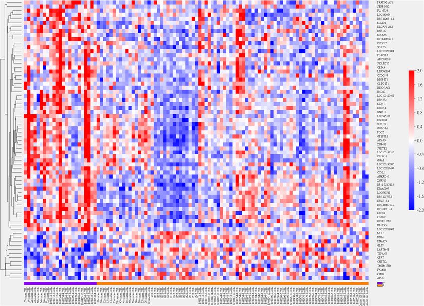
www.nature.com/scientificreports/
primarily relies on the identification of microcalcification, which represents ectopic mammary mineralization.
Other distinguishingly mammographic features telling malignant from benign conditions include speculated
mass, focal asymmetry, architectural distortions, and lesions with rapid p rogressions4.
Histologically there are two kinds of microcalcifications; type I microcalcification is made up of calcium
oxalate (CaC2O4·2H2O) while for type II, hydroxyapatite (Ca10(PO4)·6H2O) is the main composition5,6. In appear-
ance, type I microcalcification is amber while type II is gray to white. Under haematoxylin and eosin (H&E)
dyes, type II microcalcification appears purple, whereas type I is rarely stained. Inspected with light microscopy,
type I microcalcification is partial transparent while type II is opaque. Illuminated with polarized light, type I
microcalcification is birefringent, which is not true for type I I7. The major clinical significance of type I and II
differentiation comes from the observation that type I microcalcification is accompanied with predominately
benign lesions while associated lesions are half benign and half malignant for type II m icrocalcification8.
The absolute number, morphology, and distributions of microcalcifications on mammography are major
deterministic features to tell suspicious from benign lesions, and standardized algorithm has been purposed to
enhance diagnostic accuracy and reduce inter-observer variability such as the Breast Imaging-Reporting and
Data System (BI-RADS) acronym4,9. From literature reviews, candidate genes involving in microcalcification
formation and genes with molecular aberrations have been reported, namely SPARC, SPP1, IBSP, NFKB1, NFKB2,
REL, RELA, RELB, FOS, MYC, IL1B, CXCR4, MMP-1, CTGF, FGF5, and IL11 (more details in “Methods” sec-
tion, ref. 10–25).
Whether microcalcification is merely a radiographic phenomenon during rapid mammary evolution or
is itself an active process contributing to breast carcinogenesis remains inconclusive6,10. The high-throughput
microarrays are advocated to capture whole-transcriptome during a single hybridization, and breast cancer is one
of the most extensively assayed human malignancies. Ironically, rarely has it been reported regarding messenger
RNA (mRNA) abundance associated with microcalcification. In current study, we evaluated the transcriptional
profiles associated with microcalcification for Taiwanese breast cancer with a multi-gene signature derived.
Materials and methods
This study was reviewed and approved by Institute Review Board of Cathay General Hospital. All research was
performed in accordance with relevant guidelines/regulations. Informed consent was obtained from all partici-
pants after explanation by investigators (CCH and CSH). Some breast cancer samples which had been reported
previously were also enrolled in current study11. Microarray data in current study was deposited in Gene Expres-
sion Omnibus (GEO) with the access number GSE146558.
Breast cancer samples. Breast cancer samples were prospectively collected during surgery. Cancerous
breast tissues were snap-frozen and stored in liquid nitrogen at − 80 °C with RNAlater reagent (Qiagen, German-
town, MD) within 30 min after surgery. Frozen samples were cut into 1–2-mm thick slices, and slices with more
than 90% of cancer content were selected for microarray experiments.
Pathological features were retrieved from chart reviews. Estrogen receptor (ER) and progesterone receptor
(PR) positivity was defined as the presence of at least 10% of nuclei with positive immunohistochemical (IHC)
stains. Patients with low ER and PR IHC stains (defined as 1–9% of positive nuclei staining) were not enrolled
as patients with low hormone receptor status were excluded in previous s tudy11. American Society of Cancer
Research (ASCO) and College of American Pathologists (CAP) guidelines were followed for determining human
epidermal growth factor receptor 2 (HER2) status: IHC 3+ and IHC 2+ with fluorescence in situ hybridization
(FISH) amplification were considered as HER2 overexpression. The presence of microcalcification, accompa-
nied ductal carcinoma in situ (DCIS), and comedo-necrosis was ascertained from pathological report. In brief,
microcalcification was detected as magenta to purple granules from routine H&E-stained sections based on the
morphology12. All pathological examinations were carried out by one qualified pathologist (CYL) and more than
90% of cancerous tissue was a prerequisite for downstream experiments.
Microarray experiment. Total RNA was extracted from frozen specimens using TRIzol reagent (Invit-
rogen, Carlsbad, CA). RNA was purified using RNeasy mini kits (Qiagen, Valencia, CA) and RNA integration
was checked by gel electrophoresis. The Affymetrix GeneChip Human Genome U133 plus 2.0 (Thermo Fisher
Scientific, Santa Clara, CA) was used for microarray experiments. Hybridization and scanning were performed
according to the standard Affymetrix protocol. Image scanning was performed using a GeneChip Scanner 3000
(Thermo Fisher Scientific, Santa Clara, CA) with scanned images processed by GeneChip Operating Software
and Affymetrix’s Microarray Suite software to generate detection p values. The Robust Multichip Average (RMA)
algorithm, which consisted three steps of background adjustment, quantile normalization, and final summariza-
tion, was applied for perfect match probe signals within the study13. For multiple probe sets corresponding to
single gene, probe sets were reduced to one per HUGO (Human Genome Organisation) gene symbol by using
the most variable probe set measured by inter-quadrant range across all a rrays14.
Relevant genes for microcalcification. Through extensive literature reviews, genes relevant to the for-
mation, distinguishing features or molecular aberrations of microcalcification were identified. Using IHC stain-
ing, Scimeca et al. postulated that epithelial-mesenchymal transition (EMT) may take place during breast car-
rocess10. They identified
cinogenesis, and the capacity of producing microcalcifications is acquired during such p
vimentin, bone morphogenic protein-2 (BMP-2), β2-microglobulin, β-catenin, and osteopontin (OPN) as tissue
mineralization or mesenchymal phenotype markers investigated with IHC. Through microarray experiments,
Bellahcène et al. demonstrated ectopic mRNA transcription of bone extracellular matrix proteins including bone
sialoprotein (BSP), OPN, and osteonectin (OSN) in osteotropic breast cancers15. It deserves notice that BSP is
Scientific Reports | (2020) 10:18276 | https://doi.org/10.1038/s41598-020-74982-1 2
Vol:.(1234567890)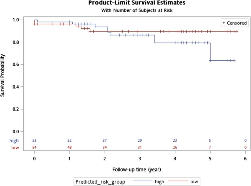
www.nature.com/scientificreports/
supposed to initiate hydroxyapatite deposition and recruit osteoclasts before crystal resorption during the early
phase of bone matrix mineralization16. In addition, BSP expression, when assayed by both immunoperoxidase
and polyclonal antibodies, was correlated with bone metastases and poor s urvival16–18.
Downstream effectors of hydroxyapatite include protein kinase C, nuclear factor-κB, proto-oncogenes c-fos
and c-myc, all of which are associated with subsequent mitogenesis and proliferation from in vitro cell culture
system and with corresponding antibodies19. In addition, up-regulation of matrix metalloproteinases (MMP)-2,
-9, and -13 by hydroxyapatite has been observed in mammary epithelial cell lines by Western blotting20. Over-
expression of MMPs interrupts basement membrane integration, further translating in situ lesion into invasive
cancer21. Other members of MMP family may involve in extracellular matrix degradation and acquiring invasive
ell22. Besides, hydroxyapatite increases transcription of IL-1β, which subsequently increases MMP-1
ability as w
and cyclooxygenase (COX)-2 expression (subjected to Northern analysis), with the latter catalyzing prostaglandin
(PG) E2 production20,23.
Since recent studies have suggested a connection between genes involved in osteoblast differentiation and
breast carcinogenesis, we also evaluated CXCR4, MMP-1, CTGF, FGF5, and IL11, all of which were suggested
by Kang et al. as being interrogated in breast cancer osteolytic metastasis through microarray profiling and
Northern blot confirmation24. These candidate genes encode secreted and cell surface proteins. CXCR4 is a
bone-homing chemokine receptor, while FGF5 and CTGF are angiogenesis growth factors and IL-11 induces
osteoclast from progenitor c ells25–28. Supplementary Table 1 detailed relevant genes of mammary microcalcifica-
tion from literature reviews.
All relevant genes were retrieved and the corresponding Affymetrix probe sets were annotated with NetAffx29.
Boxplot of RMA-normalized expression values of each candidate gene was created between arrays with and
without microcalcification with unpaired two-samples Wilcoxon rank sum test. A public domain microarray
dataset (GSE2109) with clinical microcalcification status reported was evaluated as an additional validation study.
Multi‑gene signature for microcalcification. Genes with differential mRNA abundance based on the
existence of microcalcification were identified, using two-sample T-test with permutation P values estimated
from 10,000 random permutations to correct for multiple testing from high-dimensional microarray data.
Nominal significance level of each univariate test was set to 0.001 to reduce type I error. The same procedure of
filtering significant genes was repeated for DCIS as well as comedo-type DCIS. Gene ontology (GO) terms of
cellular component, molecular function, and biological process were investigated. Only GO classes and parent
classes with at least 5 observations in the selected subset and with an observed versus expected (O/E) ratio of at
least 2 were reported. The Gene Set Comparison Tool implemented in the BRB Array Tools was used for pathway
analysis while a GO category was evident if the corresponding LS or KS resampling P value was below 0.00530.
For multi-gene signature construction, filtered genes were used to predict clinical phenotype by multiple
methods including compound covariate predictor, diagonal linear discriminative analysis, 3 nearest neighbors,
nearest centroid, and support vector machine with default penalty of LIBSVM. Clinical phenotype (histopa-
thology proved microcalcification) was regarded as the gold standard when predictive accuracy was evaluated
through leave one out cross-validation. Two-way hierarchical clustering of filtered genes and breast cancer
samples was performed with average linkage and Euclidean distance. All bioinformatics works were conducted
with the BRB-ArrayTools30.
Breast cancer risk predictive model. A breast cancer risk predictive model was built based on super-
vised principal component r egression31. Candidates genes were those identified as being prognostic by univari-
ate Cox regressions. Significant genes with an α level < 0.001 were used for principal components (supergenes)
synthesis, with the first principal component incorporated into the breast cancer risk predictive model. Relapse-
free survival, with tumor recurrence or metastasis as the first relapse event, was predicted with clinical ER, HER2
status as covariates, and a continuous prognostic index score, which was calculated for each patient by the first
principal component score. The high- and low-risk groups were dichotomized by the 50th percentile of estimated
prognostic index score.
Results
Study population. Breast cancers diagnosed and operated between 2010 and 2014 with curative intention
were recruited. A total of 109 breast cancers were successfully assayed with Affymetrix Human Genome U133
Plus 2.0 microarrays. Molecular subtyping with Prediction Analysis of Microarray 50 (PAM50) single sample
predictor (SSP) tabulating with IHC results and relapse-free survival between centroid-based luminal-A, lumi-
nal-B, basal-like, and HER2-enriched subtype (log-rank test: 0.07) were detailed in Table 1 and Supplementary
Fig. 1, respectively32.
The presence of microcalcification was documented for each subject. Eighty-six (79%) of the 109 breast can-
cers had microcalcification identified from histopathological examination. Of these 86 patients, 81 (94%) were
with coexistent DCIS. On the other hand, only 13 (57%) of the 23 patients without microscopic microcalcification
had their invasive tumors co-grown with DCIS (χ2-test, P < 0.001, Table 2). High-grade necrotic (comedo type)
DCIS were observed in 44 (54%) of the 81 breast cancers with histologically confirmed microcalcifications and
coexistent DCIS whereas only 15% (n = 2) of the 13 breast cancers with DCIS but without histological microc-
alcification, the DCIS lesions were of comedo type (χ2-test, P < 0.01, Table 3).
mRNA abundance of microcalcification‑relevant genes. Figure 1 displayed boxplots of candidate
microcalcification-relevant genes from literature reviews. There was no significant transcriptional discrepancy
of SPARC, SPP1, IBSP, NFKB1, NFKB2, REL, RELA, RELB, FOS, MYC, IL1B, CXCR4, MMP-1, CTGF, FGF5,
Scientific Reports | (2020) 10:18276 | https://doi.org/10.1038/s41598-020-74982-1 3
Vol.:(0123456789)
www.nature.com/scientificreports/
Single sample predictor
IHC results Basal-like HER2-enriched Luminal-A Luminal-B
HR+/HER2+ 3 5 5 3
HR−/HER2− 4 3 28 18
HR+/HER2+ 5 10 1 0
HR−/HER2− 7 3 0 0
Table 1. Distributions of IHC results and PAM50 molecular subtypes. Some patients without clinical HER2
status or HER2:2 + but without ISH testing were discarded. HR hormone receptor, HER2 human epidermal
growth receptor type 2.
Breast cancer
Histopathology Without DCIS With DCIS Total
Without microcalcification 10 (43.5%) 13 (56.5%) 23
With microcalcification 5 (5.8%) 81 (94.2%) 86
Table 2. Distributions of DCIS and histopathology-confirmed microcalcification.
Breast cancer with DCIS
Histopathology Non-comedo DCIS Comedo DCIS Total
Without microcalcification 11 (84.6%) 2 (15.4%) 13
With microcalcification 37 (45.7%) 44 (54.3%) 81
Table 3. Distributions of comedo type DCIS and histopathology-confirmed microcalcification.
and IL11 between breast cancer patients with and without histology-confirmed microcalcification (unpaired
two-samples Wilcoxon rank sum test, all P values > 0.001). Supplementary Fig. 5 showed lack of transcriptional
difference among these genes from the public domain expO dataset (GSE2109), of which 265 breast cancers with
known microcalcification status were assayed.
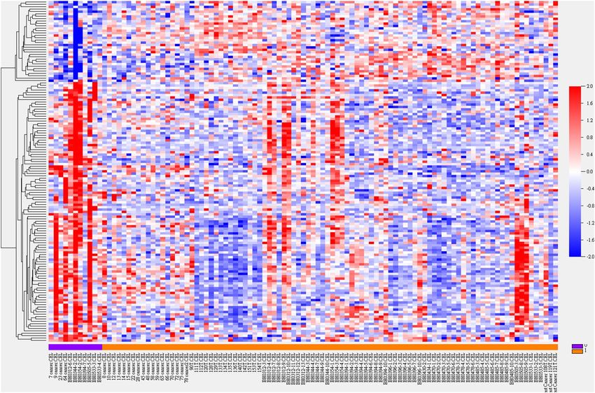
Multi‑gene signature for breast cancer microcalcification. Sixty-nine genes with differential mRNA
abundance pertaining pathological diagnosis of microcalcification passed the filter criterion (Fig. 2). Higher
mRNA transcription of GNRH1, GGA1(Lysosome), CLDN15 and lower mRNA transcription of QPRT (Nicoti-
nate/Nicotinamide), LAPTM4B (Lysosome), and DNAJC5 (HSP40) were significantly coincided with the pres-
ence of microcalcification (Supplementary Fig. 2). Enriched GO terms were Golgi apparatus (cellular compo-
nent), glycosaminoglycan, aminoglycan metabolism, and protein ubiquitination (biological process) with O/E
ratios of 2.34, 7.85 and 5.64 reported (all resampling P values < 0.005), indicating an active secretory process.
There were 143 genes with differential mRNA abundance between breast cancer with and without co-growth
of DCIS including XRCC2 (homologous recombination), GNRH1, CACNA1B (calcium signaling, MAPK path-
way), PRKAA1 (mTOR pathway), DUSP22 (MAPK pathway), MKNK1 (MAPK/mTOR pathway), and SSBP1
(homologous recombination, mitochondria). Figure 3 and Supplementary Fig. 3 depicted clustering heatmap
and volcano plot, respectively. Enriched GO pathways with resampling P values less than 0.005 were endosome
membrane (cellular component, O/E ratio: 2.32), serine-type endo-peptidase activity, carbohydrate binding,
calcium ion binding (molecular function, O/E ratios: 6.05, 4.17, 2.16), and Golgi vesicle transport (biological
process, O/E ratio: 4.81). If comedo type DCIS was selected as classifying variable, 11 significant genes were
identified, including CCDC183, SLMO1, SLC6A5, CES4A, APOD, FMO1 (P450 pathway), and QPRT.
Due to highly correlated phenotype of DCIS and microcalcification (χ2-test, P < 0.001), it was intuitive to
adopt the intersection of genes with differential mRNA abundance from these two clinical variables. A Bayes-
ian compound covariate predictor classifier was built, and the intersection (18 probe sets) provided the best
predictive accuracy with cross-validated Receiver Operating Characteristic (ROC) area under the curve (AUC)
achieving 0.713 (Supplementary Fig. 4 and Supplementary Table 2).
Breast cancer risk predictive model. The intersection of 18 probe sets was further used in relapse-free
survival prediction with the 50th percentile of prognostic index score constructing high-/low risk groups. Ten
genes were selected by fitting penalized Cox proportional hazards model. The results of leave-one-out cross-
validation with clinical ER and HER2 status incorporated as covariates were detailed in Table 4 while Fig. 4 dis-
played Kaplan–Meier plot. Five-year relapse free survival was 91% for low-risk and 83% for high-risk group (log-
rank test, P = 0.10). Supplementary Table 3 detailed predicted risk groups and clinical data for survival analysis.
Scientific Reports | (2020) 10:18276 | https://doi.org/10.1038/s41598-020-74982-1 4
Vol:.(1234567890)www.nature.com/scientificreports/
Figure 1. Boxplots of microcalcification-relevant genes from literature reviews. Each plot includes mRNA
abundance for one gene stratified by the class variable in X-axis (0: without microcalcification and 1: with
microcalcification). The Y-axis represents log intensity and the title shows gene symbol. All comparisons were
insignificant with P values > 0.001 (Wilcoxon rank sum test).
Scientific Reports | (2020) 10:18276 | https://doi.org/10.1038/s41598-020-74982-1 5
Vol.:(0123456789)www.nature.com/scientificreports/
Figure 2. Heatmap of hierarchical clustering of 69 genes with differential mRNA abundance pertaining
pathology-confirmed microcalcification. Sample name and class variable (0: without microcalcification (purple)
and 1: with microcalcification (brown)) were detailed in X-axis. Gene symbols were listed in Y-axis.
Discussion
There remains an unsolved debate on whether mammographic microcalcification is a passive phenomenon
resulted from degraded cell debris during mammary gland proliferation or is itself an active process interven-
ing in breast carcinogenesis. For instance, Scimeca et al. hypothesized that microcalcification is the product of
mesenchymal-epithelial transformation (EMT), and vimentin, BMP-2, β2-microglobulin, β-catenin, and OPN
were proposed as representative IHC m arkers10. In addition to hydroxyapatite, they also found magnesium-
substituted hydroxyapatite in vicinity of malignant breast lesions with energy dispersive X-ray microanalysis.
Acquiring osteoblast-like phenotype through mesenchymal transformation resulted in microcalcifications.
As early as 1972, Stegner et al. had suggested that mammary calcifications were produced by tumor secretions
rather than degenerated cells33. As milk is abundant in calcium, it is essential for mammary epithelial cells to
concentrate calcium i ons34. Another possible explanation of ectopic mammary calcifications came from Holme
et al. as they argued that aberrant calcium metabolism induced breast microcalcification d epositions35. It’s not a
coincidence that Bellahcène et al. using polyclonal antibodies and immunoperoxidase techniques, identified three
bone matrix proteins, namely OPN, OSN, and BSP, which were synthesized by cells with osteoblastic linage and
were expressed in human breast cancers, highlighting the importance of bone matrix mineralization in breast
carcinogenesis while BSP was postulated to initiate hydroxyapatite formation16,17. In addition, aberrant breast
BSP expression also correlated with osteotropic bone metastases when measured with immunoperoxidase and
specific anti-BSP antibodies from breast cancer cell lines and primary breast cancers metastasizing to bone16,18.
Initially we subdivided Taiwanese breast cancer samples based on pathology-proved microcalcification and
tested whether there existed any mRNA abundance discrepancy in these relevant genes from literature reviews.
These candidate genes were grouped into bone matrix proteins (SPARC, SPP1, IBSP), hydroxyapatite-induced
downstream calcium-dependent mitogens (NFKB1, REL, RELA, RELB, NFKB2, FOS, MYC), hydroxyapatide-
induced autocrine IL1B, and osteoblast differentiation associated genes (CXCR4, MMP-1, CTGF, FGF5, and
IL11)19,24. None of these candidate genes showed significant mRNA transcriptional difference between Taiwan-
ese breast cancers with and without histopathogically proved microcalcification. To make sure our experiment
results were not incidental findings, we also consulted the expO dataset (GSE2109) while the lack of tran-
scriptional difference further augmenting our negative findings regarding literature-retrieved relevant genes of
Scientific Reports | (2020) 10:18276 | https://doi.org/10.1038/s41598-020-74982-1 6
Vol:.(1234567890)www.nature.com/scientificreports/
Figure 3. Heatmap of hierarchical clustering of 143 genes with differential mRNA abundance pertaining breast
cancer with co-grown DCIS. Sample name and class variable (0: without DCIS (purple) and 1: with DCIS
(brown)) were detailed in X-axis. Gene symbols were listed in Supplementary Information File due to limited
space.
Coefficient % CV support Probe set Symbol
0.1 90.83 1552845_at CLDN15
1.156 100 1560112_at WDFY2
− 0.423 99.08 1569320_at GPBP1L1
− 0.145 97.25 1569484_s_at MDN1
− 0.792 100 207987_s_at GNRH1
0.104 95.41 215203_at GOLGA4
− 0.314 99.08 217671_at DSERG1
1.231 100 232804_at AP000330.8
− 0.968 100 239556_at LOC645513
0.483 100 244840_x_at DOCK4
Table 4. Genes used in relapse-free survival prediction. CV cross-validation.
microcalcification. Therefore, it is necessary to develop a novel multi-gene signature for microcalcification to
define the prognostic relevance of ectopic breast mineralization.
Under stringent statistical testing (10,000 permutations with a nominal significance level of 0.001), 69 and
143 genes with mRNA transcriptional discrepancy based on the presence of pathological microcalcification and
DCIS were identified. Most of these genes, when inspected individually, did not deliver explicit biological inter-
pretations regarding calcium metabolism or breast precancerous lesion. The multi-gene GO analyses, however,
did infer active secretory processes such as Golgi apparatus (cellular component), and biological pathways of
glycosaminoglycan, aminoglycan metabolism, and protein ubiquitination pronounced in microcalcification-
associated genes. Enriched GO terms for accompanied DCIS differentiated genes included endosome cellular
component, serine-type endopeptidase, carbohydrate binding, calcium ion binding functional pathways as well
Scientific Reports | (2020) 10:18276 | https://doi.org/10.1038/s41598-020-74982-1 7
Vol.:(0123456789)www.nature.com/scientificreports/
Figure 4. Kaplan–Meier plot for five-year relapse-free survival stratified by high-/low- risk defined by the
50th percentile of prognostic index score calculated from 10 intersection genes pertaining pathological
microcalcification and DCIS. X-axis: survival time in year; survival was right-censored at five years.
as Golgi vesicle transport process, all of which further reinforced the clinical interconnection and coexistence
of microcalcification and DCIS.
It was straightforward to develop a robust and concise multi-gene signature from the intersectional genes
pertaining microcalcification and DCIS. We believed transcriptional profiles in terms of mRNA abundance
were more sensitive for early detection of pathogenic microcalcification formation and undelaying in situ lesion
and a Bayesian compound covariate predictor was proposed with satisfactory performance. Ten genes were
further selected to synthesize prognostic index score and relapse-free survival predictive model, which provided
independent prognostic power in additional to ER and HER2 status. Although survival discrepancy between
high- and low-risk group was of borderline significance (P = 0.10), a prognostic trend inherited from microc-
alcification/DCIS intersection genes was prominent. Constitutional genes with increased breast cancer relapse
risk were claudin 15, WDFY2, golgin A4, DOCK4 while HCG27 and PLAC8L1 were among 18 signature signs
not endorsed by prognostic index score.
There were some limitations of current study. First, the undifferentiated mRNA abundance of literature-sug-
gested microcalcification relevant genes may be biased by Affymetrix probe design. Some candidate genes were
reported in IHC assays, immunoperoxidase as well as dispersive X-ray, and transcriptional/translational discrep-
ancy between mRNA and protein measurement inevitably compromised comparability across studies. Second,
bone matrix proteins were up-regulated in metastatic breast cancers, especially those with bone metastases. Our
samples were of early stage breast cancers without distant metastasis during surgery and might not have these
osteotropic genes up-regulated. Third, multi-gene signature for osteolytic bone metastasis from breast cancer
cell lines had been published, further studies to recruit more clinical samples including advanced breast cancers
with bone metastases should be initiated to elucidate microcalcification deposition and osteotropic metastatic
mechanism driven by calcium m etabolism15,36. Another unanswered argue might arise from whether multi-gene
signature can outperform abnormal mammographic readings such as those debrided by BIRADS categories 0,
4, 5 in terms of sensitivity, specificity, and accuracy. Indeed, the oversensitivity and low positive predictive value
of mammography resulted in high and unnecessary callbacks and biopsies. A minimally invasive multi-gene
signature-based testing from liquid biopsy might be an alternative to current screening modality. Lastly, Current
study did not decipher the impact of molecular subtyping (such as IHC4) upon the presence of microcalcifica-
tion, and future studies with more samples enrolled can evaluate their impact upon mammary c alcification37.
It deserves notice that the number of cases for microarray experiments was modest only, but details were
provided for individual patient (e.g., pathological microcalcification and DCIS status, survival time, relapse
indicator, and relevant clinical parameters). Further validation studies for the proposed signature could be
Scientific Reports | (2020) 10:18276 | https://doi.org/10.1038/s41598-020-74982-1 8
Vol:.(1234567890)www.nature.com/scientificreports/
conducted by using the most updated method of mRNA quantitation and independent breast cancer patient
samples. Nevertheless, our study suggested that mammary microcalcification is not only the earliest detectable
radiological sign for breast cancer screening but the phenomenon, ectopic breast mineralization, to some degree,
may reflect the underling events during mammary carcinogenesis. Prognostic relevance of the proposed signature
might result from relevant biological processes that contribute to the molecular heterogeneity of human breast
cancers. Future prospective studies to evaluate the prognostic significance of microcalcification are warranted.
Data availability
Microarray data in current study was deposited in Gene Expression Omnibus (GEO) with the access number
GSE146558.
Received: 17 May 2020; Accepted: 29 September 2020
References
1. Elmore, J. G., Armstrong, K., Lehman, C. D. & Fletcher, S. W. Screening for breast cancer. JAMA 293, 1245–1256 (2005).
2. Yen, A. M. et al. Population-based breast cancer screening with risk-based and universal mammography screening compared with
clinical breast examination: a propensity score analysis of 1429890 Taiwanese women. JAMA Oncol. 2, 915–921 (2016).
3. U.S. Preventive Services Task Force. Final recommendation statement: breast cancer: screening. U.S. Preventive Services Task Force
https: //www.usprev entiv eserv icest askfo
rce.org/Page/Docume nt/Recomm
endat ionSt ateme ntFin
al/breast -cancer -screen
ing1 (2019).
4. Rao, A. A., Feneis, J., Lalonde, C. & Ojeda-Fournier, H. A pictorial review of changes in the BI-RADS fifth edition. Radiographics
36, 623–639 (2016).
5. VanHouten, J. N. Calcium sensing by the mammary gland. J. Mammary Gland Biol. Neoplasia 10, 129–139 (2005).
6. Morgan, M. P., Cooke, M. M. & McCarthy, G. M. Microcalcifications associated with breast cancer: an epiphenomenon or biologi-
cally significant feature of selected tumors?. J. Mammary Gland Biol. Neoplasia 10, 181–187 (2005).
7. Frappart, L. et al. Structure and composition of microcalcifications in benign and malignant lesions of the breast: study by light
microscopy, transmission and scanning electron microscopy, microprobe analysis, and X-ray diffraction. Hum. Pathol. 15, 880–889
(1984).
8. Büsing, C., Keppler, U. & Menges, V. Differences in microcalcification in breast tumours. Virchows Arch. (Pathol. Anat.) 393,
307–313 (1981).
9. Spak, D. A., Plaxco, J. S., Santiago, L., Dryden, M. J. & Dogan, B. E. BI-RADS fifth edition: a summary of changes. Diagn. Interv.
Imaging 98, 179–190 (2017).
10. Scimeca, M. et al. Microcalcifications in breast cancer: an active phenomenon mediated by epithelial cells with mesenchymal
characteristics. BMC Cancer 14, 286 (2014).
11. Huang, C. C. et al. Concurrent gene signatures for han chinese breast cancers. PLoS ONE 8, e76421 (2013).
12. Rosen, P. P., Hoda, S. A., Brogi, E. & Koerner, F. C. Rosen’s Breast Pathology 4th edn. (Lippincott Williams & Wilkins, Philadelphia,
2015).
13. Irizarry, R. A. et al. Exploration, normalization, and summaries of high density oligonucleotide array probe level data. Biostatistics
4, 249–264 (2003).
14. Bruford, E. A. et al. Guidelines for human gene nomenclature. Nat. Genet. 52, 754–758 (2020).
15. Bellahcène, A. et al. Transcriptome analysis reveals an osteoblast-like phenotype for human osteotropic breast cancer cells. Breast
Cancer Res. Treat. 101, 135–148 (2007).
16. Bellahcène, A., Merville, M. P. & Castronovo, V. Expression of bone sialoprotein, a bone matrix protein, in human breast cancer.
Cancer Res. 54, 2823–2826 (1994).
17. Bellahcène, A. & Castronovo, V. Increased expression of osteonectin and osteopontin, two bone matrix proteins, in human breast
cancer. Am. J. Pathol. 146, 95–100 (1995).
18. Bellahcène, A., Kroll, M., Liebens, F. & Castronovo, V. Bone sialoprotein expression in primary human breast cancer is associated
with bone metastases development. J. Bone Miner. Res. 11, 665–670 (1996).
19. McCarthy, G. M. et al. Molecular mechanism of basic calcium phosphate crystal-induced activation of human fibroblasts. Role of
nuclear factor kappab, activator protein 1, and protein kinase c. J. Biol. Chem. 273, 35161–35169 (1998).
20. Morgan, M. P., Cooke, M. M., Christopherson, P. A., Westfall, P. R. & McCarthy, G. M. Calcium hydroxyapatite promotes mitogen-
esis and matrix metalloproteinase expression in human breast cancer cell lines. Mol. Carcinog. 32, 111–117 (2001).
21. Nelson, A. R., Fingleton, B., Rothenberg, M. L. & Matrisian, L. M. Matrix metalloproteinases: biologic activity and clinical implica-
tions. J. Clin. Oncol. 18, 1135–1149 (2000).
22. Lochter, A. et al. Matrix metalloproteinase stromelysin-1 triggers a cascade of molecular alterations that leads to stable epithelial-
to-mesenchymal conversion and a premalignant phenotype in mammary epithelial cells. J. Cell Biol. 139, 1861–1872 (1997).
23. Rutter, J. L., Benbow, U., Coon, C. I. & Brinckerhoff, C. E. Cell-type specific regulation of human interstitial collagenase-1 gene
expression by interleukin-1 beta (IL-1 beta) in human fibroblasts and BC-8701 breast cancer cells. J. Cell. Biochem. 66, 322–336
(1997).
24. Kang, Y. et al. A multigenic program mediating breast cancer metastasis to bone. Cancer Cell 3, 537–549 (2003).
25. Taichman, R. S. et al. Use of the stromal cell-derived factor-1/CXCR4 pathway in prostate cancer metastasis to bone. Cancer Res.
62, 1832–1837 (2002).
26. Giordano, F. J. et al. Intracoronary gene transfer of fibroblast growth factor-5 increases blood flow and contractile function in an
ischemic region of the heart. Nat. Med. 2, 534–539 (1996).
27. Moussad, E. E. & Brigstock, D. R. Connective tissue growth factor: what’s in a name?. Mol. Genet. Metab. 71, 276–292 (2000).
28. Manolagas, S. C. Role of cytokines in bone resorption. Bone 17(2 Suppl), 63S-67S (1995).
29. Thermo Fisher Scientific. NetAffx Analysis Center. Thermo Fisher Scientific. https: //www.affyme trix. com/analys is/index. affx (2017).
30. Simon, R. et al. Analysis of gene expression data using BRB-ArrayTools. Cancer Inform. 3, 11–17 (2007).
31. Bair, E. & Tibshirani, R. Semi-supervised methods to predict patient survival from gene expression data. PLOS Biol. 2, 511–522
(2004).
32. Parker, J. S. et al. Supervised risk predictor of breast cancer based on intrinsic subtypes. J. Clin. Oncol. 27, 1160–1167 (2009).
33. Stegner, H. & Pape, C. Beitrag zur feinstruktur der sogenanntan mikrokalzifikation in mammatumoren. Zentralbl. Allg. Path. 115,
106–112 (1972).
34. Ahmed, A. Calcification in human breast carcinomas: ultrastructural observations. J. Pathol. 117, 247–251 (1975).
35. Holme, T. et al. Is mammographic microcalcifiication of biological significance?. Eur. J. Surg. Oncol. 19, 250–253 (1993).
36. Yibin, K. et al. A multigenic program mediating breast cancer metastasis to bone. Cancer Cell 3, 537–549 (2003).
Scientific Reports | (2020) 10:18276 | https://doi.org/10.1038/s41598-020-74982-1 9
Vol.:(0123456789)www.nature.com/scientificreports/
37. Cuzick, J. et al. Prognostic value of a combined estrogen receptor, progesterone receptor, Ki-67, and human epidermal growth
factor receptor 2 immunohistochemical score and comparison with the Genomic Health recurrence score in early breast cancer.
J. Clin. Oncol. 29, 4273–4278 (2011).
Acknowledgements
This study contained materials presented at San Antonio Breast Cancer Symposium 2015 and Annual Scientific
Meeting of Taiwan Surgical Society 2016. The study was funded partially by Ministry of Science and Technol-
ogy, Taiwan, with the Grant Numbers NSC102-2314-B-281-003-MY3 and MOST 107-2314-B-075 -073 -MY3.
Author contributions
H.T.T. and C.SH. drafted the manuscript. C.C.T. and S.H.T. analyzed the results and correlated with clinical
data. C.Y.L. performed pathological exams and ascertained accurate diagnosis. C.J.H. and Y.S.H. established
bioinformatics analytical pipelines. L.M.T. and C.C.H. finalized the manuscript. C.C.H. initiated the and took
responsibility of study execution. All authors reviewed the manuscript and consented the publication.
Competing interests
The authors declare no competing interests.
Additional information
Supplementary information is available for this paper at https://doi.org/10.1038/s41598-020-74982-1.
Correspondence and requests for materials should be addressed to L.-M.T. or C.-C.H.
Reprints and permissions information is available at www.nature.com/reprints.
Publisher’s note Springer Nature remains neutral with regard to jurisdictional claims in published maps and
institutional affiliations.
Open Access This article is licensed under a Creative Commons Attribution 4.0 International
License, which permits use, sharing, adaptation, distribution and reproduction in any medium or
format, as long as you give appropriate credit to the original author(s) and the source, provide a link to the
Creative Commons licence, and indicate if changes were made. The images or other third party material in this
article are included in the article’s Creative Commons licence, unless indicated otherwise in a credit line to the
material. If material is not included in the article’s Creative Commons licence and your intended use is not
permitted by statutory regulation or exceeds the permitted use, you will need to obtain permission directly from
the copyright holder. To view a copy of this licence, visit http://creativecommons.org/licenses/by/4.0/.
© The Author(s) 2020
Scientific Reports | (2020) 10:18276 | https://doi.org/10.1038/s41598-020-74982-1 10
Vol:.(1234567890)You can also read


