Mutational spectrum and classification of novel mutations in patients with metastatic gastrointestinal stromal tumours
←
→
Page content transcription
If your browser does not render page correctly, please read the page content below
INTERNATIONAL JOURNAL OF ONCOLOGY 56: 1468-1478, 2020

      Mutational spectrum and classification of novel mutations in
       patients with metastatic gastrointestinal stromal tumours
           ALENKA BOMBAC1,2, BRANKO ZAKOTNIK3, MARINA BUCIC1, VITA SETRAJCIC DRAGOS1,
              BARBARA GAZIC4, VIDA STEGEL1, GASPER KLANCAR1 and SRDJAN NOVAKOVIC1

                     1
                         Department of Molecular Diagnostics, Institute of Oncology; 2Faculty of Medicine,
                         University of Ljubljana; 3Division of Medical Oncology and 4Department of Pathology,
                                       Institute of Oncology Ljubljana, Ljubljana 1000, Slovenia

                                      Received October 24, 2019; Accepted February 28, 2020

                                                     DOI: 10.3892/ijo.2020.5028

Abstract. In total, ~85% of malignant gastrointestinal                in Slovenian patients are comparable with those in other
stromal tumours (GISTs) harbour activating mutations in one           European populations. In the present group of patients anal-
of the genes KIT or PDGFRA, while 10‑15% of all GISTs                 ysed, the most frequently mutated region was exon 11 in the
have no detectable KIT or PDGFRA mutations, but could                 KIT gene, responsible for coding juxtamembrane domain of
have alterations in genes of the succinate dehydrogenase              KIT protein. In this region, eight novel mutations were iden-
complex or in BRAF, PIK3CA or rarely RAS family genes.                tified and classified as likely pathogenic driver variants. In
The clinical benefit of tyrosine kinase inhibitors, such as           addition, the present study identified 6 patients with secondary
imatinib, depends on the GIST genotype, therefore molecular           KIT mutation and 1 patient with double mutant GIST, who
characterization of GIST has a crucial role in overall manage-        had two different mutations in PDGFRA exon 14.
ment of GIST. The aim of the present study was to molecularly
characterize a cohort of 70 patients with metastatic GISTs            Introduction
from the Slovenian Cancer Registry (National Cancer
Registry) treated between January 2002 and December 2011.             Gastrointestinal stromal tumours (GISTs) are the most
Exons 9, 11, 13 and 17 of the KIT gene and exons 12, 14               common type of soft tissue sarcomas that arise from the
and 18 of the PDGFRA gene were analysed by direct Sanger              mesenchymal cells of the gastrointestinal tract and have char-
sequencing. All KIT/PDGFRA wild‑type GISTs were tested                acteristic histological and molecular features (1). According to
for the presence of mutations in hot spot regions of KRAS,            the literature, most GISTs are driven by activating mutations in
NRAS, BRAF, PIK3CA and AKT1 genes. Novel variants were                the proto‑oncogenes KIT and PDGFRA (2). In total, ~85% of
characterized and classified using Cancer Genome Interpreter          malignant GISTs harbour activating mutations in one of the
and according to The American College of Medical Genetics             genes, KIT or PDGFRA, while 10‑15% of all GISTs have no
and Genomics/Association for Molecular Pathology guide-               detectable KIT or PDGFRA mutations (2‑4).
lines. In total, 60 (85.7%) patients had mutations in KIT                 Receptors KIT and platelet derived growth factor
and 2 (2.9%) in PDGFRA. Whereas, 8 (11.4%) patients with              receptor α (PDGFRA) have a similar structure and activa-
GIST had no mutation in either of the analysed genes. The             tion mechanism, as they belong to the same sub‑family, the
majority of GIST cases (n=52) had a mutation in KIT exon 11,          type III receptor tyrosine kinases (5). Their typical structure is
where 40 different mutations were detected. Eight of the              composed of an extracellular (EC) domain, a transmembrane
variants were novel: c.1652_1672del, c.1653_1660delinsAA,             domain, an intracellular juxtamembrane (JM) domain and
c.1665_1672delinsCC, c.1668_1686del, c.1676_1720del,                  a cytoplasmic kinase domain, which is divided by a kinase
c.1715_1756dup, c.1721_1765dup, and c.1722_1766dup.                   insert into two catalytic parts, tyrosine kinase domain I (TK I)
Mutation frequencies of KIT and PDGFRA genes observed                 and tyrosine kinase domain II (TK II) (6).
                                                                          Gain‑of‑function mutations in KIT or PDGFRA gene
                                                                      cause functional changes in KIT or PDGFRA receptor, which
                                                                      lead to constitutive, ligand‑independent phosphorylation of
                                                                      the receptor and activation of downstream to KIT/PDGFRA
Correspondence to: Professor Srdjan Novakovic, Department of
Molecular Diagnostics, Institute of Oncology, Ljubljana, Zaloska 2,   signalling pathways (RAS/RAF/MAPK, PI3K/AKT/mTOR
Ljubljana 1000, Slovenia                                              and JAK/STAT3), ultimately increasing cell proliferation and
E‑mail: snovakovic@onko‑i.si                                          inhibiting apoptosis (6,7).
                                                                          In GIST, 67% of KIT mutations occur in exon 11
Key words: gastrointestinal stromal tumour, KIT mutations,            (JM domain). The mutation frequency in other regions of
imatinib                                                              the KIT receptor is markedly lower; mutations in exon 9
                                                                      (EC domain) occur in 8‑10% of GISTs, whilst exons 13 and 17
                                                                      (TK I and TK II) are mutated in ~1% of all GISTs (8‑10).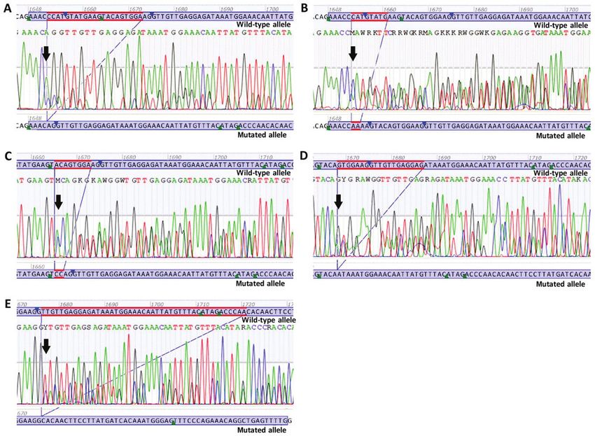
BOMBAC et al: MUTATIONAL SPECTRUM IN GIST                                           1469

PDGFRA mutations occur in ~8% of GISTs, of which exon 14                As controls for immunohistochemistry (IHC), normal
(TK I) and exon 18 (TK II) are mutated in 6‑7% of the GISTs,        appendiceal tissue samples were obtained from 5 patients
and exon 12 (JM domain) in 1% (11,12).                              (3 females, 2 males; age, 57‑74 years; median, 63 years)
     GISTs without a mutation in the KIT or PDGFRA genes are        undergoing various surgical procedures. In addition, tumour
historically known as wild‑type (WT) GIST; however, their           tissue samples were obtained from 10 patients (6 females,
molecular biology is very heterogeneous. KIT/PDGFRA WT              4 males; age, 42‑83 years; meadian, 60 years) with histo-
GISTs can have alterations in genes of the succinate dehydro-       pathologically confirmed CD117‑positive non‑metastatic
genase complex or in BRAF, PIK3CA, or rarely RAS family             GIST. All patients included as IHC controls were treated at
genes (13,14). BRAF V600E mutation has been reported in             the Institute of Oncology Ljubljana between January 2002 and
7‑15% of KIT/PDGFRA WT GISTs, meanwhile RAS and                     December 2011.
PIK3CA mutant GISTs are reported to be very rare (15‑18).               All collected tumour samples were evaluated and
Even more rare are recently reported NTRK fusions in these          were histopathologically examined under a microscope by
patients, however they are important for treatment deci-            pathology specialists, who confirmed the diagnosis using
sions (19).                                                         immunohistochemical staining for CD117 antigen expression.
     Before the discovery of tyrosine kinase inhibitor (TKI)        IHC analysis of CD117 was performed on 2‑4 µm FFPE tissue
imatinib, no effective therapy was available for the treatment      sections, dried at 56˚C for 2 h. Heat‑mediated epitope retrieval
of unresectable, metastatic or recurrent GIST. The overall          was performed in a microwave oven using home‑brew pH 9.0
survival (OS) time of such patients was 9‑20 months (20,21). With   Tris‑EDTA buffer for 10 min at 100˚C at high pressure.
imatinib, the median OS time of patients with advanced GISTs        Detection was performed in the semi‑automated IHC system
is significantly longer and is reported to be ~55‑57 months (21).   LabVision™ Autostainer AS480. Endogenous peroxidases
Although the majority of KIT/PDGFRA‑mutant GIST                     were blocked using Dako REAL™ Peroxidase blocking solution
patients (90%) achieve clinical benefit from imatinib, primary      (cat. no. S2023; Dako; Agilent Technologies, Inc.) for 8 min at
resistance frequently occurs in GISTs that are KIT/PDGFRA WT,       room temperature. CD117 was detected using a commercially
have KIT exon 9 mutation or have a substitution D842V in gene       available mouse polyclonal antibody CD117 (cat. no. A4502;
PDGFRA (22,23). Furthermore, secondary resistance develops          Dako; Agilent Technologies, Inc.; 1:100), which was added for
in 40‑50% of patients with GIST within 2 years of successful        30 min at room temperature. The antibody was diluted using
treatment due to secondary KIT/PDGFRA mutations or acti-            Dako REAL™ antibody diluent (cat. no. S2022; Dako; Agilent
vating mutations of another downstream signalling pathway,          Technologies, Inc.). Primary antibody was visualised using
such as the RAS/RAF/MAPK or PI3K/AKT/mTOR path-                     2‑step polymer detection system Dako REAL™ EnVision™
ways (18,22,23). Imatinib efficacy greatly depends on tumour        Detection system, Peroxidase/DAB+ Rabbit/ Mouse
genotype; therefore, molecular characterization of GIST is a very   (cat. no. K5007; Dako; Agilent Technologies, Inc.). Ready
important step in optimizing GIST clinical treatment (21,23).       to‑use secondary antibody Dako REAL™ EnVision™/HRP
     The aim of the present study was to molecularly charac-        Rabbit/Mouse (cat. no. K5007; Dako; Agilent Technologies,
terize a cohort of 70 patients with metastatic GISTs from the       Inc.) was added for 30 min at room temperature. Chromogene
Slovenian Cancer Registry (National Cancer Registry) treated        Dako REAL™ DAB+ Chromogen (cat. no. K5007; Dako;
between period 2002 and 2011. The spectrum of KIT and               Agilent Technologies, Inc.) was diluted at 1:50, according to
PDGFRA mutations was reported, as well as the spectrum              manufacturer's protocol and incubated twice for 7.5 min each
of hot spot mutations in KRAS, NRAS, BRAF, PIK3CA and               at room temperature.
AKT1 genes in patients with GIST.                                       Tissue sections were viewed under a light microscope
                                                                    (magnification, x10). The section of the FFPE slides that
Materials and methods                                               contained the highest number of tumour cells (always >70% in
                                                                    all GIST samples) was marked and used for DNA extraction.
Patients. In total, 70 patients with metastatic KIT‑positive            The present study was approved by the Institutional
GIST, treated with TKIs at the Institute of Oncology                Review Board of the Institute of Oncology Ljubljana (permis-
Ljubljana (Ljubljana, Slovenia) between January 2002 and            sion no. OIRIKE00036) and by the National Medical Ethics
December 2011, were recruited and included in the present           Committee of Republic of Slovenia (permission no. 1090216).
analysis.                                                           Individual patient consent was waived for this study as it was
    Formalin‑fixed, paraffin‑embedded (FFPE) primary                a retrospective study, the research involved no risk to the
tissue samples were mainly obtained from the Department             subjects, and the institutional informed consent forms for
of Pathology, Institute of Oncology Ljubljana (72%) and             treatment included consent for the use of patient's data, mate-
also from pathology departments from other hospitals and            rials and/or test results for research purposes. All procedures
clinics in Slovenia (University Medical Centre Ljubljana,           followed in the present study were therefore in accordance
Ljubljana; University Medical Centre Maribor, Maribor;              with the ethical standards of the responsible committees on
Izola General Hospital, Izola; and Celje General Hospital,          human experimentation (institutional and national) and the
Celje). All patients were treated with TKIs at the Institute        Helsinki Declaration of 1975, as revised in 2013.
of Oncology Ljubljana. In total, 12 of the 70 samples were
obtained from patients after the start of imatinib therapy.         Molecular analysis. Mutational analyses of KIT, PDGFRA,
Clinicopathological characteristics of the patients, as well as     KRAS, NRAS and BRAF genes were performed by the
prognostic risk groups according to Miettinen et al (4), are        Department of Molecular Diagnostic at the Institute of
presented in Table I.                                               Oncology Ljubljana (Ljubljana, Slovenia). Genomic DNA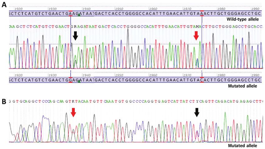
1470                                 INTERNATIONAL JOURNAL OF ONCOLOGY 56: 1468-1478, 2020

Table I. Clinicopathological characteristics of 70 patients with    extracts were assessed using a NanoDrop spectrophotometer
gastrointestinal stromal tumour.                                    (Thermo Fisher Scientific, Inc.).
                                                                        All GISTs (n=70) were examined for somatic muta-
Clinicopathological characteristic                     n (%)        tions in KIT (exons 9, 11, 13 and 17) and PDGFRA
                                                                    (exons 12, 14 and 18) by polymerase chain reaction (PCR) ampli-
Sex                                                                 fication and direct Sanger sequencing. For PCR amplification,
 Male                                                32 (45.7)      Invitrogen™EXPRESS SYBR™ GreenER™ qPCR Supermix,
 Female                                              38 (54.3)      universal (Invitrogen; Thermo Fisher Scientific, Inc.), which
Age                                                                 includes Platinum™ Taq DNA Polymerase, was used. PCR
 Median, years                                       65             reactions were run on an Applied Biosystems™ 7900HT Fast
 Range, years                                        34‑89          Real‑Time PCR system (Applied Biosystems; Thermo Fisher
 ≤60 years                                           28 (40.0)      Scientific, Inc.) with the following thermocycling conditions:
                                                                    Activation at 50˚C for 2 min; followed by denaturation at
 >60 years                                           42 (60.0)
                                                                    95˚C for 10 min; 20 cycles of denaturation at 95˚C for 35 sec,
Curative resection                                                  annealing at 64˚C for 45 sec and elongation at 72˚C for 1 min;
 Yes                                                 13 (18.6)      followed by another 30 cycles of denaturation at 95˚C for 15 sec,
 No                                                  57 (81.4)      annealing at 55˚C for 45 sec and elongation at 72˚C for 1 min.
Imatinib therapy                                                    Direct Sanger sequencing reactions were performed using a
 Yes                                                 70 (100.0)     Big Dye Terminator v3.1 (Applied Biosystems; Thermo Fisher
 No                                                   0 (0.0)       Scientific, Inc.) and run on an ABI 3500 Genetic Analyzer
                                                                    (Applied Biosystems; Thermo Fisher Scientific, Inc.). Specific
Outcome
                                                                    primers for target exon amplification and sequencing reactions
 Alive                                               21 (30.0)      were designed using Primer3 web software (version 0.1g;
 Dead                                                49 (70.0)      http://biotools.nubic.northwestern.edu/Primer3.html) and are
Primary tumour location                                             shown in Table SI.
 Oesophagus                                           2     (2.9)       Furthermore, all patients with KIT/PDGFRA WT GIST
 Stomach                                             33    (47.1)   were tested for the presence of hot spot mutations in KRAS,
 Small intestine                                     25    (35.7)   NRAS, BRAF, PIK3CA and AKT1. Testing was performed
 Rectum                                               6     (8.6)   using Colorectal Cancer Mutation Detection Panel v1.3
 Extraluminal                                          4    (5.7)   (EntroGen, Inc.), a quantitative PCR‑based assay, according
                                                                    to the manufacturer's protocol. A list of all hot spot mutations
Mitotic index, /50 HPF                                              that are included in the Colorectal Cancer Mutation Detection
 ≤5                                                  22 (31.4)      Panel are presented in Table SII.
 5.1‑10                                              11 (15.7)
 >10                                                 37 (52.9)      Sequence analysis. DNA sequences were analysed using
Primary tumour size, cm                                             Sequencing Analysis software v5.4 software (Applied Biosystems;
 0‑5                                                  6 (8.5)       Thermo Fisher Scientific, Inc.), ChromasLite software v2.1.1
 5.1‑10                                              23 (32.9)      (Technelysium Pty Ltd.) and GeneRunner software (http://www.
 >10                                                 37 (52.9)      softpedia.com/get/Science‑CAD/Gene‑Runner.shtml). Mutation
                                                                    nomenclature followed the recommendations of the Human
 Unknown                                              4 (5.7)
                                                                    Genome Variation Society v19.01 (http://varnomen.hgvs.org/).
Risk classificationa                                                Reference sequences used in the present study were taken
 High                                                49 (70.0)      from the National Centre for Biotechnology Information data-
 Intermediate                                        11 (15.7)      base (https://www.ncbi.nlm.nih.gov/): KIT (NM_000222.2),
 Low                                                  9 (12.9)      PDGFRA (NM_006206), KRAS (NM_004985), NRAS
 Very low                                             1 (1.4)       (NM_002524), BRAF (NM_004333), PIK3CA (NM_006218.2)
Morphology                                                          and AKT1 (NM_005163.2).
 Spindle cells                                       47 (67.1)
                                                                    Literature search of detected variants. A literature search for
 Epithelioid cells                                    8 (11.5)
                                                                    all detected variants found in the present study was performed
 Mixed cells                                         15 (21.4)
                                                                    using Alamut Visual v2.11 software (SOPHiA GENETICS). To
Risk groups for gastrointestinal tumours were adapted from
a                                                                   perform a search for the references of every variant detected,
Miettinen et al (4).                                                a gene name and a nucleotide change (c.notation) were typed
                                                                    into Alamut Visual browser, which is linked to Google.

                                                                    Online in silico analysis and bioinformatics tools.
of 70 sporadic GISTs was isolated from FFPE tissues, using          In silico mutation prediction analyses were performed
macrodissection and standard procedures, according to the           using the online bioinformatics tools: Align GVGD
manufacturer's protocol of the QIAmp DNA FFPE Tissue                (http://agvgd.hci.utah.edu/) MutationTaster2 (http://www.muta-
kit (Qiagen GmbH). The quality and concentration of DNA             tiontaster.org), and Cancer Genome Interpreter (https://www.
BOMBAC et al: MUTATIONAL SPECTRUM IN GIST 1471
Figure 1. Sanger sequencing electropherograms of double mutant gastrointestinal stromal tumour. (A) Forward sequence. Black arrow is indicating the
nucleotide change c. 1936A>G p.(Lys627Glu) in PDGFRA exon 14. Red arrow is indicating the nucleotide change c.1975A>C p.(Asn659His) in PDGFRA
exon 14. (B) Reverse sequence. Black arrow is indicating the nucleotide change c. 1936A>G p.(Lys627Glu) in PDGFRA exon 14. Red arrow is indicating the
nucleotide change c.1975A>C p.(Asn659His) in PDGFRA exon 14.
Table II. Mutational status and mutation location of all 70
cancergenomeinterpreter.org/analysis). Their pathogenicity patients with GIST from the Slovenian Cancer Registry.
was assessed according to The American College of Medical
Genetics and Genomics/Association for Molecular Pathology Mutation genotype Number of GISTs
(ACMG/AMP) criterions using Genetic Variant Interpretation
Tool (http://www.medschool.umaryland.edu/Genetic_ KIT/PDGFRA‑mutant GIST 62
Variant_Interpretation_Tool1.html/). KIT (exon 9) 6
KIT (exon 11) 52
Results
KIT (exon 13) 5a
KIT (exon 17) 3b
Genotype analysis
KIT/PDGFRA mutant GISTs. Among 70 metastatic GIST PDGFRA (exon 12) 0
tumours, 62 (88.6%) KIT/PDGFRA mutant GISTs were iden- PDGFRA (exon 14) 1c
tified. Of the analysed GISTs, 60 (85.7%) had a mutation in PDGFRA (exon 18) 1
KIT and only 2 (2.9%) had a mutation in PDGFRA. Of the Wild‑typed GIST 8
60 patients with KIT mutant GISTs, 6 (10%) developed a
secondary mutation in exon 13 or 17 of the KIT gene. These
a
Two GISTs with primary mutation and three GISTs with secondary
mutation. bThree GISTs with secondary mutation. cOne GIST with
secondary mutations were found in the patients whose tissue
two primary mutations. dWild‑type for KIT, PDGFRA, BRAF, KRAS,
samples were obtained following imatinib treatment.
NRAS, PIK3CA and AKT1. GIST, gastrointestinal tumour.
Among the 60 patients with KIT mutation, KIT exon 11 was
mutated in 52 patients (86.7%), harbouring 40 different vari-
ants. KIT exon 9 was mutated in 6 cases (10%) and all carried a
frequent six nucleotides duplication, c.1504_1509dup p.(Ala502_
Tyr503dup). KIT exon 13 was mutated in 5 cases (8.3%); (Fig. 1). Mutational status and mutation location of all 70 GISTs
2 patients had a primary mutation and 3 patients had a secondary are presented in Table II. All detected alterations and their
mutation. KIT exon 17 was mutated in 3 cases (5%), where all status of pathogenicity in 62 KIT/PDGFRA mutant GISTs from
3 patients had a secondary mutation (Table II). the Slovenian Cancer Registry are presented in Table SIII.
As aforementioned, mutations in PDGFRA were found
in 2 cases (2.9%). The first patient had a single nucleotide KIT/PDGFRA wild‑type GIST. All eight KIT/PDGFRA WT
change in exon 18, c.2525A>T p.(Asp842Val), while the second GISTs had no mutations in hot spot regions of KRAS (codons
patient had two different single nucleotide changes, c.1936A>G 12/13/59/61/117/146), NRAS (codons 12/13/59/61/117/146),
p.(Lys627Glu) and c.1975A>C p.(Asn659His), both in exon 14. BRAF (codon 600), PIK3CA (codons 542/545/1047) or AKT1
The latter case was a rare example of double mutant GIST (codon 17).1472
Table III. Classification of eight novel heterozygous variants in KIT exon 11.
ACMG/AMP Classification by Cancer Genome
Location of the alteration In silico prediction Genetic Variant Interpretation Tool Interpreter
‑‑‑‑‑‑‑‑‑‑‑‑‑‑‑‑‑‑‑‑‑‑‑‑‑‑‑‑‑‑‑‑‑‑‑‑‑‑‑‑‑‑‑‑‑‑‑‑‑‑‑‑‑‑‑‑‑‑‑‑‑‑‑‑‑‑‑‑‑‑‑‑‑‑‑‑‑‑‑‑‑‑‑‑‑‑‑‑‑‑‑‑‑‑‑‑‑‑‑‑‑‑‑ ‑‑‑‑‑‑‑‑‑‑‑‑‑‑‑‑‑‑‑‑‑‑‑‑‑‑‑‑‑‑‑‑‑‑‑‑‑‑‑‑‑‑‑‑‑‑‑‑‑‑‑‑‑‑‑‑‑‑‑‑‑‑‑‑‑‑‑‑‑‑‑‑‑‑‑‑‑‑‑‑‑‑‑‑‑‑‑‑‑‑‑‑‑‑‑‑‑‑‑ ‑‑‑‑‑‑‑‑‑‑‑‑‑‑‑‑‑‑‑‑‑‑‑‑‑‑‑‑‑‑‑‑‑‑‑‑‑‑‑‑‑‑‑‑‑‑‑‑‑‑‑‑‑‑‑‑‑‑‑‑‑‑‑‑ ‑‑‑‑‑‑‑‑‑‑‑‑‑‑‑‑‑‑‑‑‑‑‑‑‑‑‑‑‑‑‑‑‑‑‑‑‑‑‑‑‑‑‑‑
Nucleotide change Amino ccid change Align GVGD MutationTaster2 Criterion Pathogenicity Oncogenic prediction
(c.notation) (p.notation)
c.1652_1672del p.(Pro551_Lys558delinsGln) Class C65 Prediction disease causing PM1, PM2, Likely Predicted driver: tier 1
(GV: 0.00 ‑ GD: 75.14) ‑ long InDel. PM4, PP3 pathogenic (IV)
Model: complex_aae,
prob: 0.999999999999935
c.1653_1660delinsAA p.(Met552_Glu554delinsLys) Class C65 Prediction disease causing. PM1, PM2, Likely Predicted driver: tier 1
(GV: 0.00 ‑ GD: 94.49) Model: complex_aae, PM4, PP3 pathogenic (IV)
prob: 0.895033008598755
c.1665_1672delinsCC p.(Trp557_Lys558del) N/A Prediction polymorphism. PM1, PM2, Likely Predicted driver: tier 2
Model: complex_aae, PM4 pathogenic (IV)
prob: 0.981440878256269
c.1668_1686del p.(Trp557*) N/A Prediction disease causing. PM1, PM2, Likely Predicted driver: tier 2
Model: complex_aae, prob: 1 PM4, PP3 pathogenic (IV)
c.1676_1720del p.(Val559_Thr574delinsAla) Class C55 Prediction disease PM1, PM2, Likely Predicted driver: tier 1
(GV: 0.00 ‑ GD: 64.43) causing ‑ long InDel. PM4, PP3 pathogenic (IV)
Model: complex_aae,
prob: 0. 999999948048988
c.1715_1756dup p.(Pro585_Arg586ins14) N/A Prediction polymorphism PM1, PM2, Likely Predicted passenger
Model: complex_aae, PM4 pathogenic (IV)
INTERNATIONAL JOURNAL OF ONCOLOGY 56: 1468-1478, 2020
prob: 0.999999947411599
c.1721_1765dup p.(Arg588_Leu589ins15) N/A Prediction polymorphism. PM1, PM2, Likely Predicted passenger
Model: complex_aae, PM4 pathogenic (IV)
prob: 0.999999998406041
c.1722_1766dup p.(Gln575_Leu589dup) Class C65 Prediction polymorphism PM1, PM2, Likely Predicted driver: tier 2
(GV: 0.00 ‑ GD: 112.44) Model: complex_aae, PM4 pathogenic (IV)
prob: 0.999999998406041
Pathogenicity was determined using ACMG/AMP criterions. N/A, not avaliable.BOMBAC et al: MUTATIONAL SPECTRUM IN GIST 1473
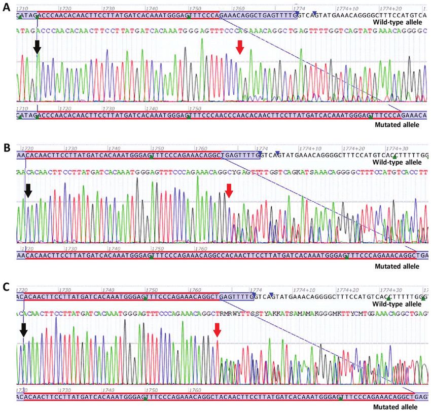
Figure 2. Sanger sequencing electropherograms of novel deletions or deletion/insertions. (A) Novel deletion c.1652_1672del p.(Pro551_Lys558delinsGln).
(B) Novel deletion/insertion c.1653_1660delinsAA p.(Met552_Glu554delinsLys). (C) Novel deletion/insertion c.1665_1672delinsCC p.(Trp557_Lys558del).
(D) Novel deletion c.1668_1686del p.(Trp557*). (E) Novel deletion c.1676_1720del p.(Val559_Thr574delinsAla). Black arrows are indicating the start of
deletions or deletion/insertions.
Novel variants. The present study detected 49 different vari- two adenine bases at the same position. At the protein level,
ants in total, of which eight were novel. A reference search p.(Met552_Glu554delinsLys) results in the deletion of three
for all detected variants (n=49) was performed using Google amino acids in the JM domain of the KIT protein from amino
browser trough Alamut Visual software, on the 19th of acids 552 to 554, combined with the insertion of a lysine (Lys)
January 2019. at the same site (Fig. 2B).
All eight novel heterozygous variants, which had not been c.1665_1672delinsCC p.(Trp557_Lys558del) is a deletion
reported previously, were detected in exon 11 of gene KIT of eight nucleotides in exon 11 of the KIT gene, from posi-
(Figs. 2 and 3). The precise effect of these eight novel vari- tion c.1665 to position c.1672, and an insertion of two cytosine
ants on KIT protein function is unknown; however, based on bases at the same position. At the protein level, p.(Trp557_
Cancer Genome Interpreter and ACMG/AMP criterions, all of Lys558del) results in the deletion of two amino acids in the
them were classified as likely pathogenic. The classification of JM domain of the KIT protein from amino acids 557 to 558
all eight novel variants found in KIT exon 11 is presented in (Fig. 2C).
Table III and their descriptions are provided in the following c.1668_1686del p.(Trp557*) is a deletion of 19 nucleotides
paragraphs. in exon 11 of the KIT gene, from position c.1668 to posi-
c.1652_1672del p.(Pro551_Lys558delinsGln) is a deletion tion c.1686. At the protein level, p.(Trp557*) results in the
of 21 nucleotides in the exon 11 of the KIT gene, from position premature termination of the KIT protein at amino acid 557.
c.1652 to position c.1672. At the protein level, p.(Pro551_ Due to the loss of the protein kinase domain, the mutation
Lys558delinsGln) results in the deletion of eight amino acids in c.1668_1686del p.(Trp557*) is predicted to lead to a loss of
the JM domain of the KIT protein from amino acids 551 to 558, KIT protein function (Fig. 2D).
combined with the insertion of a glutamine (Gln) at the same c.1676_1720del p.(Val559_Thr574delinsAla) is a dele-
site (Fig. 2A). tion of 45 nucleotides in exon 11 of the KIT gene, from
c.1653_1660delinsAA p.(Met552_Glu554delinsLys) is position c.1676 to position c.1720. At the protein level,
a deletion of eight nucleotides in exon 11 of the KIT gene, p.(Val559_Thr574delinsAla) results in the deletion of
from position c.1653 to position c.1660, and an insertion of 16 amino acids in the JM domain of the KIT protein from1474 INTERNATIONAL JOURNAL OF ONCOLOGY 56: 1468-1478, 2020
Figure 3. Sanger sequencing electropherograms of novel duplications. (A) Novel duplication c.1715_1756dup p.(Pro585_Arg586ins14). (B) Novel duplication
c.1721_1765dup p.(Arg588_Leu589ins15). (C) Novel duplication c.1722_1766dup p.(Gln575_Leu589dup). Black arrows are indicating the first duplicated
nucleotide. Red arrows are indicating the last duplicated nucleotide.
amino acids 559 to 574, combined with the insertion of an c.1722_1766dup p.(Gln575_Leu589dup) is a duplication of
Alanine (Ala) at the same site (Fig. 2E). 45 nucleotides in exon 11 of the KIT gene, from position c.1722
c.1715_1756dup p.(Pro585_Arg586ins14) is a duplication of to position c.1766. At the protein level, p.(Gln575_Leu589dup)
42 nucleotides in exon 11 of the KIT gene, from position c.1715 indicates the duplication of 15 amino acids (Gln 575 through
to position c.1756. At the protein level, p.(Pro585_Arg586ins14) Leu 589) in the JM domain of the KIT protein, starting with
indicates the insertion of 14 amino acids (Asn, Pro, Thr, Gln, Gln at position 590 (Fig. 3C).
Leu, Pro, Tyr, Asp, His, Lys, Trp, Glu, Phe and Pro) in the JM
domain of the KIT protein, starting with Asparagine (Asn) at Discussion
position 586 (Fig. 3A).
c.1721_1765dup p.(Arg588_Leu589ins15) is a duplica- Genotype analysis of 70 patients from the Slovenian Cancer
tion of 45 nucleotides in exon 11 of the KIT gene, from Registry was performed in the present study to determine
position c.1721 to position c.1765. At the protein level, the spectrum and frequency of KIT and PDGFRA altera-
p.(Arg588_Leu589ins15) indicates the insertion of 15 amino tions in Slovenian patients with metastatic GISTs, as well as
acids (Pro, Gln, Leu, Pro, Tyr, Asp, His, Lys, Trp, Glu, Phe, the frequency of hot spot mutations in KRAS, NRAS, BRAF,
Pro, Arg, Asn and Arg) in the JM domain of the KIT protein, PIK3CA and AKT1 in KIT/PDGFRA WT GISTs. Known muta-
starting with Proline (Pro) at position 589 (Fig. 3B). tion status of the aforementioned genes has been proven to beBOMBAC et al: MUTATIONAL SPECTRUM IN GIST 1475
an important prognostic and predictive factor in the manage- that this alteration disrupts normal auto‑inhibitory function of
ment and treatment of patients with GIST with TKIs (21,23). the JM domain (13).
Mutation analysis performed on Slovenian patients with Single nucleotide changes in PDGFRA were identified in
GIST has confirmed a high degree of mutation heteroge- exon 18, c.2525A>T p.(Asp842Val), which is the most common
neity in KIT and PDGFRA genes, as reported in previous PDGFRA mutation and accounts for 60‑65% of all PDGFRA
studies (24‑29). With direct Sanger sequencing of exons 9, 11, mutations seen in GIST (known to be resistant to the imatinib
13 and 17 of KIT gene and exons 12, 14 and 18 of PDGFRA treatment) (12,41). Notably, 1 patient was identified with a
gene, a total of 49 different alterations were detected. In the rare example of double mutant GIST, which had two different
present study, the overall mutation rate was 88.5%, which was primary mutations in the PDGFRA gene, c.1936A>G
slightly higher than in a study from Italy (80%), yet comparable p.(Lys646Glu) and c.1975A>C p.(Asn659His), both located
to frequencies observed in studies from Germany (86.1%) in exon 14. These patient samples were collected prior to the
and Poland (85.1%), a French population‑based study initiation of TKI treatment. According to the literature, double
(KIT, 82.8%; PDGFRA, 2.1%), and in two phase III clinical mutant GISTs, harbouring two primary mutations in the
trials that enrolled patients with metastatic GIST from the PDGFRA gene, found prior to TKI therapy, are rare (42,43).
European Organisation for Research and Treatment of Cancer Most of the patients who initially respond to imatinib develop
(KIT, 86.2%; PDGFRA, 1.6%) and Cancer and Leukemia resistance after a long period of treatment. Resistance to imatinib
Group B (KIT, 84.6%; PDGFRA, 2.65%) (24‑29). has been associated with secondary heterogeneous KIT receptor
The mutation frequency in the patients analysed in the mutations in up to 90% of the patients. Secondary mutations
present study, was expectedly the highest in the KIT gene, most commonly arise in exons 13 and 14 (ATP‑binding domain)
with most mutations in exon 11 (86.7%). The highest muta- or exons 17 and 18 (the activation loop), whereas primary KIT
tion frequency in KIT exon 11 has also been reported in mutations predominantly affect the JM domain encoded by
other studies (57, 66.1 and 87.3%, respectively) (14,24,30). In exon 11 (44). The present study identified 6 patients with KIT
8 patients with GIST in the present study (11.4%), no alterations exon 13 or 17 secondary mutation. Primary tumour samples
in KIT/PDGFRA genes were found. According to the litera- from these patients were obtained after imatinib treatment,
ture, ~10‑15% of adult GISTs are KIT/PDGFRA WT (13,31). suggesting secondary mutations most probably occurred as a
All 8 patients with no alterations in KIT or PDGFRA genes response to the treatment, which has been reported in previous
were also negative for hot spot mutations in KRAS, NRAS, studies (10,41,45). In 3 out of 4 patients with secondary mutation
BRAF, PIK3CA and AKT1 genes, which are included in the in KIT exon 13 the same mutation was detected, a single nucleo-
RAS/RAF/MAPK and PI3K/AKT/mTOR signalling pathways, tide change c.1961T>C p.(Val654Ala), confirming that this is the
suggesting that the frequency of these mutations is low (32). most commonly described secondary mutation (10). Due to the
As reported by Shi et al (33), patients with KIT/PDGFRA WT existence of intra‑tumour heterogeneity in GIST, with different
GIST can harbour mutations in EGFR. Since in the present clones being sensitive/resistant to different drugs, using a combi-
cohort, only 8 patients had KIT/PDGFRA WT GIST, and since nation of different drugs could be a better approach for maximal
the likelihood of EGFR mutations in these patients is G iii) predicted passenger; and iv) polymorphism. Variants
p.(Lys642Glu) and c.1961T>C p.(Val654Ala), were identified. pathogenicity was determined according to ACMG/AMP
The substitution of amino acids Lys to Glu at codon 642, has guidelines using Genetic Variant Interpretation Tool (criterion
been rarely described in literature and it has been suggested selection PM1, PM2, PM4, PP3).1476 INTERNATIONAL JOURNAL OF ONCOLOGY 56: 1468-1478, 2020
Exon 11 encodes the JM domain of KIT protein that In conclusion, by identifying and classifying eight novel
prevents the kinase activation loop from shifting into the variants in KIT exon 11, and by detecting one double‑mutant
active state. Alterations in exon 11 predominantly disrupt GIST with two mutations in PDGFRA exon 14, the present study
the auto‑inhibitory function and trigger ligand‑independent contributes to broadening the spectrum of known mutations in
receptor activation (46,47). Since all eight novel vari- GIST tumours. Mutation frequencies of KIT and PDGFRA
ants detected in the present study were located in a gene observed in the present cohort (85.7 and 2.9%, respec-
well‑established functional domain and mutational hot spot, tively) are in accordance with previous literature. The current
ACMG/AMP criterion PM1 was applied. None of the novel results also confirm the incidence of identified alterations in the
variants are described in public databases of human varia- previously reported and most frequently altered regions of KIT
tion GnomAD (https://gnomad.broadinstitute.org/) or ExAC or PDGFRA, and suggest that the occurrence of alterations in
(https://www.re3data.org/repository/r3d100012122), hence other genes underlying GIST development, including KRAS,
ACMG/AMP criterion PM2 was applied. All of the novel NRAS, BRAF, PIK3CA and AKT1, are very rare.
variants cause protein length change due to deletion, inser- The present study is the first from Slovenia where a
tion or duplication, therefore ACMG/AMP criterion PM4 detailed characterization of gene abnormalities in patients
was applied. with metastatic GIST from the Slovenian Cancer Registry
Five of the novel variants in KIT gene detected in the present has been performed. Knowing the accurate mutation status of
study were deletions or deletions/insertions, [c.1652_1672del patients with GIST is of great value for understanding GIST
p.(P ro551_ Lys558delinsGln), c.1653_1660delinsA A molecular biology, oncogenesis and GIST subtype classifica-
p.(Met 552 _Glu554delinsLys), c.1665_1672delinsCC tion, as well for the appropriate use of specific targeted therapy
p.(Trp557_Lys558del), c.1668_1686del p.(Trp557*), and with KIT/PDGFRA TKIs.
c.1676_1720del p.(Val559_Thr574delinsAla)], localized
within the most commonly mutated region of exon 11, Acknowledgements
between codons 550 and 580. Occurrence of deletions/inser-
tions in this region of KIT receptor are described as activating Not applicable.
alterations, which are according to literature, associated
with malignant tumour behaviour (2,8,48,49). Therefore Funding
it could be predicted that newly identified variants in the
present study, [c.1652_1672del p.(Pro551_Lys558delinsGln), This study received funding from the Slovenian Research
c.1653_1660delinsA A p.(Met552 _Glu554delinsLys), Agency (grant nos. P3‑0352 and P3‑0321).
c.1676 _172 0 del p.( Va l 559_T h r 574 del i nsA la), a nd
c.1665_1672delinsCC p.(Tr p557_ Lys558del)], a re a Availability of data and materials
gain‑of‑function mutations, leading to protein activation.
Variant c.1665_1672delinsCC p.(Trp557_Lys558del) results All data generated or analyzed during this study are included
in deletion of two amino acids Trp557 and Lys558. Deletion in this published article.
of codons 557‑558 is frequently reported in other studies and
has been described to cause constitutive activation of KIT and Authors' contributions
increased ERK phosphorylation (38,50,51). However, deletion
of codons 557‑558 as a result of this specific deletion of eight AB wrote the manuscript, collected the related literature,
nucleotides from c.1665 to c.1672 coupled with insertion of performed the majority of wet lab work, and contributed to the
two cytosines is firstly described in the present study. Deletion analysis and interpretation of sequencing data, bioinformatics
of codon 557 was detected also as a result of a novel deletion analysis and classification of novel variants. BZ contributed
causing premature stop codon c.1668_1686del p.(Trp557*). to the conception of the study, acquired tumour samples and
Because of the premature termination of the KIT protein patient clinical data, and contributed to the revision of the
at amino acid 557, it was predicted that p.(Trp557*) leads manuscript. MB contributed to the wet lab experiments and
to the loss of KIT protein function. Using Cancer Genome contributed to the analysis of sequencing data. VSD contrib-
Interpreter, an evidence‑based somatic variant classifica- uted to the bioinformatics analysis, and interpretation and
tion tool, these five novel somatic variants were classified as classification of novel variants. BG performed the pathological
predicted driver (tier 1 or tier 2), with high level of pathoge- evaluation of tumour samples. VS contributed to the interpre-
nicity. Additionally, MutationTaster2 analysis predicted that tation and classification of novel variants. GK created figures
four out of these five novel variants were disease‑causing; and contributed to the interpretation and classification of novel
based on their predicted oncogenic effect the ACMG/AMP variants. SN contributed to the conception of this study and
criterion PP3 was applied. designed, reviewed, wrote and revised the manuscript. All
Three of the novel variants were duplications of 17 to authors read and approved the final manuscript.
45 nucleotides at the 3'‑end of exon 11: c.1715_1756dup
p.(Pro585_Arg586ins14), c.1721_1765dup p.(Arg588_ Ethics approval and consent to participate
Leu589ins15) and c.1722_1766dup p.(Gln575_Leu589dup).
Duplications in GISTs are known to almost exclusively appear The present study was approved by the Institute of Oncology
at the 3'‑end of KIT exon 11, they range from 3 to 57 nucleo- Ljubljana Ethic's Committee (approval no. OIRIKE00036)
tides and they are reported to have a relatively good clinical and the Republic of Slovenia National Medical Ethics
outcome (52,53). Committee (approval no. 1090216). Individual patient consentBOMBAC et al: MUTATIONAL SPECTRUM IN GIST 1477
was waived for this study as it was a retrospective study, the 17. Pantaleo MA, Nannini M, Corless CL and Heinrich MC:
Quadruple wild‑type (WT) GIST: Defining the subset of GIST
research involved no risk to the subjects, and the institutional that lacks abnormalities of KIT, PDGFRA, SDH, or RAS
informed consent forms for treatment included consent for the signaling pathways. Cancer Med 4: 101‑103, 2015.
use of patient's data, materials and/or test results for research 18. Lasota J, Felisiak‑Golabek A, Wasag B, Kowalik A, Zięba S,
Chłopek M, Wang ZF, Coates T, Kopczynski J, Gozdz S, et al:
purposes. Frequency and clinicopathologic profile of PIK3CA mutant
GISTs: Molecular genetic study of 529 cases. Mod Pathol 29:
Patient consent for publication 275‑282, 2016.
19. Drilon A, Laetsch TW, Kummar S, DuBois SG, Lassen UN,
Demetri GD, Nathenson M, Doebele RC, Farago AF,
Not applicable. Pappo AS, et al: Efficacy of larotrectinib in TRK fusion‑positive
cancers in adults and children. N Engl J Med 378: 731‑739, 2018.
20. Isozaki K and Hirota S: Gain‑of‑Function Mutations of Receptor
Competing interests Tyrosine Kinases in Gastrointestinal Stromal Tumors. Curr
Genomics 7: 469‑475, 2006.
The authors declare that they have no competing interests. 21. Nishida T, Takahashi T and Miyazaki Y: Gastrointestinal stromal
tumor: A bridge between bench and bedside. Gastric Cancer 12:
175‑188, 2009.
References 22. Fletcher JA and Rubin BP: KIT mutations in GIST. Curr Opin
Genet Dev 17: 3‑7, 2007.
23. Gramza AW, Corless CL and Heinrich MC: Resistance to tyrosine
1. Miettinen M and Lasota J: Gastrointestinal stromal tumors ‑ defi- kinase inhibitors in gastrointestinal stromal tumors. Clin Cancer
nition, clinical, histological, immunohistochemical, and Res 15: 7510‑7518, 2009.
molecular genetic features and differential diagnosis. Virchows 24. Origone P, Gargiulo S, Mastracci L, Ballestrero A, Battistuzzi L,
Arch 438: 1‑12, 2001. Casella C, Comandini D, Cusano R, Dei Tos AP, Fiocca R, et al;
2. Hirota S, Isozaki K, Moriyama Y, Hashimoto K, Nishida T, Liguria GIST Unit: Molecular characterization of an Italian
Ishiguro S, Kawano K, Hanada M, Kurata A, Takeda M, et al: series of sporadic GISTs. Gastric Cancer 16: 596‑601, 2013.
Gain‑of‑Function Mutations of c‑kit in Human Gastrointestinal 25. Penzel R, Aulmann S, Moock M, Schwarzbach M, Rieker RJ
Stromal Tumors. Science 279: 577‑580 , 1998. and Mechtersheimer G: The location of KIT and PDGFRA
3. Corless CL and Heinrich MC: Molecular Pathobiology of gene mutations in gastrointestinal stromal tumours is site and
Gastrointestinal Stromal Sarcomas. Annu Rev Pathol Mech phenotype associated. J Clin Pathol 58: 634‑639, 2005.
Dis 3: 557‑586, 2008. 26. Wozniak A, Rutkowski P, Schöffski P, Ray‑Coquard I, Hostein I,
4. Miettinen M and Lasota J: Gastrointestinal stromal tumors: Schildhaus HU, Le Cesne A, Bylina E, Limon J, Blay JY, et al:
Pathology and prognosis at different sites. Semin Diagn Tumor genotype is an independent prognostic factor in primary
Pathol 23: 70‑83, 2006. gastrointestinal stromal tumors of gastric origin: A european
5. Duensing A, Medeiros F, McConarty B, Joseph NE, Panigrahy D, multicenter analysis based on ConticaGIST. Clin Cancer Res 20:
Singer S, Fletcher CDM, Demetri GD and Fletcher JA: 6105‑6116, 2014.
Mechanisms of oncogenic KIT signal transduction in primary 27. Emile JF, Brahimi S, Coindre JM, Bringuier PP, Monges G,
gastrointestinal stromal tumors (GISTs). Oncogene 23: Samb P, Doucet L, Hostein I, Landi B, Buisine MP, et al:
3999‑4006, 2004. Frequencies of KIT and PDGFRA mutations in the MolecGIST
6. Miettinen M and Lasota J: Gastrointestinal Stromal Tumors prospective population‑based study differ from those of advanced
Review on Morphology, Molecular Pathology, Prognosis, GISTs. Med Oncol 29: 1765‑1772, 2012.
and Differential Diagnosis. Arch Pathol Lab Med 130: 28. Debiec‑Rychter M, Sciot R, Le Cesne A, Schlemmer M,
1466‑1478,2006. Hohenberger P, van Oosterom AT, Blay JY, Leyvraz S, Stul M,
7. Heinrich MC, Rubin BP, Longley BJ and Fletcher JA: Biology and Casali PG, et al; EORTC Soft Tissue and Bone Sarcoma Group;
genetic aspects of gastrointestinal stromal tumors: KIT activation Italian Sarcoma Group; Australasian GastroIntestinal Trials
and cytogenetic alterations. Hum Pathol 33: 484‑495, 2002. Group: KIT mutations and dose selection for imatinib in patients
8. Corless CL, Fletcher JA and Heinrich MC: Biology of gastroin- with advanced gastrointestinal stromal tumours. Eur J Cancer 42:
testinal stromal tumors. J Clin Oncol 22: 3813‑3825, 2004. 1093‑1103, 2006.
9. Miettinen M, El‑Rifai W, H L Sobin L and Lasota J: Evaluation 29. Heinrich MC, Owzar K, Corless CL, Hollis D, Borden EC, Fletcher
of malignancy and prognosis of gastrointestinal stromal tumors: CDM, Ryan CW, Von Mehren M, Blanke CD, Rankin C, et al:
A review. Hum Pathol 33: 478‑483, 2002. Correlation of kinase genotype and clinical outcome in the North
10. Lasota J and Miettinen M: Clinical significance of oncogenic KIT American intergroup phase III trial of imatinib mesylate for
and PDGFRA mutations in gastrointestinal stromal tumours. treatment of advanced gastrointestinal stromal tumor: CALGB
Histopathology 53: 245‑266, 2008. 150105 study by cancer and leukemia group B and southwest
11. Heinrich MC, Corless CL, Duensing A, McGreevey L, Chen CJ, oncology gr. J Clin Oncol 26: 5360‑5367, 2008.
Joseph N, Singer S, Griffith DJ, Haley A, Town A, et al: 30. Andersson J, Bümming P, Meis‑Kindblom JM, Sihto H,
PDGFRA activating mutations in gastrointestinal stromal Nupponen N, Joensuu H, Odén A, Gustavsson B, Kindblom L‑G
tumors. Science 299: 708‑710, 2003. and Nilsson B: Gastrointestinal stromal tumors with KIT
12. Corless CL, Schroeder A, Griffith D, Town A, McGreevey L, exon 11 deletions are associated with poor prognosis.
Harrell P, Shiraga S, Bainbridge T, Morich J and Heinrich MC: Gastroenterology 130: 1573‑1581, 2006.
PDGFRA mutations in gastrointestinal stromal tumors: 31. Boikos SA, Pappo AS, Killian JK, LaQuaglia MP, Weldon CB,
Frequency, spectrum and in vitro sensitivity to imatinib. J Clin George S, Trent JC, von Mehren M, Wright JA, Schiffman JD, et al:
Oncol 23: 5357‑5364, 2005. Molecular Subtypes of KIT/PDGFRAWild‑Type Gastrointestinal
13. Corless CL, Barnett CM and Heinrich MC: Gastrointestinal Stromal Tumors. JAMA Oncol 2: 922, 2016.
stromal tumours: Origin and molecular oncology. Nat Rev 32. Huss S, Pasternack H, Ihle MA, Merkelbach‑Bruse S, Heitkötter B,
Cancer 11: 865‑878, 2011. Hartmann W, Trautmann M, Gevensleben H, Büttner R,
14. Joensuu H, Hohenberger P and Corless CL: Gastrointestinal Schildhaus HU, et al: Clinicopathological and molecular features
stromal tumour. Lancet 382: 973‑983, 2013. of a large cohort of gastrointestinal stromal tumors (GISTs) and
15. Daniels M, Lurkin I, Pauli R, Erbstösser E, Hildebrandt U, review of the literature: BRAF mutations in KIT/PDGFRA
Hellwig K, Zschille U, Lüders P, Krüger G, Knolle J, et al: wild‑type GISTs are rare events. Hum Pathol 62: 206‑214, 2017.
Sp e c t r u m of K I T/ PD G F R A / BR A F mut a t io n s a n d 33. Shi SS, Wu N, He Y, Wei X, Xia QY, Wang X, Ye S, Li R, Rao Q
Phosphatidylinositol‑3‑Kinase pathway gene alterations in gastro- and Zhou X‑J: Bin, Li R, Rao Q and Zhou XJ: EGFR gene
intestinal stromal tumors (GIST). Cancer Lett 312: 43‑54, 2011. mutation in gastrointestinal stromal tumours. Histopathology 71:
16. Miranda C, Nucifora M, Molinari F, Conca E, Anania MC, 553‑561, 2017.
Bordoni A, Saletti P, Mazzucchelli L, Pilotti S, Pierotti MA, et al: 34. Nakahara M, Isozaki K, Hirota S, Miyagawa J, Hase‑Sawada N,
KRAS and BRAF mutations predict primary resistance to Taniguchi M, Nishida T, Kanayama S, Kitamura Y and Shinomura Y:
imatinib in gastrointestinal stromal tumors. Clin Cancer Res 18: A novel gain‑of‑function mutation of c‑kit gene in gastrointestinal
1769‑1776, 2012. stromal tumors. Gastroenterology 115: 1090‑1095, 1998.1478 INTERNATIONAL JOURNAL OF ONCOLOGY 56: 1468-1478, 2020
35. Lasota J, Jasinski M, Sarlomo‑Rikala M and Miettinen M: 45. Wang CM, Huang K, Zhou Y, Du CY, Ye YW, Fu H, Zhou XY and
Mutations in exon 11 of c‑Kit occur preferentially in malignant Shi YQ: Molecular mechanisms of secondary imatinib resistance
versus benign gastrointestinal stromal tumors and do not occur in in patients with gastrointestinal stromal tumors. J Cancer Res
leiomyomas or leiomyosarcomas. Am J Pathol 154: 53‑60, 1999. Clin Oncol 136: 1065‑1071, 2010.
36. Taniguchi M, Nishida T, Hirota S, Isozaki K, Ito T, Nomura T, 46. Gajiwala KS, Wu JC, Christensen J, Deshmukh GD, Diehl W,
Matsuda H and Kitamura Y: Effect of c‑kit mutation on prognosis Dinitto JP, English JM, Greig MJ, He Y‑A, Jacques SL, et al: KIT
of gastrointestinal stromal tumors. Cancer Res 59: 4297‑4300, kinase mutants show unique mechanisms of drug resistance to
1999. imatinib and sunitinib in gastrointestinal stromal tumor patients.
37. Moskalu k CA, Tia n Q, Ma rshall CR, Rumpel CA, Proc Natl Acad Sci U S A 106: 1542‑1547, 2009.
Franquemont DW and Frierson HF Jr: Mutations of c‑kit JM 47. Mol CD, Dougan DR, Schneider TR, Skene RJ, Kraus ML,
domain are found in a minority of human gastrointestinal stromal Scheibe DN, Snell GP, Zou H, Sang BC and Wilson KP:
tumors. Oncogene 18: 1897‑1902, 1999. Structural basis for the autoinhibition and STI‑571 inhibition of
38. Wardelmann E, Losen I, Hans V, Neidt I, Speidel N, Bierhoff E, c‑Kit tyrosine kinase. J Biol Chem 279: 31655‑31663, 2004.
Heinicke T, Pietsch T, Büttner R and Merkelbach‑Bruse S: 48. Roskoski R Jr: Structure and regulation of Kit protein‑tyrosine
Deletion of Trp‑557 and Lys‑558 in the juxtamembrane domain kinase ‑ the stem cell factor receptor. Biochem Biophys Res
of the c‑kit protooncogene is associated with metastatic behavior Commun 338: 1307‑1315, 2005.
of gastrointestinal stromal tumors. Int J Cancer 106: 887‑895, 49. Bachet JB, Hostein I, Le Cesne A, Brahimi S, Beauchet A,
2003. Tabone‑Eglinger S, Subra F, Bui B, Duffaud F, Terrier P, et al:
39. Martín‑Broto J, Rubio L, Alemany R and López‑Guerrero JA: Prognosis and predictive value of KIT exon 11 deletion in GISTs.
Clinical implications of KIT and PDGFRA genotyping in GIST. Br J Cancer 101: 7‑11, 2009.
Clin Transl Oncol 12: 670‑676, 2010. 50. Garner AP, Gozgit JM, Anjum R, Vodala S, Schrock A, Zhou T,
40. Van Glabbeke M; Gastrointestinal Stromal Tumor Meta‑Analysis Serrano C, Eilers G, Zhu M, Ketzer J, et al: Ponatinib inhibits
Group (MetaGIST): Comparison of two doses of imatinib for the polyclonal drug‑resistant KIT oncoproteins and shows thera-
treatment of unresectable or metastatic gastrointestinal stromal peutic potential in heavily pretreated gastrointestinal stromal
tumors: A meta‑analysis of 1,640 patients. J Clin Oncol 28: tumor (GIST) patients. Clin Cancer Res 20: 5745‑5755, 2014.
1247‑1253, 2010. 51. Wang HC, Li TY, Chao YJ, Hou YC, Hsueh YS, Hsu KH and
41. Lasota J and Miettinen M: KIT and PDGFRA mutations in Shan YS: Kit exon 11 codons 557‑558 deletion mutation promotes
gastrointestinal stromal tumors (GISTs). Semin Diagn Pathol 23: liver metastasis through the CXCL12�������������������������
/������������������������
CXCR4 Axis in gastroin-
91‑102, 2006. testinal stromal tumors. Clin Cancer Res 22: 3477‑3487, 2016.
42. Conca E, Miranda C, Dal Col V, Fumagalli E, Pelosi G, 52. Wozniak A, Rutkowski P, Piskorz A, Ciwoniuk M, Osuch C,
Mazzoni M, Fermeglia M, Laurini E, Pierotti MA, Pilotti S, et al: Bylina E, Sygut J, Chosia M, Rys J, Urbanczyk K, et al; Polish
Are two better than one? A novel double‑mutant KIT in GIST Clinical GIST Registry: Prognostic value of KIT/PDGFRA
that responds to Imatinib. Mol Oncol 7: 756‑762, 2013. mutations in gastrointestinal stromal tumours (GIST): Polish
43. Pai T, Bal M, Shetty O, Gurav M, Ostwal V, Ramaswamy A, Clinical GIST Registry experience. Ann Oncol 23: 353‑360, 2012.
Ramadwar M and Desai S: Unraveling the spectrum of KIT 53. Lasota J, Wasag B, Steigen SE, Limon J and Miettinen M:
mutations in gastrointestinal stromal tumors: An Indian Tertiary Improved detection of KIT exon 11 duplications in formalin‑fixed,
Cancer Center Experience. South Asian J Cancer 6: 113‑117, paraffin‑embedded gastrointestinal stromal tumors. J Mol
2017. Diagn 9: 89‑94, 2007.
44. Serrano C, Mariño‑Enríquez A, Tao DL, Ketzer J, Eilers G,
Zhu M, Yu C, Mannan AM, Rubin BP, Demetri GD, et al: This work is licensed under a Creative Commons
Complementary activity of tyrosine kinase inhibitors against Attribution-NonCommercial-NoDerivatives 4.0
secondary kit mutations in imatinib‑resistant gastrointestinal International (CC BY-NC-ND 4.0) License.
stromal tumours. Br J Cancer: 120: 612‑620, 2019.You can also read