Analysis of Wilson disease mutations revealed that interactions between different ATP7B mutants modify their properties - Nature
←
→
Page content transcription
If your browser does not render page correctly, please read the page content below
www.nature.com/scientificreports
OPEN Analysis of Wilson disease
mutations revealed
that interactions between different
ATP7B mutants modify their
properties
Shubhrajit Roy1,4*, Courtney J. McCann1, Martina Ralle2, Kunal Ray3, Jharna Ray4,
Svetlana Lutsenko1,5* & Samuel Jayakanthan1,5
Wilson disease (WD) is an autosomal-recessive disorder caused by mutations in the copper (Cu)-
transporter ATP7B. Thus far, studies of WD mutations have been limited to analysis of ATP7B mutants
in the homozygous states. However, the majority of WD patients are compound-heterozygous, and
how different mutations on two alleles impact ATP7B properties is unclear. We characterized five
mutations identified in Indian WD patients, first by expressing each alone and then by co-expressing
two mutants with dissimilar properties. Mutations located in the regulatory domains of ATP7B—
A595T, S1362A, and S1426I—do not affect ATP7B targeting to the trans-Golgi network (TGN) but
reduce its Cu-transport activity. The S1362A mutation also inhibits Cu-dependent trafficking from
the TGN. The G1061E and G1101R mutations, which are located within the ATP-binding domain,
cause ATP7B retention in the endoplasmic reticulum, inhibit Cu-transport, and lower ATP7B protein
abundance. Co-expression of the A595T and G1061E mutations, which mimics the compound-
heterozygous state of some WD patients, revealed an interaction between these mutants that altered
their intracellular localization and trafficking under both low and high Cu conditions. These findings
highlight the need to study WD variants in both the homozygous and compound-heterozygous states
to better understand the genotype–phenotype correlations and incomplete penetrance observed in
WD.
Copper (Cu) is a redox-active metal that is indispensable for human growth and development. Cu is required
for the activity of several enzymes, including cytochrome c oxidase, Cu/Zn-dependent superoxide dismutases,
tyrosinase, dopamine-β-hydroxylase, ceruloplasmin, and peptidyl-α-monooxygenase1. In order to ensure an
adequate supply of Cu without toxic accumulation, Cu homeostatic mechanisms are tightly regulated. Either
Cu deficiency or overabundance is associated with pathologic changes and disease, such as, Menkes disease or
Wilson disease (WD), respectively2,3. WD is a genetic disorder caused by mutations in the Cu-transporter ATP7B
that causes abnormal deposition of Cu in the liver and the brain4. WD has a spectrum of hepatic and neurologi-
cal manifestations, along with a broad range of disease o nsets5. The mechanisms behind this variability remain
poorly understood. Studies of WD patients identified over 600 ATP7B mutations6 and revealed the diverse effects
of mutations on the functional and cellular properties of the ATP7B protein. Efforts to link different phenotypic
presentations of WD to specific ATP7B mutations have not produced strong correlations and have sometimes
led to conflicting r esults7. High prevalence of compound-heterozygous mutations further complicates the task
of genotype–phenotype c orrelation8.
1
Department of Physiology, Johns Hopkins Medical Institute, Baltimore, MD, USA. 2Oregon Health and
Science University, Portland, OR, USA. 3ATGC Diagnostics Private Ltd, Kolkata, India. 4S. N. Pradhan Centre for
Neurosciences, University of Calcutta, Kolkata, India. 5These authors contributed equally: Svetlana Lutsenko and
Samuel Jayakanthan. *email: shubhrajitroy123@gmail.com; lutsenko@jhmi.edu
Scientific Reports | (2020) 10:13487 | https://doi.org/10.1038/s41598-020-70366-7 1
Vol.:(0123456789)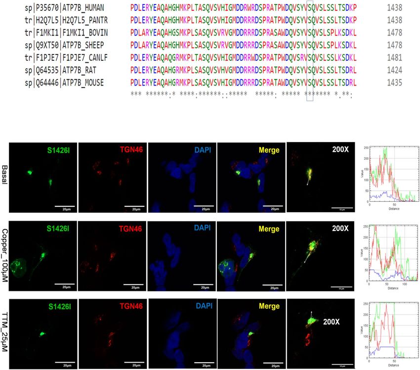
www.nature.com/scientificreports/
Figure 1. Locations of selected WD mutations in the ATP7B protein. Diagrammatic representation of the
ATP7B protein structure and the positions of the ATP7B mutations (A595T, G1061E, G1101R, S1362A, and
S1426I) in different domains.
ATP7B is a 164 kDa, multi-domain protein that transfers Cu across the cell membrane using energy generated
by ATP h ydrolysis9–11. ATP7B resides in the trans-Golgi network (TGN), where it supplies Cu for the functional
maturation of the Cu-dependent enzyme ceruloplasmin. In response to Cu elevation, ATP7B undergoes kinase-
mediated phosphorylation and moves out of the TGN to vesicles, where it sequesters excess Cu for subsequent
export across the apical membrane12. The complexes, multi-domain structure of ATP7B, combined with a lim-
ited understanding of the mechanisms underlying ATP7B targeting and trafficking, have led to difficulties in
determining the precise consequences of pathogenic mutations. In addition, ATP7B forms stable dimers both in
ligomerization18. Dimerization with
cells and in-vitro, and very little is yet known about the significance of its o
wild-type (WT) ATP7B was shown to affect the localization of ATP7B variants with mutated kinase-mediated
phosphorylation sites13 https://www.ncbi.nlm.nih.gov/pmc/articles/PMC3476272/, although the impact of
dimerization on the properties of WD mutants has not been investigated. It remains unclear whether different
ATP7B mutants expressed in the same cells act independently or influence each other’s properties.
Extensive genetic screening of ATP7B among Indian WD patients identified several novel and prevalent
missense mutations present in both homozygous and compound-heterozygous states7. In this study, we char-
acterized mutations found in different domains of ATP7B (Fig. 1). The previously reported mutations G1061E
and G1101R are located within the ATP-binding domain and were detected in both homozygous and heterozy-
gous states. Three novel mutations—A595T, S1362A and S1426I—are located in the N-terminal metal binding
domain 6 (MBD 6), trans-membrane domain 8 (TM 8), and the C-terminal domain, respectively (Fig. 1). The
S1362A mutation was found in the homozygous state, whereas A595T and S1426I were found in compound-
heterozygous WD patients.
We hypothesized that WD-causing mutations located in different domains of ATP7B would have different
impacts on its Cu-transport activity or trafficking behavior. We tested this hypothesis by characterizing the
properties of the individual ATP7B mutants in isolation, i.e., as they are found in the homozygous state. We
then modeled the compound-heterozygous state found in WD patients by co-expressing the A595T and G1061E
mutants and characterizing their cellular behavior under low and high Cu conditions.
Materials and methods
Plasmids and site‑directed mutagenesis. The plasmid pSJ101, encoding full-length ATP7B with an
N-terminal 6x-His-GFP-TEV tag (6X-His-GFP-tev-ATP7B), and another plasmid encoding full-length ATP7B
with an N-terminal Flag-tag (pcDNA5 FRT/TO plasmid; Invitrogen) were used as templates to generate the mis-
sense mutations. The ATP7B mutations were introduced using the Quick-Change site-directed mutagenesis kit
(Stratagene, La Jolla, CA) and appropriate primers (Table S1). Correct sequences and the presence of the muta-
tions were verified by sequencing the entire cDNA region. The sequencing was performed by the Johns Hopkins
School of Medicine Synthesis and Sequencing Facility.
Cell culture and transient transfection. HEK293A cells were maintained in Dulbecco’s Modified Eagle’s
Medium (DMEM; Corning Cellgro, Fisher Scientific, USA) supplemented with 1% penicillin/streptomycin and
Scientific Reports | (2020) 10:13487 | https://doi.org/10.1038/s41598-020-70366-7 2
Vol:.(1234567890)
www.nature.com/scientificreports/
10% fetal bovine serum (FBS; vol/vol). Menkes disease fibroblast (YST) cells were maintained in DMEM sup-
plemented with 1% penicillin/streptomycin, 0.5 µg/mL Puromycin (Invitrogen, Carlsbad, CA, USA), Primocin
(Invitrogen, Carlsbad, CA, USA), and 10% FBS (vol/vol) (Corning Cellgro, USA). Cell cultures were maintained
at 37 °C in a humidified incubator (5% CO2). The cells were transfected in 6-well plates for 12–18 h using Lipo-
fectamine LTX-PLUS reagent (Invitrogen, Carlsbad, CA, USA) with 1 or 2 µg of plasmid DNA and Opti-MEM
reduced serum medium (Gibco; life-technologies, USA) following the manufacturer’s protocol.
Tyrosinase activation assay for determining Cu‑transport activity. YST cells were seeded onto
sterilized, 22 × 22 mm2 glass coverslips at a density of 2.5 × 10⁵ cells per well. The cells were transfected with
either 1 µg of plasmid expressing tyrosinase (human tyrosinase protein in pcDNA 3.1 A ()myc/His) alone or
with 1 μg each of pTYR and the 6X HisGFP-tev-ATP7B plasmids (WT, S1362A, A595T, S1426I, G1101R, or
G1061E) as described above. After 16–18 h of transfection, the cells were washed twice in 0.1 M sodium phos-
phate buffer (pH 6.8) and fixed for 30 s in ice-cold acetone-methanol mix (1:1). The cells were then incubated
for 3 h at 37 °C in 0.1 M sodium phosphate buffer (pH 6.8) containing 0.4 mg/mL levo-3,4-dihydroxy-L-pheny-
lalanine (L-DOPA)14. Coverslips were mounted on slides using Fluoromount-G (Electron Microscope Sciences,
USA), and the formation of black eumelanin pigment was detected by phase contrast microscopy. A previously
characterized catalytically inactive mutant D1027A15 has been used as a negative control in the tyrosinase activ-
ity assay. Pigment intensity and area were quantified using Image-J software16, and total signal intensity was
calculated using the equation
total signal intensity = pigment intensity × pigment area.
The expression of ATP7B in YST cells were estimated using confocal microscopy by measuring GFP-fluores-
cence intensity normalized to its area.
SDS PAGE and Western blot. YST cells were transfected with 2 µg of the 6X His-GFP-TEV-ATP7B plas-
mids as described above. Following protein expression, cells were lysed for 30 min on ice using 250 µL of RIPA
lysis buffer (0.05 M Tris–HCl, pH 7.0; 0.15 M NaCl; 0.25% deoxycholic acid; 1% NP-40; and 1 mM EDTA) sup-
plemented with 1 Complete EDTA-free protease inhibitor tablet (Roche) and 100 µM of AEBSF (Sigma-Aldrich,
USA). The cells were centrifuged at 3000×g for 15 min to remove cell debris. The supernatant was collected and
used as whole cell lysate for further studies. Protein concentration was determined by BCA assay (Pierce).
Approximately 20 µg of total protein was separated by SDS-PAGE on 10% Laemmli gels (BioRad, USA).
The proteins were then transferred to PVDF membranes (Millipore, USA) using 10 mM CAPS buffer, pH 11.
Membranes were blocked for 1 h at room temperature (RT) in 5% milk diluted in phosphate-buffered saline
(PBS). The membranes were incubated in rabbit anti-ATP7B (1:500; Abcam) or mouse anti-β-actin(1:2000,
Abcam) primary antibodies for 1 h at RT and then goat anti-rabbit IgG-HRP (1:10,000; Santa Cruz) or sheep
anti-mouse IgG-HRP (1:10,000; GE Healthcare) secondary antibodies for 1 h at RT. The membranes were treated
with horseradish peroxidase (HRP) substrate for enhanced chemiluminescence (ECL; Pierce), and the protein
bands were visualized with an Amersham Imager 600 system (GE healthcare). The fluorescent band intensities
of the proteins were quantified with Image-J software (NIH). β-actin was used as a loading control.
Confocal fluorescence microscopy and trafficking studies:. HEK293 cells were seeded onto flame
sterilized, 22 × 22 mm2 glass coverslips at a density of 2 × 10⁵ cells in 6-well plates. The cells were grown to
70–80% confluency and then transiently transfected with 1 µg of DNA (WT or mutant) as described above.
After 12 h of transfection, the cells were incubated in media containing 9 µg/mL cycloheximide and either
25 µM tetrathiomolybdate (TTM; low Cu) or media containing 100 µM C uCl2 (high Cu) for 3 h. The cells were
then fixed with 4% paraformaldehyde (PFA) diluted in PBS for 15 min at RT, permeabilized with 0.5% Triton
X-100 (Sigma) for 15 min at RT, and blocked in 5% BSA in PBS for 45 min at RT. Cells were incubated with
sheep anti-TGN46 (1:300 dilution; Novus Biotech) or goat anti-calnexin (1:300; Sigma) primary antibodies for
1 h at RT and then incubated with donkey anti-sheep AlexaFluor-555 (Invitrogen) or donkey anti-goat Alex-
aFluor-633 (Invitrogen) secondary antibodies for 1 h at RT. Coverslips were mounted using 3:1 Fluoromount-
G + DAPI (Electron Microscopy Science). Stained cells were imaged using a Zeiss LSM800 microscope with a
Plan-Apochromat 63x/1.4 NA oil lens, and the images were then processed using Image-J.
Inductively coupled plasma mass‑spectrometry (ICP‑MS) for Cu measurements. YST cells were
transfected with 2 µg of the ATP7B plasmids as described above. After 16 h of transfection, the cells were treated
with 100 μM CuCl2 for 24 h and then collected by centrifugation at 10,000 rpm for 10 min. Approximately 106
cells were collected from each well and then washed once with ultrapure water. The cell pellets were resuspended
in 100 µL of concentrated nitric acid (trace element grade, Fisher Scientific) and heated at 90 °C for 2 h. The sam-
ples were cooled to RT and then diluted in 1.4 mL of 1% nitric acid. The volume of the sample was determined
before ICP-MS measurements were taken. The concentration of Cu was normalized to cell count.
Statistical analysis. All data were analyzed using Graphpad Prism 8 software unless otherwise noted. All
values are reported as means ± standard deviation (SD), as indicated, from at least 3 independent tests. Statistical
significance was determined using either unpaired Student’s t test or one-way ANOVA with Dunnett’s post-hoc
test against the control or WT, as indicated.*p < 0.05; **p < 0.01; ***p < 0.001, and ****p < 0.0001.
Scientific Reports | (2020) 10:13487 | https://doi.org/10.1038/s41598-020-70366-7 3
Vol.:(0123456789)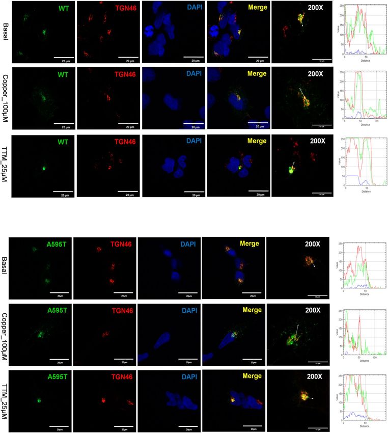
www.nature.com/scientificreports/
Figure 2. Conservation and location of Ala595 in MBD6. (A) Multiple sequence alignment using Clustal-
omega (EMBL-EBI) of the ATP7B protein showing the conservation status of the Ala595 residue among several
mammalian species (Human, chimpanzee, cow, sheep, dog, rat,andmouse). (B) The structure of MBD6 (PDB
accession 2EW9) with the Ala substituted with Thr using the mutagenesis function in PyMol. The difference in
length of the Ala and Thr side chains are indicated in yellow.
Results
Defining the cellular properties of WD-causing mutations helps to establish genotype–phenotype correlations
and facilitates the development of mutation-specific disease monitoring and therapeutic approaches. In this
study, we have characterized the expression, localization, activity, and trafficking patterns of several WD muta-
tions commonly found in the Indian population. The selected mutations are located in domains essential for
the ATP-binding and Cu-transport activities of ATP7B, as well as in the regulatory N-terminal and C-terminal
domains. We hypothesized that the consequences of the mutations will differ between those in the functional
domains and those in the regulatory domains.
A595T ATP7B mutant has reduced Cu‑transport activity but normal localization and traffick-
ing behavior. The A595T mutation was originally reported in a male WD patient in the compound-hete-
rozygous state with the G1061E mutation on the other allele (Table S2)7. The Ala residue in this position is evo-
lutionarily conserved, as evidenced by the alignment of ATP7B sequences from multiple species (Fig. 2a). A595
is located in the N-terminal domain on the β strand of MBD6, with its side chain projecting into the protein core
(Fig. 2b). Substitution of Ala (Van der Waals volume A3: 67 and accessible surface area A2: 67) with Thr (Van der
Waals volume A3: 93 and accessible surface area A 2: 102) (Table S3) increases the bulk of the side chain (Fig. 2b,
extra volume indicated in yellow) and replaces the uncharged hydrophobic Ala with a partially charged Thr in
the vicinity of Leu and Glu. Thus, the A595T change, although relatively small, could be deleterious.
To directly test this prediction, we characterized the Cu-transport activity of the A595T variant. Analysis of
Cu-transport by this and other mutants was monitored in Menkes disease fibroblast (YST) cells, which lack active
ATP7A and ATP7B. As a result, the transfer of Cu to the secretory pathway (TGN and vesicles) is impaired, and
cells produce inactive tyrosinase, a pigment-generating Cu-dependent enzyme. Expression of WT ATP7B restores
Cu delivery to tyrosinase and activates the enzyme, which can be monitored by the appearance of dark eumelanin
pigment (Fig. 3). Expression of the A595T mutant resulted in pigment formation, indicating that this mutant
has Cu-transport activity. However, the total intensity of the signal was 52.4% lower than that of WT ATP7B
(15.11159 ± 29.8 vs. 7.91 ± 23.04, respectively; Fig. 3). Quantitation of GFP signal in cells and analysis of protein
abundance indicate that the lower activity of the mutant is not the result of lower protein expression (Fig. 4).
To assess the effect of the mutants on ATP7B protein localization and trafficking, they were transfected into
HEK293 cells, which have been commonly used for studies of ATP7B trafficking. As expected, WT ATP7B was
found in the TGN under basal conditions, trafficked out of the TGN to vesicles when Cu was elevated, and does
not show a vesicular pattern when copper is low (Fig. 5a). The A595T mutant behaved very similarly. Under basal
conditions, it was targeted to the TGN, as evident by co-localization with the TGN marker (Fig. 5b). In response
to Cu elevation, co-localization decreased significantly, and the mutant was found mostly in vesicles, indicative of
trafficking. Following Cu depletion using the Cu chelator tetrathiomolybdate (TTM), the A595T mutant moved
back to the TGN (Fig. 5b). These data indicate that the A595T mutant retains normal Cu-dependent trafficking
under both low and high Cu conditions.
G1061E and G1101R ATP7B mutants do not have Cu‑transport activity and are trapped in the
ER. The G1061E and G1101R mutations are located in the nucleotide-binding domain (N-domain) of the
ATP7B protein (Fig. 1). G1061E was reported as the second most common mutation in eastern Indian WD
patients, comprising 11% of mutated alleles o bserved17. The G1061E mutation was also found in the WD patient
cohort from south India and Mediterranean r egions18,19. The residue G1061 is highly conserved (Fig. 6a), and
it is located in the α-helix in the immediate vicinity of the invariant SEHPL motif, the location of another com-
mon and well-characterized WD-causing mutation, H1069Q (Fig. 6b). G1061 is oriented towards the protein
core, and replacement of this small, neutral residue with the bulky, negatively charged Glu is likely to disrupt
protein structure. Indeed, analysis of expression and localization of the G1061E mutant in HEK293 cells revealed
that it co-localizes completely with the endoplasmic reticulum (ER) marker Calnexin under basal conditions
(Fig. 6c). Consistent with protein misfolding and retention in the ER, the protein levels of the mutant ATP7B
Scientific Reports | (2020) 10:13487 | https://doi.org/10.1038/s41598-020-70366-7 4
Vol:.(1234567890)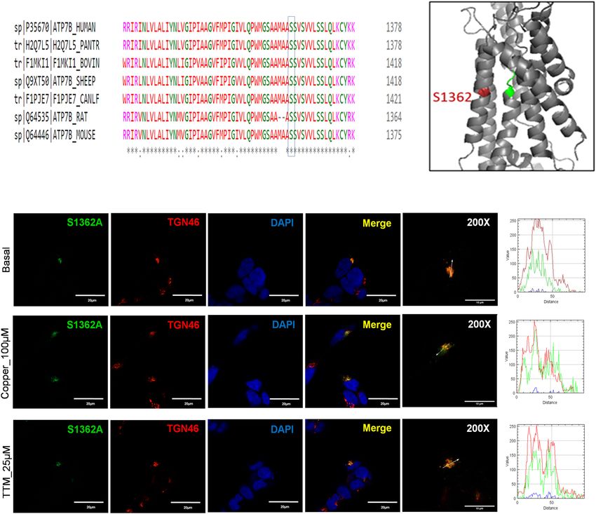
www.nature.com/scientificreports/
Figure 3. Cu-transport activity of the ATP7B mutants. (A) YST Cells were transfected with the tyrosinase-
encoding plasmid alone (Tyr, negative control) or together with wild-type (WT) ATP7B, the catalytically
inactive D1027A mutant (negative control), or the ATP7B mutants found in WD patients. Cu-transport activity
was evaluated by measuring eumelanin pigment formation (indicated with white arrows).The area (B) and
intensities (C) of the pigment were quantified. (D) Total eumelanin pigment signal (area × intensity) in cells
with the WD mutations were quantified and compared to the WT values. All values are reported as means ± SD
(Standard deviation). Significance was determined by one-way ANOVA with Dunnet’s multiple comparison test,
****p < 0.0001, n = 3.
were reduced compared to WT ATP7B (Fig. 4e, f). Moreover, tyrosinase activation assay in cells expressing this
mutation showed no pigment formation (Fig. 3a), suggesting greatly diminished Cu-transport activity. The lack
of pigment formation was similar to the result with the D1027A mutant, which is expressed normally (Fig. S1)
but is catalytically i nactive20 and therefore incapable of transporting copper and activate tyrosinase. While reten-
tion of ATP7B mutants in the ER in some cases can be overcome by increasing Cu c oncentration21, the G1061E
mutant remained trapped in the ER under both high and low Cu conditions.
The G1101R variant is another prevalent mutation in the southern and western Indian WD patient cohort18,22.
The Gly residue is invariant and is directly involved in ATP binding within the N-domain11. Replacement of
small, flexible Gly with bulky, charged Arg likely not only interferes with ATP binding, but also causes structural
perturbation within this highly conserved domain (Fig. 6a). Tyrosinase activation assay in cells expressing this
mutation showed no pigment formation, indicating the loss of Cu-transport (Fig. 3). Analysis of the G1101R
mutant expression in HEK293A cells showed that the mutant protein completely co-localized with the ER marker,
Calnexin, independently of Cu concentration (Fig. 7), suggesting that it is retained in the ER. Consistent with
these results, the protein levels of the G1101R mutant were reduced compared to WT ATP7B (Fig. 4e, f).
S1362A ATP7B mutant has reduced Cu‑transport activity and lacks Cu‑dependent traffick-
ing. The transmembrane domain (TM) of ATP7B forms the Cu translocation pathway, and the residues
within TM4–TM8 are conserved among the species (Fig. 8a). The S1362A mutation is located in TM8 (Fig. 1)
and was reported in a 21-year-old Indian male who had high urinary Cu levels and hepatic manifestations of
WD7 (Table S2). The Ser residue at position 1362 is invariant in ATP7B among various species (Fig. 8a), and it is
thought to be directly involved in Cu-binding within the membrane23,24. In the homology model of ATP7B24, the
side chain of S1362 projects into a small cavity in the immediate vicinity of the invariant Cu-binding motif CPC
(Fig. 8b). As such, the substitution of Ser with the smaller Ala is unlikely to cause major structural disruptions.
At the same time, the loss of the hydrophilic side chain of Ser may negatively affect Cu binding and transport.
Analysis of the Cu-transport activity using tyrosinase activation assay revealed significant reduction in the pig-
ment intensity in cells expressing the S1362A mutant compared to WT ATP7B (Fig. 3).The overall pigment
Scientific Reports | (2020) 10:13487 | https://doi.org/10.1038/s41598-020-70366-7 5
Vol.:(0123456789)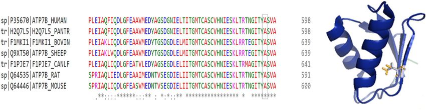
www.nature.com/scientificreports/

                                      Figure 4.  Expression of WT and mutant ATP7B proteins in YST cells. (A) The expression of GFP-tagged WT
                                      ATP7B and the Ala595Thr (A595T), Ser1362Ala (S1363A), and Ser1426Ile (S1426I) ATP7B mutants in YST
                                      cells visualized using GFP fluorescence. (B) The intensity of GFP fluorescence was normalized to the area. All
                                      values are reported as means ± SD. Significance was determined by one-way ANOVA with Dunnet’s multiple
                                      comparison test, *p < 0.05 and ***p < 0.001. (C) Representative Western blot and (D) densitometric analysis of
                                      the WT, S1362A, A595T, and S1426I protein abundances in YST cells. The band intensities were normalized to
                                      β-actin. All values are reported as means ± SD. Significance was determined by unpaired Student’s t test, n = 3.
                                      (E) Representative Western blot and (F) densitometric analysis of WT ATP7B and the Gly1101Arg (G1101R),
                                      and Gly1061Glu (G1061E) ATP7B mutant protein abundances in YST cells. The band intensities were
                                      normalized to β-actin. All values are reported as means ± SD. Significance was determined by one-way ANOVA
                                      with Dunnet’s multiple comparison test, ***p < 0.001; ****p < 0.0001, n = 3.

          Scientific Reports |   (2020) 10:13487 |              https://doi.org/10.1038/s41598-020-70366-7                                               6

Vol:.(1234567890)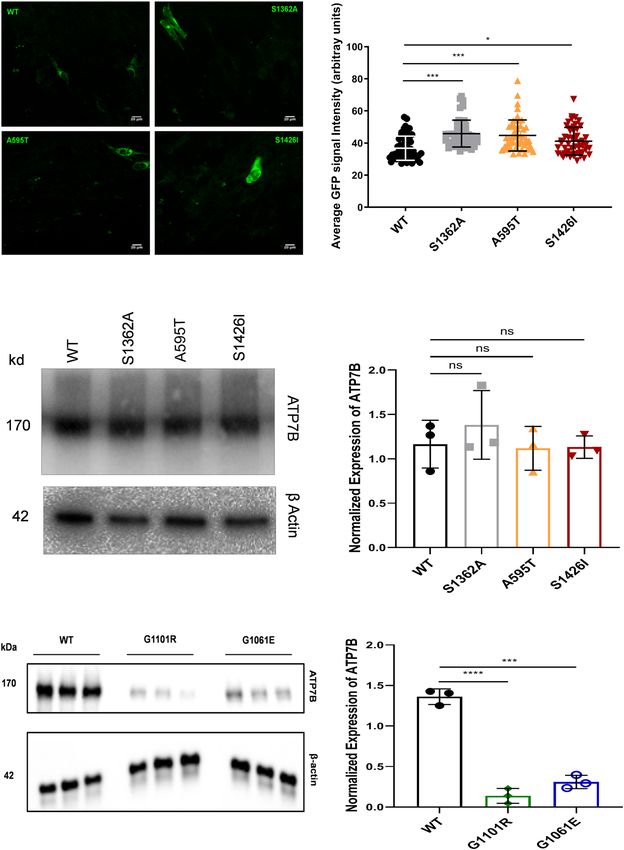
www.nature.com/scientificreports/
Figure 5. Localization of WT and Ala595Thr mutant (A595T) ATP7B in HEK293 cells in low and high Cu.
HEK293 cells expressing GFP-tagged (A) WT ATP7B or (B) A595T ATP7B mutant (green) were treated for
3 h with either basal medium (top panels), 100 µM CuCl2 (middle panels), or first with 100 µM C
uCl2 and then
with 25 µM tetra-thiomolybdate (TTM) (lower panels). TGN marker TGN46 in red, nuclei (DAPI) in blue.
Co-localization is indicated in yellow. Green signals are from GFP fluorescence.
intensity was about 11.1% of that of the WT signal (15.11159 ± 29.8 vs. 1.69 ± 1.82, respectively). The level of
protein expression was not significantly different from that of the WT ATP7B (Fig. 4).
In HEK293 cells, the S1362A mutant had normal levels of expression and showed complete co-localization
with the TGN marker, TGN46, under basal conditions (Fig. 8c). Therefore, the diminished Cu-transport activity
observed is likely due to local structural changes within the Cu-transport pathway rather than due to general pro-
tein misfolding. Even though the mutant was targeted normally, its sensitivity to elevated Cu was lost. Treatment
Scientific Reports | (2020) 10:13487 | https://doi.org/10.1038/s41598-020-70366-7 7
Vol.:(0123456789)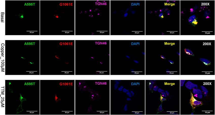
www.nature.com/scientificreports/
Figure 6. Properties of ATP7B mutants located within the N-domain. (A) Multiple sequence alignment
showing conservation of Gly1061 (G1061) and Gly1101 (G1101). (B) Location of G1061 and G1101 (shown
in red) in the N-domain (in grey). The conserved SEHPL motif containing the common H1069Q mutation is
in green. The actuator (A)-domain is in pink. (C) Localization of the G1061E ATP7B mutant in HEK293 cells
under low and high Cu conditions. HEK293 cells were transfected (12 h) with WT or G1061E mutant GFP-
ATP7B (green) and treated for 3 h with basal medium (top), medium containing 100 µM of C uCl2 (middle), or
medium containing 25 µM of tetra-thiomolybdate (TTM) (lower panel).Endoplasmic reticulum (ER) marker,
Calnexin, in red and nuclei (DAPI) in blue. Co-localization is indicated in yellow. Green signals are from GFP
fluorescence.
of cells expressing the S1362A mutant with 100 µM Cu for 3 h showed no trafficking of the mutant from the TGN
to vesicles (Fig. 8c). This indicates that the negative effect of this mutation on Cu-transport is compounded by
the inability of the mutant ATP7B to move from the TGN to vesicles to facilitate Cu export.
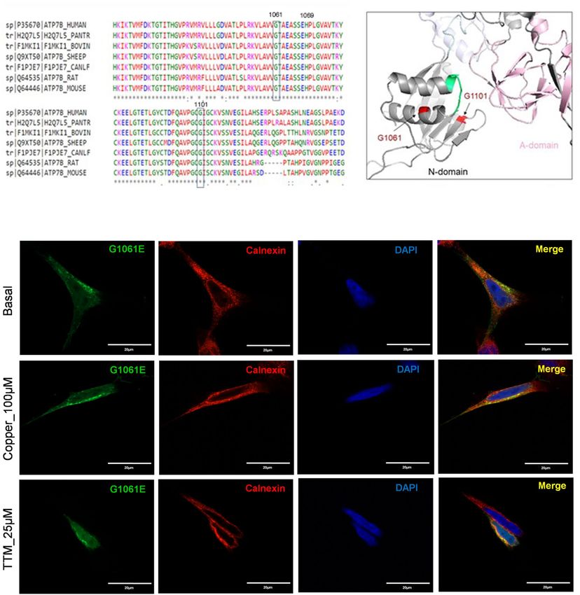
The S1426I mutant has reduced Cu‑transport activity but normal Cu‑dependent traffick-
ing. The S1426I mutation resides in the C-terminal region of ATP7B (Fig. 1). It was detected in a female
Indian WD patient in the compound-heterozygous state, with A1003V on the other allele (Table S2). The C-ter-
Scientific Reports | (2020) 10:13487 | https://doi.org/10.1038/s41598-020-70366-7 8
Vol:.(1234567890)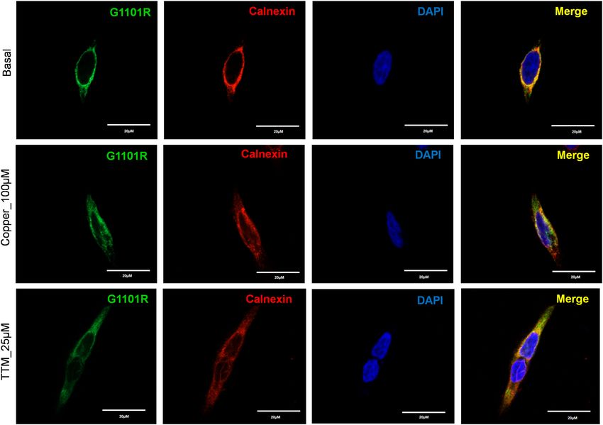
www.nature.com/scientificreports/
Figure 7. Localization of G1101R ATP7B mutant in HEK293 cells under low and high Cu conditions. HEK293
cells were transfected (12 h) with WT or G1101R mutant ATP7B GFP-ATP7B (green) and treated for 3 h with
basal medium (top), medium containing 100 µM of CuCl2 (middle), or medium containing 25 µM of tetra-
thiomolybdate (TTM) (lower panel).Endoplasmic reticulum (ER) marker, Calnexin, in red and nuclei (DAPI) in
blue. Co-localization is indicated in yellow. Green signals are from GFP fluorescence.
minus of ATP7B is predicted to be unstructured, but current data suggest that it plays important regulatory
roles by interacting with other proteins and ATP7B d omains25. Previous studies have shown that partial or
complete deletion of the C-terminal domain of ATP7B decreases protein retention in the TGN and impairs
retrograde trafficking26. Tyrosinase activation assay revealed that the S1426I mutant had reduced but measur-
able Cu-transport activity (Fig. 3). The mean pigment signal of cells expressing the S1426I mutant decreased by
66.05% compared to WT ATP7B (15.11159 ± 29.8 vs. 5.13 ± 11.52, respectively). In HEK293 cells, the S1426I
mutant is targeted to the TGN under basal conditions. Treatment with 100 µM Cu triggers the movement of the
S1426I mutant from the TGN to vesicles while under Cu depletion conditions, the mutant shows complete co-
localization with the TGN marker, TGN46 (Fig. 9).
Cu retention in YST cells expressing different ATP7B mutants. The above experiments identified
three categories of ATP7B mutants: (1) mutants that are mis-localized and inactive, (2) mutants with reduced
Cu-transport activity but normal Cu-dependent trafficking, and (3) mutants with reduced Cu-transport activ-
ity and impaired trafficking behavior. To determine whether mutants with partial transport and/or trafficking
activity affect the total cellular Cu content, YST cells were transfected with either WT ATP7B or individual
mutants and then cellular Cu content was measured using ICP-MS. Cells expressing WT ATP7B had the low-
est Cu content (123.57 ± 11.35 fg/106 cells), whereas the inactive G1061E and G1101R mutants had the highest
Cu contents (243.77 ± 18.53 and 171.11 ± 9.46 fg/106 cells, respectively). Moreover, cells expressing the S1362A
mutant had more Cu than cells expressing WT ATP7B (167.55 ± 2.68 vs. 123.57 ± 11.35 fg/106 cells, respectively).
This observation is consistent with the finding that the S1362A mutant cannot move out of the TGN towards
the plasma membrane and pump excess Cu out of the cell. Cells expressing the S1426I mutant did not show a
significant difference in Cu content compared to WT ATP7B, though it did have more variable values (Fig. 10).
Taken together, these data illustrate not only the broad range of consequences that mutations in ATP7B have
on the properties of the protein, but also the wide variation of Cu-retention phenotypes in cells expressing the
mutations in the homozygous state.
Scientific Reports | (2020) 10:13487 | https://doi.org/10.1038/s41598-020-70366-7 9
Vol.:(0123456789)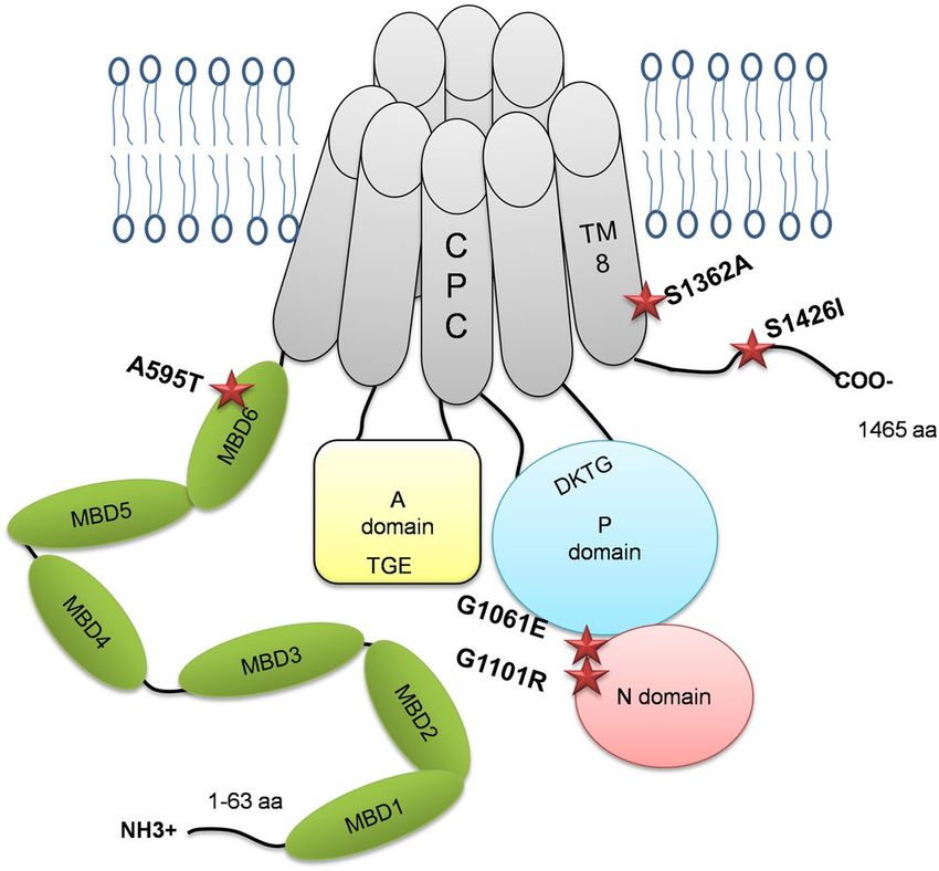
www.nature.com/scientificreports/
Figure 8. Molecular and cellular characteristics of the S1362A ATP7B mutant. (A) Multiple sequence
alignment showing conservation of Ser1362. (B) Location of Ser1362 (red) in proximity to the invariant CPC
motif (green) involved in Cu coordination within the transmembrane domain. (C) Localization of S1362A
mutant ATP7B in HEK293 cells under low and high Cu conditions. HEK293 cells were transfected (12 h) with
WT or S1362A mutant ATP7B GFP-ATP7B (green) and treated for 3 h with basal medium (top), medium
containing 100 µM of CuCl2 (middle), or medium containing 25 µM of tetra-thiomolybdate (TTM) (lower
panel). Trans-golgi network (TGN) marker, TGN46, in red and nuclei (DAPI) in blue. Co-localization is
indicated in yellow. Green signals are from GFP fluorescence.
Different ATP7B mutants interact and influence each other’s behavior. Many WD patients
are compound-heterozygous for ATP7B mutations, i.e. express different ATP7B mutants in same cell. In cells,
ATP7B forms a dimer, and heterodimerization of experimental ATP7B variants with different trafficking prop-
erties was shown to modify their localization compared to homodimeric forms15. This finding suggested that
dissimilar WD-causing mutants could influence each other localization and trafficking. To test this hypothesis,
we co-expressed the A595T and G1061E mutants in cells. This combination was previously found in a male WD
patient who had low plasma ceruloplasmin levels and high urinary Cu levels (Table S2). The G1061E mutant is
trapped in the ER when expressed alone (Fig. 7), but is targeted to the TGN under basal conditions when co-
expressed with the A595T mutant, with which it is fully co-localized (Fig. 11). In other words, the presence of an
active, better folded mutant restores the localization of an inactive and misfolded mutant. However, the dimeri-
zation of the active A595T with the inactive G1061E has a negative impact on the Cu-dependent trafficking of
ATP7B from the TGN. The A595T mutant, which when expressed alone, traffics from the TGN in response
to Cu (Fig. 5), shows no trafficking in response to Cu when co-expressed with the G1061E mutant. Given that
the loss of trafficking often reflects the loss of Cu-transport, these results suggest that the A595T mutant forms
a non-functional complex with the G1061E mutant. Lastly, under low Cu conditions, only a small fraction of
the A595T mutant was found to co-localize with the TGN marker. The majority of A595T co-localized with
Scientific Reports | (2020) 10:13487 | https://doi.org/10.1038/s41598-020-70366-7 10
Vol:.(1234567890)www.nature.com/scientificreports/
Figure 9. Localization of the S1426I ATP7B mutant in HEK293 cells under low and high Cu conditions. (A)
Multiple sequence alignment showing conservation of Ser1426Ile. (B) HEK293 cells were transfected (12 h)
with WT or S1426I mutant ATP7B GFP-ATP7B (green) and treated for 3 h with basal medium (top), medium
containing 100 µM of CuCl2 (middle), or medium containing 25 µM of tetra-thiomolybdate (TTM) (lower
panel). Trans-golgi network (TGN) marker, TGN46, in red and nuclei (DAPI) in blue. Co-localization is
indicated in yellow. Green signals are from GFP fluorescence.
the G1061E mutant in the pattern consistent with their retention in the ER. Thus, while dimerization occurs
in the ER in the absence of Cu, the proper folding of the dimer and its exit from the ER is at least partially Cu-
dependent.
Discussion
WD is a monogenetic disorder with a remarkable variety of both mutations and manifestations of disease. Previ-
ous studies have identified ATP7B mutations in different domains of the protein that had a negative impact on
ATP7B stability13, loss of Cu-transport activity14, ER retention15, and inability to traffic from the TGN to vesicles
in response to Cu e levation16,17. However, establishing clear genotype–phenotype correlations in WD has proven
difficult. The majority of disease-associated mutations in ATP7B are missense mutations, and WD patients are
commonly compound-heterozygous. In this study, we characterized the cellular properties of mutations identified
among Indian WD patients and demonstrated the importance of studying mutations in combinations observed
in compound-heterozygous WD patients. The properties of the individual mutations are summarized in Table 1.
The A595T, S1362A, and S1426I mutations are novel genetic changes, whereas the G1061E and G1101R muta-
tions were previously identified in the WD patient c ohort7. These five ATP7B mutations are located in structurally
and functionally different domains of the protein and are distributed throughout the entire ATP7B structure,
including the N-terminus, the TM domain, the ATP-binding domain, and the C-terminus. We identified a broad
Scientific Reports | (2020) 10:13487 | https://doi.org/10.1038/s41598-020-70366-7 11
Vol.:(0123456789)www.nature.com/scientificreports/
Figure 10. Cu retention in YST cells expressing ATP7B mutants. YST cells were transfected (12 h) with WT or
mutant (A595T, S1362A, S1426I, G1061E, and G1101R) ATP7B and treated for 24 h with medium containing
100 µM of CuCl2. Cellular Cu content was measured by inductively coupled plasma mass spectrometry
(ICP-MS) to evaluate Cu-export. The values were normalized to the YST cell count. All values are reported as
means ± SD. Significance was determined by unpaired Student’s t test, *p < 0.05; **p < 0.01; ***p < 0.001, n = 3.
Figure 11. Co-expression of A595T and G1061E ATP7B mutants in HEK293 cells alters their subcellular
distribution. HEK293 cells were co-transfected with GFP-ATP7B-A595T (green) and FLAG-ATP7B-G1061E
(red) and incubated in basal medium (top panels), medium containing 100 µM C uCl2 (middle panels), or
medium containing 25 µM of tetra-thiomolybdate (TTM; lower panels). Trans-golgi network (TGN) marker,
TGN46, in red and nuclei (DAPI) in blue. Co-localization is indicated in yellow. The rightmost panels show
× 200 magnified images. Green signals are from GFP fluorescence.
spectrum of consequences on ATP7B activity and cellular behavior, in agreement with previous analysis of other
WD-causing mutations27.
The N-terminal domain of ATP7B has 6 metal binding domains (MBD 1–6) that have similar structures but
different functions28. MBD1, MBD2, and MBD3 form a transient complex that receives Cu from A tox129. MBD5
and MBD6 are situated close to the TM domains of ATP7B30, and at least one of these domains must be present to
sustain Cu-transport activity31. Consistent with this, the A595T mutation, within MBD6, results in a significant
reduction in Cu-transport activity of the protein and a decrease in its ability to remove Cu from cells. However,
this missense mutation did not lead to gross misfolding or mis-localization, nor did it disrupt Cu-transport
completely, as evidenced from the fact that it was able to traffic from the TGN to vesicles in response to Cu eleva-
tion. These properties are similar to those of the previously characterized G591D and R616Q mutants, which are
Scientific Reports | (2020) 10:13487 | https://doi.org/10.1038/s41598-020-70366-7 12
Vol:.(1234567890)www.nature.com/scientificreports/
Mutation Domain Cu-transport Eumelanin signal (area × intensity) Subcellular localization Cu-dependent trafficking Intracellular Cu compared to WT
Ala595Thr MBD6 Yes 7.91 ± 23.04 Golgi and vesicles Yes High
Gly1061Glu N- domain No NA ER retained No High
Gly1101Arg N domain No NA ER retained No High
Ser1362Ala TM-8 Yes 1.69 ± 1.82 Golgi retained No High
Ser1426Ile C-terminal Yes 5.13 ± 11.52 Golgi and vesicles Yes No difference
Table 1. Properties of the individual ATP7B mutants characterized in this study. MBD metal binding domain,
Cu copper, ER endoplasmic reticulum, WT wild type.
also located in MBD632. Taken together, these data suggest that the structural integrity of A595 is important for
Cu-transport but not crucial for Cu-dependent trafficking. The impact of this mutation on trafficking kinetics,
if any, remains to be determined. Notably, the fact that the A595T mutation results in only a 50% decrease in
Cu-transport activity suggests that this mutation alone may not cause the WD phenotype but perhaps would in
combination with more deleterious mutations.
In the N-domain of ATP7B, the residues H10169, G1099, G1101, I1102, G1149, and N1150 are indispen-
sable for the binding of A TP33. Mutation of some of these residues or their neighboring residues are associated
with WD . The H1069Q substitution is a common WD-causing mutation found in Caucasian populations3,34.
11
The G1061E and G1101R mutations have been identified as prevalent missense mutations among Indian WD
patients35. Mutations at these two positions may directly impede ATP binding and disrupt protein folding, as
both have the small Gly residue replaced with bulkier, charged residues. We show that these two mutations
produce misfolded ATP7B and completely block Cu-transport to the secretory pathway. Previous studies have
found that mutations within the N-domain often disrupt protein folding and stability36. Unlike the G875R vari-
ant of ATP7B that can be rescued from ER retention by the addition of C u21, the G1061E and G1101R mutants
remain in the ER even under high Cu conditions. Taken together our data suggest that zero copper transport
activity of G1101R and G1061E observed in a tyrosinase could be a combination of their compromised function,
retention in the ER and lower level of expression. Previous studies have identified some novel therapeutics and
molecular targets in the signaling pathway that seems to be promising in rescuing ER retained and misfolded
ATP7B mutations37,38. It would be interesting to observe whether this same strategy can be applied to correct
these two ER retained ATP7B mutations like G1101R and G1061E.
Although both the G1061E and G1101R mutants have similar properties in cells, the reported clinical phe-
notypes of WD patients with these mutations are very different. The p.G1061E homozygotes had predominantly
neuropsychiatric presentations with an age of onset of 9–16 years, whereas the p.G1101R homozygotes had liver
involvement and an age of onset of 5–7 years7 (Table S2). These results caution against establishing direct links
between molecular and clinical phenotypes and support emerging evidence suggesting that other modifying
factors play a role in WD.
The trafficking of ATP7B is a complex process, and the factors regulating the exit of ATP7B from the TGN
are still not fully understood. Previous reports have demonstrated that TM1 and TM2 play a role in the Cu-
dependent trafficking of ATP7B to v esicles39. It has also been suggested that Cu-transport and the ability to traffic
are linked13. For instance, while mutation of D1027 has no negative impact on ATP7B structure, mutation of this
residue renders the protein catalytically inactive and blocks its exit from the TGN. Previous studies have found
that the S1362F-fs mutation impairs Cu-transport and phosphorylation a ctivity27. S1362A is a novel mutation in
TM8 of ATP7B found in an Indian WD patient7. Within the molecular model of ATP7B, this residue is located
in the immediate vicinity of the crucial Cu-binding CPC motif and M1359 residue of TM8 (Fig. 8b). This finding
suggests that structural changes caused by the mutation may impact Cu binding and translocation within the
domain, as well as the ability of the protein to leave the TGN. Consistent with these predictions, the tyrosinase
activation assay shows a significantly lower activity for this variant compared to WT ATP7B. The mutant also
shows complete TGN retention, even under high Cu conditions. Similar to these results, S653Y in TM1 and
G943S in TM5 were shown to result in ATP7B protein with some amount of Cu-transport activity, though the
proteins failed to exit the TGN under high Cu c onditions39,40. Taken together, these data suggest that the TM
portion of ATP7B plays an important role in its exit from the TGN, possibly through interaction with specific
lipids or other molecules.
The unstructured C-terminal region of ATP7B contains conserved signal residues (e.g. tri-leucine1454LLL1456)
important for vesicular trafficking of the p rotein41. Numerous Ser/Thr residues in this domain were found to be
phosphorylated11. Partial or complete deletion of the C-terminus destabilizes the ATP7B protein and impairs
retrograde trafficking. The L1373R mutation in the C-terminus has been associated with reduced Cu-transport
and protein expression, as well as loss of TGN retention under low Cu c onditions26. In contrast, the S1426I
mutant had normal protein expression and trafficked normally in response to Cu stimulation. Even though
its Cu-transport activity was reduced, the ability of this variant to remove Cu from cells was not significantly
compromised, indicating that this mutation is likely mild. This conclusion is consistent with the compound-
heterozygous status of the patient from which this mutation was identified.
Many WD patients are compound-heterozygous, i.e., have different ATP7B mutations on two different alleles.
Mukherjee et al. identified a WD patient with the A595T and G1061E mutations in a compound-heterozygous
state7. By characterizing these mutations both separately and together, we showed that these mutants interact,
and that this interaction in turn impacted their cellular properties. Alone, the catalytically inactive G1061E is
Scientific Reports | (2020) 10:13487 | https://doi.org/10.1038/s41598-020-70366-7 13
Vol.:(0123456789)www.nature.com/scientificreports/
retained in the ER, whereas the partially active A595T is targeted to the TGN and traffics in response to high Cu.
The co-expression of these mutants to mimic the compound-heterozygous state generated an ATP7B complex
with distinct cellular properties. Under low Cu conditions, only a small fraction of the A595T mutant was found
to co-localize with the TGN marker, and the majority was found to co-localize with the G1061E mutant in the
ER. No vesicular movement of the mutants was observed following Cu stimulation. The low levels of plasma
ceruloplasmin (Table S2) observed in the WD patient containing these mutations could be explained by this
cellular phenotype, however direct measurements of copper-transport activities of heterodimer (using tyrosinase
assay or copper retention) will be necessary to fully understand consequences of compound heterozygosity on
ATP7B properties.
Taken together, our findings suggest that future genotype–phenotype correlation studies should rely, when
appropriate, on characterization of ATP7B hetero-dimers rather than individual ATP7B mutants. Furthermore,
sometimes positive influence of mutants (such as rescue of ATP7B localization by a better folded mutant upon
dimerization—as we observed A595T and G1061E mutations) can modify the property of more deleterious
mutant producing milder phenotype. In our view, going forward the best approach for studies of ATP7B mutants
would be to use the WD patients-derived cells.
Received: 16 February 2020; Accepted: 16 June 2020
References
1. Gupta, A. & Lutsenko, S. Human copper transporters: mechanism, role in human diseases and therapeutic potential. Future Med.
Chem. 1, 1125–1142. https://doi.org/10.4155/fmc.09.84 (2009).
2. Bull, P. C., Thomas, G. R., Rommens, J. M., Forbes, J. R. & Cox, D. W. The Wilson disease gene is a putative copper transporting
P-type ATPase similar to the Menkes gene. Nat. Genet. 5, 327–337. https://doi.org/10.1038/ng1293-327 (1993).
3. Tanzi, R. E. et al. The Wilson disease gene is a copper transporting ATPase with homology to the Menkes disease gene. Nat. Genet.
5, 344–350. https://doi.org/10.1038/ng1293-344 (1993).
4. Das, S. K. & Ray, K. Wilson’s disease: an update. Nat. Clin. Pract. Neurol. 2, 482–493. https: //doi.org/10.1038/ncpneu
ro029 1 (2006).
5. Kegley, K. M. et al. Fulminant Wilson’s disease requiring liver transplantation in one monozygotic twin despite identical
genetic mutation. Am. J. Transplant. Off. J. Am. Soc. Transplant. Am. Soc. Transplant Surg. 10, 1325–1329. https://doi.org/10.111
1/j.1600-6143.2010.03071.x (2010).
6. Kenney, S. M. & Cox, D. W. Sequence variation database for the Wilson disease copper transporter, ATP7B. Hum. Mutat. 28,
1171–1177. https://doi.org/10.1002/humu.20586 (2007).
7. Mukherjee, S. et al. Genetic defects in Indian Wilson disease patients and genotype-phenotype correlation. Parkinsonism Relat.
Disord. 20, 75–81. https://doi.org/10.1016/j.parkreldis.2013.09.021 (2014).
8. Leggio, L., Addolorato, G., Loudianos, G., Abenavoli, L. & Gasbarrini, G. Genotype-phenotype correlation of the Wilson disease
ATP7B gene. Am. J. Med. Genet. Part A 140, 933. https://doi.org/10.1002/ajmg.a.31191 (2006).
9. Inesi, G., Pilankatta, R. & Tadini-Buoninsegni, F. Biochemical characterization of P-type copper ATPases. Biochem. J. 463, 167–176.
https://doi.org/10.1042/BJ20140741 (2014).
10. Inesi, G. Molecular features of copper binding proteins involved in copper homeostasis. IUBMB Life 69, 211–217. https://doi.
org/10.1002/iub.1590 (2017).
11. Lutsenko, S., Jayakanthan, S. & Dmitriev, O. Y. in Clinical and Translational Perspectives on WILSON DISEASE (eds Nanda Kerkar
& Eve A. Roberts) 33–43 (Academic Press, 2019).
12. Pierson, H. & Lutsenko, S. in Wilson Disease (eds Karl Heinz Weiss & Michael Schilsky) 23–32 (Academic Press, 2019).
13. Hasan, N. M. et al. Molecular events initiating exit of a copper-transporting ATPase ATP7B from the trans-Golgi network. J. Biol.
Chem. 287, 36041–36050. https://doi.org/10.1074/jbc.M112.370403 (2012).
14. Petris, M. J., Strausak, D. & Mercer, J. F. The Menkes copper transporter is required for the activation of tyrosinase. Hum. Mol.
Genet. 9, 2845–2851. https://doi.org/10.1093/hmg/9.19.2845 (2000).
15. Jayakanthan, S., Braiterman, L. T., Hasan, N. M., Unger, V. M. & Lutsenko, S. Human copper transporter ATP7B (Wilson disease
protein) forms stable dimers in vitro and in cells. J. Biol. Chem. 292, 18760–18774. https: //doi.org/10.1074/jbc.M117.807263 (2017).
16. McCann, C. J. et al. Single nucleotide polymorphisms in the human ATP7B gene modify the properties of the ATP7B protein.
Metallomics Integr. Biomet. Sci. 11, 1128–1139. https://doi.org/10.1039/c9mt00057g (2019).
17. Gupta, A. et al. Molecular diagnosis of Wilson disease using prevalent mutations and informative single-nucleotide polymorphism
markers. Clin. Chem. 53, 1601–1608. https://doi.org/10.1373/clinchem.2007.086066 (2007).
18. Guggilla, S. R., Senagari, J. R., Rao, P. N. & Madireddi, S. Spectrum of mutations in the ATP binding domain of ATP7B gene of
Wilson Disease in a regional Indian cohort. Gene 569, 83–87. https://doi.org/10.1016/j.gene.2015.05.031 (2015).
19. Loudianos, G. et al. Mutation analysis in patients of Mediterranean descent with Wilson disease: identification of 19 novel muta-
tions. J. Med. Genet. 36, 833–836 (1999).
20. Tsivkovskii, R., Eisses, J. F., Kaplan, J. H. & Lutsenko, S. Functional properties of the copper-transporting ATPase ATP7B (the
Wilson’s disease protein) expressed in insect cells. J. Biol. Chem. 277, 976–983. https://doi.org/10.1074/jbc.M109368200 (2002).
21. Gupta, A. et al. Cellular copper levels determine the phenotype of the Arg875 variant of ATP7B/Wilson disease protein. Proc. Natl.
Acad. Sci. U.S.A. 108, 5390–5395. https://doi.org/10.1073/pnas.1014959108 (2011).
22. Aggarwal, A. et al. Wilson disease mutation pattern with genotype-phenotype correlations from Western India: confirmation
of p.C271* as a common Indian mutation and identification of 14 novel mutations. Ann. Hum. Genet. 77, 299–307. https://doi.
org/10.1111/ahg.12024(2013).
23. Padilla-Benavides, T., McCann, C. J. & Arguello, J. M. The mechanism of Cu+ transport ATPases: interaction with CU+ chaperones
and the role of transient metal-binding sites. J. Biol. Chem. 288, 69–78. https://doi.org/10.1074/jbc.M112.420810 (2013).
24. Schushan, M., Bhattacharjee, A., Ben-Tal, N. & Lutsenko, S. A structural model of the copper ATPase ATP7B to facilitate analysis
of Wilson disease-causing mutations and studies of the transport mechanism. Metallomics Integr. Biomet. Sci. 4, 669–678. https://
doi.org/10.1039/c2mt20025b (2012).
25. Braiterman, L. T., Gupta, A., Chaerkady, R., Cole, R. N. & Hubbard, A. L. Communication between the N and C termini is required
for copper-stimulated Ser/Thr phosphorylation of Cu(I)-ATPase (ATP7B). J. Biol. Chem. 290, 8803–8819. https: //doi.org/10.1074/
jbc.M114.627414 (2015).
26. Braiterman, L., Nyasae, L., Leves, F. & Hubbard, A. L. Critical roles for the COOH terminus of the Cu-ATPase ATP7B in protein
stability, trans-Golgi network retention, copper sensing, and retrograde trafficking. Am. J. Physiol. Gastrointest. liver Physiol. 301,
G69-81. https://doi.org/10.1152/ajpgi.00038.2011 (2011).
Scientific Reports | (2020) 10:13487 | https://doi.org/10.1038/s41598-020-70366-7 14
Vol:.(1234567890)www.nature.com/scientificreports/
27. Huster, D. et al. Diverse functional properties of Wilson disease ATP7B variants. Gastroenterology 142(947–956), e945. https://
doi.org/10.1053/j.gastro.2011.12.048 (2012).
28. Lutsenko, S., Petrukhin, K., Cooper, M. J., Gilliam, C. T. & Kaplan, J. H. N-terminal domains of human copper-transporting adeno-
sine triphosphatases (the Wilson’s and Menkes disease proteins) bind copper selectively in vivo and in vitro with stoichiometry of
one copper per metal-binding repeat. J. Biol. Chem. 272, 18939–18944. https://doi.org/10.1074/jbc.272.30.18939 (1997).
29. LeShane, E. S. et al. Interactions between copper-binding sites determine the redox status and conformation of the regulatory
N-terminal domain of ATP7B. J. Biol. Chem. 285, 6327–6336. https://doi.org/10.1074/jbc.M109.074633 (2010).
30. Huang, Y. et al. Interactions between metal-binding domains modulate intracellular targeting of Cu(I)-ATPase ATP7B, as revealed
by nanobody binding. J. Biol. Chem. 289, 32682–32693. https://doi.org/10.1074/jbc.M114.580845 (2014).
31. Cater, M. A., Forbes, J., La Fontaine, S., Cox, D. & Mercer, J. F. Intracellular trafficking of the human Wilson protein: the role of
the six N-terminal metal-binding sites. Biochem. J. 380, 805–813. https://doi.org/10.1042/BJ20031804 (2004).
32. Arioz, C., Li, Y. & Wittung-Stafshede, P. The six metal binding domains in human copper transporter, ATP7B: molecular biophys-
ics and disease-causing mutations. Biomet. Int. J. Role Met. Ions Biol. Biochem. Med. 30, 823–840. https://doi.org/10.1007/s1053
4-017-0058-2 (2017).
33. Dmitriev, O. et al. Solution structure of the N-domain of Wilson disease protein: distinct nucleotide-binding environment and
effects of disease mutations. Proc. Natl. Acad. Sci. U.S.A. 103, 5302–5307. https://doi.org/10.1073/pnas.0507416103 (2006).
34. Thomas, G. R., Roberts, E. A., Walshe, J. M. & Cox, D. W. Haplotypes and mutations in Wilson disease. Am. J. Hum. Genet. 56,
1315–1319 (1995).
35. Bavdekar, A., Gupta, A. & Ray, K. in Clinical and Translational Perspectives on WILSON DISEASE (eds Nanda Kerkar & Eva Roberts)
335–343 (Academic press, 2019).
36. Dmitriev, O. Y., Bhattacharjee, A., Nokhrin, S., Uhlemann, E. M. & Lutsenko, S. Difference in stability of the N-domain underlies
distinct intracellular properties of the E1064A and H1069Q mutants of copper-transporting ATPase ATP7B. J. Biol. Chem. 286,
16355–16362. https://doi.org/10.1074/jbc.M110.198101 (2011).
37. van den Berghe, P. V. et al. Reduced expression of ATP7B affected by Wilson disease-causing mutations is rescued by pharmaco-
logical folding chaperones 4-phenylbutyrate and curcumin. Hepatology 50, 1783–1795. https://doi.org/10.1002/hep.23209 (2009).
38. Chesi, G. et al. Identification of p38 MAPK and JNK as new targets for correction of Wilson disease-causing ATP7B mutants.
Hepatology 63, 1842–1859. https://doi.org/10.1002/hep.28398 (2016).
39. Braiterman, L. T. et al. Distinct phenotype of a Wilson disease mutation reveals a novel trafficking determinant in the copper
transporter ATP7B. Proc. Natl. Acad. Sci. U.S.A. 111, E1364-1373. https://doi.org/10.1073/pnas.1314161111 (2014).
40. Forbes, J. R. & Cox, D. W. Copper-dependent trafficking of Wilson disease mutant ATP7B proteins. Hum. Mol. Genet. 9, 1927–1935.
https://doi.org/10.1093/hmg/9.13.1927 (2000).
41. Lalioti, V., Hernandez-Tiedra, S. & Sandoval, I. V. DKWSLLL, a versatile DXXXLL-type signal with distinct roles in the Cu(+)-
regulated trafficking of ATP7B. Traffic 15, 839–860. https://doi.org/10.1111/tra.12176 (2014).
Acknowledgements
We thank the Johns Hopkins School of Medicine Synthesis and Sequencing Facility for their assistance. We
sincerely thank Dr. Martina Ralle for helping with the Cu measurements. These studies were supported by NIH
grant R01 GM101502 to SL; SR is supported by fellowship from the Fulbright-Nehru Scholarship Program
2018–19; USIEF.
Author contributions
S.R.: Carried out most of the experiments, analyzed the results drafted the manuscript; C.M.: carried out some
of the experiments and analyzed the results and review of the manuscript; K.R., J.R., S.L., S.J.; conceived and
designed the study, critical review of the manuscript. M.R.: helped with ICPMS experiment and data analysis.
All authors reviewed the manuscript.
Competing interests
The authors declare no competing interests.
Additional information
Supplementary information is available for this paper at https://doi.org/10.1038/s41598-020-70366-7.
Correspondence and requests for materials should be addressed to S.R. or S.L.
Reprints and permissions information is available at www.nature.com/reprints.
Publisher’s note Springer Nature remains neutral with regard to jurisdictional claims in published maps and
institutional affiliations.
Open Access This article is licensed under a Creative Commons Attribution 4.0 International
License, which permits use, sharing, adaptation, distribution and reproduction in any medium or
format, as long as you give appropriate credit to the original author(s) and the source, provide a link to the
Creative Commons license, and indicate if changes were made. The images or other third party material in this
article are included in the article’s Creative Commons license, unless indicated otherwise in a credit line to the
material. If material is not included in the article’s Creative Commons license and your intended use is not
permitted by statutory regulation or exceeds the permitted use, you will need to obtain permission directly from
the copyright holder. To view a copy of this license, visit http://creativecommons.org/licenses/by/4.0/.
© The Author(s) 2020
Scientific Reports | (2020) 10:13487 | https://doi.org/10.1038/s41598-020-70366-7 15
Vol.:(0123456789)You can also read