Real Time insight into in vivo redox status utilizing hyperpolarized 1 13C N acetyl cysteine - Nature
←
→
Page content transcription
If your browser does not render page correctly, please read the page content below
www.nature.com/scientificreports
OPEN Real‑Time insight into in vivo redox
status utilizing hyperpolarized
[1‑13C] N‑acetyl cysteine
Kazutoshi Yamamoto1, Ana Opina2, Deepak Sail2, Burchelle Blackman2, Keita Saito1,
Jeffrey R. Brender1, Ronja M. Malinowski3, Tomohiro Seki1, Nobu Oshima1, Daniel R. Crooks1,
Shun Kishimoto1, Yu Saida1, Yasunori Otowa1, Peter L. Choyke1, Jan H. Ardenkjær‑Larsen3,
James B. Mitchell1, W. Marston Linehan1, Rolf E. Swenson2 & Murali C. Krishna1,4*
Drastic sensitivity enhancement of dynamic nuclear polarization is becoming an increasingly
critical methodology to monitor real-time metabolic and physiological information in chemistry,
biochemistry, and biomedicine. However, the limited number of available hyperpolarized 13C
probes, which can effectively interrogate crucial metabolic activities, remains one of the major
bottlenecks in this growing field. Here, we demonstrate [1-13C] N-acetyl cysteine (NAC) as a novel
probe for hyperpolarized 13C MRI to monitor glutathione redox chemistry, which plays a central part
of metabolic chemistry and strongly influences various therapies. NAC forms a disulfide bond in the
presence of reduced glutathione, which generates a spectroscopically detectable product that is
separated from the main peak by a 1.5 ppm shift. In vivo hyperpolarized MRI in mice revealed that
NAC was broadly distributed throughout the body including the brain. Its biochemical transformation
in two human pancreatic tumor cells in vitro and as xenografts differed depending on the individual
cellular biochemical profile and microenvironment in vivo. Hyperpolarized NAC can be a promising
non-invasive biomarker to monitor in vivo redox status and can be potentially translatable to clinical
diagnosis.
Cells normally exist in a fine balance between reductive and oxidative states. When this balance is disrupted,
either by external environmental stimuli or by abnormal metabolic states, the cellular integrity is compromised.
To maintain the oxidative balance, the cells employ a variety of compartmentalized antioxidant systems to elimi-
nate reactive oxygen species before damage can occur. Chief among these is glutathione/glutathione disulfide
(GSH/GSSG) redox pair, which serves to maintain thiol redox balance through the NADPH-dependent reduction
of glutathione disulfide (GSSG), and also serves as a primary control point in the coupled reactions that main-
alance1–3. In general, imbalance of redox state is also closely linked to the genesis and
tain intracellular redox b
progression of numerous pathological conditions, including cancer, aging, diabetes, obesity, neurodegeneration,
age-related retinopathy, cochlear degeneration, and chronic inflammatory d iseases1–4. Particularly, malignant
tumors frequently accumulate large amounts of glutathione as a countermeasure as the high rate of aerobic
glycolysis found in many cancers can result in oxidative s tress5.
There is therefore a strong interest in determining the GSH/GSSG balance in vivo. Furthermore, imaging
redox environment of GSH/GSSH balance can be a powerful diagnostic strategy for non-invasively detecting
cancer tissues, in particular, and assessing their early readout of therapeutic responses for ionizing radiation and
some pharmaceuticals6,7. Measurements are complicated by the fact that glutathione is primarily intracellular
and likely varies within a tumor due to metabolic heterogeneity8,9. As previously reported, 13C labeled dehy-
droascorbic acid has been used to probe the GSH/GSSG balance indirectly in preclinical studies10–12. Unfortu-
nately, dehydroascorbic acid causes transient respiratory arrest at relatively low concentrations (10 mg kg−1) in
mice10 as well as pancreatic toxicity13, conditions which may limit its translational potential. Hyperpolarized
spin trap probes based on DMPO have been developed but are limited to detecting ROS p roduction14,15. Toxicity
concerns have also been expressed for the lanthanide based redox sensitive PARACEST MRI contrast agents16,17.
1
Center for Cancer Research, National Cancer Institute, National Institutes of Health, Bethesda, MD 20892,
USA. 2Chemistry and Synthesis Center, National Heart, Lung, and Blood Institute, National Institutes of Health,
Rockville, MD 20850, USA. 3Department of Electrical Engineering, Technical University of Denmark, 2800 Lyngby,
Denmark. 4Radiation Biology Branch, Center for Cancer Research, National Cancer Institute, National Institutes of
Health, Building 10, Room B3B35, Bethesda, MD 20892‑1002, USA. *email: cherukum@mail.nih.gov
Scientific Reports | (2021) 11:12155 | https://doi.org/10.1038/s41598-021-90921-0 1
Vol.:(0123456789)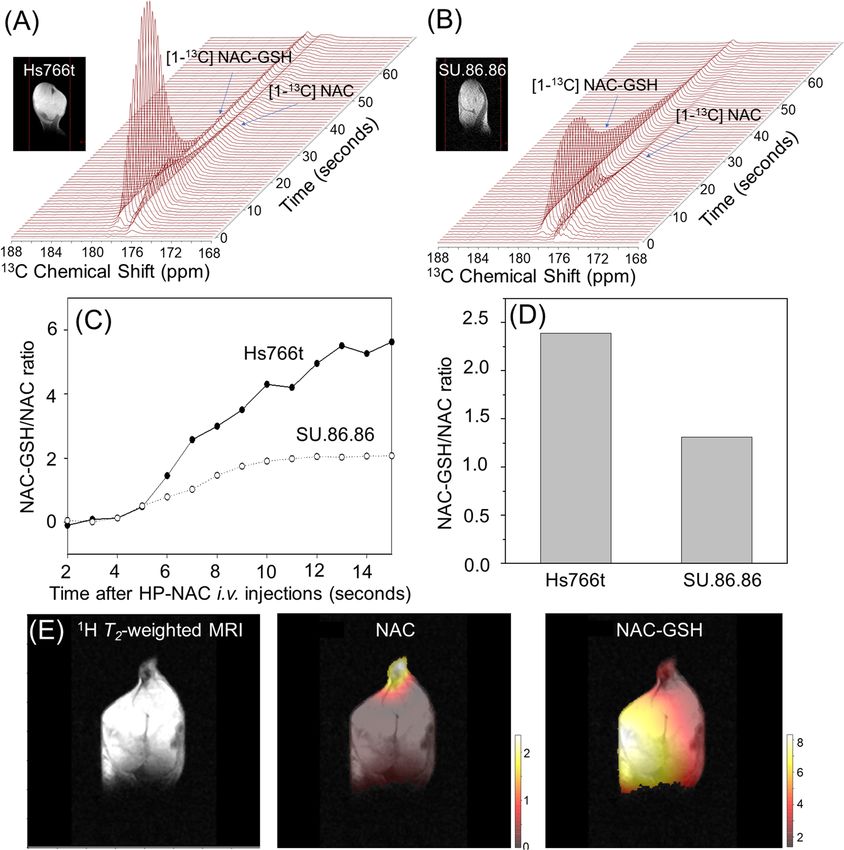
www.nature.com/scientificreports/
Figure 1. Optimizing sample conditions for hyperpolarized in vivo NMR/MRI experiments with N-acetyl
cysteine. (A) Synthetic scheme of [1-13C] NAC. (B) Hyperpolarization build-up curves of [1-13C] NAC showing
the drastic improvement of polarization using the optimized condition of a NaOH solution vs DMSO solutions.
(C) 13C NMR spectra of unlabeled NAC at 1 T NMR confirm the pH dependence of polarization. An asterisk (*)
is from a referencing standard of 13C Urea. (D) Dynamic spectra of hyperpolarized [1-13C] NAC in PBS buffer at
3 T MRI indicates a T1 relaxation time of 19.6 s.
Fluorescent techniques based either on the intrinsic fluorescence of NADH/NAD or specific probes for GSH/
GSSH18 have proven effective for monitoring the redox environment preclinically and for tumors that lie close
to the surface, for example melanoma and head and neck cancers19, but widespread adoption is hindered by the
limited penetration of light in the visible/IR region of the EM s pectrum20.
Here, we demonstrate N-acetyl cysteine (NAC)21, the acetylated derivative of the amino acid l-cysteine and
a precursor of glutathione as a promising novel probe to monitor redox status which overcomes the potential
safety disadvantages of dehydroascorbic acid21–23. We successfully designed stable 13C isotope labeled NAC with
a long life time (T1 spin lattice relaxation) of hyperpolarization, and show tissue dependent redox transformation
in human pancreatic tumor xenografts utilizing the cutting-edge technologies of both hyperpolarized [1-13C]
NAC and metabolic 13C MRI, taking advantage of the drastic sensitivity enhancement ~ 105 fold increase via
hyperpolarization24–26. The biodistribution of hyperpolarized [1-13C] NAC and its biochemical transformation
during the rapid imaging allows us to monitor important early reactions of thiol biochemistry in vivo.
Results and discussion
Our preliminary hyperpolarized NMR experiments on natural abundance NAC indicated that only the [1-13C]
NAC peak can be observed out of two potentially detectable carbonyl groups in NAC structure as shown in 13C
NMR spectra (Supplementary Fig. S1), since the scalar relaxation from adjacent 14 N-nuclei shortens both the T1
and T2 relaxation times of the [4-13C] peak27. In addition to the relaxation characteristics, the [4-13C] position in
NAC is more distal from the redox active sulfhydryl group, therefore, an efficient synthetic scheme was developed
using commonly available starting materials to label NAC only in the [1-13C] position with relatively high yield
by acetylation of [1-13C] L-cysteine (Fig. 1A). Briefly, [1-13C] l-cysteine was reacted with acetic anhydride in
the presence of sodium acetate as the base in deoxygenated tetrahydrofuran28. Isolating the resulting product
by crystallization was not successful as previously r eported29, however the product could be purified by HPLC
to afford [1-13C] NAC as a white, hygroscopic powder in 64% yield. The use of either HCl gas or concentrated
aqueous HCl to convert the sodium salt to the free acid gave similar yields.
To validate [1-13C] NAC as an imaging probe, we first determined the sample conditions for polarization
enhancement and the T1 longitudinal relaxation time in vitro. Samples using a standard solvent of D MSO25 polar-
ized poorly (Fig. 1B), possibly because the anhydrous solvent favors the formation of intermolecular hydrogen
bonds between two protonated carboxylic acid, which would increase the dipolar coupling associated with the
carbonyl carbon and shorten T1 relaxation30. To reduce intermolecular association, [1-13C] NAC was titrated to
a neutral pH of 7.5 using 5 M NaOH. The resulting solution of 3.2 M [1-13C] NAC became a homogenous self-
glassing solution when frozen, a particular advantage for in vivo applications which typically require as highly
concentrated solution as possible to achieve maximum sensitivity and to avoid complications from additives
Scientific Reports | (2021) 11:12155 | https://doi.org/10.1038/s41598-021-90921-0 2
Vol:.(1234567890)
www.nature.com/scientificreports/
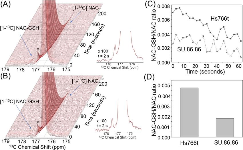
Figure 2. Real-time monitoring NAC metabolism in in cell NMR spectroscopy of tumor cell lines. In cell
dynamic 13C NMR spectra of hyperpolarized [1-13C] NAC at 1 T NMR on 20 × 106 cells of human pancreatic
tumor cell lines of Hs766t (A, left) and SU.86.86 (B, left). Expanded spectra with 100 times magnifications at
2 s after the hyperpolarized [1-13C] NAC injections in Hs766t (A, right) and SU.86.86 (B, right) cells. (C) Time
dependence of NAC-GSH/NAC peak intensity ratio after mixing HP-NAC with PDAC cells. (D) Comparison of
the ratios of NAC-GSH to NAC between Hs766t and SU.86.86 cell lines. A chemical shift peak around 177 ppm
indicated with asterisk (*) is assigned as the dimeric form of NAC.
which may be potentially toxic or interfere with the metabolic processes being s tudied31,32. This polarizing solu-
tion shows efficient polarization build-up (Fig. 1C), reaching half of the equilibrium polarization in 11,000 s,
similar to other hyperpolarized probes being considered for clinical u se25. An improvement in polarization
kinetics and equilibrium polarization values may be possible with optimization of polarization and glassing
conditions31,33,34. This solution remained stable overtime at both neutral and acidic pH (Fig. 1(C)). The polariza-
tion was much weaker at pH 2.5, suggesting a possible role for hydrogen bonds among NAC clusters in reducing
the equilibrium polarization25,30. The T1 relaxation time at 3 T of the 3.2 M [1-13C] NAC solution was determined
to be 19.6 s by the decay dynamics of 13C MR signal (Fig. 1D).
These excellent optimized conditions allowed us to use [1-13C] NAC for in cell NMR and in vivo MRI. In
cell dynamic 13C NMR spectra of hyperpolarized [1-13C] NAC at 1 T NMR spectrometer on human pancreatic
ductal adenocarcinoma (PDAC) cell lines, which have one of the worst prognoses among common cancers and
need effective diagnostic approaches35–37, Hs766t (Fig. 2A) and SU.86.86 (Fig. 2B), in both cases showed three
distinct peaks, a major peak at 176.5 ppm and two peaks at 176.8 and 177.5 ppm. The major peak was immedi-
ately identified as [1-13C] NAC on the basis of the 13C NMR spectrum of a pure phantom sample (Fig. 3B). The
peak at 176.8 ppm was assigned as an oxidized NAC-NAC dimer in a similar manner (Fig. 3B, Supplementary
Figs. S4 and S5)22. The peak at 177.4 ppm was tentatively identified as the oxidized NAC-GSH dimer based on
the 13C NMR spectrum of an authentic sample (Fig. 3(B), Supplementary Fig. S6). To confirm this assignment,
metabolomics approaches based on Mass Spectrometry (MS) were used. Tumor xenografts were treated with
unlabeled and [ 13C3,15 N]-labeled NAC, extracted according to published protocols and analyzed by LC/MS. The
data were collected using scanning quadrupole data-independent acquisition, which gives fragmentation infor-
mation for precursor peaks to aid in identification. The NAC metabolite was traced by first identifying retention
times (rt) and m/z pairs which are unique to the labeled sample relative to the unlabeled sample and therefore
indicate conversion products of the labeled probe (Fig. 3A). Peaks shifted by 4 Da with identical retention
times correspond to labeled products. A 471/467 m/z pair with a rt of 4.47 min confirmed the third product at
177.4 ppm was the oxidized NAC-GSH dimer, which was further supported by fragmentation analysis (Fig. 3A).
Figure 2 indicates hyperpolarized [1-13C] NAC can produce NAC-glutathione (NAC-GSH) in cell cultures.
The rapid kinetics of this reaction suggest that hyperpolarized NAC can permeabilize through cell membranes
without active transport38,39, and chemical reactions of hyperpolarized NAC with GSH can be observed within
the lifetime of this hyperpolarized 13C probe. The time-dependence of the NAC-GSH/NAC peak intensity ratio
Scientific Reports | (2021) 11:12155 | https://doi.org/10.1038/s41598-021-90921-0 3
Vol.:(0123456789)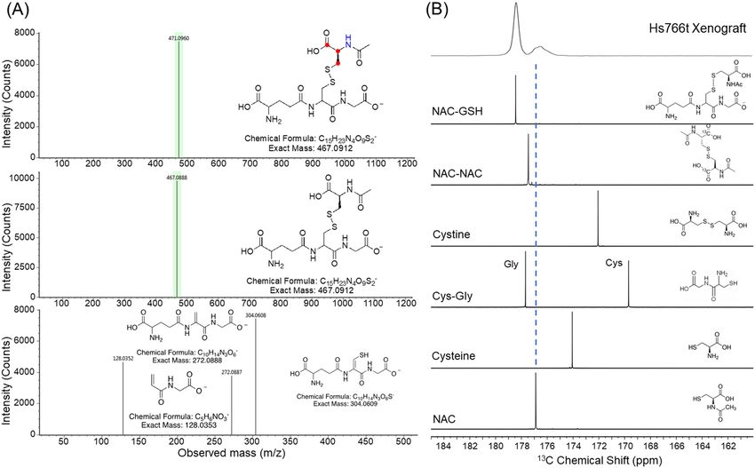
www.nature.com/scientificreports/
Figure 3. Identification of products from hyperpolarized [1-13C] NAC. (A) ESI–MS spectra of SU.86.86 tumor
extracts with (top) and without (middle) isotope labeling in NAC. 13C labeled atoms are indicated in red, 15N
labeled atoms are indicated in blue in the NAC-GSH structure. High energy ESI–MS spectrum of NAC-GSH
with possible fragment identifications (bottom). (B) 13C NMR spectra of synthesized model compounds at
400 MHz, pH 7.5 that represent potential products in comparison to the spectrum from the hyperpolarized
[1-13C] NAC MRS experiments in Hs766t tumor xenograft at 20 s after the iv injection (top).
after mixing hyperpolarized NAC with human PDAC cell lines (Fig. 2C) and the area under the curve ratio
(Fig. 2D) suggest a higher potential for NAC oxidation with glutathione in SU.86.86 cells. The potential for NAC
to be oxidized by glutathione depends on the GSH/GSSG balance, as NAC is not oxidized by GSH40. Lower
concentrations of NAC-GSH in SU.86.86 is consistent with previous metabolomics experiments41,42, as the reli-
ance of SU.86.86 on the TCA cycle depletes NAD+ and therefore shifts the equilibrium of the GSH/GSSG redox
buffer system towards GSH.
Furthermore, to test the effectiveness of [1-13C] NAC as an imaging probe in vivo, real-time dynamic 13C MR
spectra of hyperpolarized [1-13C] NAC were acquired from mice bearing tumor xenograft. We first conducted
13
C two-dimensional chemical shift imaging (CSI) experiments in both a healthy mouse body and head after
intravenous (iv) injection of hyperpolarized [1-13C] NAC solution through a tail vein cannula as shown in Sup-
plementary Fig. S2. Hyperpolarized [1-13C] NAC was globally distributed throughout the mouse body within
30 s after the injection of hyperpolarized solutions, with higher concentrations of [1-13C] NAC in the liver,
kidney, and heart region. Conversely, lower signal was observed in the lung region (Supplementary Fig. S2A).
Although the blood–brain barrier (BBB) permeability of NAC is subject to controversy, the presence of hyper-
polarized [1-13C] NAC in the normal mouse brain indicates the possibility that membrane-permeable NAC may
penetrate the blood–brain barrier and be retained in the brain (Supplementary Fig. S2B)38. However, the current
experimental design cannot distinguish intercellular or interstitial conversion from partial volume effects arising
from circulating NAC in blood vessels and this interpretation should be viewed with caution in light of the fast
timescales involved38,43.
Metabolites of in vivo hyperpolarized [1-13C] NAC were not observed in the liver and kidney regions of these
normal mice, suggesting that the enzymatic conversion of NAC was below the detection level in the absence
of any imposed oxidative stress either focally or globally, although in vitro enzymatic assays of hyperpolarized
NAC incubated with acylase 1 resulted in immediate production of cysteine (Supplementary Fig. S3). To test
[1-13C] NAC in a tumor environment, mouse leg xenografts of Hs766t and SU.86.86 were prepared. The single
voxel MRS signal for NAC-GSH is much stronger in the xenografts (Fig. 4A,B), consistent with higher cellular
density in vivo. In other aspects, the in vivo data (Fig. 4C,D) resembles the in vitro data of the corresponding
cell cultures (Fig. 2C,D). Similar to the in vitro results, NAC-GSH is rapidly formed in both tumors and the
amount of NAC-GSH formed is higher in Hs766t than in SU.86.86 tumors. Encouraged by these results, we also
confirmed that NAC-GSH formation could be imaged as shown in Fig. 4E. Using chemical shift imaging, it can
be seen that NAC-GSH formation is highest in the tumor and lowest in the surrounding muscle and leg regions
Scientific Reports | (2021) 11:12155 | https://doi.org/10.1038/s41598-021-90921-0 4
Vol:.(1234567890)www.nature.com/scientificreports/
Figure 4. Real-time observation of NAC metabolism effectively probes redox status in tumors in vivo.
Dynamic 13C MR spectra of hyperpolarized [1-13C] NAC at 3 T MRI on human pancreatic tumor xenografts
of Hs766t (A) and SU.86.86 (B). Differences in the conversions reflect the redox status of each tumor. (C) Time
dependence of NAC-GSH/NAC peak intensity ratio after the iv injection of HP-NAC. (D) Comparison of the
ratios of NAC-GSH to NAC between Hs766t and SU.86.86 tumor xenografts. (E) Site-specific differences in
chemical conversions of hyperpolarized [1-13C] NAC by 13C Chemical shift imaging in Hs766t xenografts.
while the distribution of non-converted NAC was observed dominantly in the leg area, which is consistent with
higher overall glutathione concentrations in the tumor regions (Fig. 4)41,42.
Interestingly, the 13C chemical shifts of both NAC and its reaction products, NAC-GSH, have a pH depend-
ence (Supplementary Fig. S7), although this may require a high field magnet and/or well optimized shimming
conditions to adequately resolve. This could be advantageous to identify the site-specific differences in pH, in
heterogeneous tumors or differentiate the components inside and outside of tumor cells on the experiments
with high field magnet with optimized shimmed conditions. Using suitable animal disease models, and clinical/
biological targets, hyperpolarized [1-13C] NAC can be used to probe the enzymatic activities or oxidative stress
throughout the body. As for practical clinical subjects, NAC is a widely used in clinical practices as a beneficial
antioxidant and suggested as a potential therapeutic agent in the treatment of cancer, drug toxicity, heart disease,
Scientific Reports | (2021) 11:12155 | https://doi.org/10.1038/s41598-021-90921-0 5
Vol.:(0123456789)www.nature.com/scientificreports/
HIV infection, cystic fibrosis, liver detoxification, Alzheimer disease, diabetes, and other diseases related to
oxidative stress22.
Conclusions
In summary, we have demonstrated the application of a novel hyperpolarized 13C probe based on a widely used,
FDA-approved pharmaceutical agent to assess oxidative stress in human tumor cells and xenografts non-inva-
sively utilizing hyperpolarized 13C MRS imaging. State-of-the-art hyperpolarized 13C MRS allows us to obtain
real-time monitoring of in vivo physiological process and progression of diseases through changes in metabolic
flux7,8,13–16,28. As we have described in this study, in order to design effective in vivo hyperpolarized MRI probes,
successful hyperpolarized isotope labeled biomolecules have to exhibit the following requirements: (a) suitable
biocompatibility and nontoxicity, (b) the availability of an organic synthesis scheme for the production of iso-
tope labeled probes at high yields, (c) long spin lattice T1 relaxation times, (d) efficient nuclear spin polarization
with high concentrations of substrates, (e) the ability to monitor biologically or clinically relevant mechanisms
of metabolic pathways and/or physiological processes, (f) rapid distribution of the hyperpolarized probes to
the targeted imaging regions, (g) adequate chemical shift differences between original injected substrates and
metabolic products, (h) detectable MR signals in both injected probes and the products. We demonstrate here
hyperpolarized NAC potentially satisfies all of these requirements. The biodistribution of hyperpolarized [1-13C]
NAC demonstrates that significant signal levels can be observed in globally in a mouse, including the heart, liver,
kidneys, brain, muscle, and lungs. Hyperpolarized [1-13C] NAC can potentially be used for probing free radical
scavengers, antioxidant, and enzymatic activities, including acylases, which catalyze the deacetylation of NAC
to produce cysteines (Supplementary Fig. S3). Although cysteine-containing NAC has been also considered
as a precursor of glutathione, in this study the formation of GSH was not detectable in our hyperpolarized MR
spectra, most likely due to the relatively short observation window in hyperpolarized experiments and the pos-
sible indirect mechanism of the GSH synthesis after iv injections in in vivo44. Further studies to investigate the
detailed metabolic pathways of hyperpolarized 13C NAC using deuterated analogs to enhance T1 relaxation time
and other metabolomics approaches are in progress in our laboratory. These findings in this study can promote
strategic labeling schemes of biocompatible pharmaceuticals for hyperpolarized MRI to monitor key metabolic
reactions. Our current work expands the hyperpolarization of FDA-approved pharmaceutical compounds to
image in situ metabolic activities and/or MRI contrast agents, which may be relatively smoothly translatable to
high impact clinical applications with proven biocompatibilities.
Methods
Synthesis of [1‑13C] N‑acetyl cysteine. All commercially available reagents were used as received unless
otherwise noted. [1-13C] l-cysteine and D2O were purchased from Cambridge Isotope Laboratories, Inc (Tewks-
bury, MA). Liquid chromatography mass spectrometry (LC–MS) was performed on an Agilent 1200 Series Mass
Spectrometer equipped with LC/MSD TrapXCl Agilent Technologies instrument. Preparative RP-HPLC analysis
was performed on an Agilent 1200 Series instrument equipped with a multi-wavelength detector. 1H and 13C-
NMR were recorded on a Varian 400 MHz NMR spectrometer.
N‑Acetyl cysteine‑[1‑13C] 1. [1-13C] l-cysteine 2 (0.50 g, 4.1 mmol) and sodium acetate
trihydrate (1.11 g, 8.2 mmol) was dissolved in a degassed THF: water (90:10 v/v, 10 mL) solution and was stirred
at room temperature for 20 min under nitrogen. The reaction was cooled to 0 °C and acetic anhydride (0.44 g,
4.3 mmol) was added dropwise. The reaction was stirred for 16 h at room temperature under nitrogen. The
clear solution was cooled and acidified to pH 1 with concentrated HCl. The solvent was evaporated in vacuo
and the product purified by RP-HPLC. Purification was performed using an Agilent Prep C18 column (5 µm,
50 × 100 mm) with a flow rate of 50 mL/min. A linear gradient of 5–35% acetonitrile with 0.1% TFA was used to
elute the product 1 as a white, hygroscopic powder after lyophilization (0.41 g, 64%). 1H-NMR (400 MHz, D 2O):
δ 2.08 (3H, s, CH3), 2.99 (2H, m, CH2SH), 4.63 (1H, m, NHCH). 13C-NMR (400 MHz, D 2O): δ 23.45 (CH3), 27.41
(CH2SH), 57.51 (d, 1JC-C = 232 Hz, NHCH), 173.66 ( CH3C = O), 176.89 (COOH). m/z (ESI–MS +): 165.0 [M+H]+.
Hyperpolarized 13C MRI. NaOH (5 M) was added to [1-13C] NAC powder and OX063 to produce a 3.2 M
[1-13C] NAC solution with 17 mM OX063 at pH of 7.5. 35 mL of 3.2 M [1-13C] NAC with 17 mM OX063
was hyperpolarized using the SPINlab (GE Healthcare) for 3–4 h, and the scans were performed using the
Philips Achieva 3 T MRI. 13C two dimensional spectroscopic chemical shift images (CSIs) were acquired with
a 28 × 28 mm, field of view in a 10 mm axial slice through the head, a matrix size of 14 × 14, spectral width of
3333 Hz, repetition time of 86 ms, and excitation pulse width a flip angle of 3° for the mouse head, and with a
32 × 32 mm, field of view in a 10 mm coronal slice through the body, a matrix size of 16 × 16, spectral width of
3333 Hz, repetition time of 85 ms, and excitation pulse with a flip angle of 10° for the mouse body. CSIs were
acquired 30 s after the beginning of the hyperpolarized [1-13C] NAC injections.
Scientific Reports | (2021) 11:12155 | https://doi.org/10.1038/s41598-021-90921-0 6
Vol:.(1234567890)www.nature.com/scientificreports/
LC/MS methods for identification of product. Chemicals: [13C3, 15N]-NAC was purchased from Cam-
bridge Isotope Laboratories, Inc (Tewksbury, MA). NAC, formic acid and ammonium formate was purchased
from Sigma-Aldrich (St. Louis, MO). LC–MS acetonitrile was purchased from Fisher Scientific. Liquid chro-
matography/mass spectrometry analysis was performed on a Waters Acquity UPLC coupled to a Waters Xevo
Q-ToF quadruple time of flight mass spectrometer operating in electrospray ionization (ESI) in negative mode.
The capillary and sampling cone voltages were set to 1.5 kV and 10 V, respectively. Source and desolvation tem-
peratures were set to 120 °C and 450 °C, respectively, and the cone and desolvation gas flows were set to 50.0 and
800.0 L/h, respectively. To maintain mass accuracy, leucine enkephalin was used at a concentration of 2 ng/mL
in 50:50 acetonitrile/water containing 0.1% formic acid and injected at a rate of 10 μL/min. Data was acquired
using SONAR (scanning quadrupole data-independent acquisition) in continuum mode. In low-energy MS1
mode, the quadrupole was scanned between 50 -1200 m/z, with a quadrupole transmission width of ~ 50 Da,
with a collision cell energy of 10 eV. In high-energy MS2 mode, the collision cell energy was ramped between
20 and 30 Da. The analytes were separated by HILIC chromatography on an Xbridge BEH Amide (2.5 μm, 2.1
× 100 mm) column. Chromatographic separation was achieved with 95:5 water:acetontrile containing 10 mM
ammonium formate, pH 3 (A) and 95:5 acetonitrile:water containing 10 mM ammonium formate, pH 3 (B).
Gradient elution, with a flow rate of 0.340 mL/min, began at 95% B, then decreased to 50% B from 0.0 to 3.4 min,
50–5% B from 3.4 to 5.39 min, held at 5% B from 5.39 to 6.37 min, then returned to initial conditions (95%B) in
0.20 min. The column was equilibrated at 95% B for 4.43 min before the next injection. The column temperature
was maintained at 40 °C in a column oven.
Cell culture and animal studies. All of the animal experiments were conducted in compliance with the
Guide for the Care and Use of Laboratory Animal Resources and ARRIVE guidelines, and experimental proto-
cols were approved by the Animal Care and Use Committee, National Cancer Institute (NCI-CCR-ACUC)45,46.
The human pancreatic ductal adenocarcinoma (PDAC) cell lines, Hs776t, and SU.86.86 cells, were purchased
from Threshold Pharmaceuticals (Redwood City, CA). Human pancreatic tumor inoculated mice were gener-
ated by subcutaneous injection of 3 × 10⁵ cells into the right hind legs of mice. Detailed conditions for cell culture
and xenograft tumor development were as described p reviously47. Athymic nude mice were obtained from the
Frederick Cancer Research Center, Animal Production (Frederick, MD). Both respiration (60–90 breaths per
min) and temperature (35–37 °C) were maintained at a normal physiological range and monitored continuously
during the animal experiment using the adjusted anesthesia, isoflurane.
Extraction of metabolites from tumors. 13
C, 15N labeled NAC ( [13C3, 15N] cysteine) was purchased from
Cambridge Isotope Laboratories, Inc (Tewksbury, MA). Unlabeled NAC was purchased from Sigma-Aldrich (St.
Louis, MO). 2.76 mg of either 13C, 15N labeled NAC ( [13C3, 15 N] cysteine) or unlabeled NAC was intravenously
injected to track metabolites of NAC in xenograft tumors. Mice were euthanized in 2 min after the tail vein injec-
tions. The tumors were rapidly removed and flash frozen in the liquid nitrogen, then they were stored at − 80 °C.
The metabolites were extracted from the obtained tumors using a previously reported p rocedure48. The resulting
lyophilized aqueous metabolite extracts were used for the MS for metabolomic analysis.
Received: 21 February 2021; Accepted: 10 May 2021
References
1. Meister, A. & Anderson, M. E. Glutathione. Annu. Rev. Biochem. 52, 711–760. https://d oi.o
rg/1 0.1 146/a nnure v.b
i.5 2.0 70183.0 03431
(1983).
2. Zhu, J. & Thompson, C. B. Metabolic regulation of cell growth and proliferation. Nat. Rev. Mol. Cell. Biol. 20, 436–450. https://doi.
org/10.1038/s41580-019-0123-5 (2019).
3. Cook, J. A. et al. Oxidative stress, redox, and the tumor microenvironment. Semin. Radiat. Oncol. 14, 259–266. https://doi.org/10.
1016/j.semradonc.2004.04.001 (2004).
4. Badgley, M. A. et al. Cysteine depletion induces pancreatic tumor ferroptosis in mice. Science 368, 85–89. https://doi.org/10.1126/
science.aaw9872 (2020).
5. Cairns, R. A., Harris, I. S. & Mak, T. W. Regulation of cancer cell metabolism. Nat. Rev. Cancer 11, 85–95. https://doi.org/10.1038/
nrc2981 (2011).
6. Trachootham, D., Alexandre, J. & Huang, P. Targeting cancer cells by ROS-mediated mechanisms: A radical therapeutic approach?.
Nat. Rev. Drug Discov. 8, 579–591. https://doi.org/10.1038/nrd2803 (2009).
7. Wang, J. & Yi, J. Cancer cell killing via ROS: To increase or decrease, that is the question. Cancer Biol. Ther. 7, 1875–1884. https://
doi.org/10.4161/cbt.7.12.7067 (2008).
8. Kuppusamy, P. et al. Noninvasive imaging of tumor redox status and its modification by tissue glutathione levels. Cancer Res. 62,
307–312 (2002).
9. Ilangovan, G. et al. In vivo measurement of regional oxygenation and imaging of redox status in RIF-1 murine tumor: Effect of
carbogen-breathing. Magn. Reson. Med. 48, 723–730. https://doi.org/10.1002/mrm.10254 (2002).
10. Timm, K. N. et al. Assessing oxidative stress in tumors by measuring the rate of hyperpolarized [1-13C]dehydroascorbic acid
reduction using 13C magnetic resonance spectroscopy. J. Biol. Chem. 292, 1737–1748. https://doi.org/10.1074/jbc.M116.761536
(2017).
11. Bohndiek, S. E. et al. Hyperpolarized [1-13C]-ascorbic and dehydroascorbic acid: Vitamin C as a probe for imaging redox status
in vivo. J. Am. Chem. Soc. 133, 11795–11801. https://doi.org/10.1021/ja2045925 (2011).
12. Keshari, K. R. et al. Hyperpolarized 13C dehydroascorbate as an endogenous redox sensor for in vivo metabolic imaging. Proc.
Natl. Acad. Sci. U S A 108, 18606–18611. https://doi.org/10.1073/pnas.1106920108 (2011).
13. Patterson, J. W. & Lazarow, A. Sulfhydryl protection against dehydroascorbic acid diabetes. J. Biol. Chem. 186, 141–144 (1950).
Scientific Reports | (2021) 11:12155 | https://doi.org/10.1038/s41598-021-90921-0 7
Vol.:(0123456789)www.nature.com/scientificreports/
14. Shoda, S. et al. Imaging of hydroxyl-radical generation using dynamic nuclear polarization-magnetic resonance imaging and a
spin-trapping agent. Anal. Chem. 92, 14408–14414. https://doi.org/10.1021/acs.analchem.0c02331 (2020).
15. Saito, K. et al. Synthesis and evaluation of (13)C-labeled 5–5-dimethyl-1-pyrroline-N-oxide aimed at in vivo detection of reactive
oxygen species using hyperpolarized (13)C-MRI. Free Radic. Biol. Med. 131, 18–26. https://doi.org/10.1016/j.freeradbiomed.2018.
11.013 (2019).
16. Do, Q. N., Ratnakar, J. S., Kovacs, Z. & Sherry, A. D. Redox- and hypoxia-responsive MRI contrast agents. ChemMedChem 9,
1116–1129. https://doi.org/10.1002/cmdc.201402034 (2014).
17. Hancu, I. et al. CEST and PARACEST MR contrast agents. Acta Radiol. 51, 910–923. https://doi.org/10.3109/02841851.2010.
502126 (2010).
18. Jiang, X. et al. Quantitative real-time imaging of glutathione. Nat. Commun. 8, 16087. https://doi.org/10.1038/ncomms16087
(2017).
19. Wu, C., Gleysteen, J., Teraphongphom, N. T., Li, Y. & Rosenthal, E. In-vivo optical imaging in head and neck oncology: Basic
principles, clinical applications and future directions. Int. J. Oral Sci. 10, 10. https://doi.org/10.1038/s41368-018-0011-4 (2018).
20. Ntziachristos, V., Pleitez, M. A., Aime, S. & Brindle, K. M. Emerging technologies to image tissue metabolism. Cell Metab. 29,
518–538. https://doi.org/10.1016/j.cmet.2018.09.004 (2019).
21. Samuni, Y., Goldstein, S., Dean, O. M. & Berk, M. The chemistry and biological activities of N-acetylcysteine. Biochim. Biophys.
Acta. 1830, 4117–4129. https://doi.org/10.1016/j.bbagen.2013.04.016 (2013).
22. Atkuri, K. R., Mantovani, J. J., Herzenberg, L. A. & Herzenberg, L. A. N-Acetylcysteine: A safe antidote for cysteine/glutathione
deficiency. Curr. Opin. Pharmacol. 7, 355–359. https://doi.org/10.1016/j.coph.2007.04.005 (2007).
23. Cotgreave, I. A. N-acetylcysteine: Pharmacological considerations and experimental and clinical applications. Adv. Pharmacol.
38, 205–227 (1997).
24. Ardenkjaer-Larsen, J. H. et al. Increase in signal-to-noise ratio of > 10,000 times in liquid-state NMR. Proc. Natl. Acad. Sci. U S A
100, 10158–10163. https://doi.org/10.1073/pnas.1733835100 (2003).
25. Keshari, K. R. & Wilson, D. M. Chemistry and biochemistry of 13C hyperpolarized magnetic resonance using dynamic nuclear
polarization. Chem. Soc. Rev. 43, 1627–1659. https://doi.org/10.1039/c3cs60124b (2014).
26. Kurhanewicz, J. et al. Hyperpolarized (13)C MRI: Path to clinical translation in oncology. Neoplasia 21, 1–16. https://doi.org/10.
1016/j.neo.2018.09.006 (2019).
27. Cho, A., Eskandari, R., Granlund, K. L. & Keshari, K. R. Hyperpolarized [6-(13)C, (15)N3]-arginine as a probe for in vivo arginase
activity. ACS Chem. Biol. 14, 665–673. https://doi.org/10.1021/acschembio.8b01044 (2019).
28. Martin, T. A., Corrigan, J. R. & Waller, C. W. N-acylation of cysteine. J. Org. Chem. 30, 2839–2840. https://doi.org/10.1021/jo010
19a509 (1965).
29. Amoyaw, P. N. A. et al. Synthesis of 13C-labeled derivatives of cysteine for magnetic resonance imaging studies of drug uptake
and conversion to glutathione in rat brain. J. Label. Compd. Radiopharm. 54, 607–612. https://doi.org/10.1002/jlcr.1904 (2011).
30. Hundshammer, C., Grashei, M., Greiner, A., Glaser, S. J. & Schilling, F. pH dependence of T1 for (13) C-labelled small molecules
commonly used for hyperpolarized magnetic resonance imaging. ChemPhysChem 20, 798–802. https://doi.org/10.1002/cphc.
201801098 (2019).
31. Brender, J. R. et al. Trehalose as an alternative to glycerol as a glassing agent for in vivo DNP MRI. Magn. Reson. Med. 85, 42–48.
https://doi.org/10.1002/mrm.28405 (2021).
32. Marco-Rius, I. et al. Monitoring acute metabolic changes in the liver and kidneys induced by fructose and glucose using hyper-
polarized [2-(13) C]dihydroxyacetone. Magn. Reson. Med. 77, 65–73. https://doi.org/10.1002/mrm.26525 (2017).
33. Capozzi, A. et al. Gadolinium effect at high-magnetic-field DNP: 70% (13)C polarization of [U-(13)C] glucose using trityl. J. Phys.
Chem. Lett. 10, 3420–3425. https://doi.org/10.1021/acs.jpclett.9b01306 (2019).
34. Ludwig, C., Marin-Montesinos, I., Saunders, M. G. & Gunther, U. L. Optimizing the polarization matrix for ex situ dynamic nuclear
polarization. J. Am. Chem. Soc. 132, 2508–2509. https://doi.org/10.1021/ja909984w (2010).
35. Garrido-Laguna, I. & Hidalgo, M. Pancreatic cancer: From state-of-the-art treatments to promising novel therapies. Nat. Rev. Clin.
Oncol. 12, 319–334. https://doi.org/10.1038/nrclinonc.2015.53 (2015).
36. Hidalgo, M. Pancreatic cancer. N. Engl. J. Med. 362, 1605–1617. https://doi.org/10.1056/NEJMra0901557 (2010).
37. Adamska, A., Domenichini, A. & Falasca, M. Pancreatic ductal adenocarcinoma: Current and evolving therapies. Int. J. Mol. Sci.
https://doi.org/10.3390/ijms18071338 (2017).
38. Bavarsad Shahripour, R., Harrigan, M. R. & Alexandrov, A. V. N-acetylcysteine (NAC) in neurological disorders: Mechanisms of
action and therapeutic opportunities. Brain Behav. 4, 108–122. https://doi.org/10.1002/brb3.208 (2014).
39. Mazor, D. et al. Red blood cell permeability to thiol compounds following oxidative stress. Eur. J. Haematol. 57, 241–246. https://
doi.org/10.1111/j.1600-0609.1996.tb01370.x (1996).
40. Noszal, B., Visky, D. & Kraszni, M. Population, acid-base, and redox properties of N-acetylcysteine conformers. J. Med. Chem. 43,
2176–2182. https://doi.org/10.1021/jm9909600 (2000).
41. Kishimoto, S. et al. Imaging of glucose metabolism by 13C-MRI distinguishes pancreatic cancer subtypes in mice. Elife https://
doi.org/10.7554/eLife.46312 (2019).
42. Matsumoto, S. et al. Metabolic and physiologic imaging biomarkers of the tumor microenvironment predict treatment outcome
with radiation or a hypoxia-activated prodrug in mice. Cancer Res. 78, 3783–3792. https://doi.org/10.1158/0008-5472.CAN-18-
0491 (2018).
43. Sheffner, A. L. et al. Metabolic studies with acetylcysteine. Biochem. Pharmacol. 15, 1523–1535. https://doi.org/10.1016/0006-
2952(66)90197-3 (1966).
44. Zhou, J. et al. Intravenous administration of stable-labeled N-acetylcysteine demonstrates an indirect mechanism for boosting
glutathione and improving redox status. J. Pharm. Sci. 104, 2619–2626. https://doi.org/10.1002/jps.24482 (2015).
45. National Research Council (US) Committee for the Update of the Guide for the Care and Use of Laboratory Animals. Guide for
the Care and Use of Laboratory Animals. (National Academies Press (US), 2011).
46. Percie du Sert, N. et al. Reporting animal research: Explanation and elaboration for the ARRIVE guidelines 2.0. PLoS Biol. 18,
e3000411. https://doi.org/10.1371/journal.pbio.3000411 (2020).
47. Yamamoto, K. et al. Molecular imaging of the tumor microenvironment reveals the relationship between tumor oxygenation,
glucose uptake, and glycolysis in pancreatic ductal adenocarcinoma. Cancer Res. 80, 2087–2093. https://doi.org/10.1158/0008-
5472.CAN-19-0928 (2020).
48. Crooks, D. R., Fan, T. W. & Linehan, W. M. Metabolic labeling of cultured mammalian cells for stable isotope-resolved metabo-
lomics: Practical aspects of tissue culture and sample extraction. Methods Mol. Biol. 1928, 1–27. https://doi.org/10.1007/978-1-
4939-9027-6_1 (2019).
Acknowledgements
This study was supported by the intramural research program at NCI/NIH. The content of this publication does
not necessarily reflect the views or policies of the Department of Health and Human Services, nor does mention
of trade names, commercial products, or organizations imply endorsement by the U.S. Government.
Scientific Reports | (2021) 11:12155 | https://doi.org/10.1038/s41598-021-90921-0 8
Vol:.(1234567890)www.nature.com/scientificreports/
Author contributions
K.Y. and K.S. conducted the experiments. A.O. and D.S. synthesized stable isotope labeled probes A.O., D.S.
and B.B. carried out analysis of 13C labeled materials K.Y. analyzed the data with processing support from J.R.B.
Y.S. and Y.O. contributed technical support and analytical measurements. R.M.M., T.S., N.O., D.R.C., S.K. con-
tributed to sample and animal experiment preparations under the supervision of W.M.L., J.H.A.-L., and M.C.K.
K.Y. and M.C.K. conceived the original idea and designed/planned the experiments. P.L.C., J.H.A.-L., J.B.M.,
W.M.L., R.E.S., and M.C.K. supervised each sections of project. M. C. K. supervised the whole project. K.Y. and
M.C.K. wrote the manuscript with support from J.R.B., and all authors provided feedback and contributed to
the final manuscript.
Funding
Open Access funding provided by the National Institutes of Health (NIH).
Competing interests
The authors declare no competing interests.
Additional information
Supplementary Information The online version contains supplementary material available at https://doi.org/
10.1038/s41598-021-90921-0.
Correspondence and requests for materials should be addressed to M.C.K.
Reprints and permissions information is available at www.nature.com/reprints.
Publisher’s note Springer Nature remains neutral with regard to jurisdictional claims in published maps and
institutional affiliations.
Open Access This article is licensed under a Creative Commons Attribution 4.0 International
License, which permits use, sharing, adaptation, distribution and reproduction in any medium or
format, as long as you give appropriate credit to the original author(s) and the source, provide a link to the
Creative Commons licence, and indicate if changes were made. The images or other third party material in this
article are included in the article’s Creative Commons licence, unless indicated otherwise in a credit line to the
material. If material is not included in the article’s Creative Commons licence and your intended use is not
permitted by statutory regulation or exceeds the permitted use, you will need to obtain permission directly from
the copyright holder. To view a copy of this licence, visit http://creativecommons.org/licenses/by/4.0/.
© The Author(s) 2021
Scientific Reports | (2021) 11:12155 | https://doi.org/10.1038/s41598-021-90921-0 9
Vol.:(0123456789)You can also read


